. . . .
. . . .
Clinical significance of dynamic pulmonary
vascular resistance in two populations at risk of
pulmonary arterial hypertension
Charlien Gabriels
1, Patrizio Lancellotti
2, Alexander Van De Bruaene
1,
Damien Voilliot
2, Pieter De Meester
1, Roselien Buys
3, Marion Delcroix
4,
and Werner Budts
1*
1
Department of Cardiology, University Hospitals Leuven, Herestraat 49, Leuven B-3000, Belgium;2
Department of Cardiology, GIGA Cardiovascular Sciences, University Hospital Sart Tilman Lie`ge, Lie`ge B-4000, Belgium;3
Department of Rehabilitation Sciences, University Hospitals Leuven, Leuven, Belgium; and4
Department of Pneumology, University Hospitals Leuven, Leuven, Belgium
Received 25 September 2014; accepted after revision 17 November 2014; online publish-ahead-of-print 18 December 2014
Aims Patients at risk of pulmonary arterial hypertension (PAH) may present with abnormal dynamic pulmonary vascular resist-ance (PVR) during exercise. However, its clinical significresist-ance remains unclear. The present study aimed at analysing the meaning of dynamic PVR in two populations at risk of PAH: secundum atrial septal defect (ASD) and systemic sclerosis (SSc).
Methods and results
Adult patients with corrected ASD were consecutively selected from the database of Pediatric and Congenital Heart Disease of the University Hospitals Leuven. Patients with SSc were consecutively selected from the rheumatology data-base of the University Hospital Lie`ge. At inclusion, all underwent a rest and bicycle stress echocardiography to obtain baseline right heart characteristics and dynamic PVR. Routine follow-up echocardiography was performed. Twenty-eight patients with corrected ASD (mean age 41 + 17 years, 79% female) were followed for a median time of 3.7 [inter-quartile range (IQR) 2.9 – 4.1] years. No patient developed PAH. Dynamic PVR was significantly associated with right atrial dilatation at latest follow-up (Spearman’s r 0.51, P ¼ 0.013). Forty-five SSc patients (mean age 54 + 13 years, 76% female) were followed for a median time of 2.4 (IQR 0.8 – 2.9) years. Thirteen patients (30%) developed PAH. Dynamic PVR was the only independent predictor of PAH (hazards ratio 1.22, 95% confidence interval 1.01 – 1.47). No significant right heart morphometric changes occurred.
Conclusion Dynamic PVR predicted PAH development in patients with SSc, whereas dynamic PVR was associated with right heart
morphometric changes after ASD closure. The predictive role of dynamic PVR might depend on the underlying disease type. Larger studies are needed to confirm this hypothesis.
-Keywords atrial septal defect † systemic sclerosis † pulmonary vascular disease † stress echocardiography
Introduction
Pulmonary arterial hypertension (PAH), characterized by a progres-sive increase in pulmonary vascular resistance (PVR), is associated with considerable morbidity and mortality.1Unfortunately, PAH is
usually detected when patients develop symptoms and pulmonary vascular disease (PVD) has already evolved to an advanced (irrevers-ible) stage.2,3Hence, treatment is often initiated too late, and results
are unsatisfactory. Therefore, developing an early detection method for mild PVD is crucially important.
In the last years, many studies have demonstrated an abnormal in-crease in pulmonary artery pressure (PAP) and/or PVR during exer-cise testing in different populations at risk of PAH development.4–12 This so-called exercise-induced PAH has been associated with decreased oxygen uptake during exercise and impaired exercise capacity.13–15It has been suggested that the slope of a PAP-flow
*Corresponding author. Tel:+32 16344369; Fax: +32 16344240, E-mail: werner.budts@uzleuven.be
plot, defining dynamic PVR, provides a better representation of PVR.16However, the clinical significance of dynamic PVR remains unclear. Does a high-dynamic PVR represent a stable self-contained entity or could it equal an early stage in the ‘disease spectrum of PAH’?14,15,17
The aetiology of PVD is very diverse and explains why the pace of disease progression might differ substantially between several types of PVD. Idiopathic PVD and PVD associated with connective tissue disease usually evolve fast, whereas PVD secondary to shunt-related congenital heart disease (CHD) is mostly avoided in case of early closure of septal defects in childhood.1,18,19However, after paediat-ric age, there is no evidence that closure of septal defects can prevent PVD progression, and prognosis may even be worsened by late closure.20This different pace of PVD progression might lead to a dif-ferent predictive role of dynamic PVR.
This study aimed at determining the significance of dynamic PVR in two populations at risk of developing PAH: patients with secundum atrial septal defect (ASD), mostly closed later in life, and patients with systemic sclerosis (SSc).
Methods
Patient selection
Patients with secundum ASD closure were consecutively selected from the database of Pediatric and Congenital Heart Disease of the University Hospitals Leuven from March to November 2009. Minimum age at inclu-sion was 18 years, and patients had to be able to perform a bicycle stress test. Patients with associated complex diseases (coronary artery disease, significant valvular diseases other than mild tricuspid or mitral regurgita-tion, pulmonary disease, history of pulmonary embolism, concomitant CHD) were excluded. Patients with a history of arrhythmias or current arrhythmias were also excluded because of practical and safety reasons. Similarly, patients with SSc were consecutively selected from the rheuma-tology database of the University Hospital of Lie`ge from January 2008 to November 2012.21All patients signed informed consent, after the
Univer-sity Hospitals Institutional Review Boards approved the study protocol.
Rest and bicycle stress echocardiography
At inclusion, all patients underwent complete transthoracic echocardio-graphic exam at rest in the supine position, followed by bicycle stress echocardiography in semi-supine position. For the ASD group, a Vivid 7 or 9 ultrasound system (General Electric Vingmed Ultrasound, Horten, Norway), for the SSc group a Vivid 9 ultrasound system (General Electric Healthcare, Little Chalfont, UK), was used. Methodo-logical details and baseline characteristics have already been pub-lished.6,21 Bicycle stress echocardiography was performed in a semi-supine position, with the exercise table tilted 20 – 308 to the left. Patients started at an initial workload of 25 W, which was increased by 25 W every 2 min, until the maximal tolerated workload. A 12-lead ECG and blood pressure were recorded at rest and at every 2 min during exercise. Analyses were performed by a single observer using cus-tomized software (Echopac). All measurements were calculated in tripli-cate and presented as means. Measurements were performed and analysed according to the 2006 guidelines on chamber quantification.22 Basic echocardiogram mainly focused on right heart geometry [right atrial (RA) diameter, right ventricular (RV) diameter, grade of tricuspid valve regurgitation (TR)] and tricuspid regurgitation velocity (TRV), from which the pressure difference between RV and RA was calculated using the simplified Bernoulli equation. Adding RA pressure (RAP)
resulted in the estimate of systolic PAP (sPAP). RAP was calculated from diameter and breath-induced variability of inferior vena cava (IVC). For all patients, due to normal IVC diameter and decrease in IVC diameter .50% after deep inspiration, RAP was estimated at 5 mmHg at rest and at 10 mmHg at peak exercise, similar to the method-ology of other studies.13,17,23–25
Total PVR was calculated as the ratio of sPAP and left ventricular cardiac output (flow, calculated from the velocity time integral in the left ventricular outflow tract), at rest and at peak exercise. The slope of the sPAP-flow plot was obtained and determined as dynamic PVR, using linear regression analysis with two measurement points (rest and peak exercise). Dynamic PVR thus represents change in sPAP per unit change in cardiac output from rest to peak exercise.
RA and RV dilatation was graded normal (0/3), mild (1/3), moderate (2/3) or severe (3/3), based on measurements in apical four-chamber view (RA: long-axis diameter and area at end-systole; RV: short-axis diam-eter at the mid-ventricular level and area at end-diastole).22TR was graded semi-quantitatively using colour Doppler echocardiography (x/4).26
Follow-up
All patients were followed systematically. Rest transthoracic echocardio-gram at latest follow-up was considered the study endpoint, with focus on right heart geometry and the presence of PAH. RA and RV change were graded (0 ¼ no increase in dilatation grade, 1 ¼ increase in dilatation of 1 grade, 2 ¼ increase in dilatation of 2 grades). TR change was defined simi-larly based on the increase in regurgitation grade. PAH at rest was diag-nosed when estimated sPAP ≥37 mmHg (TRV ≥2.9 m/s + RAP 5 mmHg), following ESC/ERS guidelines on pulmonary hypertension.1
Statistical analysis
Statistical analyses were performed using the software program SPSS (version 22.0, Chicago, IL, USA). For continuous variables, data are reported as means + standard deviation, or as medians [inter-quartile range (IQR)] if the assumption of normal distribution was not valid. Levene’s test was performed to assess for equal variances. Discrete vari-ables are presented as frequencies and/or proportions. Comparison of two means was done by two-tailed Student’s t-test. Analysis of paired data was done by two-tailed paired Student’s t-test or by related-samples Wilcoxon signed-rank test, when normality was not assumed. Compari-son of several means was done by one-way analysis of variance. PearCompari-son’s correlation coefficient and Spearman’s rank correlation coefficient were calculated to estimate the correlation between dynamic PVR and PAP or right heart morphometry, respectively. Univariate and multivariate Cox-regression analysis was performed to identify predictors of PAH de-velopment. Only those factors that are significant predictors in univariate analysis will be included in multivariate analysis. In case of strong collinear-ity (r . 0.5), only one of the collinear variables was retained for multivari-ate analysis. All tests were two sided, and a P-value of ,0.05 was considered statistically significant.
Results
Patient baseline characteristics
Twenty-eight patients with corrected ASD (mean age at inclusion 41 + 17 years, 79% female patients) were selected and followed for a median time of 3.7 (IQR 2.9 – 4.1) years. Forty-five patients with SSc (mean age at inclusion 54 + 13 years, 76% female patients) were selected and followed for a median time of 2.4 (IQR 0.8 – 2.9) years. Patient baseline characteristics are summarized in Table1. None of the patients died during follow-up.
Dynamic PVR in patients with corrected
ASD: associated morphometric changes of
the right heart
Mean dynamic PVR was 5.6 + 2.4 mmHg/L/min. No patient devel-oped PAH during follow-up. RA diameter and TR severity evolved significantly during follow-up (Table2).
Dynamic PVR was only associated with greater RA dilatation grade at latest follow-up, and a trend towards association with RA change over time was noted (Table3). RA dilated significantly more during follow-up in patients with higher dynamic PVR at inclusion (mean dynamic PVR 5.2 + 2.1 vs. 6.8 + 2.7 vs. 10.8 mmHg/L/min, P ¼ 0.047) (Figure1).
Dynamic PVR in patients with SSc:
predictor of PAH
Mean dynamic PVR was 7.3 + 4.2 mmHg/L/min. Thirteen patients (30%) developed PAH during follow-up. No morphometric changes of the right heart were noted.
Dynamic PVR, age at latest follow-up, sPAP at rest and sPAP and total PVR at peak exercise were significantly associated with PAH de-velopment (Table4). Patients with higher dynamic PVR (steeper slopes) and a higher sPAP at rest developed PAH. Because bivariate correlation analysis showed a strong correlation between dynamic
PVR and sPAP at peak exercise (Pearson’s correlation coefficient 0.595, P ¼ 0.001) and between dynamic PVR and total PVR at peak exercise (Pearson’s correlation coefficient 0.862, P , 0.001), sPAP and total PVR at peak exercise were excluded from the multivariate model. Multivariate Cox-regression analysis showed that dynamic PVR was the only independent predictor of PAH (Table4). Mean dynamic PVR in the PAH group was significantly higher than in the non-PAH group (11.0 vs. 5.8 mmHg/L/min, P ¼ 0.002).
Comparison between both populations
Patients with SSc and corrected ASD were divided based upon the development of PAH or RA change over time, respectively (Figure2). Whereas mean dynamic PVR differed significantly in the PAH vs. non-PAH group of SSc patients (P ¼ 0.002) and in the group of corrected ASD patients with vs. without RA change (7.6 vs. 5.1 mmHg/L/min, P ¼ 0.042), there was no significant difference in mean dynamic PVR of SSc patients with PAH vs. corrected ASD patients with RA change (P ¼ 0.144). Likewise, mean dynamic PVR of SSc patients without PAH vs. corrected ASD patients without RA change did not differ significantly (P ¼ 0.487). This is visually represented in Figure3.
. . . . Table 1 Patient baseline characteristics
Corrected ASD (n 5 28)
SSc (n 5 45) Age at inclusion (years) 41 + 17 54 + 13 Age at SSc diagnosis (years) 49 + 15 Age at ASD closure (years) 34 + 20
ASD defect size (mm) 17 + 5 Shunt ratio prior to ASD
closure (Qp : Qs)a
1.9 (1.5 – 2.5) sPAP prior to ASD closure
(mmHg)a
26 + 8
Gender (male : female) 6 : 22 11 : 34 BMI (kg/m2) 26 + 5 24 + 5 SBP rest (mmHg) 119 + 16 131 + 22 DBP rest (mmHg) 71 + 11 75 + 11 HR rest (bpm) 63 + 10 73 + 13 RA dilatation (x/3) 0 (0 – 0) 0 (0 – 0) RV dilatation (x/3) 0 (0 – 1) 0 (0 – 0) TR (x/4) 1 (0 – 1.5) 1 (0 – 1) sPAP rest (mmHg) 27 + 6 25 + 7 Total PVR rest (mmHg/L/min) 6.3 + 1.7 6.9 + 2.4 Total PVR peak exercise
(mmHg/L/min)
5.8 + 1.6 6.5 + 2.5
Continuous variables are presented as mean + standard deviation, discrete variables as ratio or as median (inter-quartile range).
ASD, atrial septal defect; SSc, systemic sclerosis; BMI, body mass index; SBP, systolic blood pressure; DBP, diastolic blood pressure; HR, heart rate; RA, right atrium; RV, right ventricle; TR, tricuspid regurgitation; sPAP, systolic pulmonary artery pressure; PVR, pulmonary vascular resistance.
a
Invasive measurement.
. . . . Table 2 Changes in echocardiographic variables of the corrected ASD group
Baseline Latest follow-up P-value RA diameter (mm) 45 + 5 48 + 6 0.013 RA dilatation (x/3) 0 (0 – 0) 0 (0 – 1) 0.058 RV dilatation (x/3) 0 (0 – 1) 0 (0 – 1) 0.317 TR (x/4) 1 (0 – 1.5) 1 (1 – 2) 0.005 sPAP rest (mmHg) 27 + 6 28 + 7 0.427 Continuous variables are presented as mean + standard deviation, discrete variables as median (inter-quartile range).
For abbreviations, see Table1.
Significant P-values (P , 0.05) are indicated in bold.
. . . . Table 3 Bivariate correlation analysis with dynamic PVR in the corrected ASD group
Spearman’s correlation coefficient P-value RA dilatation at latest FU (x/3) 0.509 0.013 RA change during FU (Dx/3) 0.386 0.069 RV dilatation at latest FU (x/3) 20.100 0.658 RV change during FUa TR severity at latest FU (x/4) 20.067 0.761 TR change during FU (Dx/4) 20.084 0.702 sPAP rest at latest FU (mmHg) 0.145b 0.553 FU, follow-up. For the remaining abbreviations, see Table1.
a
RV dilatation did not change over time.
b
Pearson’s correlation coefficient.
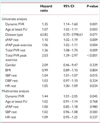
Dynamic PVR in the combined group of
corrected ASD and SSc
Dynamic PVR in the combined group of corrected ASD and SSc remained an independent predictor of PAH in multivariate analysis (Table 5). Based on ROC curve analysis (AUC 0.856), a cut-off value for dynamic PVR at 9.74 mmHg/L/min was determined as pre-dictor for PAH development, with sensitivity and specificity of 63 and
93%, positive and negative predictive value of 0.67 and 0.92, respect-ively (Figure3). Positive and negative likelihood ratios were 9.33 and 0.40. This cut-off value was validated in the subgroup of SSc patients (AUC 0.837, cut-off value of 9.65 mmHg/L/min, sensitivity 62%, and specificity 90%).
Discussion
This study showed an association between dynamic PVR and PAH de-velopment in patients with SSc, whereas dynamic PVR in corrected ASD patients was associated with right heart morphometric changes. . . . .
Table 4 Cox regression analysis for PAH in patients with SSc
Hazard ratio 95% CI P-value Univariate analysis
Dynamic PVR 1.23 1.04 – 1.45 0.015 Age at latest FU 1.06 1.02 – 1.10 0.007 sPAP rest 1.15 1.07 – 1.24 ,0.001 sPAP peak exercise 1.13 1.07 – 1.20 ,0.001 Total PVR rest 1.25 0.99 – 1.56 0.054 Total PVR peak exercise 1.67 1.15 – 2.41 0.006 Gender 2.41 0.53 – 11.03 0.256 BMI 1.03 0.93 – 1.14 0.616 SBP rest 1.02 0.99 – 1.05 0.342 DBP rest 1.01 0.94 – 1.08 0.842 HR rest 1.01 0.97 – 1.06 0.587 Multivariate analysis Dynamic PVR 1.22 1.01 – 1.47 0.038 Age at latest FU 1.00 0.95 – 1.06 0.933 sPAP rest 1.15 0.98 – 1.35 0.083 All variables refer to baseline measurements, unless otherwise specified. PAH, pulmonary arterial hypertension; SSc, systemic sclerosis; CI, confidence interval; PVR, pulmonary vascular resistance; FU, follow-up; sPAP, systolic pulmonary artery pressure; BMI, body mass index; SBP, systolic blood pressure; DBP, diastolic blood pressure; HR, heart rate
Figure 1 Boxplot of dynamic PVR by RA change during follow-up in patients with corrected ASD.
Figure 2 Mean dynamic PVR in patients with corrected ASD and SSc, differentiated by the development of PAH or RA change over time.
Figure 3 Boxplot of dynamic PVR per disease type and the pres-ence or abspres-ence of PAH or RA change during follow-up.
The choice for dynamic PVR in the
prediction model
Right heart catheterization is the gold standard for PAH diagnosis. However, non-invasive assessment techniques such as stress echo-cardiography and cardiac magnetic resonance imaging are evolving as useful tools for early PAH detection.8,27Although the sensitivity of transthoracic echocardiography at rest for early PAH diagnosis is rather limited, stress echocardiography could be more promising to predict PAH development through detection of mild PVD. In add-ition, exercise capacity correlates with outcome in patients with moderate to severe PAH.28
Our research group already developed a standardized technique for the detection of mild PVD by bicycle stress echocardiography (by measuring dynamic PVR) in patients with ASD and normal PAP at rest.6Some studies were able to show that PAP measurements during rest and stress echocardiography correlated sufficiently well with haemodynamic invasive measurements.7,17,29,30
Therefore, we focused more on dynamic PVR in our study. We believe that dynamic PVR may better represent PVD than total PVR at peak exercise.11Dynamic PVR integrates more information about patient haemodynamics (sPAP and cardiac output throughout the entire exercise). Moreover, PVR at peak exercise is flow depend-ent, whereas dynamic PVR is not.16Furthermore, it is difficult to measure sPAP at peak exercise instead of immediately after because of the rapid decline during recovery.31Dynamic PVR will be less influ-enced by delayed measurement. In addition, in a recently published
study of patients with SSc, sPAP at peak exercise was not predictive of PAH development.24
Dynamic PVR: association with PAH and
right heart morphometric changes
In patients with corrected ASD, dynamic PVR was significantly asso-ciated with RA dilatation at latest follow-up. Long-standing volume overload of RV and pulmonary circulation in patients with secundum ASD causes PVD in the long run.32After late ASD closure, mild PVD may even persist. We hypothesized that mild PVD, identified by high-dynamic PVR, related to morphometric changes of the right heart. We showed earlier that high PVR during exercise is related to persistent TR after ASD closure.33The present data fit with the morphometric hypothesis. However, a longer follow-up time could have showed greater morphometric changes and maybe PAH develop-ment.34No ASD patients developed PAH during follow-up. However, high-dynamic PVR could be an early sign of PAH development, before evident RV remodelling has taken place and symptoms occur. Moreover, by excluding patients with a history of or current arrhythmias, we might have induced a selection bias. The risk of arrhythmia is indeed increased in patients with late ASD closure, and this subgroup of ASD patients could be at greater risk of PVD and PAH.
In contrast, no morphometric changes could be detected in SSc patients, but PAH developed in 30%. This could be explained by the rapid PAH development in SSc patients, probably before right heart morphometric changes are observed.19Dynamic PVR was identified as an independent predictor of PAH development. Recent studies in patients with SSc without clinically suspected PAH or elevated PAP at rest analogously showed that an inappropriate PAP increase during exercise was related to PAH development.10,24
Interestingly, Figure 2 showed that mean dynamic PVR in SSc patients with PAH development and in corrected ASD patients with RA change over time were within the same range. Moreover, SSc patients without PAH and corrected ASD patients without RA change during follow-up also presented similar mean dynamic PVR. This suggests a different predictive value for dynamic PVR depending on the underlying disease type. However, our data suggest that a more severe underlying predisposing risk factor for PAH develop-ment (connective tissue disease vs. shunt-related CHD) per se is not enough to develop PAH, but that a certain degree of PVD, repre-sented by a steeper dynamic PVR slope, is needed.
Future challenges
Dynamic PVR aetiology can be considered multicausal, including demo-graphic variables (age, obesity, and physical health status), genetic predisposition (gender), and environmental factors (pulmonary vascu-lar overload and vasoconstriction, left ventricuvascu-lar dysfunction, and pulmonary vascular distensibility).8,9,16,23,32,35–40 Therefore, clear threshold values for exercise-induced PAP and dynamic PVR for the prediction of PAH development are needed. The next step would be the treatment of pathologically high dynamic PVR to prevent PAH development and its (morphometric related) complications.41
Limitations
We simplified the way of slope calculation, which was based on two measurement points (rest and peak exercise), to enable an equal . . . .
Table 5 Cox regression analysis for PAH in the combined group of corrected ASD and SSc
Hazard ratio 95% CI P-value Univariate analysis Dynamic PVR 1.35 1.14 – 1.60 0.001 Age at latest FU 1.07 1.02 – 1.11 0.003 Disease type 63.82 0.70 – 5798.61 0.071 sPAP rest 1.10 1.02 – 1.19 0.009 sPAP peak exercise 1.06 1.02 – 1.11 0.004 Total PVR rest 1.36 1.08 – 1.70 0.009 Total PVR peak exercise 2.03 1.39 – 2.97 ,0.001 Gender 2.09 0.46 – 9.47 0.339 BMI 0.99 0.89 – 1.10 0.804 SBP rest 1.04 1.01 – 1.07 0.015 DBP rest 1.03 0.97 – 1.10 0.324 HR rest 1.05 1.00 – 1.09 0.034 Multivariate analysis Dynamic PVR 1.44 1.01 – 2.05 0.045 Age at latest FU 1.02 0.91 – 1.14 0.768 sPAP rest 1.00 0.85 – 1.18 0.980 SBP rest 1.02 0.96 – 1.08 0.583 HR rest 1.09 0.95 – 1.25 0.237 All variables refer to baseline measurements, unless otherwise specified. ASD, atrial septal defect. For the remaining abbreviations, see Table4.
slope calculation in both populations. More measurement points per patient, as methodologically examined earlier by our research group,6probably gives a more precise estimation of dynamic PVR. However, multipoint sPAP-flow plots were well described by a linear or slightly curvilinear approximation in earlier studies.16,31,42 In addition, there was a strong linear correlation between the slopes of the corrected ASD group, measured with 2 vs. 5 measure-ment points per patient (slope-2-points ¼ 1.26× slope-5-points + 0.4; P , 0.001). Furthermore, echocardiographic measurements were not confirmed by right heart catheterization. Therefore, it is not excluded that some patients had combined precapillary and post-capillary pulmonary hypertension. Moreover, analyses were limited by the relatively small sample size, possibly resulting in a lack of power. Lastly, no control group was implemented because of the dif-ficulty to match with both patients with corrected ASD and SSc (lack of homogeneity). For the corrected ASD group, data of a control group are available.6
Conclusions
Dynamic PVR was identified as an important predictor for PAH de-velopment in patients with SSc, whereas dynamic PVR was associated with morphometric changes of the right heart in patients after secun-dum ASD closure. Dynamic PVR might have a different predictive value depending on the underlying disease type or as a result of a dif-ferent pace of disease progression in patients with SSc opposed to corrected ASD. Larger studies are needed to confirm these findings and to clearly define a diagnostic and prognostic relevant cut-off of dynamic PVR.
Conflict of interest: None declared
Funding
This work was supported by a grant of the Agency for Innovation by Science and Technology (IWT) in Flanders, Belgium [C.G.]. M.D. is sup-ported by grants of the Companies Actelion, GlaxoSmithKline, Pfizer, Bayer and United Therapeutics for research and consultancy in the domain of pulmonary hypertension. W.B. and P.L. received a research grant from Actelion Belgium. No funding resource was involved in the study design, data collection and interpretation, writing, and submission of the present study.
References
1. Galie` N, Hoeper MM, Humbert M, Torbicki A, Vachiery JL, Barbera JA et al. Guide-lines for the diagnosis and treatment of pulmonary hypertension: the Task Force for the Diagnosis and Treatment of Pulmonary Hypertension of the European Society of Cardiology (ESC) and the European Respiratory Society (ERS), endorsed by the International Society of Heart and Lung Transplantation (ISHLT). Eur Heart J 2009; 30:2493 – 537.
2. Yong G, Khairy P, De Guise P, Dore A, Marcotte F, Mercier LA et al. Pulmonary ar-terial hypertension in patients with transcatheter closure of secundum atrial septal defects: a longitudinal study. Circ Cardiovasc Interv 2009;2:455 – 62.
3. Heath D, Edwards JE. The pathology of hypertensive pulmonary vascular disease; a description of six grades of structural changes in the pulmonary arteries with special reference to congenital cardiac septal defects. Circulation 1958;18:533 – 47. 4. Lange SA, Braun MU, Schoen SP, Strasser RH. Latent pulmonary hypertension in
atrial septal defect: dynamic stress echocardiography reveals unapparent pulmonary hypertension and confirms rapid normalisation after ASD closure. Neth Heart J 2013; 21:333 – 43.
5. Gru¨nig E, Janssen B, Mereles D, Barth U, Borst MM, Vogt IR et al. Abnormal pulmon-ary artery pressure response in asymptomatic carriers of primpulmon-ary pulmonpulmon-ary hyper-tension gene. Circulation 2000;102:1145 – 50.
6. Van De Bruaene A, La Gerche A, Prior DL, Voigt JU, Delcroix M, Budts W. Pulmon-ary vascular resistance as assessed by bicycle stress echocardiography in patients with atrial septal defect type secundum. Circ Cardiovasc Imaging 2011;4:237 – 45. 7. Himelman RB, Stulbarg M, Kircher B, Lee E, Kee L, Dean NC et al. Noninvasive
evalu-ation of pulmonary artery pressure during exercise by saline-enhanced Doppler echocardiography in chronic pulmonary disease. Circulation 1989;79:863 – 71. 8. Suzuki K, Akashi YJ, Manabe M, Mizukoshi K, Kamijima R, Kou S et al. Simple exercise
echocardiography using a Master’s two-step test for early detection of pulmonary arterial hypertension. J Cardiol 2013;62:176 – 82.
9. Gru¨nig E, Weissmann S, Ehlken N, Fijalkowska A, Fischer C, Fourme T et al. Stress Doppler echocardiography in relatives of patients with idiopathic and familial pulmonary arterial hypertension: results of a multicenter European analysis of pulmonary artery pressure response to exercise and hypoxia. Circulation 2009; 119:1747 – 57.
10. D’Alto M, Ghio S, D’Andrea A, Pazzano AS, Argiento P, Camporotondo R et al. In-appropriate exercise-induced increase in pulmonary artery pressure in patients with systemic sclerosis. Heart 2011;97:112 – 7.
11. Huez S, Roufosse F, Vachie´ry JL, Pavelescu A, Derumeaux G, Wautrecht JC et al. Iso-lated right ventricular dysfunction in systemic sclerosis: latent pulmonary hyperten-sion? Eur Respir J 2007;30:928 – 36.
12. Collins N, Bastian B, Quiqueree L, Jones C, Morgan R, Reeves G. Abnormal pulmon-ary vascular responses in patients registered with a systemic autoimmunity database: Pulmonary Hypertension Assessment and Screening Evaluation using stress echo-cardiography (PHASE-I). Eur J Echocardiogr 2006;7:439 – 46.
13. Oelberg DA, Marcotte F, Kreisman H, Wolkove N, Langleben D, Small D. Evaluation of right ventricular systolic pressure during incremental exercise by Doppler echo-cardiography in adults with atrial septal defect. Chest 1998;113:1459 – 65. 14. Tolle JJ, Waxman AB, Van Horn TL, Pappagianopoulos PP, Systrom DM.
Exercise-induced pulmonary arterial hypertension. Circulation 2008;118:2183 – 9. 15. Kovacs G, Maier R, Aberer E, Brodmann M, Graninger W, Kqiku X et al. Pulmonary
arterial hypertension therapy may be safe and effective in patients with systemic sclerosis and borderline pulmonary artery pressure. Arthritis Rheum 2012;64: 1257 – 62.
16. Argiento P, Vanderpool RR, Mule` M, Russo MG, D’Alto M, Bossone E et al. Exercise stress echocardiography of the pulmonary circulation: limits of normal and sex dif-ferences. Chest 2012;142:1158 – 65.
17. Steen V, Chou M, Shanmugam V, Mathias M, Kuru T, Morrissey R. Exercise-induced pulmonary arterial hypertension in patients with systemic sclerosis. Chest 2008;134: 146 – 51.
18. McLaughlin VV, Presberg KW, Doyle RL, Abman SH, McCrory DC, Fortin T et al. Prognosis of pulmonary arterial hypertension: ACCP evidence-based clinical prac-tice guidelines. Chest 2004;126:78S – 92S.
19. Hsu VM, Chung L, Hummers LK, Wigley F, Simms R, Bolster M et al. Development of pulmonary hypertension in a high-risk population with systemic sclerosis in the Pul-monary Hypertension Assessment and Recognition of Outcomes in Scleroderma (PHAROS) cohort study. Semin Arthritis Rheum 2014;44:55 – 62.
20. Kutty S, Hazeem AA, Brown K, Danford CJ, Worley SE, Delaney JW et al. Long-term (5- to 20-year) outcomes after transcatheter or surgical treatment of hemodynam-ically significant isolated secundum atrial septal defect. Am J Cardiol 2012;109: 1348 – 52.
21. Voilliot D, Magne J, Dulgheru R, Kou S, Henri C, Laaraibi S et al. Determinants of exercise-induced pulmonary arterial hypertension in systemic sclerosis. Int J Cardiol 2014;173:373 – 9.
22. Lang RM, Bierig M, Devereux RB, Flachskampf FA, Foster E, Pellikka PA et al. Recom-mendations for chamber quantification. Eur J Echocardiogr 2006;7:79 – 108. 23. Ha JW, Choi D, Park S, Shim CY, Kim JM, Moon SH et al. Determinants of
exercise-induced pulmonary hypertension in patients with normal left ventricular ejection fraction. Heart 2009;95:490 – 4.
24. Codullo V, Caporali R, Cuomo G, Ghio S, D’Alto M, Fusetti C et al. Stress Doppler echocardiography in systemic sclerosis: evidence for a role in the prediction of pul-monary hypertension. Arthritis Rheum 2013;65:2403 – 11.
25. Lancellotti P, Magne J, Donal E, O’Connor K, Dulgheru R, Rosca M et al. Determi-nants and prognostic significance of exercise pulmonary hypertension in asymptom-atic severe aortic stenosis. Circulation 2012;126:851 – 9.
26. Lancellotti P, Tribouilloy C, Hagendorff A, Popescu BA, Edvardsen T, Pierard LA et al. Recommendations for the echocardiographic assessment of native valvular regurgi-tation: an executive summary from the European Association of Cardiovascular Imaging. Eur Heart J Cardiovasc Imaging 2013;14:611 – 44.
27. Badesch DB, Champion HC, Sanchez MA, Hoeper MM, Loyd JE, Manes A et al. Diag-nosis and assessment of pulmonary arterial hypertension. J Am Coll Cardiol 2009;54: S55 – 66.
28. Oudiz RJ. The role of exercise testing in the management of pulmonary arterial hypertension. Semin Respir Crit Care Med 2005;26:379 – 84.
29. Naeije R, Torbicki A. More on the noninvasive diagnosis of pulmonary hypertension: Doppler echocardiography revisited. Eur Respir J 1995;8:1445 – 9.
30. Freeman ML, Landolfo C, Safford RE, Keller CA, Heckman MG, Burger CD. Non-invasive assessment of right heart function and hemodynamics during exercise in patients with pulmonary arterial hypertension. South Med J 2013;106:141 – 6. 31. Argiento P, Chesler N, Mule` M, D’Alto M, Bossone E, Unger P et al. Exercise stress
echocardiography for the study of the pulmonary circulation. Eur Respir J 2010;35: 1273 – 8.
32. Jategaonkar S, Scholtz W, Schmidt H, Fassbender D, Horstkotte D. Cardiac remod-eling and effects on exercise capacity after interventional closure of atrial septal defects in different adult age groups. Clin Res Cardiol 2010;99:183 – 91.
33. De Meester P, Van De Bruaene A, Herijgers P, Voigt JU, Vanhees L, Budts W. Increased pulmonary artery pressures during exercise are related to persistent tri-cuspid regurgitation after atrial septal defect closure. Acta Cardiol 2013;68:365 – 72. 34. Gabriels C, De Meester P, Pasquet A, De Backer J, Paelinck BP, Morissens M et al. A different view on predictors of pulmonary hypertension in secundum atrial septal defect. Int J Cardiol 2014;176:833 – 40.
35. Mo¨ller T, Brun H, Fredriksen PM, Holmstrøm H, Peersen K, Pettersen E et al. Right ventricular systolic pressure response during exercise in adolescents born with atrial or ventricular septal defect. Am J Cardiol 2010;105:1610 – 6.
36. Mandegar M, Fung YC, Huang W, Remillard CV, Rubin LJ, Yuan JX. Cellular and mo-lecular mechanisms of pulmonary vascular remodeling: role in the development of pulmonary hypertension. Microvasc Res 2004;68:75 – 103.
37. Lam CS, Borlaug BA, Kane GC, Enders FT, Rodeheffer RJ, Redfield MM. Age-associated increases in pulmonary artery systolic pressure in the general popu-lation. Circulation 2009;119:2663 – 70.
38. Sachweh JS, Daebritz SH, Hermanns B, Fausten B, Jockenhoevel S, Handt S et al. Hypertensive pulmonary vascular disease in adults with secundum or sinus venosus atrial septal defect. Ann Thorac Surg 2006;81:207 – 13.
39. McQuillan BM, Picard MH, Leavitt M, Weyman AE. Clinical correlates and reference intervals for pulmonary artery systolic pressure among echocardiographically normal subjects. Circulation 2001;104:2797 – 802.
40. Bossone E, Rubenfire M, Bach DS, Ricciardi M, Armstrong WF. Range of tricuspid regurgitation velocity at rest and during exercise in normal adult men: impli-cations for the diagnosis of pulmonary hypertension. J Am Coll Cardiol 1999;33: 1662 – 6.
41. Van De Bruaene A, Jansen K, De Meester P, Delcroix M, Voigt JU, Gabriels C et al. Bosentan for mild pulmonary vascular disease in Asd patients (the BOMPA trial): a double-blind, randomized controlled, pilot trial. Int J Cardiol 2013;168: 5081 – 2.
42. Reeves J, Taylor A. Pulmonary Hemodynamics and Fluid Exchange in the Lungs During Exercise. Lung Biology in Health and Disease. New York: Oxford University Press; 1996. p107 – 34.
IMAGE FOCUS
. . . .
doi:10.1093/ehjci/jev016
Online publish-ahead-of-print 24 February 2015
Three-dimensional echocardiography guidance in case of papillary
fibroelastoma complicating transaortic valve implantation
Renuka Jain*, Tanvir Bajwa, Daniel O’Hair, and Bijoy K. Khandheria
Aurora Cardiovascular Services, Aurora Sinai/Aurora St. Luke’s Medical Centers, 2801 W. Kinnickinnic River Parkway, Suite 840, Milwaukee, WI 53215, USA *Corresponding author. Tel:+1 414 649 3909; Fax: +1 414 649 3551, E-mail: publishing159@aurora.org
A 77-year-old man with severe aortic stenosis pre-sented for transcatheter aortic valve replacement (TAVR). His clinical history was significant for severe chronic obstructive lung disease (COPD) and prior treatment of lung cancer with surgery and chest radiation. Given his co-morbidities and frailty, he was classified as prohibitive risk for surgi-cal aortic valve replacement. Given the anaesthesia risks in the setting of COPD, the plan was for a trans-esophageal echocardiogram (TEE) prior to implant-ation in the cardiac catheterizimplant-ation laboratory.
On initial pre-procedure TEE images, a large mobile echodensity was noted near the aortic valve, with a stalk at the base of the non-coronary cusp (Panel A, Supplementary data online, Videos S1 and S2), the appearance most consistent with a
fibroelastoma. The decision was made to proceed with TAVR implantation, using 3-dimensional TEE guidance to avoid excessive manipu-lation in the aortic root so there would be minimal disruption to the fibroelastoma. Under 3D TEE guidance, the wire was visualized as it was advanced through the aortic valve (Panel B, Supplementary data online, Video S3). A Medtronic 31 mm CoreValve (Minneapolis, MN, USA) was maneuvered into position and deployed with the valve ‘waist’ at the level of the fibroelastoma, essentially trapping the fibroe-lastoma in the non-coronary cusp (Panel C, Supplementary data online, Video S4). It could be visualized with minimal mobility after TAVR implantation. Minimal paravalvular regurgitation was noted (Panel D). The patient was uneventfully discharged 6 days later. This case illus-trates the use of real-time 3-dimensional TEE to aid in a complex cardiac catheterization procedure treating both severe aortic stenosis and a fibroelastoma in a patient who was not a surgical candidate.
Supplementary data are available at European Heart Journal – Cardiovascular Imaging online.