The n e w e n g l a n d j o u r n a l of m e d i c i n e
o r i g i n a l a r t i c l e
The Effects of Strontium Ranelate on the
Risk of Vertebral Fracture in Women
with Postmenopausal Osteoporosis
Pierre J. Meunier, M.D., Christian Roux, M.D., Ph.D., Ego Seeman, M.D.,Sergio Ortolani, M.D., Janusz E. Badurski, M.D., Tim D. Spector, M.D., Jorge Cannata, M.D., Adam Balogh, M.D., Ernst-Martin Lemmel, M.D., Stig Pors-Nielsen, M.D., René Rizzoli, M.D., Harry K. Genant, M.D.,
and Jean-Yves Reginster, M.D.*
From the Department of Rheumatology and Bone Diseases, Edouard Herriot Hospital, Lyons, France (P.J.M.); the Department of Rheumatology, Cochin Hospital, René Des-cartes University, Paris (C.R.); the Endocrine Unit, Austin Hospital, University of Mel-bourne, MelMel-bourne, Australia (E.S.); the Center for Metabolic Bone Disease, Istituto Auxologico Italiano, Milan, Italy (S.O.); the Center for Osteoporosis and Osteo-Artic-ular Diseases, Bialystok, Poland (J.E.B.); the Department of Rheumatology, St. Thomas’ Hospital, London (T.D.S.); the Mineral and Bone Department, Central Hospital As-turias, Oviedo, Spain (J.C.); the Department of Obstetrics and Gynecology, University of Debrecen, Medical and Health Sciences Center, Debrecen, Hungary (A.B.); Max Grundig Klinik, Innere Medizin–Rheuma-tology, Bühl, Germany (E.-M.L.); the De-partment of Clinical Physiology, Hillerød Hospital, Hillerød, Denmark (S.P.-N.); the Division of Bone Diseases, Department of Internal Medicine, University Hospital, Geneva (R.R.); the Osteoporosis and Arthri-tis Group, University of California, San Fran-cisco (H.K.G.); and the World Health Or-ganization Collaborating Center for Public Health Aspects of Osteoarticular Disorders, Department of Epidemiology and Public Health, University of Liège, Liège, Belgium (J.Y.R.). Address reprint requests to Dr. Meu-nier at the Department of Rheumatology and Bone Disease, Pavillon F, Edouard Her-riot Hospital, 69437 Lyons CEDEX 03, France, or at pierre.meunier@laennec.univ-lyon1.fr. *Investigators participating in the study
are listed in the Appendix. N Engl J Med 2004;350:459-68.
Copyright © 2004 Massachusetts Medical Society.
b a c k g r o u n d
Osteoporotic structural damage and bone fragility result from reduced bone formation and increased bone resorption. In a phase 2 clinical trial, strontium ranelate, an orally active drug that dissociates bone remodeling by increasing bone formation and decreas-ing bone resorption, has been shown to reduce the risk of vertebral fractures and to in-crease bone mineral density.
m e t h o d s
To evaluate the efficacy of strontium ranelate in preventing vertebral fractures in a phase 3 trial, we randomly assigned 1649 postmenopausal women with osteoporosis (low bone mineral density) and at least one vertebral fracture to receive 2 g of oral strontium ranelate per day or placebo for three years. We gave calcium and vitamin D supplements to both groups before and during the study. Vertebral radiographs were obtained annu-ally, and measurements of bone mineral density were performed every six months. r e s u l t s
New vertebral fractures occurred in fewer patients in the strontium ranelate group than in the placebo group, with a risk reduction of 49 percent in the first year of treatment and 41 percent during the three-year study period (relative risk, 0.59; 95 percent confi-dence interval, 0.48 to 0.73). Strontium ranelate increased bone mineral density at month 36 by 14.4 percent at the lumbar spine and 8.3 percent at the femoral neck (P<0.001 for both comparisons). There were no significant differences between the groups in the incidence of serious adverse events.
c o n c l u s i o n s
Treatment of postmenopausal osteoporosis with strontium ranelate leads to early and sustained reductions in the risk of vertebral fractures.
ertebral fractures, a serious con-sequence of osteoporosis, lead to acute and chronic back pain, spinal deformity, and a reduction in survival equivalent to that caused by hip fractures.1 The health care burden in financial
terms is substantial.2 Vertebral deformities predict
further vertebral fracture, even within one year, and also predict nonvertebral fractures.1,3,4
The bone fragility that characterizes osteoporo-sis after menopause results both from an imbalance in bone remodeling at the cellular level, which caus-es bone rcaus-esorption to exceed bone formation, and from an increase in the rate of remodeling at the tissue level.5 Antiresorptive therapies reduce the rate
of bone remodeling and lower the fracture rate by 30 to 50 percent. Antiresorptive agents, however, do not increase bone tissue mass. Instead, the increase in bone mineral density observed in clinical trials of antiresorptive drugs is the result of a more com-plete secondary mineralization of the existing (but reduced) bone tissue mass.6-10 Restoration of bone
tissue mass and bone structure is not achieved with antiresorptive drugs and requires the use of anabol-ic agents.11,12
Strontium ranelate is a new orally active agent consisting of two atoms of stable strontium and an organic moiety (ranelic acid). It stimulates the for-mation of new bone tissue and decreases bone re-sorption, as has been shown in vitro and in experi-ments in animals.13-16 Strontium ranelate prevents
bone loss in ovariectomized rats, increases bone mass in osteopenic animals, and increases bone strength in normal animals.13,17,18 To date, no
del-eterious effects on the primary or secondary
miner-alization of bone in laboratory animals18,19 or
humans20 have been reported. Results from a
two-year placebo-controlled, phase 2, dose–response study involving 353 postmenopausal women with osteoporosis suggested that ingestion of 2 g a day of oral strontium ranelate reduced the incidence of vertebral fractures during the second year of treat-ment and simultaneously increased bone mineral density.20 In order to confirm these results, we
de-signed the Spinal Osteoporosis Therapeutic Inter-vention study to test the safety of strontium rane-late and its efficacy against vertebral fracture when given orally at a dose of 2 g per day for three years in postmenopausal women with osteoporosis and a history of vertebral fracture.
s t u d y s u b j e c t s
We recruited postmenopausal women from Novem-ber 1996 through July 1998 at 72 centers in 11 Euro-pean countries and Australia for this prospective, randomized, double-blind, placebo-controlled trial. Women were eligible for the study if they were at least 50 years old, had been postmenopausal for at least five years, had had at least one fracture con-firmed by spinal radiography (after minimal trau-ma), and had a lumbar-spine bone mineral density of 0.840 g per square centimeter or less (measured with Hologic instruments). Women were ineligible if they had severe diseases or conditions that could interfere with bone metabolism or if they used anti-osteoporotic treatments (fluoride salts and bis-phosphonates taken for more than 14 days within the previous 12 months, or estrogen, calcitonin, or calcitriol taken for more than 1 month in the previ-ous 6 months). All participants provided written in-formed consent before enrollment in the study; the study was approved by the institutional review board at each center.
t r e a t m e n t p r o t o c o l a n d f o l l o w - u p
Throughout the study, subjects received daily cal-cium supplements at lunchtime (up to 1000 mg of elemental calcium, depending on their dietary cal-cium intake), to maintain a daily calcal-cium intake above 1500 mg, and vitamin D (400 to 800 IU, de-pending on the base-line serum concentration of 25-hydroxyvitamin D). After a run-in period of 2 to 24 weeks, depending on the severity of the defi-ciency of calcium and vitamin D, the subjects were randomly assigned to receive 2 g a day of strontium ranelate (two packets a day of a powder that they mixed with water) or placebo powder for 3 years. Subjects were instructed to take the study drug once daily, at bedtime, or twice daily (one packet 30 min-utes before breakfast, and one at bedtime). Most subjects (87 percent) chose the once-daily regimen. Three lateral radiographs of the spine (thoracic and lumbar radiographs and an image of the thora-columbar junction) were obtained at base line and annually, according to standardized procedures, and if there were indications of symptomatic vertebral fracture (acute back pain, a decrease in body height of at least 1 cm, or both). At base line,
s t r o n t i u m r a n e l a t e a n d v e r t e b r a l f r a c t u r e i n p o s t m e n o p a u s a l o s t e o p o r o s i s
rior radiographs of the spine were also obtained. All radiographs were assessed at a central facility; radiologists were told the time sequence of each ra-diograph but were unaware of the treatment as-signment.
Two methods of assessing vertebral fracture were used. First, a semiquantitative visual assess-ment of each vertebra, from T4 to L4, was performed by the same reader throughout the study. The semi-quantitative grading scale was as follows: grade 0, normal; grade 1, a decrease in the height of any ver-tebra of 20 to 25 percent; grade 2, a decrease of 25 to 40 percent; and grade 3, a decrease of 40 percent or more.21,22 For primary analysis, a new fracture
was defined by a change in the score of a vertebra from grade 0 at base line to a subsequent grade of 1 or more. Second, quantitative assessment was also performed: anterior, middle, and posterior verte-bral heights were measured for each vertebra, from T4 to L4. A new fracture was defined by a decrease in height of at least 15 percent (or 3 mm) on a ver-tebra graded 0 at base line and with a grade on the semiquantitative scale of 1, 2, or 3.23
Nonvertebral fractures were confirmed by a ra-diologic evaluation or by a report from a hospital-ization. (The study did not have sufficient power for adequate statistical comparison of the two groups.) Skull, face, finger, toe, and coccygeal fractures were not considered to be osteoporotic fractures. Stand-ing height was measured at base line and every six months with a Harpenden stadiometer, according to a standardized procedure.
Bone mineral density at the lumbar spine and proximal femur was measured by dual-energy x-ray absorptiometry at base line and at six-month inter-vals (Hologic). All the scans were analyzed centrally. A quality-control program was conducted
through-out the study.24 Lumbar-spine bone mineral
densi-ty, adjusted for the strontium content of the bone, was calculated at month 36 according to the
fol-lowing formula20: adjustment factor =
1÷[1+(esti-mated bone strontium content ¬ 0.61)], with bone strontium content defined as the value measured in bone-biopsy samples obtained in some subjects at month 36.
Blood and urine samples were collected at base line, three months, and six months, and then ev-ery six months; the samples were stored at ¡80°C and analyzed centrally. Biochemical tests were per-formed by Bio Analytical Research Corporation (BARC) according to standard methods. Serum
con-centrations of bone-specific alkaline phosphatase (a marker of bone formation) were measured with an immunoradiometric assay (Tandem-R Ostase, Hybritech), serum concentrations of C-telopep-tide cross-links (a marker of bone resorption) with an enzyme-linked immunosorbent assay (Serum CrossLaps, Osteometer Biotech), parathyroid hor-mone with an immunoradiometric assay (N-tact, DiaSorin), 25-hydroxyvitamin D with a radionoassay (DiaSorin), and calcitonin with an immu-noradiometric assay (Biosource). Also measured was 1,25-dihydroxyvitamin D, by means of a
radi-Figure 1. Study Populations.
Randomized sample, 1649 patients Strontium ranelate, 828
Placebo, 821
Safety sample, 1640 patients Strontium ranelate, 826
Placebo, 814
9 Excluded: no treatment received
198 Excluded: no radiograph after base line
Analysis at 1 yr, 1385 patients Strontium ranelate, 686
Placebo, 699
Completed follow-up at 3 yr, 1260 patients Strontium ranelate, 628
Placebo, 632 Full analyzed sample
(intention-to-treat population), 1442 patients Strontium ranelate, 719
oreceptor assay (DiaSorin). The strontium con-tent of serum and bone was measured with induc-tively coupled plasma-emission spectrophotometry (BARC), and the results were released only after the randomization code had been broken.
Biopsies of transiliac bone were carried out after tetracycline double labeling in 20 consenting pa-tients to assess bone strontium content and safety-related histomorphometric variables.
s t a t i s t i c a l a n a l y s i s
The main efficacy analysis was performed on an in-tention-to-treat basis and included patients who underwent randomization, who had taken at least one packet of treatment, and for whom at least one spinal radiograph was obtained after base line. Kap-lan–Meier product-limit estimates of the incidence of new vertebral fractures were calculated at the time each year when radiography was performed. An un-adjusted Cox model served as the main statistical analysis for the comparison of groups and the esti-mation of relative risks and 95 percent confidence intervals.
Two-sided Student’s t-tests were used to
com-pare percent changes from base line in independent samples, the Pearson chi-square test was used to compare the numbers of patients with at least two new vertebral fractures and the numbers of patients with a specific adverse event, and 95 percent confi-dence intervals were determined for the differences between the groups with respect to mean changes in serum calcium, phosphorus, and parathyroid hor-mone levels. For percent changes from the base-line value in bone mineral density at each subsequent visit, a step-down hierarchical procedure was per-formed on the basis of the increasing treatment ef-fect over time. The mean values in the two groups at each visit were compared with the use of one-sid-ed Student’s t-tests with a type I error rate of 2.5 per-cent. The P values presented correspond to a two-sided test at the 5 percent threshold. (One-two-sided P values were doubled.)
The data and assessments collected in this study were held by Servier, and statistical analyses were performed by Servier. Data concerning bone min-eral density, biochemical markers and other bio-chemical variables, and the evaluation of spinal ra-diographs were collected centrally by independent investigators and then transferred to Servier for sta-tistical analysis. The authors had access to all the data and take responsibility for the veracity of the analyses.
This study was coordinated and organized un-der the control of an independent advisory com-mittee, whose members were not directly involved in the study, and the international coordinator (Dr. Meunier), who monitored the scientific quality of the studies, patient compliance and adherence to the protocol, results, and conclusions. A steering committee planned the study, its conduct, and the statistical analysis, and it oversaw any scientific or technical issues. The members of an adverse-events committee were independent of the sponsor and of other committees.
s t u d y s u b j e c t s
Of 1649 women who underwent randomization, 87.4 percent (1442 women) made up the popula-tion for the intenpopula-tion-to-treat analysis (Fig. 1). The base-line characteristics of the two groups were similar both in the intention-to-treat population (Table 1) and among all patients randomly assigned to treatment groups (data not shown). In the inten-tion-to-treat population, 87.4 percent of the
place-r e s u l t s
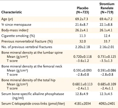
* Plus–minus values are means ±SD. There were no significant differences be-tween the groups.
† The body-mass index is the weight in kilograms divided by the square of the height in meters.
Table 1. Base-Line Characteristics of the 1442 Patients in the Intention-to-Treat
Population.* Characteristic Placebo (N=723) Strontium Ranelate (N=719) Age (yr) 69.2±7.3 69.4±7.2 Yr since menopause 21.6±8.7 22.1±8.8 Body-mass index† 26.2±4.1 26.1±4.1 Cigarette smoking (%) 11.3 12.4
Previous nonvertebral fracture (%) 32.0 33.7
No. of previous vertebral fractures 2.20±2.18 2.16±2.01
Bone mineral density at the lumbar spine Mean (g/cm2) T score 0.720±0.118 ¡3.6±1.2 0.731±0.125 ¡3.5±1.3 Bone mineral density at the femoral neck
Mean (g/cm2) T score 0.591±0.093 ¡2.8±0.8 0.591±0.086 ¡2.8±0.8 Bone mineral density of the total hip
Mean (g/cm2) T score 0.6811±0.113 ¡2.4±1.1 0.685±0.109 ¡2.4±1.1 Serum bone-specific alkaline phosphatase
(ng/ml)
12.8±4.9 12.3±4.5
s t r o n t i u m r a n e l a t e a n d v e r t e b r a l f r a c t u r e i n p o s t m e n o p a u s a l o s t e o p o r o s i s
bo group and 87.3 percent of the strontium ranelate group completed three years of follow-up. v e r t e b r a l f r a c t u r e s , b o d y h e i g h t ,
a n d n o n v e r t e b r a l f r a c t u r e s
At the end of the first year of treatment, there was a 49 percent lower risk of a new vertebral fracture in the strontium ranelate group than in the placebo group (incidence, 6.4 percent vs. 12.2 percent; rela-tive risk, 0.51; 95 percent confidence interval, 0.36 to 0.74; P<0.001), and a 52 percent lower risk of symptomatic fracture (3.1 percent vs. 6.4 percent; relative risk, 0.48; 95 percent confidence interval, 0.29 to 0.80; P=0.003). Over the entire three-year study period, the strontium ranelate group had a 41 percent lower risk of a new vertebral fracture than the placebo group (20.9 percent vs. 32.8 percent; relative risk, 0.59; 95 percent confidence interval, 0.48 to 0.73; P<0.001) (Fig. 2). On the basis of these data, 9 patients would need to be treated for three years with strontium ranelate in order to prevent 1 patient from having a vertebral fracture (95 per-cent confidence interval, 6 to 14).
Quantitative assessment confirmed by the semi-quantitative evaluation of vertebral fractures showed that 17.7 percent of patients receiving strontium ranelate for three years and 28.4 percent of patients receiving placebo had one new vertebral fracture that met the study criteria (relative risk in the stron-tium ranelate group, 0.58; 95 percent confidence interval, 0.46 to 0.73; P<0.001). The proportion of patients with more than one new vertebral fracture over the three-year period was 6.4 percent in the strontium ranelate group and 9.8 percent in the pla-cebo group (relative risk, 0.64; 95 percent confi-dence interval, 0.44 to 0.93; P=0.02). Symptomatic vertebral fractures were detected in 192 patients (11.3 percent of the strontium ranelate group and 17.4 percent of the placebo group), corresponding to a 38 percent lower risk over a period of three years in the strontium ranelate group than in the control group (relative risk, 0.62; 95 percent confidence in-terval, 0.47 to 0.83; P<0.001).
Over the three-year treatment period, fewer pa-tients had height loss of at least 1 cm in the stron-tium ranelate group (30.1 percent) than in the pla-cebo group (37.5 percent, P=0.003). Back pain was reported by 17.7 percent of the women in the stron-tium ranelate group and by 21.3 percent in the pla-cebo group (P=0.07). Nonvertebral fractures oc-curred in 234 women (112 in the strontium ranelate group and 122 in the placebo group) over the study
period (relative risk, 0.90; 95 percent confidence interval, 0.69 to 1.17).
b o n e m i n e r a l d e n s i t y , s e r u m s t r o n t i u m
l e v e l s , a n d m a r k e r s o f b o n e t u r n o v e r
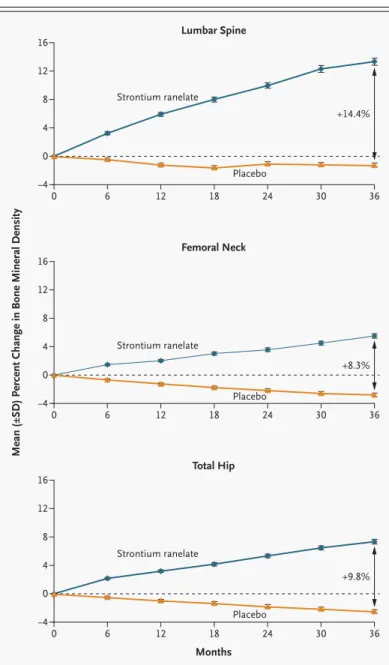
Bone mineral density was similar at base line in the two groups and increased continuously at the spine, femoral neck, and total hip in the strontium rane-late group over the three-year period, with no trend toward a plateau (Fig. 3). Over a period of three years, bone mineral density in the strontium rane-late group had increased from base line by 12.7 per-cent at the lumbar spine, 7.2 perper-cent at the femoral neck, and 8.6 percent at the total hip (P<0.001 for all three comparisons with base-line values), corre-sponding to differences between the placebo and the treatment groups at three years of 14.4 percent, 8.3 percent, and 9.8 percent, respectively. At three years, the bone mineral density at the lumbar spine, adjusted for the strontium content, showed an in-crease over the base-line value of 6.8 percent in the
Figure 2. Proportion of Patients in the Intention-to-Treat
Population Who Had One or More New Vertebral Frac-tures, Assessed According to the Semiquantitative Method.
Analysis at month 12 was restricted to patients with as-sessable radiographs at base line and at month 12 (686 patients in the strontium ranelate group and 699 in the placebo group). The relative risk of fracture in the treat-ment group at 12 months was 0.51 (95 percent confidence interval, 0.36 to 0.75; P<0.001). Analysis for the three-year period was restricted to patients with assessable radio-graphs at base line and after base line (719 patients in the strontium ranelate group and 723 in the placebo group). The relative risk of fracture over 36 months was 0.59 (95 percent confidence interval, 0.48 to 0.73; P<0.001).
Placebo Strontium ranelate
Probability of Vertebral Fracture (%)
35 40 30 10 5 15 20 25 0 12 Months 36
strontium ranelate group and a decrease of 1.3 per-cent in the placebo group (P<0.001); these changes correspond to a treatment-related increase of 8.1 percent.
At base line, the median serum strontium con-centration was 0.3 µmol per liter in both groups. At three months, the median serum strontium con-centration in the treated group had risen to 117.9
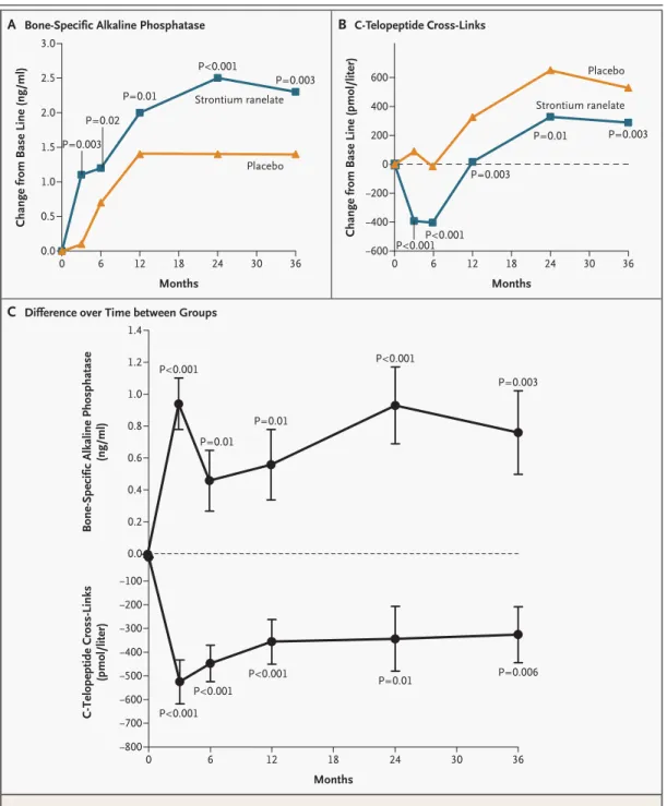
µmol per liter and then reached a plateau. At the third month of therapy, the serum concentration of bone-specific alkaline phosphatase was higher in the strontium ranelate group than in the placebo group (a treatment-related increase of 8.1 percent, P<0.001), and this difference persisted at each eval-uation during the three years (Fig. 4A and 4C). The concentration of serum C-telopeptide cross-links was lower in the strontium ranelate group than in the placebo group at month 3 (a treatment-related difference of 12.2 percent, P<0.001) and at each sub-sequent evaluation during the three years (P<0.001) (Fig. 4B and 4C).
h i s t o m o r p h o m e t r i c f e a t u r e s o f b o n e
Bone biopsies were performed at 24, 36, or 48 months in 20 consenting patients, resulting in 14 samples that could be assessed. All biopsy speci-mens consisted of lamellar bone. None of the biop-sies showed evidence of osteomalacia or any sign of a primary mineralization defect. As compared with the placebo group, the strontium ranelate group had no increase in osteoid thickness (P=0.64) or in the mineralization lag time (P=0.76) and no decrease in the mineral apposition rate (P=0.93).
a d v e r s e e v e n t s a n d m e t a b o l i c r e s u l t s
During the three years of the study, the rate of com-pliance with therapy was 83 percent in the stron-tium ranelate group and 85 percent in the placebo group. The rates of adverse events, serious adverse events, and withdrawal due to adverse events were similar in the two groups. Diarrhea was the most frequent gastrointestinal adverse event (occurring in 6.1 percent of the strontium ranelate group and 3.6 percent of the placebo group, P=0.02). How-ever, this effect disappeared after the first three months. There was a lower incidence of gastritis, as diagnosed clinically by the investigators, in the strontium ranelate group than in the placebo group (3.6 percent vs. 5.5 percent, P=0.07). Serum calci-um concentrations were lower and sercalci-um phos-phate concentrations higher in the strontium rane-late group than in the control group at month 3, with a plateau thereafter (95 percent confidence interval for the difference between the groups, ¡0.32 to ¡0.20 mg per deciliter [¡0.08 to ¡0.05 mmol per liter] for calcium and 0.25 to 0.37 mg per deciliter [0.08 to 0.12 mmol per liter] for phosphorus). There was a slight reduction in serum parathyroid hormone from the level at month 6 in both groups (decrease
Figure 3. Effects of Strontium Ranelate on Bone Mineral Density in All
Pa-tients Receiving 2 g a Day of Oral Strontium Ranelate.
P<0.001 for all comparisons, with the use of a step-down hierarchical procedure.
Lumbar Spine
Femoral Neck
Total Hip
Mean (±SD) Percent Change in Bone Mineral Density
12 8 0 16 4 ¡4 0 6 12 18 24 30 36 0 6 12 18 24 30 36 0 6 12 18 24 30 36 Placebo Strontium ranelate 12 8 0 16 4 ¡4 Strontium ranelate 12 8 0 16 4 ¡4 Months Strontium ranelate Placebo Placebo +14.4% +8.3% +9.8%
s t r o n t i u m r a n e l a t e a n d v e r t e b r a l f r a c t u r e i n p o s t m e n o p a u s a l o s t e o p o r o s i s
Figure 4. Strontium Ranelate–Induced Changes in Serum Biochemical Markers of Bone Metabolism.
Panel A shows absolute changes from base-line values in bone-specific alkaline phosphatase, Panel B shows absolute changes from base-line values in C-telopeptide cross-links, and Panel C shows differences over time in biochemical markers between the two groups. Data shown are mean (±SE) values in the strontium ranelate group minus mean val-ues in the placebo group. Comparisons were performed with analyses of covariance in which base-line valval-ues were used as covariates.
Change from Base Line (ng/ml)
2.0 2.5 1.5 1.0 0.5 0.0 0 6 12 18 24 30 36 Months Strontium ranelate Placebo 3.0
Bone-Specific Alkaline Phosphatase
(ng/ml) C-Telopeptide Cross-Links (pmol/liter) 1.0 1.2 0.8 0.6 0.4 0.2 ¡100 ¡200 ¡300 ¡400 ¡500 ¡600 ¡700 ¡800 0.0 0 6 12 18 24 30 36 Months 1.4
Change from Base Line (pmol/liter)
200 400 0 ¡200 ¡400 ¡600 0 6 12 18 24 30 36 Months Strontium ranelate Placebo 600 A B C
Bone-Specific Alkaline Phosphatase C-Telopeptide Cross-Links
Difference over Time between Groups
P=0.003 P=0.02 P=0.01 P<0.001 P<0.001 P<0.001 P<0.001 P<0.001 P<0.001 P<0.001P<0.001 P=0.003 P=0.003 P=0.01 P=0.01 P=0.01 P=0.01 P=0.003 P=0.003 P=0.006
from base-line to six-month values, ¡3.4±10.7 pg per milliliter in the strontium ranelate group and ¡1.3±15.8 pg per milliliter in the placebo group). No changes were observed for serum 25-hydroxyvi-tamin D, 1,25-dihydroxyvi25-hydroxyvi-tamin D, or calcitonin.
Serum creatine kinase concentrations in the skeletal muscle increased in some patients; with a high concentration defined as a value that was at least twice the upper limit of the normal range (145 IU per liter), high levels were detected in 3.4 per-cent of patients in the strontium ranelate group at least once during the study and in 1.8 percent of patients in the placebo group. No increase in mus-cular symptoms or other biologic abnormalities were observed. Most of the increases in creatine phosphokinase were transient, and in more than 88 percent of the patients who had high concentra-tions, the values returned to the normal range dur-ing treatment.
Prevention of fractures is the primary aim of anti-osteoporotic treatment. In the present study, stron-tium ranelate ingested daily reduced the risk of new vertebral fractures by 49 percent at one year and by 41 percent over a three-year period among post-menopausal women with osteoporosis. Although data from direct comparisons with other antios-teoporotic treatments are lacking, the reduction in the risk of vertebral fracture seems similar to the
reduction reported with alendronate (47 percent),6
5 mg of risedronate (49 percent),7 60 mg of
ralox-ifene (30 percent),8 and parathyroid hormone (65
percent after 21 months of treatment).12 The
meth-ods of assessing vertebral fractures, involving both semiquantitative and morphometric evaluations, were similar to the methods used in these other re-ports.6-8,12 Strontium ranelate also reduced the risk
of multiple vertebral fractures and the risk of symp-tomatic fractures. In this group of women, who had established osteoporosis and a history of fractures, the number needed to treat to prevent one fracture was nine.
Most antiresorptive agents prevent bone destruc-tion by reducing the rate of bone remodeling, as reflected by a decrease in both markers of bone re-sorption (more than 50 percent with bisphospho-nates and about 30 percent with raloxifene) and markers of bone formation (about 50 percent with
bisphosphonates and 20 percent with raloxifene).25
Treatment with parathyroid hormone increases both
bone formation and bone resorption.26 When
para-thyroid hormone and alendronate are combined, there is, unexpectedly, no potentiation of their
ef-fects on biochemical bone markers.27 The
mecha-nism of action of strontium ranelate is probably dif-ferent from those of these drugs. Each time the patients were evaluated during our study, bone for-mation had increased in the group assigned to stron-tium ranelate, on the basis of serum concentrations of bone-specific alkaline phosphatase, and bone re-sorption had decreased, on the basis of serum con-centrations of C-telopeptide cross-links, as com-pared with the values in the placebo group. The changes in biochemical markers of bone resorption and formation were most pronounced during the first six months; the dissociation between the bone markers was evident throughout the study. The mechanisms for the apparent dissociation between reduced bone resorption and increased bone forma-tion are not yet understood, but they probably
dif-fer from the mechanisms of current treatments.6-9
Any metal with an atomic number higher than that of calcium can be expected to influence bone
mineral density.28 In experiments, strontium in
bone correlated with strontium in plasma.29 In this
study, strontium ranelate increased bone mineral density at three years by 14.4 percent at the lumbar spine, by 8.3 percent at the femoral neck, and by 9.8 percent at the total hip, and bone strength is directly proportional to bone mineral density. Al-though data from directly comparable trials are not available, the treatment effect after adjustment for the strontium content of bone was an 8.1 percent increase in bone mineral density at the lumbar spine at 3 years, which compares favorably with a 9 per-cent increase with parathyroid hormone (20 µg) at 21 months and with the increases with other drugs (5.9 percent with risedronate, 6.2 percent with alen-dronate, and 2.6 percent with raloxifene). Moreover, this increase is consistent with the results of pre-vious phase 2 studies involving strontium rane-late.20,30 In summary, strontium ranelate given
oral-ly at a dose of 2 g daioral-ly appears to reduce the risk of vertebral fractures rapidly, effectively, and safely among postmenopausal women with osteoporosis.
Supported by Servier.
Drs. Balogh, Meunier, Nielsen, Ortolani, Reginster, Roux, See-man, and Spector report having received consulting fees from Servi-er; Drs. Meunier and Roux consulting fees from Eli Lilly; Dr. Balogh lecture fees from Lilly Hungaria; Dr. Genant consulting and lecture fees from Servier, Wyeth Laboratories, Eli Lilly, GlaxoSmithKline, Novartis, Pfizer, and Procter & Gamble and grant support from Pfi-zer, Procter & Gamble, and Eli Lilly; Dr. Ortolani consulting fees from Roche and Janssen, lecture fees from Italfarmaco, and grant
s t r o n t i u m r a n e l a t e a n d v e r t e b r a l f r a c t u r e i n p o s t m e n o p a u s a l o s t e o p o r o s i s
support from Servier, Novartis, and TEVA Pharmaceutical Indus-tries; Dr. Meunier lecture fees from Servier, Eli Lilly, Merck, Procter & Gamble, and Aventis; Dr. Reginster consulting fees from Novar-tis, Negma Laboratories, Eli Lilly, Wyeth Laboratories, Amgen, GlaxoSmithKline, Roche, Merck Pharmaceuticals, Nycomed, NPS Pharmaceuticals, and Theramax, lecture fees from Merck, Eli Lilly, Rotta Pharmaceuticals, Institut Biochimique, Novartis, Servier,
Merck, Teijin, Anelis, Theramex, Nycomed, and Novo Nordisk, and grant support from Bristol-Myers Squibb; Dr. Rizzoli consult-ing fees from Novartis and Wyeth Laboratories, lecture fees from Merck, Servier, and Aventis, and grant support from Proskalia and Servier; Dr. Roux consulting and lecture fees from Procter & Gam-ble and lecture fees from Novartis and Eli Lilly; and Dr. Spector lec-ture fees from Procter & Gamble.
a p p e n d i x
The Spinal Osteoporosis Therapeutic Intervention study investigators were as follows: Australia: J. Graham, Ashford Specialist Centre, Ash-ford; K.W. Ng, St. Vincent’s Hospital, Fitzroy; R. Prince, Sir Charles Gairdner Hospital, Nedlands; J. Prins, Princess Alexandra Hospital, Wooloongabba; E. Seeman, Austin and Repatriation Medical Centre, Heidelberg; J. Wark, Royal Melbourne Hospital, Parkville; Belgium: J.Y. Reginster, Brull Hospital, Liege; J.P. Devogelaer, Catholic University of Louvain, Brussels; J.M. Kaufman, University of Gent, Gent; F. Rae-man, Jan Palfijin Ziekenhuis, Merksem; M. Walravens, Medical Centre, Tessenderloo; Denmark: S. Pors-Nielsen, Hillerød Hospital, Hill-erød; H. Beck-Nielsen, University Hospital Osteoporosis Centre, Odense; P. Charles, Aarhus Amtssygehus, Aarhus; O.H. Sorensen, Osteoporosis Research Center, Hvidovre; France: P.J. Meunier, Edouard Herriot Hospital, Lyons; J.P. Aquino, Medical Clinic La Porte Verte, Versailles; C. Benhamou, La Madeleine Hospital, Orleans; F. Blotman, Lapeyronie Hospital, Montpellier; O. Bonidan, E. Muller Hospital, Mulhouse; P. Bourgeois, Pitié-Salpétrière Hospital, Paris; M.C. De Vernejoul Lariboisière Hospital, Paris; J. Dehais, Pellegrin-Tondu Hospi-tal, Bordeaux; P. Fardellone, Nord HospiHospi-tal, Amiens; A. Kahan, Cochin HospiHospi-tal, Paris; J.L. Kuntz, Hautepierre HospiHospi-tal, Strasbourg; C. Marcelli, Regional Hospital and University Centre, Caen; A. Prost, Hôtel Dieu Hospital, Nantes; B. Vellas, La Grave-Casselardit Hospital, Toulouse; G. Weryha, Brabois Hospital, Vandoeuvre; Germany: E.M. Lemmel, Max Grundig Clinic, Buhl; D. Felsenberg, Benjamin Franklin Clinical University, Berlin; J. Hensen, Medical Clinic 1, Erlangen; H.P. Kruse, Medical Clinical University, Hamburg; W. Schmidt, St. Josef Hospital, Bochum; J. Semler, Immanuel Hospital, Berlin; G. Stucki, Clinic for Physical Medicine and Rehabilitation, Munich; Greece: C. Phenekos, Red Cross Hospital, Athens; Hungary: A. Balogh, University of Debrecen, Medical and Health Sciences Centre, Debrecen; R. De Chatel, Semmelweis Orvostudomanyi Egyetem, Budapest; Italy: S. Ortolani, Centre Auxologico Italiano, Milan; S. Adami, Hospital and Clinical Centre of Medecine, Valeggio sul Mincio; G. Bianchi, Hospital La Colletta, Arenzano; M.L. Brandi, General Hospital Careggi, Flo-rence; D. Cucinotta, S. Orsola Malpighi Hospital, Bologna; C. Fiore, Medical Clinic, Vittorio Emanuel Hospital, Catania; C. Gennari, Insti-tute of Medicine Clinic, New General Hospital Le Scotte, Siena; G. Isaia, University of General Medicine, Turin; G. Luisetto, University In-stitute of Semeiotica Medica, Padua; R. Passariello, InIn-stitute of Radiology, La Sapienza University, Rome; M. Passeri, Parma University, Parma; G. Rovetta, E. Bruzzone Institute, Genoa; L. Tessari, San Raffaele Hospital, Milan; Poland: J.E. Badurski, Center for Osteoporosis and Osteo-Articular Diseases, Bialystok; K. Hoszowski, Medical Centre, Warsaw; R.S. Lorenc, Osteoporosis Centre of Krajowe, Warsaw; A. Sawicki, Warsawian Center of Osteoporosis and Calcium Metabolism Osteomed, Warsaw; Spain: A. Díez, El Mar Hospital, Barcelona; J.B. Cannata, General Hospital of Asturias, Oviedo; M. Díaz Curiel and A. Rapado, Jiménez Díaz Foundation, Madrid; J. Gijon and A. Torrijos, La Paz Hospital/Hospital de Rehabilitacion y Traumatologia, Madrid; J.M. Padrino, Hospital 12 de Octubre, Madrid; A. Roces Varela, Hos-pital Nuestra Sra de la Candelaria, Santa Cruz de Tenerife, Canary Islands; Switzerland: J.P. Bonjour and R. Rizzoli, Geneva University Hospi-tal, Geneva; United Kingdom: T. D. Spector, Department of Rheumatology, St. Thomas’ Hospital, London; M. Clements, North London Clin-ical Studies Centre, Mount Vernon Hospital, Northwood; D.V. Doyle, Silverthorn Centre, Chingford; P. Ryan, Medway Hospital Trust, Gillingham; I.G. Smith, Synexus Ltd. Clinical Research Centre, Manchester; R. Smith, Banbury Clinical Studies Centre, Banbury.
r e f e r e n c e s
1. Nevitt MC, Ettinger B, Black DM, et al. The association of radiographically detected vertebral fracture with back pain and func-tion: a prospective study. Ann Intern Med 1998;128:793-800.
2. Ray NF, Chan JK, Thamer M, Melton LJ III. Medical expenditures for the treatment of osteoporotic fractures in the United States in 1995: report from the National Osteoporo-sis Foundation. J Bone Miner Res 1997;12: 24-35.
3. Lindsay R, Silverman SL, Cooper C, et al. Risk of new vertebral fracture in the year fol-lowing a fracture. JAMA 2001;285:320-3.
4. Kotowicz MA, Melton LJ III, Cooper C, Atkinson EJ, O’ Fallon WM, Riggs BL. Risk of hip fracture in women with vertebral frac-ture. J Bone Miner Res 1994;9:599-605.
5. Seeman E. Pathogenesis of bone fragil-ity in women and men. Lancet 2002;359: 1841-50.
6. Black DM, Cummings SR, Karpf DB, et al. Randomised trial of effect of alendro-nate on risk of fracture in women with ex-isting vertebral fractures. Lancet 1996;348: 1535-41.
7. Reginster JY, Minne HW, Sorensen OH, et al. Randomised trial of the effects of ris-edronate on vertebral fractures in women with established postmenopausal osteoporo-sis. Osteoporos Int 2000;11:83-91.
8. Ettinger B, Black DM, Mitlak BH, et al. Reduction of vertebral fracture risk in post-menopausal women with osteoporosis treat-ed with raloxifene: results from a 3-year ran-domized clinical trial. JAMA 1999;282:637-45. [Erratum, JAMA 1999;282:2124.]
9. Lufkin EG, Wahner HW, O’Fallon WM, et al. Treatment of postmenopausal osteopo-sosis with transdermal estrogen. Ann Intern Med 1992;117:1-9.
10.Boivin GY, Chavassieux PM, Santora AC, Yates J, Meunier PJ. Alendronate increases bone strength by increasing the mean de-gree of mineralization of bone tissue in os-teoporotic women. Bone 2000;27:687-94.
11.Chavassieux PM, Arlot ME, Reda C, Wei L, Yates AJ, Meunier PJ. Histomorphometric assessment of the long-term effects of alen-dronate on bone quality and remodeling in patients with osteoporosis. J Clin Invest 1997; 100:1475-80.
12.Neer RM, Arnaud CD, Zanchetta JR, et al. Effect of parathyroid hormone (1-34) on fractures and bone mineral density in post-menopausal women with osteoporosis. N Engl J Med 2001;344:1434-41.
13.Marie PJ, Amman P, Boivin G, Rey C. Mechanisms of action and therapeutic po-tential of strontium in bone. Calcif Tissue Int 2001;69:121-9.
14.Marie PJ, Garba MT, Hott M, Miravet L. Effect of low doses of stable strontium on bone metabolism in rats. Miner Electrolyte Metab 1985;11:5-13.
15.Canalis E, Hott M, Deloffre P, Tsou-deros Y, Marie PJ. The divalent strontium salt S12911 enhances bone cell replication and bone formation in vitro. Bone 1996;18: 517-23.
16.Buehler J, Chappuis P, Saffar JL, Tsou-deros Y, Vignery A. Strontium ranelate in-hibits bone resorption while maintaining bone formation in alveolar bone in monkeys (Macaca fascicularis). Bone 2001;29:176-9.
17.Marie PJ, Hott M, Modrowski D, et al. An uncoupling agent containing strontium prevents bone loss by depressing bone
re-sorption and maintaining bone formation in estrogen-deficient rats. J Bone Miner Res 1993;8:607-15.
18.Grynpas MD, Marie PJ. Effects of low doses of strontium on bone quality and quan-tity in rats. Bone 1990;11:313-9.
19.Boivin G, Deloffre P, Perrat B, et al. Stron-tium distribution and interactions with bone mineral in monkey iliac bone after strontium salt (S 12911) administration. J Bone Miner Res 1996;9:1302-11.
20.Meunier PJ, Slosman DO, Delmas PD, et al. Strontium ranelate: dose-dependent ef-fects in established postmenopausal verte-bral osteoporosis — a 2-year randomized placebo controlled trial. J Clin Endocrinol Metab 2002;87:2060-6.
21.Genant HK, Wu CY, van Kuijk C, Nevitt MC. Vertebral fracture assessment using a semiquantitative technique. J Bone Miner Res 1993;8:1137-48.
22.Genant HK, Jergas M, Palermo L, et al.
Comparison of semiquantitative visual and quantitative morphometric assessment of prevalent and incident vertebral fractures in osteoporosis. J Bone Miner Res 1996;11: 984-96.
23.Wu CY, Li J, Jergas M, Genant HK. Com-parison of semiquantitative and quantitative techniques for the assessment of prevalent and incident vertebral fractures. Osteoporos Int 1995;5:354-70.
24.Slosman DO, Provvedini DM, Meunier PJ, et al. The use of different dual X-ray ab-sorptiometry brands in a multicenter clini-cal trial. J Clin Densitom 1999;2:37-44.
25.Delmas PD. Markers of bone turnover for monitoring treatment of osteoporosis with antiresorptive drugs. Osteoporos Int 2000; 11:Suppl 6:S66-S76.
26.Lindsay R, Nieves J, Formica C, et al. Ran-domised controlled study of effect of para-thyroid hormone on vertebral-bone mass and fracture incidence among postmenopausal
women on oestrogen with osteoporosis. Lan-cet 1997;350:550-5.
27.Black DM, Greenspan SL, Ensrud KE, et al. The effects of parathyroid hormone and alendronate alone or in combination in postmenopausal osteoporosis. N Engl J Med 2003;349:1207-15.
28.Nielsen SP, Slosman D, Sorensen OH, et al. Influence of strontium on bone miner-al density and bone minerminer-al content measure-ments by dual X-ray absorptiormetry. J Clin Densitom 1999;2:371-9.
29.Dahl SG, Allain P, Marie PJ, et al. Incor-poration and distribution of strontium in bone. Bone 2001;28:446-53.
30.Reginster JY, Deroisy R, Dougados M, Jupsin I, Colette J, Roux C. Prevention of ear-ly postmenopausal bone loss by strontium ranelate: the randomized, two-year, double-masked, dose-ranging, placebo-controlled PREVOS trial. Osteoporos Int 2002;13:925-31.