THE PENICILLIN RECEPTOR IN STREPTOMYCES
*
J.-M. Ghuysen, M. Leyh-Bouille, J.-M. FrBre,I.
Dusart, andA. Marquet
Service de Microbiologie, FacultP de Mkdecine Institirt de Botanique, Universite' de LiPge
Liige, Belgiutn
H. R. Perkins and M. Nieto
t
National Institute for Medical Research Mill Hill, London N.W.7 I A A , EnglandINTRODUCTION
Penicillin kills bacteria by suppressing or decreasing the efficiency of the membrane-bound transpeptidase, which during the last steps of the wall synthesis catalyzes cross-linking between the peptide units of the nascent peptidoglycan and makes the polymer insoluble.l, 2 In addition to this specific receptor, other penicillin-binding sites also occur within the bacterial membrane~.3-~ These sites, or at least most of them, seem to be irrelevant as far as peptidoglycan synthesis is concerned.4 Although involved in antibiotic specificity, they do not appear to be the killing target of penicillin.
At present, no transpeptidase has been isolated from bacterial membranes and characterized. For a long time, the technical limitation to such an achieve- ment has resided in the lack of a suitable assay for transpeptidase activity. Cell-free particulate multienzyme preparations obtained from various bacteria were shown to catalyze the in vitro utilization of the nucleotide precursors UDP-N-acetylglucosamine and UDP-N-acetylmuramyl pentapeptide for the entire sequence of peptidoglycan synthesis, which includes the peptide cross- linking.g-15 These assays, however, were devised in such a way that they did not allow measurement of the transpeptidation reaction per se. Another ap- proach was undertaken through a joint effort between our laboratories and was based on the development of well-defined systems of peptide donors and ac- ceptors that could be used by bacterial transpeptidases for transpeptidation reactions and, hence, would allow these enzymes to be operative and tested independently of the preceding biosynthetic sequential reactions. Streptornyces sp were chosen as a model, because they had the property, probably unique in the bacterial world, of spontaneously excreting an enzyme that appeared to be a soluble form of the membrane-bound transpeptidase.
*
This work was supported in part by grants (to J.M.G.) from the Fonds de la Recherche Fondamentale Collective (no. 1000),
Brussels, Belgium and the Jnstitut pour 1'Encouragement de la Recherche Scientifique dans 1'Industrie et I'Agriculture(nos. 1699 and 2013), Brussels, Belgium.
P Present address: Centro de Investigaciones Biologicas, Instituto de Biologia Celular, Madrid 6, Spain.
Ghuysen et al. : Streptoniyces Penicillin Receptor 237 THE DD-CARBOXYPEPTIDASE-TRANSPEPTIDASE SYSTEM IN
Streptomyces STRAIN R61 Structure of the Wall Peptidoglycan
In Streptomyces R61 1'; (as well as in strains
K11
li and albus G 1s and inClostridium perfringens I $ ) , the wall peptidoglycan is composed of L-alanyl-D-
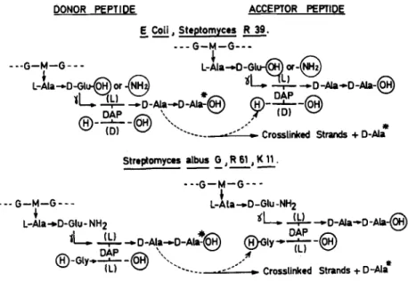
isoglutaminyl-( L,) -LL-diaminopimelyl-( L ,) -D-alanine tetrapeptides that are in- terlinked through an additional glycine residue that extends from the C-terminal D-alanine of one peptide to the (L,) amino group of LL-diaminopimelic acid of another peptide. Because of this structure, the transpeptidation reaction in Streptomyces R61 must occur as shown in FIGURE 1.
DONOR PEPTIDE ACCEPTOR PEPTIDE & Coil, Steptomyces
R.
--
FIGURE 1. Transpeptidation reaction in various bacteria, G, N-acetylglucosamine;
M, N-acetylmuramic acid; DAP, diaminopimelic acid. The a-carboxyl group of D-
glutamic acid is free in Esclierichia coli and is substituted by an amide in Strepto-
rnyces R39.
Ac,-L-Lys-D-Ala-D-A In and Gly-Gly as Substrates f o r Transpeptidation by Streptomyces R61
Streptomyces R61 possesses a transpeptidase activity that catalyzes the trans- fer of Na,NE-diacetyl-i.-lysyl-D-alanine from the tripeptide donor N=,NE-diacetyl- L-lysyl-D-alanyl-D-alanine to the dipeptide acceptor glycylglycine, which yields free D-alanine and the tetrapeptide Na,NE-diacetyl-L-lysyl-D-alanylglycylglycine. This transpeptidase activity was located within the plasma membrane *l but was also excreted in the external medium during growth.16 With the membrane- bound enzyme, liberation of D-alanine from the tripeptide donor was strictly
238
AnnalsNew
York Academy of Sciencesdependent on the presence of the acceptor glycylglycine in the reaction mixture. The culture filtrate, however, could also hydrolyze the tripeptide donor alone into D-alanine and the dipeptide Na.N'-diacetyl-L-lysyl-D-alanine; that is, it exhibited both a m-carboxypeptidase activity and a transpeptidase activity. The only difference between the two reactions was that in transpeptidation the enzyme catalyzed a reaction with the free amino group of glycylglycine to complete peptide cross-linking, whereas in hydrolysis the enzyme catalyzed a reaction with water:
Ac,-L-Lys-D-Ala-D-Ala
+
Gly-Gly + D-Ala+
Ac2-L-Lys-D-Ala-Gly-Gly,
Ac,-L-Lys-D-Ala-D-Ala+
H,O + D-Ala+
Ac,-L-Lys-D-Ala.It can be argued that integration of the enzyme in the hydrophobic environment of the membrane only allowed transpeptidation to occur, whereas water was accessible to the exocellular enzyme.
Characterization of the R61 Membrane-Bound Transpeptidase that Acts on Ace-L-Lys-n-Ala-DAla and Gly-Gly as the
Physiological Transpeptidase
The in vivo susceptibility of Streptomyces R61 toward a series of penicillins and cephalosporins (LD,, values) was tested under conditions in which j3- lactamase activity was not involved. The in vivo susceptibility was found to correlate well with the in vitro susceptibility of the membrane-bound trans- peptidase toward the same antibiotics 21, z 2 (ID,, values, TABLE 1 ). Membranes isolated from Streptornyces R61 bound a maximum of 25 pmoles of [14C]benzyl- penicillin per milligram of protein (as determined after treatment of the mem- branes with a large excess of [14C]benzylpenicillin and removal of the unbound antibiotic by washings with buffer). Pretreatment of the membrane by non- radioactive 6-aminopenicillanic acid, benzylpenicillin, ampicillin, or carbenicillin at concentrations equivalent to those that reduced by 50% the activity of the rnembrane-bound transpeptidase (i.e., the ID,, values) inhibited by 50% the uptake of [14C]benzylpenicillin.3i
The above experiments did not establish whether the transpeptidase was the only binding site for penicillin. They demonstrated, however, that penicillins were competing for the same membrane component(s) and that the membrane- bound transpeptidase, as revealed by using the above tripeptide donor and dipeptide acceptor for transpeptidation reactions, was indeed the killing target of penicillin. Na,NE-diacetyl-L-lysyl-D-alanyl-D-alanine was, therefore, an analog of the donor site, and glycylglycine was an analog of the glycyl-Lr.-diamino- pimelyl acceptor site involved in transpeptidation in vivo (FIGURE 1 ) . One should also note that both the cross-linking accomplished by transpeptidation from the artificial donor-acceptor system and the cross-linking produced by transpeptidation in vivo resulted in the synthesis of the same D-alanylglycine linkage in an endo position (FIGURE 1 )
.
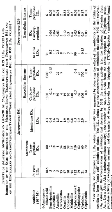
TABLE 1 EFFECTS OF &LACTAM ANTl8lOTICS ON GROWTH OF strepton2yces R61 AND R39 (LDjo VALUES) AND INHIBITION BY THE SAME ANTIBIOTICS OF THE MEMBRANE-BOUND TRANSPEPTIDASE FROM Streptomyces R61 AND OF THE EXOCELULLAR DD-CARBOXYPEPTIDASE-TRANSPEPTIDASE FROM Strepfonlyces R61 AND R39 (IDs, VALUES)
:'
Antibiotic (10% M) 6-Aminopeni- cillanic acid Benzylpenicillin Penicillin VAmpicillin Carbenicillin Oxacillin Cloxacillin Methicillin Cephalosporin
C
Cephaloglycine Cephalexin Cephalothin
Streptomyces R6 1 E xo ce ll ul ar E nz ym e T ra ns - M em br an e ID, Carbow- Trans- Membrane In Vivo peptidase peptidase peptidase LDa ID, LDm ID, ID, 18.5 80 5 2 5 18 5 8 33 34 47 88
so
39 44 35 112 510 29 62 21 60 14 70 4.3 0.4 3.6 1.5 1 1.9 0.8 0.8 4.5 2.2 2.9 5 1300 1300 0. 12 0. 15 2 30 22 6 9 19 70 140 0.5 0.4 66 76 600 500 0.8 0.8-_
Streptomyces R39 E xo ce ll ul ar E nz ym e ID, Trans- In Vivo Carboxy- peptidase LD, peptidase ID;, 51 0.7 1.5 2.5 8 7 5 7 90 1 0.15 1 5 0.04 0.04 0.03 2 0.12 0.3 0.4 0.05 0.05 0.3 0.03 6 0.07 0.05 0.04 2 0.33 0.46 0.5 0.06 0.07 0.17 0.05 "c
*
For details, see Reference 21. LD, values: sensitivity was measured by observing the effect of th e an ti bi ot ic s on the ability of conidia to form "single cell" colonies on plates. The LDm values give th e co nc en tr at io ns of antibiotics that allow 50% cell survival. ID, values give the concentrations of antibiotics that decrease by 50% the en zy m e ac ti vi ty (i.e., th e li be ra ti on of palanine from Acz-L-Lys-D-Ala-pAla) (c ar bo xy pe pt id as e ac ti vi ty ), the transfer of Aca-L-Lys-D-Ala from the ab ov e tr ip ep ti de to ['%]glycine (trans- peptidase activity of exocellular en zy m es ), a nd the transfer of Acz-L-Lys-D-Ala from tripeptide to ~ 'C lg ly cy lg ly ci ne (m em br an e-bo un d tr an sp ep ti da se a ct iv it y) . h) W a240
Annals New
York
Academy
of Sciences
Characterization of the Exocellirlar R61 Transpeptidase that Acts on Ac,-L-Lys-D-Ala-D-Ala and Gly-Gly as a Soluble Form
of the Membrane-Bound Transpeptidase
Urea ( 2 M, in the presence of 10 mM ethylenediaminetetraacetate) solu- bilized the membrane-bound transpeptidase. Strikingly, the solubilized prepara- tion exhibited carboxypeptidase activity, in addition to the transpeptidase ac- t i ~ i t y . ~ ~ The enzyme released from the membrane has not yet been isolated, but the exocellular enzyme was purified to protein homogeneity.23 It consisted of a single polypeptide chain, with a molecular weight of 38,000 and an isoelectric point at pH 4.7. The acid residues (20% ) outnumbered the basic ones (5% )
.
The protein contained about three half-cystine residues out of a total of 350 amino acid residues. Approximately 50% of the residues were nonpolar. The turnover number was 3500 molecules of D-alanine liberated from Na,NE-diacetyl- L-lysyl-D-alanybalanine per molecule of enzyme per minute [carboxypeptidase assay, in 0.005 M phosphate buffer (pH 7.5)].
As
observed with the urea- treated membrane enzyme, the purified exocellular enzyme was shown to function either as carboxypeptidase or as transpeptidase; this depended upon the availability of nucleophiles (H,O or Gly-Gly or other NH,-R).24-20 The pro- portion of the exocellular enzyme activity that could be channeled into the transpeptidation and the hydrolysis pathways depended upon the environmental conditions.2G Transpeptidation was increased and hydrolysis was decreased by increasing the pH of the reaction mixture and the concentration of acceptor in it (within certain limits). Replacement of part of the water of the reaction mixture by a solvent of low polarity [such as a mixture of water, ethylene glycol, and glycerol, 30:45:25 (v:v:v) ] preferentially decreased the carboxypeptidase activity of the enzyme, so that transpeptidation then largely superseded hy- drolysis. The specificity profile of the exocellular enzyme for acceptors (with a,c-Ac,-L-Lys-D-Ala-D-Ala as donor) (TABLE 2 ) 25 closely resembled that of themembrane-bound enzyme.” Finally, irrespective of the penicillins and cepha- losporins used, inhibition of the transpeptidase activity of the exocellular enzyme always occurred at those antibiotic concentrations that inhibited its carboxy- peptidase activity 21 (TABLE 1 )
.
As
a whole, the above experiments strongly suggested that the exocellular transpeptidase-DD-carboxypeptidase was a soluble form of the “physiological” membrane-bound transpeptidase. There were, however, quantitative differences between the two enzymes, especially with respect to their susceptibility to8-
lactam antibiotics (TABLE 1). It could be argued that the enzyme conformation in the hydrophobic environment of the membrane was unlike that in an aqueous solvent and that the observed dissimilarities resulted from a change in orientation of one or more amino acid residues in or near the catalytic center(s) and/or penicillin binding site.Kinetics of Concotnitant Transfer and Hydrolysis Reactions Catalyzed by the Exocellular R61 Enzyme
From a theoretical analysis of the kinetic parameters of the various possible enzyme mechanisms in which bimolecular transfer reactions occur concomi- tantly with the hydrolysis of the donor molecule, it appeared that the ratios v,/ vHy (T, transpeptidation; Hy, hydrolysis) versus the concentration of the
Ghuysen et al. : Streptomyces Penicillin Receptor 24 1 donor [D] and that of the acceptor [A] are the most useful parameters for making a choice of mechanism." If the reactions occur randomly or proceed through an ordered mechanism in which the donor binds first to the enzyme, the ratio vT/vIrr is independent of [D]. With mechanisms in which the acceptor
TABLE 2
Streptomyces R61, K11, R39, AND albiis G
TRANSPEPTIDATION BY EXOCELLULAR DD-CARBOXYPEPTIDASES FROM
Transpeptidation (% )
Acceutor:Donor
Molar Ratio -i- R61 K11 R39 S. albris G a-Amino Acids ["Clgl ycine o-["C]alanine ~-["C]alanine rneso-[3H]diaminopimelic acid
*
Mono["C]acetyl-~~- diaminopimelic acid Diueutides 1 : l 10: 1 1:1 10: 1 10: 1 1:l 10: 1 I : l 13.7 48 45 30 0 7.5 50 35 0 0 0 16.0 18.2 7.4 48 45 0 0 i-(~]glycylg~ycine 1:l 18.4 0 10: 1 0 0 Glycyl-~-["C]alanine 1 : l 3.6 4.0 0 Glycyl-~-["C]alanine 1 : l 25.5 24.6 0 ~-Glycyl-a-acetyl-~-[SH]lysine 1:l 18.2 0 0 a-GIy~yl-a'-['~C]acetyl- 0.56: 1 4.8 0 0 LL-diaminopimelic acid 4.1 : 1 2.7 0 0 D-Alanyl [14C]glycine 1 : l 3.6 3.4 0 ~-Alanyl-~-['~C]alanine 1 : l 2.6 2.1 0 ~-Alanyl-~-["C]alanine 1:l 0.4 0 ~-Alanyl-~-["C]alanine 1:l 0.2 0.2 0*' The D center of meso-diaminopimelic acid was the one that acted as a transpepti-
dation ac~eptor.'~
t The donor was diacetyl-L-lysyl-D-alanyl-D-atanine, 50 nmol, in a final volume of 30pl. Incubation: 1 hr, 37" C. The values represent the percentage of the donor converted to the transpeptidation product AcrL-Lys-D-Ala-acceptor. For details, see
Reference 25.
binds first to the enzyme, the ratio v,IvH, is a function of [D], according to the equation
vrr
-
a+b[DJ---
'HY c+
d[D] 'where a, b, c, and d are constants. Irrespective of the mechanisms involved in the reaction, the ratio v,/v,, is directly proportional to [A], except if an additional acceptor binding site occurs on the enzyme, which leads to the formation of an [enzyme-acceptor,-donor] complex nonproductive for trans- peptidation and an [enzyme-H20-acceptor-donor] complex that remains pro-
242
VT30
-
vHY 21:U
Annals New York Academy of Sciences
4
A = mcso-DAP
I
A=
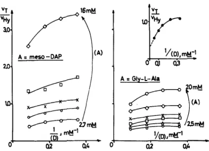
Gly-L-MaFIGURE 2. Concomitant transfer and hydrolysis reactions catalyzed by the exocel- lular R61 enzyme. Plots of vT/vHy vs 1/[D] for various concentrations of acceptor. Tri- peptide donor and acceptor were incubated at 37" C in 4 rnM sodium phosphate buf- fer (pH 7.2) (final volume=35 1.1) with R61 enzyme. With Gly-L-Ala, 125 units of enzyme were used. With meso-diaminopimelic acid, 250 units were used, and the reaction mixture was 32 mM in NaCI. Maximal utilization of the tripeptide donor was 20-2596. The reaction rates were similar with both acceptors and were linear with time. Concentrations of mesodiaminopimelic acid were:
0
,
2.7; 0, 4; X,5.3; U, 8; and
0,
16 mM. Concentrations of Gly-L-Ala were:0,
2.5; A , 3.5;m,
5;
x,
6.7;0,
10; and0
, 20 mM. For the curve enclosed in the right upper part of the Figure, the highest concentration of tripeptide donor utilized was increased up to 19.3 mM; Gly-L-Ala (20 mM) was incubated with 80 units of enzyme for 80 min. (From Frkre et aLX By permission of The Biochemical Journal.)ductive for hydrolysis. In the latter case, the ratio vT/vHY is a function of [A], according to the equation
e tA1
vT
-
V H ~
-
/[A1+
gvHy
=-+g,
f
where e, f, and g are also constants.1.e.. ~
VT e e[Al
When Na,NE-diacetyl-~-lysyl-~-alanyl-~-alanine (i.e., the donor) and either rneso-diaminopimelic acid or glycyl-L-alanine (i.e., the acceptor) were exposed
to the exocellular R61 enzyme, initial rate measurements showed that both
acceptors behaved as noncompetitive inhibitors of the hydrolysis reaction.26
Furthermore, the ratios vT/vHY were not independent of [D] but significantly
decreased as [D] increased
(FIGURE
2), which suggested an ordered pathwayGhuysen et al. : Streptomyces Penicillin Receptor 243 +D
YEA
-
EAD + transpeptidation +A/
E\
+H,O +DEH,O ___* EH,OD ___b hydrolysis.
\
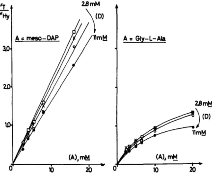
At any concentration of tripeptide donor, the ratio vT/ vIig was directly propor- tional to the concentration of the acceptor meso-diaminopimelic acid (FIGURE 3). In marked contrast, high concentrations of glycyl-L-alanine inhibited the transpeptidation pathway (FIGURE 3), in a way that fitted the equation
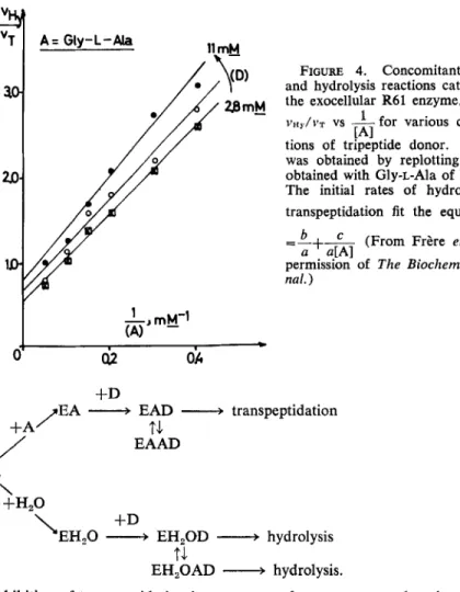
(FIGURE 4) which suggested a more complex mechanism:
A t mcs0-0AP
FIGURE 3. Concomitant transfer and hydrolysis reactions catalyzed by the exocel- lular R61 enzyme. Plots of V T / V H ~ vs [A] for various concentrations of tripeptide donor. For conditions, see FIGURE 2. Concentrations of acceptor (either meso- diaminopimelic acid or Gly-L-Ala were:
,O,
2.8;x,
3.7;0,
5.5; and 0, 11 mM. (From Frhe et al.” By permission of The Biochemical Journal.)244
Annals New
York Academy of Sciences
FIGURE 4. Concomitant transfer and hydrolysis reactions catalyzed by the exocellular R61 enzyme. Plots of
1
vH,./vT vs ~ for various concentra-
tions of tripeptide donor. FIGURE 4 was obtained by replotting the data obtained with Gly-L-Ala of FIGURE 3. The initial rates of hydrolysis and transpeptidation fit the equation 5 =-+L b (From Frkre et al.” By
a a[Al
permission of The Biochemical Joirr-
nal.) [A1 V T +D EAD --+ transpeptidation EAAD
FA
-
t.l
E\
+H,O +D\EH,O + EH,OD
--+
hydrolysist-l
EH,OAD 4 hydrolysis.
Inhibition of transpeptidation by an excess of acceptors was also observed with peptides other than glycyl-L-alanine (legend of TABLE 2) and especially with the natural peptide that undergoes transpeptidation in Streptomyces R61, that is, L-alanyl-D-isoglutaminyl-( L1) ;glycyl-( L ~ ) ;LL-diaminopimelic It is pos- sible that this feature reflects a control mechanism involved in the functioning of the enzyme in vivo. In the above proposed mechanisms, H,O was considered as an alternative nucleophile acceptor. One should note, however, that the kinetics were not incompatible with nonsymmetrical pathways, in which the acceptor would bind first to the enzyme in the transpeptidation reaction, whereas the donor would bind first to the enzyme in the hydrolysis reaction.
The Exocellular R61 Enzyme as a Model for Carboxypeptidase Activity In carboxypeptidase assays [peptide donor: -R,-R,-R, (OH)
+
H,O + -R,-R,(OH)+
R,], the enzyme exhibited a considerable specificity for pcptides that ended in the tripeptide sequence L-R,-D-Ala-D(OH) and for theGhuysen et al. : Streptornyces Penicillin Receptor 245 occurrence of a long aliphatic side chain at the L-R, position 16, 2s (TABLE 3).
The C-terminal o-residue was preferentially D-alanine. Peptides that were good substrates (such as Na,N~-diacetyl-L-lysy1-Balanyl-D-alanine) had
K,,,
values (10-15 mM) as high as the poor substrates, but the good substrates had much higher V,,, values. The enzyme thus appeared to possess a binding surface that was not very specific, as it was even able to bind molecules that were only remotely analogous to the substrate. It was suggested that the alignment of the catalytic groups induced on binding would be incorrect or unfavorable for poor substrates, which would thus decrease the V,,,,, greatly or even prevent enzyme action.The carboxypeptidase activity of the enzyme was inhibited by peptide ana- logs of the standard substrate Na,l\;e-diacetyl-L-lysyl-D-alanyl-~alanine 2o
(TABLE 4). Kinetically, the inhibition by acetyl-D-alanyl-Daspartic acid was competitive. These studies (TABLES 3 & 4) indicated that residues R, and
R,
were mainly concerned with the binding to the enzyme, whereas size, shape, and charge on residue L-R, appeared to be decisive for enzymatic activity. Acetyl-racemic-cyclodiaminoadipic acid was an inhibitor (TABLE 4), and its binding to the enzyme was stronger than that of acetyl-D-alanyl-Dalanine (as judged by the inhibition caused by both of them), which suggested that the enzyme combined only with peptides that have a cis configuration in the C-terminal amide linkage. A possible sequence of events that lead to hydrolysis
TABLE 3
INFLUENCE OF THE LENGTH, SIZE, AND CHARGE OF THE SIDE CHAIN
OF THE L-R, RESIDUE ON CARBOXYPEPTIDASE ACTIVITY
C-TERMINAL D-ALANINE) :Y.
OF EXOCELLULAR s f ~ ~ p t O ~ ? t y C C S ENZYMES (RELEASE OF THE
Activity (% ) t
R6 1 KI 1 R39 S. albris G Substrates i. Enzyme Enzyme Enzyme Enzyme
1. Ac-L-Ala-D-Ala-D-Ala 1.4 1.2 0.2 0.8 2. R'-L-homoSer-D-Ala-o-Ala 3 2 6.6 5 3. N",NY-Ac,-~-DAB I-D-Ala-o-AIa 8.5 9 15.5 55 4. N",Na-Ac2-L-Orn-o-Ala-D-Ala 45 55 83 55 5. N",N'-Ac.-L-Lys-D-Ala-D-Ala 100 100 100 100 6. N"-Ac-L-Lys-D-Ala-D-Ala 0.5 0.3 730 1 8. R'-L-Lys-D-Ala-D-Ala 80 100 340 11 7. Rb(L)-meso-DAP-( L)-D-Ala-D-Ala 0.5 1 400 8 Gly$
*
For details, see Ref. 16, 17, 28 and 32.I- R" = UDP-MurNAc-Gly-D-Glu-7; R" = UDP-MurNAc-L-Ala-D-Glur; R' =N"- [~-1,4-GlcNAc-MurNAc-~-Ala-~-Glu(NH~)~]. Peptides 1-5 were compared on the basis of nequiv of D-Ala liberated per milligram of enzyme per hour at 37" C (pep- tide concentration: 0.45 mM. Peptides 5-8 were compared on the basis of enzyme efficiency (i.e., V,,,,,x,Km).
$ Turnover numbers (molecule D-Ala released/min/molecule enzyme) for standard peptide Ac,-L-Lys-D-Ala-D-Ala
=
3500 for purified R61 enzyme (MW 38,000)" and 850 for purified R39 enzyme (MW 55,000).::'246
Annals
New York Academy of
Sciences TABLE 4ACTIVXTY OF THE EXOCELLULAR R61 AND S. albus G ENZYMES IN THE PRESENCE
OF INHIBITORS (CARBOXYPEPTIDASE ASSAY)
*
Molar
Ratio % Inhibition Inhibitor
Activity as ___ R61 S. albrcs G Compound Substrate % Substrate Enzyme Enzyme Ace-~-Lys-~-Ala-~-Ala Ac-D-Ala-D- Ala Ac-D-Ala-D-Glu ae-Suc*-L-Lys-D-Ala-D-Glu Ac-Gly-~-Ala-~-Glu Ac-D- Ala-D-Asp Ac*-L-Lys-D-Glu-D-Ala a- Ac-L-Lys-D-Glu-D-Ala Acet yl-racemic-cyclodiaminoadipic acid
*
For details, see Reference 29.100 0- 1 0 0 0 0 0 0 0 10 10 10 10-12 10-11 14 11.6 17 0 10 25 3 2-46 25 38 34-36 33-35 26 0 10-20 88 88-96 84 59 87 67 52
was According to the model, the enzyme would select the small proportion of cis isomer, hence displacing the configuration equilibrium. The free enthalpy of binding would be mainly provided by the interaction of the two C-terminal residues, whereas the side chain of the L-R, residue would induce a conformational change in the enzyme that, in turn, would cause the previously cis-amide linkage to adopt a configuration intermediate between cis and trans and to thus lose all double-bond character. Subsequently, cleavage of the C-terminal dipeptide bond would occur with the help of catalytic groups of the active site thus formed in the enzyme. The conformational change in the enzyme together with the interactions between enzyme groups and substrate would have to supply an energy of about 30-40 kJ1mol (energy barrier between cis and trans configurations), According to this model, distortion of the amide bond only occurs after the substrate is bound to the enzyme and as a conse- quence of the binding. Conversely, the change in the enzyme only occurs in the presence of bound substrate.
The Exocellular R61 Enzyme as a Model for Transfer Reaction In transpeptidation reactions (Ac,-L-Lys-DAla-D-Ala
+
acceptor + DAla+
Ac,-L-Lys-D-Ala-acceptor) that occur in competition with the unavoidable hydrolysis of the tripeptide donor ( Ac,-L-Lys-D-Ala-D-Ala+
H,O + Ac,-L-L~s- D-Ala+
D-Ala), the enzyme utilized, with varying efficiency, a wide range of compounds (TABLES 2 & S), such as glycine, D-amino acids (but not L-amino acids), peptides with N-terminal glycine or Balanine, o-amino acids, amino- hexuronic acids, 6-aminopenicillanic acid, and ~-cycloserine.~~TABLE 2 shows that of the dipeptides that function as acceptors the most effective were those with N-terminal glycine. This sequence resembles that found in the cross-links of corresponding peptidoglycan. With the most effective acceptor, glycyl-L-alanine, at an acceptor:donor ratio of 1 : 1, 25% of the donor
Ghuysen et al. : Streptomyces Penicillin Receptor 247 was converted to transpeptidation product. Replacement of the glycine residue by L-alanine prevented acceptor function. Certain substances that were not glycyl peptides and had amino groups that were not at pasymmetric centers were, however, able to function as acceptors (TABLE 5). For instance, a-amino acids were recognized by the enzyme; the shortest (C,) and longest (C,) examples studied were more effective than the intermediate chain lengths. Simple amines were not tested as acceptors, but glucosamine did not function. The spacing between the amino group and the carboxyl group was certainly not the only factor in determining the effectiveness of an acceptor, because, apart from the fact that L symmetry at the amino terminal prevented action,
glycylglycine was a much better acceptor than 5-aminovaleric acid, which has about the same separation between the free carboxyl and amino groups.
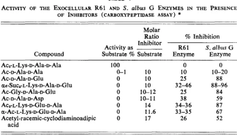
Compounds that were not simple peptides or amino acids also functioned as acceptors. Among these were the lactams of nieso or racemic diaminoadipic acids, which in a sense are fixed-conformation analogs of Dalanyl-L-alanine and of D-alanyl-D-alanine, respectively (on the reasonable assumption that molecules with the amino function at the L asymmetric center were in neither case accep
tors).25 The conformation of the diaminoadipic acid lactam is not known, but representations of possible conformers deduced from relatively strainless ar- rangements of atomic models are shown in FIGURE 5. The diametrical arrange-
w P 00 TABLE 5 COMPOUNDS OTHER THAN PEPTIDES AS ACCEPTORS FOR TRANSPEPTIIDATION BY EXWELLULAR DD-CARBOXYPEPTIDASES- TRANSPEPTIDASES FROM strCpfOi?1yCCS R61 AND R39 '$ ?
2
Acceptor Diaminoadipic Acids Lactam of nieso-diaminoadipic acid meso-Diaminoadipic acid Lactam of racemic (DD+LL) diamino- adipic acid Racemic (DD+LL) diaminoadipic acid A m in o S ug ar s 2-Amino-2-deoxy-~-glucuronic acid 2-Amino-2-deoxy-~-galacturonic acid 2-~ino-2-deoxy-~-glucose 2-Amino-3-0-(~-1 '-carboxyethyl) - 2-de ox y-~ -g lu co se (m ur am ic acid) 1 Acceptor Acceptor: Concen- Donor Enzyme tr at io n M ol ar Source (mM) Ratio (strain) 9.3 6.7 13.4 8.3 8.3 8.3 8.3 8.3 2.8 t R6 1 R39 4 R6 1 R39 4" R6 I R39 2.5 t R6 1 R39 5 R6 I R39 5 R6 1 R39 5 R6 1 R39 R6 1 5 R39 E. v) Percentage of Total R ad io ac ti vi ty i n Ratio of Transpep-3
Transpep- Hydrol- tidation 8 tidation ysis Residual toHy- 4ii
Product Product Donor drol ysis 12.3 7.2 2.5 1.1 15.2 5.6 54.4 63.0 8.1 18.5 0 0 0 0 0 70.5 17.2 0.17R
86.5 6.3 0.08 DJ 76 .8 20 .7 0.03 91.5 7.4 0.01F
64 .7 20 .1 0. 23SL
E5?
87 .3 7. 1 0.06 12.0 30.3 4.8 17.2 18.3 3.8 70.9 21 .0 0. 11 48.5 33.0 0.38 c) %w-Amino Acids 3-Aminopropionic acid 4-A m in ob ut yr ic a ci d 5-A m in ov al er ic a ci d 6-Aminohexanoic acid 6-Amino-2-acetamido-hexanoic acid (a- acetyl-L-1 ysine ) Antibiotics 6-Aminopenicillanic acid D-Cycloserine 8.3 8.3 8.3 8.3 8.3 0 1.67 8.3 0 1.67 8.3 0 1.67 8.3 16.7 16.7 5 R6 1 R3 9 5 R6 1 R3 9 5 R61 R39 5 R6 1 R39 R6 I 5 R3 9 0 R6 1 1 5 0 R39 1 5 0 R61 1 5 10 10 R3 9 6.1 0 3.7 0 2.2 0 5.4 0 2.4 0 0 1.3 2.5 0 0 0 0 28.0 65.5 75.9 16.7 77.5 74.6 76.9 73.0 77.0 59. I 26.2 56.2 22.0 11.4 70.7 16.4 0.08 21.7 0.05 20.9 0.03 ~
9
21.6 0.07 Gz
0 3 20.6 0.03E
Q F..
18.0 39.6 0.02 71.3 0.10 Ic) 9 .O m 71.0x
99.2x
20.02
15.8 0.50 12 .5 2. 98 cd 12.7 6.7 CDe.
12.6 0.24 -7s
h..
e. e-
*
From Perkins et al." i Conditions as i n TABLE 2. The donor was di['lC]acety~-~-~ysyI-~-a~any~-D-a~anine. Hydrolysis pr od uc t re fe rs to di["C]acetyl-L-lysyl- $ Calculated for the proportion of the compound that has a free amino group on an asymmetric D center, on the assumption that 5'p
CD 0 n, malanine. For details, se e R ef er en ce 25. transpeptidation will not occur at a n L center.2
h, P W250 Annals New York
Academy
of Sciences
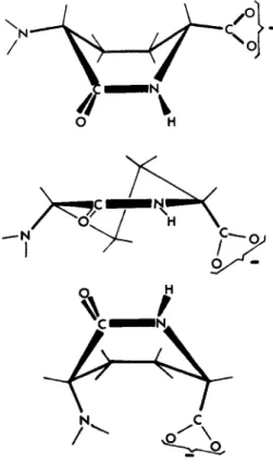
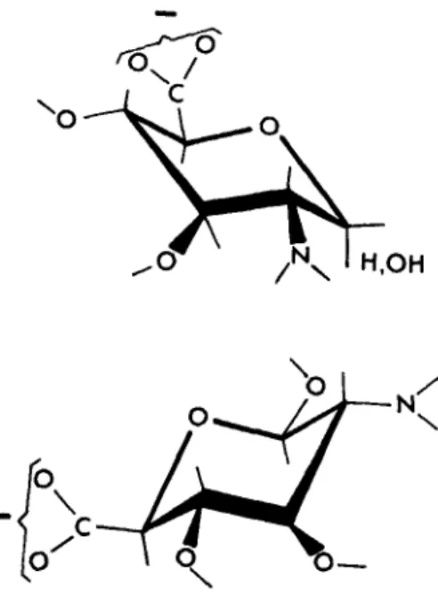
ment of the carboxyl and amino groups around a six-membered ring at once suggested an analogy with aminohexuronic acids. Again, the actual conforma- tion of these compounds has not been determined, but FIGURE 6 shows D-
glycosaminuronic acid in the C, conformation of Reeves, as found for salts of /3-Pglucopyranuronic acid.”; The lower diagram gives the same conformation drawn to bring the amino and carboxyl groups into positions comparable to those in FIGURE 5. In fact, glucosaminuronic acid and galactosaminuronic acid were both good acceptors for transpeptidation by the soluble enzyme from strain R61 (TABLE 5 ) . Ultimately, these observations must fit into any definition of the acceptor site for this enzyme.
FIGURE 6. PGlucosaminuronic acid.
Another molecule of fixed conformation with amino and carboxyl groups separated by about the same distance is 6-aminopenicillanic acid, and this too functioned as a rather poor acceptor. Evidently, this antibiotic could occupy the acceptor site on the R61 enzyme; but, of course, the penicillins are acylated at the 6-position, and it is most unlikely, therefore, that they could occupy a similar position on the enzyme.
The function of D-cycloserine as an acceptor is of considerable interest. Presumably, it functions, as in its other activities, as an analog of D-alanine, which is itself an excellent acceptor (TABLE 2 ) . If, indeed, D-cycloserine can also serve as an acceptor for transpeptidation in vivo, this may represent an additional facet of its mode of action.
Ghuysen et
al.
: Streptornyces Penicillin Receptor25
1Action of Benzylpenicillin on the R61 Membrane-Bound Transpeptidase Fixation of 25 pmoles of [14C]benzylpenicillin per milligram of protein of isolated membrane (and removal of the unbound benzylpenicillin) completely suppressed the transpeptidase activity (see above). When the penicillin-treated membranes were resuspended at 37" C in 17 mM phosphate buffer (pH K O ) ,
however, release of the bound radioactivity occurred spontaneously and was virtually complete after 3 hr, after which the recovery of the transpeptidase activity was also complete.3i Hence, benzylpenicillin binds firmly but not irre- versibly to the membranes. The type of binding between penicillin and the membrane site(s) is under current study.
Kinetics of Inhibition of the Exocellular R61 Enzyme by Penicillin inhibition of the exocellular R61 enzyme by benzylpenicillin was fully reversible by dialysis 30 or by treatment with p-la~tamase.~' Large concentra- tions of P-mercaptoethanol30 and iodoacetate S T neither impaired the enzyme
activity nor prevented its inhibition by benzylpenicillin, which suggested that sulfhydryl groups were involved neither in catalytic activity nor in penicillin inhibition.
Kinetically, inhibition of carboxypeptidase activity (Ac,-L-Lys-D-Ala-D- Ala
+
H 2 0 -+ D-Ala+
Ac,-L-Lys-D-Ala) by penicillin was competitive. TheK i
values (lo-' M) were 0.75 for benzylpenicillin,ls 33 for penicillin V, and100 for carbenicillin and cephalothin.3'
Kinetically, inhibition of concomitant transfer and hydrolysis reactions (Ac,-L-Lys-D-Ala-D-Ala
+
meso-diaminopimelic acid (meso-DAP)+
H,O --* D-Ala
+
Ac,-L-Lys-D-Ala-meso-DAP+
Ac,-L-Lys-D-Ala) by penicillin V was competitive with respect to the donor, both in the hydrolysis and in the trans- peptidation pathways (K, = 6 X 1 0 - 7 M),
and was noncompetitive with respect to the acceptor in the transpeptidation pathway( K L
= 9x
10-7 M).3' it should be remembered that the acceptor is by itself n noncompetitive inhibitor of the hydrolysis reaction (see above). meso-Diaminopimelic acid (and not a peptide) was used in these assays to avoid inhibition of the transpeptidation by excess of acceptor.Conforinational Change in the Exocellular R61 Enzyme upon Binding of Berizylpenicillin
Two techniques, fluorescence and circular dichroism, proved especially useful to study directly the following interaction: enzyme
+
benzylpenicillin K f*
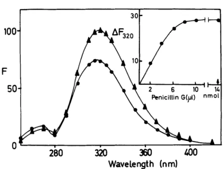
[enzyme-ben~ylpenicillin].~~ The quenching of the fluorescence of the en- K,zyme (at 320 nm) induced by binding of benzylpenicillin (FIGURE 7) was used to calculate the amount of enzyme-benzylpenicillin complex formed. One mole of benzylpenicillin was bound per 35-39,000 g of enzyme, with an asso- ciation constant
K,
of 1.1x
los liter.mo1-l (K,=
reverse of the concentration of benzylpenicillin at which half of the maximum quenching of the fluorescence is produced). These results were in good agreement with both the molecular25 2
100,
F
50
Annals
New
York Academy
ofSciences
/.--+-
30
Penicillin
G(yI)
nmoPenicillin
G(yI)
nmo280
320
360
360
400
400
Wavelength (nml
FIGURE 7. Emission spectrum of (A) R61 enzyme (95% pure, 2 ml of 26.5 pg/
ml), and ( 0 ) the same enzyme solution saturated with benzylpenicillin. Excitation was at 273 nm, and the experiment was performed at 25" C in 10 mM sodium phos- phate buffer (pH 7.0). Fluorescence intensity (F) is expressed in arbitrary units; the maximum emission was considered as 100. Insert: same experiment. Binding of benzylpenicillin to the enzyme as followed by fluorescence quenching at 320 nm
(AFaZ0). The sodium benzylpenicillin solution (0.232 mM) was in 10 mM sodium phosphate buffer (pH 7.0). After 10 pl of this solution had been added, a large excess
of benzylpenicillin ( 5 fil of 2.32 mM) was added to ensure saturation. (From Nieto et a/." By permission of The Biochemical Journal.)
weight obtained by analytical centrifugation and by polyacrylamide gel elec- trophoresis 23 and the K i obtained kinetically la and indicated that one mole of
enzyme bound one mole of benzylpenicillin.
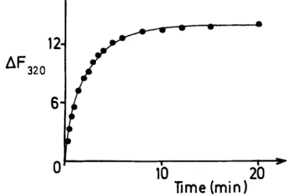
The combination between benzylpenicillin and the enzyme was relatively slow and reached equilibrium after about 10 min (FIGURE 8). In the first 3-4 rnin, when the forward reaction to the enzyme-benzylpenicillin complex was dominant, straight lines in second-order kinetic plots were obtained. The forward reaction rate constant ( K f ) so obtained was 1.1
x
lo4 liter.mol-l.sec-l, and the reverse rate constant ( K , = K , / K , ) was 1 X 10⁴*sec1 [in 0.01 mM Tris-HC1 buffer (pH 7.3)]. Computer simulations of the kinetics of the reaction gave second-order plots that paralleled those obtained e~perimentally.~~ In the absence of covalent bond formation (see above), this time dependence sug- gested that benzylpenicillin produced a conformational change in the enzyme with a Gibb's free energy change of approximately 45 kJ/mol (at 25" C).The near ultraviolet circular dichroism of the R61 enzyme was extensively affected by benzylpenicillin so (FIGURE 9). According to the studies of Chen
Ghuysen
etal.
: Streptomyces Penicillin Receptor25
3 conformationat response of the enzyme. The conformational change was not very extensive, because it was not reflected in the far ultraviolet circular dichro- ism, which was unaltered in the presence of benzylpenicillin. The variations in the near ultraviolet circular dichroism were used in the same manner as the quenching of the fluorescence to construct titration plots, and they gave identical results. Fluorescence was preferred, however, because of its higher sensitivity. Finally, an effect of benzylpenicillin on the conformation of the R61 enzyme was also indicated by its influence on the temperature of transition in the heat denaturation in 3.6 M guanidine-HC1. The antibiotic-saturated enzyme denaturated both faster and at a lower temperaturev,,
= 0.5 at 31"C
(f, = the fraction of protein in its native state)] than the untreated protein (f,, = 0.5 at 39" C ) (FIGURE 10).Znfluence of High Salt Concentrations and of Donor, Acceptor, and Inhibitor Peptides on Binding of Benzylpenicillin t o the
Exocellular R61 Enzyme
High concentrations of salts (ammonium acetate, sodium phosphate, at pH 7.0) quenched the fluorescence of the R61 enzyme.
K,
values (at 25" C ) were 4 and 8 liter.mol-', and maximum quenching was 11 and 25%, respectively. The quenching was very rapid, and no time dependence could be observed. The anion was probably responsible for the interaction, because the quenching was independent of the cation used. If the enzyme was first saturated with benzyl- penicillin and then exposed to salts, the quenching produced by the latter andAF
32
0
FIGURE 8. Time course of the binding of sodium benzylpenicillin to the R61 en- zyme. To a solution of the enzyme ( 7 . 5 ~ lo-' M) in 10 mM sodium phosphate buf- fer (pH 7.0), antibiotic was added to a concentration of 7.6x lo-' M, and the quenching of the fluorescence at 320 nm (AFa,,) was followed with time. Other conditions were the same as in FIGURE 7 . (From Nieto et al.so By permission of The Biochemical Journal.)
254
X
(nm)0
q0
290FIGURE 9. Near and far ultraviolet circular dichroism of the R61 enzyme alone
(-) and saturated with benzylpenicillin (.
.
.). Combination of the enzyme with benzylpenicillin did not alter the spectrum in the far ultraviolet, after correction for the dichroism of the antibiotic. [AE] is AE/c, where A E is the measured value of thc dichroism as the difference in absorption for left- and right-hand circularly polarized light in a light path of 1 cm and c is the concentration of protein in mg/ml. Experi- ments were performed at pH 7.0 and 21" C. (From Nieto e f al." By permission ofThe Biochemical Journal.)
FIGURE 10. Influence of benzylpeni- cillin on the thermal transition of the enzyme in 3.6 M guanidinium hydro- chloride. The experiments were per- formed in 10 mM phosphate buffer (pH 7.0); fn is the fraction of enzyme
in its native state. For explanations, see text. (From Nieto et al." By per-
mission of The Biochernical Journal.)
20
40
60
Ghuysen et
al.
: Streptotngces Penicillin Receptor 255
the apparent association were decreased (for ammonium acetate, K,, became 1.3 1iter.mol-*, and maximum quenching was 6-7%) .3O
The standard tripeptide donor, Na,Ne-diacetyl-L-lysyI-D-alanyl-D-alanine, and the hydrolysis product, Na,Ne-diacetyl-L-lysyl-D-alanine, were no better ligands than phosphate. Acceptors such as glycine or glycyl-L-alanine did not affect the fluorescence of the enzyme.s'
The tripeptide donor and the dipeptide acetyl-D-alanyl-D-glutamic acid (a competitive inhibitor of the enzyme acting as carboxypeptidase; TABLE 4) at concentrations five times their
K,,,
and K i values, respectively, had very little effect on both theK,
value and the rate constantsK,
andK,
of the interaction between the enzyme and benzylpenicillin. The same small effect was also ob- served with the hydrolysis product Na,N~-diacetyl-r.-lysyl-~-alanine, which was neither a substrate nor an inhibitor of the enzyme. The two acceptor mole- cules, glycine and glycyl-L-alanine, at concentrations that should saturate the acceptor site of the enzyme had no effect on either K , or the rate constants.30Noticeable decreases of the
K,
and K , values of the enzyme-benzylpenicillin interaction occurred in 0.19 M sodium phosphate ( K , L = 2.3 X lo7; Kf = 3.9 X lo3), 1 M ammonium acetate (K, = 2.6 X lo0), and 3.6 M guanidine- HCl ( K , = 2.7x
loc;K f
= 1.3x
102) (at 25" C and at neutral pH in all cases). The observed decreases of the K , values were mainly attributable to a slowing down of the forward reaction, whereas the K , values changed rela- tively little. Hence, the affinity of the enzyme for benzylpenicillin was still very high under conditions where the enzyme was inactive (carboxypeptidase activity fell to 30% in 0.1 M buffer l o and was zero in 0.6-0.7 M guanidine- HCl)Mechanism of Inhibition of the Exocellular R61 Enzyme by Benzylpenicillin Both kinetics of inhibition by benzylpenicillin and optical studies clearly demonstrated that benzylpenicillin and acceptor peptide bound at different sites on the enzyme. With regard to the peptide donor, the situation was less clear-cut. Kinetically, the inhibition was competitive, which suggested that ternary (enzyme-donor-benzylpenicillin) complexes did not form. Competitive kinetics do not, however, necessarily exclude an inhibitory mechanism other than a direct competition between substrate and inhibitor for the same site on the free enzyme. Saturating concentrations of donor substrate did not appre- ciably interfere with the binding of benzylpenicillin. The antibiotic bound to the enzyme under conditions where the latter was totally inactive (i.e., in the presence of guanidine-HC1)
.
Furthermore, the mode of binding of benzylpeni- cillin and donor to the enzyme was very different, as reflected in the time dependence. Thus, the most straightforward conclusion seemed that the sites for donor and benzylpenicillin were totally, or at least partially, different but not totally independent; that is, the binding of penicillin affected the donor binding site, although the reverse might not be true.Whatever the exact relationship between donor and benzylpenicillin binding sites, the mode of action of benzylpenicillin rested upon the demonstration that it produced a conformational change in the enzyme with liberation of a Gibb's free energy of about 45 kJ/mol (at 25" C). If ternary (enzyme-donor-benzyl- penicillin) complexes are not formed, the penicillin-treated enzyme, in order to become again enzymatically active, would have to return to its normal confor-
25
6
Annals New York Academy
of Sciences
mation; that is, it would have to overcome an energy barrier of 45 kJ/mol without any external supply of energy, which the enzyme is obviously unable to do. On the contrary, if (enzyme-donor-benzylpenicillin) complexes are allowed to form, there would be some external release of energy (through the binding of the donor peptide). However, the energy barrier between the conformation of the benzylpenicillin-saturated enzyme and that of the enzymatically produc- tive complex in its activated state would then be 45
+
35 kJ/mol (the 35 kJ represents the energy barrier between cis and trans configurations of the C- terminal amide bond that is to be cleaved; see above). This 80 kJ/mol energy barrier would be more than twice that required when the antibiotic is not present, and, presumably, the enzyme would again be unable to overcome it. Irrespective of the case, benzylpenicillin would, therefore, inhibit the enzyme, because it freezes it in a conformation that prevents activity.THE
DD-CARBOXYPEPTIDASE-TRANSPEPTIDASE
SYSTEM
INStreptornyces STRAIN R39 Structure of the Wall Prptidoglycan
In Streptomyces R39, the wall peptidoglycan is composed of L-alanyl-D- isoglutaminyl-( L) -meso-diaminopimelyl-( L) -D-alanine tetrapeptides that are di- rectly interlinked through C-terminal D-alanyl-(D) -mesa-diaminopimelic acid 1inkages.lV The same structure was found in many gram-negative bacteria (except that the a-carboxyl group of D-glutamic acid was not amidated) and in many bacilli.20 Because of this structure, the transpeptidation reaction in Streptornyces R39 must occur as shown in FIGURE 1. Note that the interpeptide bond made by transpeptidation is in an a-position to a free carboxyl group.
The Exocellular R39 Transpeptidase-m-Carboxypeptidase
The exocellular R39 enzyme consists of a single polypeptide chain. Its molecular weight is about 55,000. Turnover number with regard to its carboxy- peptidase activity upon Na9N'-diacetyl-L-lysyl-D-alanyl-D-alanine was 850.37 The transpeptidase activity of the R39 enzyme relative to its carboxypeptidase activity was also enhanced by raising the pH, by increasing the concentration of acceptor (within certain limits), and by decreasing the water content of the reaction mixtures.'@ High concentrations in K,HPO,, however, that suppressed the activity of the R6 1 enzyme, considerably favored the transpeptidase activity of the R39 enzyme. For this reason, most of the transpeptidation reactions with this enzyme were performed in 0.5 M K,HPO, (ionic strength 1.5).19 Penicillins and cephalosporins inhibited the transpeptidase activity of the R39 enzyme at those concentrations that inhibited its carboxypeptidase activ- ity (TABLE 1 )
.
Its susceptibility toward most of the &lactam antibiotics was higher than that of the exocellular R61 enzyme. A similar difference was observed between the LD,,, values for the two corresponding strains (TABLE 1). Kinetics of inhibition of the enzymic activities of the R39 enzyme by penicillin appeared to be a much more complex phenomenonI7 than the inhibition of the R61 enzyme, and they are currently being studied.Ghuysen et al. : Streptomyces Penicillin Receptor
257
Specificity Profile of Exocellular R39 Enzyme for Carboxypeptidase Activity
The R39 enzyme, similar to the R61 enzyme, exhibited a considerable speci- ficity for a C-terminal L-R,-D-Ala-D(OH) sequence and for a long aliphatic side chain at the L-R, residue.li,z8 The occurrence of ionized groups at the end of this side chain, however, differently influenced the activity of the two enzymes (TABLE 3 ) . The KnL values of the R39 enzyme varied from 10 pM for the best substrate so far studied (the pentapeptide L-Ala-y-DGlu-( L)-meso- DAP-(L)-D-Ala-D-Ala, vide infra) to 2.5 mM for poor substrates. The
K,,,
value for the standard substrate Ac?-L-Lys-D-Ala-DAla was 0.8 mM. The V,,, values were relatively little affected by the substrates. None of the peptides that were inhibitory for the R61 enzyme (TABLE 4) had any effect on the carboxypeptidase activity of the R39 enzyme.*g In fact, some of them were good substrates for this enzyme.
Specificity Profile of Exocellular R39 Enzyme. for Transpeptidase Activity
The most striking peculiarity of the R39 enzyme was its inability to catalyze transpeptidation reactions when --amino acids, peptides, and 6-aminopenicillanic acid, instead of glycine or a-D-amino acids, were tested as possible acceptors 24p 2B
(TABLES 2 & 5 ) . Clearly, this enzyme was unable to synthesize peptide bonds in an endo position. In fact, this peculiarity correlates very well with an im- portant structural feature of the wall peptidoglycan of Streptomyces R39 : the interpeptide bond made by transpeptidation is at a C-terminal position (FIGURE 1 ) . The only effective acceptor for the R39 enzyme so far studied that had no free carboxyl group was D-cycloserine.25 This antibiotic, however, behaves as a zwitterion, with pK values of 4.4 and 1.4. At alkaline pH, the charge properties would be similar to those of an a-amino acid, and function as an acceptor could be rationalized. The basic requirement of the R39 enzyme for acceptor was, therefore, an amino group located on a D center in a-position
to a free carboxyl group. The influence exerted by the side chain was then studied with the help of peptides that were either similar or identical to the natural peptide L-alanyl-y-Disoglutaminyl- ( L) -meso-diaminopimelyl-( L) -D-ala-
nine (TABLE 6 ) .
Kinetics of Concomitant Transfer and Hydrolysis Reactions Catalyzed by the Exocellular R39 Enzyme
The amide-free pentapeptide L-alanyl-y-D-glutamyl- ( L) -meso-diaminopime-
lyl-(L)-D-alanyl-D-alanine (peptide 1 in TABLE 6) was a substrate for the R39 enzyme, but its fate depended upon its concentration in the reaction mixture (FIGURE 11).
K,,
values were approximately 10 pM for hydrolysis and 10 mM for transpeptidation, respectively. At concentrations lower than 200 p M , there- fore, only hydrolysis into tetrapeptide occurred, whereas at higher concentra- tions, dimerization through the formation of a C-terminal Dahyl-(D) -meso- diaminopimelic acid interpeptide bond (FIGURE 1 ) progressively increased in yield and hydrolysis was inhibited.a7Ghuysen et
al.
: Streptomyces Penicillin Receptor 2594
260
Q040
Annals
New York Academy
of Sciences
f
I f
1
i7
FIGURE 11. Transfer and hydrolysis reactions catalyzed by the exocellular R39 enzyme when acting on the pentapeptide L-alanyl-y-u-glutamyl-( L)-meso-diaminopi-
melyl-(L)-D-alanyl-D-alanine. Right: pentapeptide ( 1.8 pM-15 pM) was incubated at 37" C for 1 min in 15 pl (final volume) of 0.5 M K2HPOI in the presence of 3
units of R39 enzyme. Left: pentapeptide (300-3400 pM) was incubated at 37" C for 30 min in 15 pl (final volume) of 0.5 M KzHPO, in the presence of 3 units of
R39 enzyme. Hy, hydrolysis of the pentapeptide into tetrapeptide and free D-alanine; Tendo, "endogenous" transpeptidation leading to dimerization (see system 1 in TABLE 6) ; P, total reaction (transpeptidation+hydrolysis) estimated by measuring the release of free D-Ala, which is the product common to both reactions. Rates of the reactions (v) were expressed in pmoles of products formed per minute. Radioactive pentapeptide (~-[1C]A~a-u-["C]Ala) was used. It was a gift of Dr. P. Reynolds, Cambridge, England.
Ghuysen et
al.
: Streptomyces Penicillin Receptor26
1 The question of whether the occurrence of an amide group on the u- carboxyl group of D-glutamic acid in the natural peptide was important for the transpeptidation reaction was investigated by studying the transfer of Na,Ne- diacetyl-L-lysyl-D-alanyl from Na,Nf-diacetyl-L-lysyl-D-alanyl-D-alanine to the ( D) amino group of meso-diaminopimelic acid in the following peptides: thenonamidated disaccharide tetrapeptide
8-
1,4-GlcNAc-MurNAc-~-a1anyl-~-~- glutamyl-(L) -meso-diaminopimelyl-( I.) -D-alanine, the corresponding disaccha-ride-free tetrapeptide, the tripeptide L-alanyl-y-D-glutamyl-( I.) -meso-diamino- pimelic acid, the amidated tripeptide L-alanyl-y-D-glutamyl-( L) -meso-diamino- pimelic acid-(D) -amide, and the amidated tetrapeptide L-alanyl-D-isoglutaminyl-
(L) -meso-diaminopimelyl-( L ) -D-alanine 19 (peptides 2-4 in TABLE 6). With the three nonamidated acceptors, the yield of transpeptidation, at a given concentration of donor, was maximal at about the same concentration of acceptor. At this concentration of acceptor, the hydrolysis of the tripeptide donor, when compared to that which occurred in the absence of acceptor, was decreased to an extent greater than could be accounted for by transpeptidation. A further increase in acceptor concentration caused additional inhibition of the hydrolysis but did not affect the transpeptidation reaction. The effect of amidation of the carboxyl group on the D carbon of meso-diaminopimelic acid
was drastic, in that it completely prevented the peptide from being recognized by the enzyme. This DAP-amidated peptide was not a substrate for transpepti- dation and did not inhibit the hydrolysis of the tripeptide donor. The effect of amidation of the a-carboxyl group of D-glutamic acid conferred on the acceptor a new property. At a given concentration of tripeptide donor, maximal trans- peptidation only occurred within a range of concentrations of acceptor. At higher concentrations of acceptor, both transpeptidation and hydrolysis reactions were progressively inhibited, until eventually they were completely abolished (FIGURE 12). This inhibition by excess acceptor ( a property also exhibited by the R61 enzyme with some peptide acceptors) again pointed to the possible existence of control mechanisms that might be involved in the regulation of the activity of the enzyme in vivo. Other possible control mechanisms were also revealed by these studies.1!’ They included inhibition of transpeptidation by the transpeptidation product and hydrolysis of the transpeptidation product into intact peptide acceptor and dipeptide Na,N‘-diacetyl-L-lysyl-D-alanine through the carboxypeptidase activity of the enzyme (hydrolysis of the C-terminal Dalanyl-( D) -meso-diaminopimelic acid made by transpeptidation)
.
Replacement of the tripeptide donor in the above system, Ac,-L-Lys-DAla- D-Ala and Glu-amidated tetrapeptide, by the pentapeptide donor r.-alanyl--y- D-glUtamyl-( L) -meso-diaminopimelyl-( L ) -D-alanyl-malanine (at concentrations
lower than 200 pM to avoid “endogenous” transpeptidation; see above) gave rise to similar results with respect to kinetics of hydrolysis and of transpepti- dation. The transpeptidation product, of course, was the dimer in which a Glu-amidated tetrapeptide and a nonamidated tetrapeptide were linked to- gether, that is, a dimer that, if one excepts the lack of one amide group, was identical to that found in the completed wall peptidoglycan of Streptomyces R39. With both systems (Ac,-L-Lys-D-Ala-D-Ala
+
Glu-amidated tetrapeptide or nonamidated pentapeptide $- Glu-amidated tetrapeptide), the influences ex-erted by the concentration of the donor [D] and the concentration of the acceptor [A] on the ratios vrI./vIIr suggested for the reactions an ordered mechanism in which the acceptor binds first and the occurrence on the enzyme of an additional inhibitory binding site for the Glu-amidated acceptor.37 A
262
Annals New York Academy
of Sciences
(D)=
26i'py
'2
1
-3
3
4
FIGURE 12. Concomitant transfer and hydrolysis reactions catalyzed by the exocel- Mar R39 enzyme with the system tetrapeptide acceptor (r-alanyl-D-isoglutaminyl-
( L) -,neso-diarninopirnelyl-(L) -D-alanine) and tripeptide donor N",N'-diacetyl-L-lysyl-
D-alanyl-D-alanine. Influence of concentration of acceptor at two concentrations of
donor (267 and 1393 pM, respectively) on hydrolysis of the donor (Hy), transpepti-
dation (T), and residual donor (RD). Acceptor (320-4800 pM) and donor (267 or
1393 pM) were incubated for 30 rnin at 37" C in IS pl (final volume) of 0.5 M
KzHPOI in the presence of 34 units of R39 enzyme. Under these conditions, at opti- mal concentrations of acceptor (700 and 1600 pM, respectively), 30 and 150 pmole
of donor are transformed into transpeptidation product per minute, which depends
upon the concentration of donor used (267 and 1363 pM, respectively); that is, the rate of transpeptidation is proportional to the concentration of donor.
Ghuysen et
al.
: Streptornyces Penicillin Receptor263
similar situation had been found with the exocellular R61 enzyme when acting on the system Ac,-L-Lys-mAla-D-Ala
+
Gly-L-Ala (see above).THE DD-CARBOXY PEPTIDASE-TRANSPE PTIDASE SYSTEM IN Streptornyces STRAINS K l l AND albus G
Insofar as could be judged, the exocellular enzyme from Streptomyces K11 exhibited properties very similar, if not identical, to those of the R61 enzyme with respect to both carboxypeptidase and transpeptidase activities 1 7 9 2 4 , 2 5 ,
(TABLES 2 & 3). Strains K11 and R61 had wall peptidoglycans of identical structure (FIGURE 1) but differed from each other in their ability to produce )6-lactamase.21~ 22
The exocellular DD-carboxypeptidase from S. ulbus G did not affect trans- peptidation with any of the acceptors tried 21, 25 (TABLE 2 ) . It was also
exceptional in other respects: it was a cationic protein (even at pH 8 ) , lysed bacterial walls or cells in which the interpeptide bonds are C-terminal malanyl-D linkages, and was virtually not affected by penicillin.28, 31-33 The carboxy- peptidases from Streptornyces R61,
K1
1, and R39 exerted endopeptidase activity (see above) on isolated peptide dimers and oligomers but were not bacterio- lytic agents. It is possible that their anionic properties prevented them from combining with the bacterial walls and exerting a lytic activity. Possibly, the true transpeptidase of S. alhus G is not the same as the isolated carboxypepti- dase.The S . ulbus G DD-carboxypeptidase, although not inhibited by penicillins and cephalosporins, was inhibited by peptide analogs 29 of the standard substrate
aeAc2-L-Lys-D-Ala-D-Ala to an extent that was even greater than that observed with the penicillin-sensitive R61 enzyme (TABLE 4 ) . Altogether, these obser- vations and the fact that the R39 enzyme which exhibited the highest sensi- tivity to penicillin was not inhibited by these peptide analogs (see above) are another demonstration that binding sites for penicillin in sensitive enzymes are probably independent of the binding sites for the donor substrates.
CONCLUSIONS
Depending upon the bacterial species, the independence between the sub-
strates and penicillin sites on the DD-carboxypeptidases-transpeptidases might be somewhat pronounced. Enzymes that have lost their acceptor and/ or penicillin binding site(s) may exist. Enzymes may also exist in which binding of peni- cillin would not affect binding of the donor and even of the subsequent en- zymatic catalysis. All would depend on whether the conformational change induced by penicillin involves peptide chains in the enzyme that need con- formational freedom for normal catalysis. Activation of catalysis by penicillin can even be easily envisaged by assuming that the conformation induced in the protein by penicillin binding would result in a more favorable alignment of the catalytic groups. Penicilloylation of reactive side chains in some enzymes could be a secondary event and could occur after the conformation change has occurred, if, as a consequence of it, a susceptible side chain were placed in a spatially favorable position. Finally, the present proposal of the mode of action of penicillin also explains many facts unaccounted for where simple