Université de Montréal
New Grafted PLA-g-PEG Polymeric Nanoparticles Used to
Improve Bioavailability of Oral Drugs.
par
Mohamed Mokhtar
Faculté de pharmacie
Thèse présentée à la Faculté de pharmacie en vue de l’obtention du grade de Ph.D.
en Science Pharmaceutiques option Technologie Pharmaceutique
February, 2015 © Mohamed Mokhtar, 2015
Université de Montréal Facluté des études supérieures
Cette thèse intitulée:
New Grafted PEG-g-PLA Polymeric Nanoparticle used to Improve
Bioavailability of Oral Drugs.
Présentée par Mohamed Mokhtar
a été évaluée par un jury composé des personnes suivantes :
Dr. Valérie Gaëlle, Roullin , Président-rapporteur Dr. Patrice Hildgen, Directeur de recherche Dr. Patrick Gosselin, Co-directeur de recherche
Dr. François-Xavier Lacasse, Membre du jury Dr. Charles Ramassamy, Examinateur externe Dr. Gaétan Mayer, Représentant du doyen de la FES
Résumé
Les nanoparticules (NPs) de polymère ont montré des résultats prometteurs pour leur utilisation comme système de transport de médicaments pour une libération contrôlée du médicament, ainsi que pour du ciblage. La biodisponibilité des médicaments administrés oralement pourrait être limitée par un processus de sécrétion intestinale, qui pourrait par la suite être concilié par la glycoprotéine P (P-gp). La dispersion de la Famotidine (modèle de médicament) à l’intérieur des nanoparticules (NPs) pegylées a été évaluée afin d’augmenter la biodisponibilité avec du polyéthylène glycol (PEG), qui est connu comme un inhibiteur de P-gp. L’hypothèse de cette étude est que l’encapsulation de la Famotidine (un substrat de P-gp) à l’intérieur des NPs préparées à partir de PEG-g-PLA pourrait inhiber la fonction P-gp. La première partie de cette étude avait pour but de synthétiser quatre copolymères de PEG greffés sur un acide polylactide (PLA) et sur un squelette de polymère (PLA-g-PEG), avec des ratios de 1% et 5% (ratio molaire de PEG vs acide lactique monomère) de soit 750, soit 2000 Da de masse moléculaire. Ces polymères ont été employés afin de préparer des NPs chargés de Famotidine qui possède une faible perméabilité et une solubilité aqueuse relativement basse. Les NPs préparées ont été analysées pour leur principaux paramètres physicochimiques tels que la taille et la distribution de la taille, la charge de surface (Potentiel Zeta), la morphologie, l’efficacité d’encapsulation, le pourcentage résiduel en alcool polyvinylique (PVA) adsorbé à la surface des NPs, les propriétés thermiques, la structure cristalline et la libération du médicament. De même, les formules de NPs ont été testées in vitro sur des cellules CaCo-2 afin dʼévaluer la perméabilité bidirectionnelle de la Famotidine. Généralement, les NPs préparées à
partir de polymères greffés PLA-g-5%PEG ont montré une augmentation de la perméabilité du médicament, ce par l’inhibition de l’efflux de P-gp de la Famotidine dans le modèle CaCo-2 in vitro. Les résultats ont montré une baisse significative de la sécrétion de la Famotidine de la membrane basolatéral à apical lorsque la Famotidine était encapsulée dans des NPs préparées à partir de greffes de 5% PEG de 750 ou 2000 Da, de même que pour d’autres combinaisons de mélanges physiques contenant du PEG5%. La deuxième partie de cette étude est à propos de ces NPs chargées qui démontrent des résultats prometteurs en termes de perméabilité et d’inhibition d’efflux de P-gp, et qui ont été choises pour développer une forme orale solide. La granulation sèche a été employée pour densifier les NPs, afin de développer des comprimés des deux formules sélectionnées de NPs. Les comprimés à base de NPs ont démontré un temps de désintégration rapide (moins d’une minute) et une libération similaire à la Famotidine trouvée sur le marché. Les résultats de l’étude du transport de comprimés à base de NPs étaient cohérents avec les résultats des formules de NPs en termes d’inhibition de P-gp, ce qui explique pourquoi le processus de fabrication du comprimé n’a pas eu d’effet sur les NPs. Mis ensemble, ces résultats montrent que l’encapsulation dans une NP de polymère pegylé pourrait être une stratégie prometteuse pour l’amélioration de la biodisponibilité des substrats de P-gp.
Mots-clés : Poly(ethylene glycol), P-glycoprotéine, Nanoparticules, Perméabilité, CaCo-2, Acide polylactique.
Abstract
Polymeric nanoparticles (NPs) have shown promising results to be used as drug delivery carriers for controlled drug release and for targeting. The bioavailability of orally administrated drugs might be limited by an intestinal absorbed secretion process, which could in turn be mediated by P-glycoprotein (P-gp). The dispersion of Famotidine (drug model) within PEGylated nanoparticles (NPs) was evaluated in order to enhance the bioavailability using polyethylene glycol (PEG), which is known as a P-gp inhibitor. The hypothesis of this study is that the encapsulation of Famotidine into NPs prepared from PEG-g-PLA could inhibit the P-gp function. The first part of the study aimed to synthesize four copolymers of PEG grafted on a polylactide acid (PLA) polymer backbone (PLA-g-PEG) with ratios of 1 % and 5 % (molar ratio of PEG vs lactic acid monomer) of either 750 or 2000 Da molecular weights. These polymers have been used to prepare NPs loaded with Famotidine, which has low permeability and relatively low aqueous solubility. Prepared NPs were analyzed for their main
physicochemical parameters such as size and size distribution, surface charge, morphology, encapsulation efficiency, percentage of residual poly(vinyl alcohol) (PVA) adsorbed atthe surface of the NPs, thermal properties, crystalline structure and drug release. Also, NP formulations were tested in vitro on CaCo-2 monolayer cells in order to assess the bidirectional permeability of Famotidine. Generally speaking, NPs prepared from PLA-g-5%PEG grafted polymers showed improved drug permeability by inhibiting the P-gp efflux of Famotidine in the CaCo-2 in vitro model. Results showed a very significant decrease in the secretion of Famotidine from basolateral to
750 or 2000 Da PEG, as well as in other combinations of physical mixtures containing PEG5%. The second part of the study is about those loaded NPs which showed promising results in terms of permeability and inhibition of P-gp efflux and which were selected to develop a solid oral dosage form. Dry granulation was used to densify NPs in order to develop tablets of the two selected NP formulations. NP based tablets showed a fast disintegration time (less than one minute) and a similar release profile as marketed Famotidine. The transport study of NP based tablets showed consistent results with NP formulations in terms of P-gp inhibition, which explains why the tablet’s manufacturing process had no effect on NPs. Our results demonstrated that the presence of hydrophilic polymers (PEG) on the surface of NPs grafted at 5 % PEG, irrespective to molecular weights, provides an effective way to control the interface between NPs and the biological systems (P-gp in this case) as they are designed to interact with. Taken together, these results show that encapsulation in a PEGylated polymeric NP could be a promising strategy to improve the oral bioavailability of P-gp substrates.
Keywords : Poly(ethylene glycol), P-glycoprotein, Nanoparticles, Permeability, CaCo-2, Poly(lactic) acid.
Table of content
List of Tables ... x
List of Figures ... xii
Abbreviations ... xiv
Contribution des auteurs ... xx
Chapter One ... 2
Introduction ... 2
1. Introduction ... 3
1.1. Structure and Physiology of GIT ... 4
1.2. Route and Barrier to Drug Absorption ... 5
1.2.1. GIT Environment ... 6
1.2.2. Mucus Layer (unstirred water layer) ... 7
1.2.3. Tight Junctions and Paracellular Route ... 8
1.2.4. M-cells ... 9
1.2.5. Transcellular Route and Membrane Transporter (Permeability-glycoprotein) .... 10
1.2.5.1. Transcellular Route ... 10
1.2.5.2. Membrane Transporter P-gp ... 10
1.3. P-gp Inhibitors ... 13
1.4. Nanoparticles as Effective Oral Drug Carrier ... 15
1.4.1. Polymeric Nanoparticles ... 16
1.4.2. Liposomes and Lipid-Based Nanocarriers ... 17
1.4.3. Micelles ... 18
1.4.4. Hydrogel Nanocarrier ... 19
1.4.5. Dendrimers ... 20
1.5. NanoCarrier Translocation and Drug Bioavailability ... 20
1.6. Optimizing Nanocarrier Physiochemical Properties for Oral Delivery ... 22
1.6.1. Increased Drug Dissolution, Absorption, and Protection from Degradation .... 22
1.6.2.2. Mucus-Penetrating Nanocarriers ... 24
1.6.3. Translocation Across Tight Junctions ... 25
1.6.4. Translocation Across Enterocytes ... 25
1.6.5. Translocation Across M-cells ... 27
1.7. Poly (Ethylene Glycol) (PEG) Used as P-gp Inhibitor ... 28
1.8. Targeting ... 30
1.8.1. Passive Targeting ... 32
1.8.2. Active Targeting ... 33
1.9. Oral Dosage Form ... 34
1.9.1. Direct Compression ... 36
1.9.2. Wet Granulation ... 37
1.9.3. Dry Granulation ... 37
Chapter Two ... 40
Hypothesis and Objectives ... 40
2. Hypothesis and Objectives of the study ... 41
2.1. Hypothesis ... 41
2.2. The objectives of the study ... 42
References ... 46
Experimental Works ... 63
Chapter Three. Design of PEG-grafted-PLA nanoparticles as oral permeability enhancer for P-gp substrate drug model Famotidine ... 64
3.1. Abstract ... 66
3.2. Introduction ... 66
3.3. Materials and methods ... 69
3.3.1. Materials ... 70
3.3.2. Polymer synthesis ... 70
3.3.3. Polymer characterisation ... 71
3.3.3.1. Nuclear magnetic resonance (NMR) ... 71
3.3.3.2. Gel permeation chromatography (GPC) ... 72
3.3.3.3. Grafting efficiency ... 72
3.3.3.5. Differential scanning calorimetry (DSC) ... 74 3.3.4. NP preparation ... 75 3.3.4.1. Emulsion-solvent evaporation ... 75 3.3.4.2. Nanoprecipitation ... 75 3.3.5. NPs characterisation ... 76 3.3.5.1. Particle size ... 76
3.3.5.2. Surface charge (zeta potential) ... 77
3.3.5.3. Determination of residual PVA ... 77
3.3.5.4. Particle morphology ... 78
3.3.5.5. Encapsulation efficiency and drug loading ... 78
3.3.5.6. X-Ray powder diffraction (XRPD) ... 79
3.3.5.7. Drug release ... 80
3.3.5.8. Cytotoxicity ... 80
3.3.5.9. In vitro permeability evaluation by bidirectional Caco-2 cells monolayer ... 82
3.3.5.9.1. Culture of Caco-2 cells ... 82
3.3.5.9.2. Caco-2 cells monolayer ... 82
3.3.5.9.3. Transport experiments ... 83
3.3.6. Data analysis ... 84
3.4. Results and discussion ... 85
3.4.1. Polymer synthesis and characterisation ... 85
3.4.1.1. NMR ... 85
3.4.1.2. GPC ... 85
3.4.1.3. Grafting efficiency ... 86
3.4.1.4. Fourier transform infrared FTIR ... 88
3.4.1.5. DSC ... 90
3.4.2. NPs characterisation ... 90
3.4.2.1. Particle size ... 90
3.4.2.2. Surface charge ... 92
3.4.2.3. Residual PVA determination ... 95
3.4.2.6. FTIR ... 98
3.4.2.7. DSC ... 99
3.4.2.8. XRPD ... 101
3.6. References ... 110
Chapter Four :Tablet Formulation of Famotidine loaded P-gp inhibiting Nanoparticles using PLA-g-PEG Grafted Polymer. ... 120
4.1. Abstract ... 122
4.2. Introduction ... 122
4.3. Materials and Methods ... 124
4.3.1. Materials ... 124
4.3.2. Polymer Synthesis and Characterization ... 124
4.3.3. Nanoparticles Preparation and Characterization ... 125
4.3.3.1. Particle Size and Size Distribution ... 126
4.3.3.2. Surface Charge (Zeta Potential) ... 126
4.3.3.3. Drug Loading and Encapsulation Efficiency ... 127
4.3.4. Tablet formulation ... 127
4.3.4.1. Excipients Selection ... 128
4.3.4.2. Tablet Formulation ... 128
4.3.4.3. Dry Granulation ... 129
4.3.4.4. Tablet Compression ... 129
4.3.4.5. Tablet Formulation Characterization ... 130
4.3.4.5.1. Bulk & Tapped Density, Powder Flow ... 130
4.3.4.5.2. Particle Size and Size Distribution of Powder Blends ... 131
4.3.4.5.3. Tablet Weight, Thickness and Hardness ... 131
4.3.4.5.4. Tablet Disintegration ... 131
4.3.4.5.5. Thermal Properties by Differential Scanning Calorimetry (DSC) ... 132
4.3.4.5.6. Crystallinity of Famotidine in Tablets Formulations by XRPD ... 132
4.3.4.6. Drug Release ... 133
4.3.4.6.1. Tablet Release Stability ... 133
4.3.4.7. Drug Dissolution ... 134
4.3.4.8.1. Cell Culture ... 134
4.3.4.8.2. Transport Study ... 135
4.3.4.9. Data Analysis ... 137
4.4. Results and Discussion ... 137
4.4.2. Nanoparticles Characterization ... 140
4.4.3. Tablet Formulation ... 142
4.4.4. Dry Granulation ... 143
4.4.5. Formulation Characterization ... 143
4.4.5.1. Bulk, Tapped Density and Powder Flow ... 143
4.4.5.2. Particle Size and Size Distribution of Powder blends ... 146
4.4.6. Tablets Characterization ... 147
4.4.7. NPs Size Analysis Before and After Tablet Compression ... 149
4.4.8. Thermal Properties by Differential Scanning Calorimetry (DSC) ... 149
4.4.9. Crystallinity of Famotidine in Tablets Formulations by X-Ray Powder Diffraction (XRPD) ... 151 4.4.10. Drug Release ... 153 4.4.11. Stability Study ... 155 4.4.12. Drug Dissolution ... 155 4.4.13. Transport Study ... 157 4.5. Conclusion ... 159 4.6. References ... 161 Chapter Five ... 170
General Discussion, Conclusion and Perspectives ... 170
5.1. General Discussion ... 171
References ... 178
List of Tables
Table 3.1Molecular weight, PDI, grafting percentage and thermal properties (n=3) of polymers evaluated during this study. ... 88 Table 3.2 Particle size, PI, drug loading, and thermal properties Famotidine and different NPs (n=3) ... 92 Table 3.3 Residual PVA content of NPs prepared by emulsion evaporation as well as zeta potential of blank and drug-loaded NPs prepared by emulsion evaporation or nanoprecipitation Methods (n=3) ... 95 Table 3.4 Results of bidirectional transport of different NP formulations and PMs of Famotidine across Caco-2 cell monolayer (n=3) ... 107 Table 4.1 Molecular weights, PDI and DSC results for PEG-PLA grafted polymers ... 139 Table 4.2 The mean particles size, PI, Zeta Potential and Drug Loading of Two NPs Formulations (n=3) ... 141 Table 4.3 Powder flow characterization of NPs, powder blend and dry granulated powder . 145 Table 4.4 Hausner ratio and Carr's index interpretation(20)... 146 Table 4.5 Tablets characterization of Commercial Famotidine, F1 and F2 NPs formulations ... 148 Table 4.6 The mean particles size values of NPs tablet formulations before and after tablet manufacturing (n=3) ... 149 Table 4.7 DSC characterization of Tablet formulations (n=3) ... 150
Table 4.8 Bidirectional results from Marketed Famotidine,F1&F2 NPs tablets formulations, pure Famotidine and tablet physical mixtures across CaCo-2 monolayer cells ... 159
List of Figures
Figure 1.1 Scheme of Gastro-Intestinal-Tract. Adapted from(1). ... 3
Figure 1.2 Structure of GIT barrier: drug and NC transportation pathways. ... 6
Figure 1.3 Schematic representation of some elements of epithelial cells of GIT: ... 8
Figure 1.4 Different mechanism involved in P-gp inhibition and Drug-substrate binding pocket of P-gp. Adapted from(25, 28). ... 13
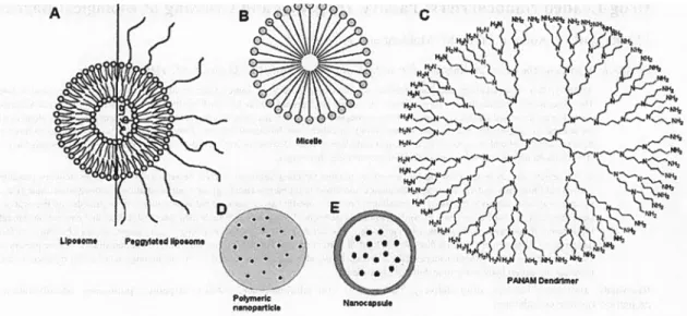
Figure 1.5 Different Types of NCs (A) Micelles, (B) Liposomes, (C) Dendrimers, Poymeric nanoparticles and Nanocapsules With a permission from (8). ... 16
Figure 1.6 PEG NCs coverage. (A) Brush configuration and (B) Mushroom configuration. With a permission from (8). ... 30
Figure 1.7 Powder blend flow properties and different techniques for compression into tablet. Adapted from(113). ... 38
Figure 2.1 Famotidine Structure ... 42
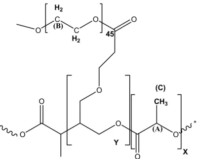
Figure 3.1 Structure of PLA-g-PEG polymer ... 73
Figure 3.2 FTIR Spectra of PLA, PEG and different PLA-g-PEG polymers ... 89
Figure 3.3 Tapping mode AFM image (topography) of NPs (PLA-g-5%PEG2000Da), scan size 1µmx1µm ... 97
Figure 3.4 Thermogram of Famotidine and four loaded NPs formulations ... 100
Figure 3.5 X- Ray diffractograms of Famotidine and four loaded NPs formulations ... 102
Figure 3.6 Percentage of Released Famotidine from different loaded NPs formulations (n=3) ... 104
Figure 3.7 Bidirectional transport study of Famotidine across Caco-2 from different NPs formulations and (PM) combinations (n=3) ... 108
Figure 4.1 NMR for grafted PLA-g-5%PEG2000Da polymer ... 139
Figure 4.2 FTIR Spectrum of pure PLA, PEG and two grafted polymers ... 140
Figure 4.3 Particle size distribution of F1,F2 and Placebo formulations ... 147
Figure 4.4 Thermograms of Famotidine from different tablet formulations ... 151
Figure 4.5 X-ray Diffractograms of Famotidine from different tablet formulations ... 152
Figure 4.6 Percentage of Famotidine Released at pH 7.4 from different tablet formulations 154 Figure 4.7 Famotidine Dissolution Profile from Different Tablet Formulations ... 156
Abbreviations
ABC ATP-Binding Cassette
AFM Atomic Force Microscope AGE Allyl Glycidyl Ether
ANOVA Analysis of Variance
A˚ Angstrom
AP Apical
AI Asymmetric Index
ATP Adenosine triphosphate
BCS Biopharmaceutical Classification System
BL Basolateral
BSA Bovine serum albumin
ºC Degree Celsius
CDCl3 Deuteriated chloroform
CO2 Carbon dioxide
CTB Cholera toxin B
Da Dalton
D2O Deuterium oxide
DCM DiChloroMethane
DMAP DiMethylAminoPyrudine
DMF DiMethylFormamide
DMSO DiMethyl SulfOxide
DL Drug Loading
DLS Dynamic Light Scattering DSC Differential scanning calorimetry EE Encapsulation efficiency
EPR Enhanced permeability and retention
EDTA Ethylene Diamine Tetra Acetic acid
FMT Famotidine
FBS Fetal bovine serum
FAE Follicle-Association Epithelium FDA Food Drug Administration
FTIR Fourier transform infrared spectroscopy GIT Gastro Intestinal tract
GPC Gel permeation chromatography h Hour
HBSS Hanks balanced supplement solution HDPE High Density Polyethylene
HCl Hydrochloric acid
1H NMR Proton nuclear magnetic resonance HPLC High performance liquid chromatography
HPH High Pressure Homogenizer
IV Intravenous
kDa Kilo Dalton
MePEG Methoxy Poly Ethylene Glycol Mg Magnesium
Mn Number average molecular weight
MPS Mononuclear phagocytic system
MTT 3-(4,5-dimethylthiazol-2-yl)-2,5-diphenyltetrazolium bromide
Mw Weight average molecular weight
NaCl Sodium chloride
NaOH Sodium hydroxide
NBD Nucleotide Binding Domains
NC Nanocarrier
N/D Not Detected
NPs Nanoparticles
NEAA Non Esstential Amino Acid
O/W oil in water
PSD Particle Size Distribution
Papp Apparent permeability coefficient
PAMAM Poly(amido amine)
PBS Phosphate buffered saline PCL Poly(ε-caprolactone)
PCS Photon Correlation Spectroscopy
PDI Polydispersity index
PEG Poly(ethylene glycol)
PEG-g-PLA Poly(ethylene glycol)-grafted-poly(D,L-lactide) PEG1%-g-PLA Poly(ethylene glycol)1%-grafted-poly(D,L-lactide) PEG5%-g-PLA Poly(ethylene glycol)5%-grafted-poly(D,L-lactide)
PLA Poly(lactic acid) PLGA Poly(lactic-co-glycolic acid)
PM physical Mixtures
PP Peyer's patches
PPG Polypropylene Glycol
PS Polystyrene
PVA Polyvinyl alcohol
RGD Arginylglycylaspartic acid
SD Standard deviation
SDP Size Distribution Processor SLN Solid Lipid Nanoparticle
SLC Solute Carrier
TEER Trans epithelial electrical resistance
Tg Glass transition temperature TM-AFM Tapping mode atomic force microscope TMS Tetra Methyl Silane
USP United States Pharmacopeia
UV Ultraviolet
wt Weight
Remerciements
This work was done at Pharmaceutics technology department, Faculty of Pharmacy, University of Montreal. I would like to express my appreciation to my supervisor, Dr. Patrice Hildgen and co supervisor, Dr. Patrick Gosselin for their continuous support, advice, guidance and being available and willing to help me in the challenges during my study. Also I would like to thanks Dr. Patrice has given me the freedom and reliable basis necessary to advance this project and to develop as an independent researcher. I really value his valuable criticism throughout of my doctoral studies without which it would have been difficult to finish this thesis. I am sincerely grateful to all my co-supervisor, Dr. Patrick Gosselin, for his remarkable support, advices and being available contribution to this work.
Special thanks are due to Jean Michel Rabanel for his technical assistance, valuable discussion and suggestions during my study which I really appreciate. I would like to express my deep thankfulness to all my lab colleagues for discussions, friendship and for creating a pleasant atmosphere during these years.
Finally, I would like to express my gratitude to my parents, my wife, and my daughters for their endless support and love. It is their sacrifice and understanding that allowed me to work hard to finish this thesis.
I also express my gratitude for COREALIS Pharma, BMP innovation and Ministry of Higher Education, Libya for their financial support is gratefully acknowledged.
Contribution des auteurs
Cette thèse se base sur deux articles dont sont soumis. La contribution de chaque auteur est présentée ci-dessous
Chapitre 3: Design of PEG-grafted-PLA nanoparticles as oral permeability enhancer for P-gp substrate drug model Famotidine.
Mohamed Mokhtar*, Patrick Gosselin, Lacasse, François-Xavierand Patrice Hildgen
Tous les travaux expérimentaux ont été faites par Mohamed Mokhtar. Chaque auteur a participé à révision du manuscrit.
Chapitre 4: Tablet Formulation of Famotidine loaded P-gp inhibiting Nanoparticles using PEG-PLA Grafted Polymer.
Mohamed Mokhtar*, Patrick Gosselin, Lacasse, François-Xavierand Patrice Hildgen
Tous les travaux expérimentaux ont été faites par Mohamed Mokhtar. Chaque auteur a participé à révision du manuscrit.
Chapter One
Introduction
1. Introduction
The majority of drugs on the market are formulated for the oral route, most of them aimed at the blood circulation for the systemic action. It is the most convenient and safe administration route, particularly for chronic delivery, but it poses a number of challenges for formulators in terms of bioavailability (fraction of drug actually reaching the circulation) due to degradation by enzymes and harsh pH conditions, low solubility of some drugs and limited absorption by the gastro intestinal tract (GIT) epithelium(1). The GIT (Figure 1.1) is approximately 6 m in length with varying diameters. It consists of the oesophagus, the stomach, the small intestine (the major digestive organ) and large intestine or colon. The luminal surface is not smooth, with deep, circular fold about 1 cm high, villi, mucosal projections about 1 mm long, and microvilli (plasma membrane microprojections), increasing the surface area of absorption to about 200 cm2. Orally administrated drug molecules must stay for enough time in intestinal lumen and resist harsh gastric and intestinal environments to be efficiently absorbed by intestinal cells via different mechanisms(1-3).
1.1.
Structure and Physiology of GIT
The structure of GIT wall (at the small intestinal level) is comprised of four main histological layers. The first layer, the serosa, is the outer layer of epithelial and supporting connective tissue. The second layer contains two layers of smooth muscle, a thinner outer layer, with longitudinally orientated muscle fibers and a thicker inner layer, with fibers orientated in a circular pattern. The third layer, submucosa is a connective tissue layer consisting of some secretory tissues richly supplied with blood and lymphatic vessels, including the lacteal, a wide lymph capillary at the centre of the villi. The fourth layer, the mucosa, is composed of: the muscularis mucosa, the mucosa, connective tissue and epithelium(1). The intestinal epithelium acts as a physical and physiological barrier to drug absorption. It mainly consists of absorptive enterocytes and mucus producing Goblet cells, endocrine and Paneth cells spread along the epithelium. The GIT epithelium is covered by a layer of mucus. Immuno-competent cells, such as B and T lymphocytes and dendritic cells are located beneath the epithelium(2, 4). The small intestine wall possesses a rich blood network and the GIT blood circulation is nearly a third of cardiac output flow, underlining the importance of exchanges between the GIT lumen and blood circulation. The lymphatic system plays an important part in fat absorption from GIT. The areas of lymphoid tissue close to the epithelial surface called Peyer's patches or follicle-association epithelium (FAE) and serve as immune sampling ports in the intestine. The FAE, separating organized, mucosa associated tissue from the lumen, is composed of enterocytes and M cells. M-cells are found in the follicle-epithelium of Peyer's patches. Microflod M-cells are important in local immune response to pathogens(5) and could
be harness to deliver macromolecules and oral vaccines(6). M cells disorganized brush border at the apical membrane have reduced microvilli and thinner surface mucus. Moreover, M cells have specific surface-adhesion molecules which might be important features for active targeting(1, 2, 4-7).
1.2.
Route and Barrier to Drug Absorption
Drugs molecules encounter many barriers in GIT (Figure 1.2) once released from their dosage forms after they have dissolved in the gastrointestinal fluids. Drug molecules must be in solution and not bound to food or other material within GIT, chemically stable to withstand the GIT pH and resist to enzymatic degradation in the lumen. Finally drug molecules need to diffuse across the mucus (unstirred water layer) and across the gastrointestinal membrane, the main cellular barrier in order to reach blood circulation(3). The main drug absorption pathways from the GIT include carrier-mediated transcellular transport, vesicular transport, passive paracellular transport through entrocytes, and lymphatic uptake by M-cells.
Figure 1.2 Structure of GIT barrier: drug and NC transportation pathways.
on the left,enterocytes, on the right M-cell. A mucus layer is found on the apical side of enterocytes and M-cell.(1) Paracelluar route. (2) Transcelluar route. (3) M-cell
phagocytosis. With a permission from (8)
1.2.1. GIT Environment
The GIT environment is characterized by peristalsis, variable pH, and the presence of surfactants (bile salts), enzymes, bacteria, food, and different types of secretions. GIT pH affects the dissolution rate and absorption of drug molecules. The GIT has a wide range of pH values in empty stomach (pH 1.2-3), small intestine (pH 5-7) and colon (pH 6 to 7.5)(9). Moreover, pH depends of some variables such as time of the day, meal volume and volume of secretions. To provide effective treatment, the delivery system should offer a good protection against these enzymes(2, 10).
1.2.2. Mucus Layer (unstirred water layer)
Mucus is a viscoelastic, translucent, aqueous, protective gel that is secreted throughout GIT and different mucosal surfaces ( pulmonary, nasal, etc.)(1). Its thickness varies from 50 to 150 μm in the stomach to 15-150 μm in intestine. It has a large water component (≈ 95%) and other constituents, which are responsible for its physical and functional properties, are largely glycoproteins called mucins(11). Mucus acts as a protective layer and a mechanical barrier. It is continually replaced from beneath as it is being removed from GIT surface through acidic, enzymatic breakdown and abrasion (peristaltic movement of food towards the colon). GIT mucus is comprised of two layers, a firmly adherent layer, slowly cleared of an unstirred layer close to the epithelium surface, and luminal mucus layer, a stirred layer, a more rapidly cleared layer(12). It is secreted continuously and digested, so for drugs to be delivered to mucosal surfaces, they need to diffuse through the unstirred mucus layers adhering to cells(1, 12, 13). Below the mucus layer, the cells present a dense of highly diverse glycoproteins and glycolipids, which form the glycocalyx. A glycocalyx (Figure 1.3) found on the apical portion of microvilli within the GIT, especially within the small intestine(3, 5, 7, 13, 14). Mucin glycoproteins are secreted in large quantities by mucosal epithelia, and cell surface mucins are a prominent feature of the apical glycocalyx of all mucosal epithelia. It provides additional surface for adsorption and includes enzymes secreted by the absorptive cells that are essential for the final steps of digestion of proteins and sugars(3, 13, 14). The glycocalyx is highly variable from tissue to tissue; for example, the glycocalyx of human intestinal microvilli tips is thick (100–500 nm) in comparison with the glycocalyx of the lateral microvilli surface (30–
60 nm)(5). Both the secreted and adherent mucosal barriers are constantly renewed and could potentially be rapidly adjusted to changes in the environment, for example, in response to microbial infection(5, 13, 14).
Figure 1.3 Schematic representation of some elements of epithelial cells of GIT: (A)Glycocalyx, (B)Mucus layer, (C)Transcellular Route, (D)Enerocytes, (E)Tigh
junction and (F)Paracellular Route. Adapted from(5).
1.2.3. Tight Junctions and Paracellular Route
In the paracellular space, the presence of junctions restricts the passage of large molecules. These include the tight junction, a protein complex that establishes links between adjacent cells through the intercellular space . Two other protein complexes, namely the adherens junctions and desmosomes further tighten cell association, without fusing cell membranes. The apical compartment of the lateral membrane
consists of 3 components: tight junctions, adherent junctions, and desmosomes(9). For desmosomes and adherent junction the adjacent cell membranes are 15 – 20 nm apart, but are in contact at tight junctions, with the intercellular spaces being completely absent. Tight junctions act as gatekeeper paracellular route, regulating drug molecule flux and prevent free movement of molecules in paracellular space. Understanding the barrier function of tight junctions is necessary for developing absorption enhancers to deliver drugs via the paracellular route. Diffusion is regulated by concentration differences and by hydrostatic pressure gradient between the two sides of the epithelium. Tight junctions are main barriers to this type of absorption(15-18).
1.2.4. M-cells
Finally, transportation by Peyer's patches, which contain M-cells found in the Follicle-association epithelium (FAE) at the base of villi, is important in the phagocytosis and endocytosis of NPs and microparticles(5). Peyer's patches are aggregations of lymphoid tissue that are widespread along the intestine and tend to increase in number in the ileum. The physiological role of M-cells is to sample antigens present in the lumen to transport them to immune cells on their basal side. They have high transcytosis activity, but represent less than 1 % of the intestinal mucosa surface. Particles could be taken up by M-cells via specific (active endocytosis) or non-specific (adsorption endocytosis) mechanism(19).
1.2.5. Transcellular Route and Membrane Transporter
(Permeability-glycoprotein)
1.2.5.1. Transcellular Route
The transcellular route comprises passive and active transport. For carrier-mediated transcellular transport (active transcellular transport), drugs are moved across membranes by protein transporters present on the apical membrane of enterocytes (generally energy-dependent). The vesicular transport route consists of fluid-phase endocytosis and receptor-mediated endocytosis (caveolae-dependent, clathrin-dependent, caveolae-calthrin-inclathrin-dependent, etc.). Vesicular transport could lead to the degradative pathway (via lysosomes), or to exocytosis, transport across the epithelium (transcytosis) to the basolateral side of enterocytes near capillaries(8, 20). For passive transcellular transport, drugs should be able to diffuse across the apical membrane, cytoplasm, and basal membrane. The surface area available for passive transcellular transport makes up 99.9% vs. 0.01 % for the passive paracellular pathway(8).
1.2.5.2. Membrane Transporter P-gp
Recently research has been focus in understanding the role of membrane transporters in drug safety and efficacy. In particular, more than 400 membrane transporters in two major super families ATP-binding cassette (ABC) and solute carrier (SLC) have been characterized and localized to tissues and cellular membrane domains in the human body(21, 22). Membrane transporters are controlling uptake and efflux of crucial compounds such as sugars, amino acids, nucleotides, inorganic ions and drugs(23). Permeability glycoprotein (P-gp) is a plasma membrane glycoprotein of about 170 kDa
that belongs to the superfamily of ATP- binding cassette (ABC) transporters expressed in cancer cells and responsible for chemotherapy resistance(23). P-gp protects cells from cytotoxic compounds and relatively lipophilic substrates by actively transporting them out of the cell against a concentration gradient, thus decreasing intracellular levels below their effective and/or toxic concentrations(23, 24). It is expressed in the apical membrane of many normal tissues such as luminal small intestine, colon, kidney proximal tubules, liver and capillary endothelial cells of the blood brain barrier(23, 24). Membrane transporter expression in the two major sites (the intestine and/or liver) can influence how much of a drug reach the systemic circulation after an oral dose, suggests that factors affecting their function will be determinants of oral drug pharmacokinetics(22-24). It has been demonstrated that the intestinal P-gp localized in the enterocytes, an ATP dependent multidrug efflux pump, can be an active secretion system or an absorption barrier by pumping drugs from the intestinal cells into the lumen(24-27). Numerous studies have demonstrated that P-gp possesses broad substrate specificity and it is involved in the transport of neutral compounds such as digoxin and cyclosporine, negatively charged carboxyl groups such as those found in atorvastatin and fexofenadine, and hydrophilic drugs such as methotrexate(28). The degree of hydrogen bonding and partitioning into the lipid membrane has been determined to be a rate-limiting step for substrate interactions with P-gp(23). There is strong evidence in the literature that P-gp obtains the energy required for transport of drug substrates across the membrane via the hydrolysis of ATP. The mechanistic basis of the transport of substrates by ABC transport proteins has been studied and focused on the characterization of the ATP hydrolysis and transport of a variety of
drug-substrates and understanding how these two activities are coupled(28). A number of innovative approaches have been used to identify the causal events at the nucleotide binding domains (NBDs) that drive conformational changes at the transport-substrate site. It has been proposed by Loo and ClarkethatP-gp binding site (Figure 1.4a) to be funnel shaped and narrow at the cytoplasmic side and the central, putative drug-substrate binding region has a diameter of 9–25 Å (also designated as the high affinity site) and approximately 50 A˚ at its widest (low affinity site)(29). Biochemical data and the structures of ABC transporters suggest the presence of a large hydrophobic cavity with remarkable plasticity of this cavity and provide insights into the conformational changes that accompany drug binding(28). It has been proposed that P-gp is composed of two symmetrical halves joined by a linker region and each half with six putative trans-membrane segments and one cytoplasmic nucleotide-binding site (NBD)(30, 31). The cytoplasmic terminus, of each half, contains the sequences for a nucleotide-binding site, responsible for ATP binding and hydrolysis. Both nucleotide binding sites of P-gp are necessary for transport of substrates out of the cell(30).
Figure 1.4 Different mechanism involved in P-gp inhibition and Drug-substrate binding pocket of P-gp. Adapted from(25, 28).
1.3.
P-gp Inhibitors
Manipulating membrane transporters level leading to increase or reduce their functions and co-administration with inhibitors are all important tools to alter the ability of membrane transporter to transport substrates (23). Therefore, intestinal absorption of drugs that are secreted by a P-gp-mediated efflux system can be improved by inhibiting the function of P-gp in the intestinal membrane and the oral bioavailability of a wide range of drugs can be increased. NPs have the ability to inhibit the effect of P-gp by
limiting the presence of free drugs in cell cytosol (drugs confined inside NPs) and/or by including gp inhibitors, such as PEG, on their surface. Moreover, inhibition of P-gp activity may be an effective way to enhance oral bioavailability and increase anticancer agent efficacy in multi-drug resistant tumors. P-gp inhibitors based on their development duration are classified in three generations. The first generation inhibitors were pharmacological actives such as verapamil, cyclosporin A, quinidine, reserpine and tamoxifen but were of limited use due to their high toxicity profile. Second generation of P-gp inhibitors came into existence to overcome toxicity problem of the first generation. D-isomer of verapamil, that is, dexverapamil, which is less cardiotoxic than verapamil and they were more potent than their parent compounds and also shown less toxicity. Development of third generation of P-gp inhibitors are superior and balanced in performance in relation to activity and toxicity with regard to their earlier counterparts. Third generation P-gp inhibitor are effective at the nanomolar concentration range for example, elacridar, tariquidar and zosuquidar and they are currently being tested(24). Various pharmaceutical excipients both from synthetic and natural sources, belonging to the categories such as surfactants, polymers and lipid excipients have demonstrated ability to inhibit the transport activity of P-gp resulting in enhanced intracellular drug accumulation (24, 28). Directed transport and the mechanisms by which excipients inhibit P-gp activity varies with the excipients types and are currently under investigation. Different mechanisms (Figure 1.4b) are said to be involved in P-gp inhibition for various class of inhibitors. P-gp can be inhibited by: i) blocking drug-binding site(s) either competitively, non-competitively or allosterically; ii) interfering ATP hydrolysis and iii) altering integrity of cell membrane
lipids(24, 28, 32). It was proposed that changes in cellular membrane fluidity may be associated with alterations in P-gp activity and the lipid phase of the membrane plays a crucial role in the multidrug resistance of P-gp(25). Moreover, P-gp drug-binding domains are present in the transmembrane segments of P-gp and P-gp is highly sensitive to the lipid environment(25). Some excipients such PEG, Cremophor El and Tween80 increase cell membrane fluidity, whereas others decrease the fluidity of the cell membrane(33, 34).
1.4.
Nanoparticles as Effective Oral Drug Carrier
A significant challenge that most drugs face is their inability to be delivered effectively. Nanotechnology offers a solution to allow for safe, high-dose, specific delivery of pharmaceuticals. Nanotechnology has the potential to transform the pharmaceutical field by offering the ability to encapsulate and strategically deliver drugs while improving their therapeutic index(35, 36). It is becoming evident that nanotechnology prove to b effective in creating new therapies but also in giving old therapies new life. NCs have attracted significant attention due to their promising applications in the fields of drug transport. A major issue in drug delivery is maintaining the stability of nanoparticles in the human body for the effective delivery of a drug to the site of action without clearance by reticuloendothelial (RES). Indeed, significant progress has already been achieved by introducing hydrophilic moieties to the nanoparticle surface, which can inhibit the adsorption of serum proteins and hemocytes, therefore maintaining stable nanoparticle circulation in the blood(35, 36).
drug delivery with different types of NCs. They do not represent an exhaustive compilation of studies produced in this field.
Figure 1.5 Different Types of NCs (A) Micelles, (B) Liposomes, (C) Dendrimers, Poymeric nanoparticles and NanocapsulesWith a permission from(8).
1.4.1. Polymeric Nanoparticles
Polymeric NPs are among the most promising strategies to improve oral delivery, because of their stability in the GIT, protection of encapsulated actives and the relative ease with which their physiochemical and drug release characteristics can be modulated(37). A variety of biodegradable and nonbiodegradable polymers have been employed for fabrication of polymeric NPs for oral drug delivery. A large variety of materials can be used to prepare NPs (matrix-structure nanospheres or capsules) and to modify their surfaces. Nanospheres are matrix systems in which the drug is physically
and uniformly dispersed, while nanocapsules are vesicular systems in which the drug is confined to a cavity surrounded by a polymer membrane(11, 38). Synthetic (polyester, acrylates, etc.) and natural materials, such as chitosan, dextran, gelatin, etc., are being used to prepare these NCs. Polymeric NPs are more stable in GIT than other NCs such as liposomes and they can protect encapsulated drug from GIT environment(37, 39, 40). Various polymeric materials enable to modulate the physicochemical characteristics (e.g. zeta potential, hydrophobicity), drug release properties (e.g. delayed, prolonged), and biological behavior (e.g. targeting, bioadhesion, improved cellular uptake) of nanoparticles(37). Nanoparticle surface can be modified by adsorption or chemical grafting of certain molecules such as poloxamers and poly(ethylene glycol) (PEG)(37). Polymeric NPs are distributed more uniformly in the GIT therefore the drug is released and absorbed more uniformly and the risk of local irritation reduced, compared to unique dosage forms(39-41). Insulin has been the focus of research for formulation in effective NCs for oral delivery. Insulin loaded poly(isobutylcyanoacrylate) nanocapsules manifest improved effects on glycemia in a diabetic rat model over oral free Insulin(37, 42). It is noteworthy that the percentage of Insulin dose actually crossing the GIT mucosa is increased but stay low (a few % of bioavailability), requiring higher insulin doses increasing potential toxic effects and underlining the limitations of these approaches(37).
1.4.2. Liposomes and Lipid-Based Nanocarriers
enclosed by a membranous lipid bilayer mainly composed of natural or synthetic phospholipids. Liposomes are formed when thin lipid films or lipid cakes are hydrated and stacks of liquid(39, 40). Liposomes can serve as efficient drug carrier to improve drug bioavailability because of their ability to pass through lipid bilayers and interact with cell membrane(43, 44). Because of liposomes structure, liposomes can be designed for water- and lipid-soluble drugs. Unfortunately, for oral drug delivery, they have limited application because of their instability in the GIT environment(43). Their recent role seems confined to increasing the bioavailability of low-solubility drugs. They appear to accumulate in mucus, prolonging their residence time but limiting their diffusion(43). They have been proposed as vaccination adjuvants and as antigen presentation to M-cells(45). Lipids and phsopholipids in the form of solid lipid nanoparticles (SLN) are an interesting alternative to polymeric matrix NCs for hydrophobic drug and protein encapsulation(43). For instance, camptothecin-loaded SLN coated with poloxamer 188 (200 nm sized particles with zeta potential around -70 mV) display increased bioavailability compared to the oral, soluble drug form(46). Similarly, insulin encapsulated in lectin-modified SLN presents increased bioavailability(47).
1.4.3. Micelles
In an aqueous medium, above a given concentration termed the critical micellar concentration (CMC), surfactants self-assemble into a colloidal dispersion of molecular aggregates called micelles (<50–100 nm)(48). Micelles are composed of a hydrophilic shell (usually PEG) and hydrophobic core(48). Micelles have been studied
as drug delivery systems to improve solubility, absorption and protect drugs molecules(9, 49). Although polymeric micelles have a longer life span than surfactant micelles, there are still some challenges facing their preparation, such as stability, increased drug loading, resistance to dilution in the GIT, and narrow size distribution required(50-53). Micelles have been mainly studied to improve the drug solubilisation of highly hydrophobic drugs, such as taxanes, for oral chemotherapy(49, 54). There are no clear indication that they can cross the mucosal epithelium other than in the form of isolated copolymers(9, 48).
1.4.4. Hydrogel Nanocarrier
Hydrogels are polymeric networks with three-dimensional configuration capable of absorbing high amounts of water or biological fluids(55). Hydrogel nanoparticles have gained considerable attention as one of the most promising NC owing to their unique potentials via combining the characteristics of a hydrogel system with a nanoparticle(55, 56). NC delivery systems, hydrogel nanospheres, fabricated from poly(methacrylic acid) (PMAA) and PEG, and loaded with the chemotherapeutic agent bleomycin, have disclosed an improved effect because of their mucoadhesive properties and P-gp inhibition(57). Negatively-charged, 700nm sized NCs of alginate and chitosan encapsulating insulin were prepared and results indicate a decrease of glycemia about 40 % in diabetic rats(56, 58). 400nm sized alginate and dextran sulphate NCs encapsulating insulin increased GIT uptake (13 % bioavailability) and
decreased glycemia in a diabetic rat model. This superior uptake comparison to free insulin seems to be linked to uptake of NCs by small intestine epithelial cells(58).
1.4.5. Dendrimers
A dendrimer is generally described as a macromolecule, which is characterized by its highly branched 3D structure that provides a high degree of surface functionality and versatility. Due to their multivalent and monodisperse character, dendrimers have attracted wide interest in the field of chemistry and biology, especially in applications like drug delivery, gene therapy and chemotherapy(59, 60). Dendrimers have hydrophilic exteriors and hydrophilic interiors, which are responsible for its unimolecular micellar nature. They form covalent as well as non-covalent complexes with drug molecules and hydrophobes, which are responsible for its solubilisation behaviour(59-61). Dendrimers have a distinctive small size and could serve as solubilizing agents for hydrophobic drugs(59). Poly(amido amino) (PAMAM) dendrimers have been reported to improve the intestinal absorption of poorly soluble drugs (propranolol) but appeared less efficient with macromolecular drugs (insulin)(60, 62). Moreover, the dendrimer dose should be kept low to reduce toxicity(62, 63). In vitro, PAMAM toxicity was diminished by surface modification with lauroyl chloride while their permeation through CaCo-2 cell monolayer was increased(62).
1.5.
NanoCarrier Translocation and Drug Bioavailability
A nanocarrier (NC) is nanomaterial being used as a transport module for another substance, such as a drug. Nanocarriers for drug delivery have become key vehicles forthe storage and delivery of drugs.Various nanocarriers including liposomes, polymers, micelles and dendrimers have been developed and explored for drug delivery(64). Drug bioavailability refers to the rate and extent at which the active drug reaches the systemic circulation. For most drugs, their pharmacological activity can be directly related to bioavailability(65). As consequence of GIT morphology and physiology, drugs must overcome GIT barriers to be absorbed efficaciously: conditions prevailing in GIT lumen favoring degradation, the barrier mucus layer and, finally, transport across the epithelium themselves toward the blood or lymphatic circulation. NC role could be limited to protection against degradation and/or enhancing drug solubilisation. In this scenario, intact NCs are confined to the GIT lumen, but contribute to increased bioavailability by releasing drugs near the epithelium, acting as permeability enhancer, favoring adhesion and penetration of mucus layer, and/or contributing to the opening of tight junctions. On the other hand, drug encapsulation could involve NCs translocation, along with the drug load, across the epithelium(66). It could even involve a role for the orally administrated NCs in drug distribution in the systemic circulation. Indeed intact NCs introduced in the blood circulation could provide the advantages of protection from metabolism, long circulation (if PEGylated), targeting and improved pharmacokinetic. While most studies have focused on blood drug levels or pharmacodynamic effects, few have followed the fate of NCs(51). However, in animal experiments, several types of NCs, administrated by oral route, have been found in blood or organs, but in general, they are poorly taken up, the order of magnitude being a few percentage of the initial dose (67). There are also stability issues, as polymeric NPs are more likely to be discerned in blood compared to
liposomes or micelles that are most likely to undergo dissociation and degradation in the GIT lumen or during translocation across the epithelium. For most of the drugs administered as oral solid dosage forms, except in case of controlled release formulations and disintegration occur rapidly. In these cases, the rate limiting processes in the absorption of dosage forms are (a) dissolution rate and (b) rate of drug permeation through the biological membrane(65). Dissolution is the rate determining step for hydrophobic, poorly water soluble drugs. In case of hydrophilic drugs with high aqueous solubilities, dissolution is rapid and the rate determining step in the absorption is often the rate of permeation through the biological membrane. Drug instability during absorption can affect its bioavailability(65). Two major stability problems resulting in poor bioavailability of an orally administered drug are degradation of the drug into inactive form, and interaction with one or more components of the dosage form or those present in the GIT to form a complex that is poorly soluble or is unabsorbable.(65)
1.6.
Optimizing Nanocarrier Physiochemical Properties for
Oral Delivery
1.6.1. Increased Drug Dissolution, Absorption, and Protection from
Degradation
NCs could offer protection to drug molecules, and could also decrease the amount of drug needed for therapeutic action, thus reducing drug side effects. This could be
particularly significant for peptide and protein delivery, such as insulin for instance. Unprotected drugs, when introduced into the GIT, might not achieve the expected pharmacological effect because of many factors, such as drug stability in GIT fluid, incomplete absorption and permeability issues. Particles in nano-scale could improve bioavailability by increasing drug absorption by augmenting dissolution rate, due to favorable surface/volume ratios and favorable physical drug states (crystalline, amorphous or dispersed molecularly) in NCs for release dissolution(10, 19, 68-70). Lastly, NCs stability is variable, and some are more sensitive than others (micelles, liposomes) to the GIT environment ( acidity, bile salts, lipids, etc.).
1.6.2. Mucus Adhesion and Penetrating Properties
1.6.2.1. Mucoadhesive Nanocarrier
Increasing the residence time of drug loaded NCs in small intestine might result in greater absorption of the drug and/or of the carrier itself. Adhesion to mucus has been proposed to extent NC residence time. For instance, mucoadhesion has been documented for NC surface modification with chitosan, the cationic charges of the polymer interacting with the negatively charged mucus(71). The creation of disulphate bonds on mucus glycoproteins with NC surface derived from thiols have been proposed as a mucoadhesion strategy(72). Broomberg et al. studied mucoadhesive ionic micelles, composed of the copolymer Pluronic® poly(acrylic acid), for oral delivery(73). The mucoadhesive properties of the copolymer linked to ionic interaction between mucin and carboxyl groups on the polymer, and the entanglement of Pluronic (PPG-PEG-PPG block) with the mucin network. Mucoadhesion is size dependent, with
maximum adhesion at around 100 nm(74). Examination of the behavior of NCs with different surfaces, i.e., PLA-PEG NPs (200 nm, zeta potential -24 mV), PS NPs (200 nm, +1 mV), and chitosan NPs (30 nm, +17 mV) revealed the effects of mucus adhesion (via interactions of hydrophobic surface with hydrophilic domains of mucus gylcoproteins) on NC uptake in vitro(75). pH-responsive NPs (250 nm diameter) of chitosan and poly(glutamic acid) encapsulating a modified insulin were prepared. It was found that the drug carrier was retained in the GIT, while modified insulin showed an increase in absorption into systemic circulation, a result consistent with the mucoadhesion properties of chitosan NCs(76). Mucoadhesion increases the residence time. On the other hand, bounding to mucin fibers are subjected to natural and continuous clearance of the mucus layer (stirred layer) with as a consequence a maximum residence of about 4-6 h(12).
1.6.2.2. Mucus-Penetrating Nanocarriers
Penetration of mucus is limited by gel porosity and interactions between NC surfaces and gel components. Conflicting data on charges required to avoid ionic interactions with mucus resulted in an attempt to develop neutral hydrophilic surface modifications(12). PEG a hydrophilic polymer, has been used to coat NPs. PEG (2kDa) minimizes interactions with anionic-charged hydrogel, allowing faster transport(77). PEGylated 100 nm sized particles show slower transport and diffusion than 200 or 500 nm sized particles through the mucus layer. This could be explained by the existence of large pores in gel, allowing large NPs to be transported, while smaller ones enter the more densely packed gel (a process similar to size exclusion
chromatography). It could also be explained by differences in the PEG layer structure in smaller NCs, with a higher radius of curvature, permitting direct interaction between the NC surface and mucus compare to larger NC(78). Indeed lowering PEG coverage to 40 % and increasing PEG length to 10 KDa, decrease NC diffusion in the mucus layer(12). PEG effect on further translocation across the epithelium by different pathways remains to be clarified(67, 75, 79).
1.6.3. Translocation Across Tight Junctions
In normal physiological conditions, translocation of NCs through tight junctions is severely limited by their relative small surface area and tightness. In fact, NC passage is dependent on junction opening. NCs made of chitosan or with chitosan on surface (and also co-administration with free chitosan) have displayed the ability to transiently open tight junctions between epithelial cells, facilitating the transport of drug molecules(16, 18). Other permeability enhancers such as synthetic peptides (Occludin) and fatty acids (Tween) have been proposed, but all of them have intrinsic toxicity and indiscriminately open the junctions to all kinds of GIT contents(15, 37).
1.6.4. Translocation Across Enterocytes
NC translocation across enterocytes can occur by transcytosis, consisting of three steps, i.e., uptake of the NC at the apical side, intracellular trafficking toward the basolateral side and exocytosis (67). Size and surface properties are the main
determinants of transport efficacy and pathway chosen by NCs. Particle size, a critical characteristic of NCs, has a direct effect on cellular uptake. It has been well-accepted in research that small sized particles could be taken up by enterocytes and M-cells, enterocytes having a maximum uptake of 50-100 nm sized particles, while M-cells can engulf particles up to the mcirometer range(5, 67, 80). Peyer's patches have up to 200-fold higher uptake of PLGA NCs than non-patch tissues(68, 81). 100 nm sized NCs diffuse in the submucosal layers while large size particles (micrometer range) are predominantly localized in epithelial lining of tissue(80). Moreover, Peyer's patch uptake predominates over enterocytes uptake, even if the former represents only about 1 % of the intestinal mucosal surface(80). The presence of hydrophilic polymers on the surface of NPs might increase transport through mucosal surface, although conflicting results have been reported(82). The adsorption of hydrophilic block-co-polymers (poloxamer) onto polystyrene NCs markedly reduces uptake in the small intestine(67). For instance, 300-nm sized PLGA particles were found to migrate from the GIT into different tissues and organs, especially the liver(83). When modified on the surface with PEG and chitosan, they migrated from the GIT into peritoneal macrophage(84). These results are consistent with the importance of polymeric NC stability as well as surface modification to cross the GIT epithelium. Endocytosis pathways depend on surface properties. PLGA particles, or chitosan NCs, for instance, are predominantly endocytosed via clathrin-dependent pathways(51). In contrast, it has been demonstrated that the bioavailability of paclitaxel loaded in lipid nanocapsules (from 25 to 135 nm) is improved across CaCo-2 cell monolayers because of this type of NC capacity to improve transport, predominantly via caveolae-dependent pathways(85,
86). Non-specific adhesion properties might improve NC localization and transport. Lectins are saccharide-binding membrane glycoproteins, which can bind reversibly to sugars either in free form or supported on other membranes. They might adhere to the GIT surface to improve absorption and permeability(87). Lectin binding was found to be favored at neutral pH and reduced under acidic pH. Tomato lectin has been reported to increase bioadhesive and conjugate with mucus gel. Surface modification with covalent attachment of tomato lectin molecules indicated widespread uptake of PS NP by enterocytes rather than by Peyer's patchs(67). Lectin conjugation with the NP surface improved NP transport across the intestinal mucosa by increasing interaction with mucus or epithelial cells(87, 88). Gliadin, another mucoadhesive agent, was conjugated with NPs carrying amoxicillin. These NPs were more effective for local treatment of Helicobacter pylori than conventional GIT therapy(89).
1.6.5. Translocation Across M-cells
The ability of M-cells to transport different materials by transcytosis (either phagocytosis or endocytosis) have made them a good target to deliver vaccines and drugs(90). Moreover, steric hindrance by mucus layer is lowered as the layer over M-cells is thinner than over enterocytes. M-M-cells in the intestine might be targeted for mucosal vaccination to transport antigens to induce mucosal immunity(6). PLA particles (in the 200 nm range), traced by fluorescence, showed initial accumulation in mucus and then moved to M-cells (in 15 min). Further examination disclosed NPs in immune cells in sub epithelial tissue, confirming barrier crossing(91). In summary,
optimal NC size seems to be below 1 μm with maximal uptake between 100 nm and 200 nm.
1.7.
Poly (Ethylene Glycol) (PEG) Used as P-gp Inhibitor
Poly(ethylene glycol) (PEG) is water soluble, uncharged, having very low order of toxicity (up to 10 mg/kg body-weight), and non-immunogenic(92). It is a commonly used pharmaceutical excipient as coating agent, plasticizing agent, stabilizer for emulsion and to enhance the solubility of drugs(92). Presently PEG is one of the most popular material to modify particulate surfaces in order to avoid recognition by cells of the mononuclear phagocyte system (MPS) and inhibit P-gp efflux(8, 35, 93). The incorporation of PEG onto the NC surface (absorbed or covalently linked) increases the NC half life in the systemic circulation and this effect is related to decrease opsonization(94, 95). PEG decreases protein interaction with NC surfaces, modifies PK and biodistribution(96), and decrease NC cellular uptake(97). Hydrophilic and flexible PEG chains act via a steric repulsion effect, rendering protein binding unfavorable and that repulsion effect depends on chain length, optimal surface density, and optimal chain configuration(96). 2 to 5 kDa seems to be the minimum length for the stealth effect and are able to reduce the absorption of opsonins and other serum proteins(8, 93, 95). Optimal protein resistance has been reported in PLA polymeric NPs with 5% w/w PEG content and PEG density for optimal conformation and efficacy translates into an inter-chain distance of around 1.5 nm on polyester NP
surfaces(93). PEG layer density which cover NCs should reach a density where PEG chains are forced to configure a mushroom/brush configuration (Figure 1.6), however without leaving uncovered hydrophobic or charged surface where opsonins can bind(35, 44). The efficacy result of PEG ‘brush’ to alter the biodistribution of biodegradable nanoparticles was composed of poly (lactic-co-glycolic acid) (PLGA) or of poly (lactic acid) (PLA) as core was demonstrated(10, 12, 93). However other configurations have been tested, the linear PEG (usually methoxy-PEG) is the standard to be used. Generally, linear PEGs are more effective than branched PEGs of equivalent size, as shown by a decrease in opsonization and macrophage uptake(98). A complex relationship of PEG chain length, carrier size (determining surface availability for PEG anchorage), and weight ratio between PEG and hydrophobic component of the carrier (e.g, PLA-PEG particles) influences the final surface properties of NCs. It was demonstrated that a free PEG, a pharmaceutical excipients, is capable of inhibiting efflux transporter (e.g., P-gp and/or MRP activity) activity in Caco-2 cell monolayer and such PEG-induced inhibition of efflux transporter activity is most likely related to changes in the microenvironment of P-gp in Caco-2 cell membranes(99). It was reported that PEG capability to inhibit the P-gp transporter function in CaCo-2 monolayer cells and in vitro model of intestinal mucosa might likely be due to changes in membrane fluidity of P-gp in CaCo-2 monolayer cells(25, 33, 100). Although the mechanism of PEG for P-gp inhibition is unclear, direct interactions of the PEG chain with substrate binding site of P-gp pump and alter phospholipids membrane fluidity have been also proposed (22, 23, 100-103). Some concerns about the importance of the microenvironment of P-gp expressed in CaCo-2 model were raised. For instance,
would it be the same environment as intestinal mucosal cells in vivo and whether P-gp in these cells react in similar way to PEG? In the case where the lipid composition of membrane in the human intestinal mucosa differs from CaCo-2 cells, it is possible that the effects of PEG on P-gp activity in vitro may be different from those observed in vivo in animal and/or human(100). Therefore, further studies (e.g., gut perfusion studies, pharmacokinetic studies in animal models) should be conducted to establish a possible in vitro/in vivo correlation. Besides mucus penetration, PEG addition to the PLA NP surface has been reported to enhance particle stability in biological fluids,
preventing protein and enzymes absorption(104).
Figure 1.6 PEG NCs coverage. (A) Brush configuration and (B) Mushroom configuration. With a permission from (8).
The main purpose in the development of nanocarriers for drug delivery application is certainly drug targeting to the desired site of action and controllable biodistribution into the body. Orally administered targeted nanoparticles have a large number of potential biomedical applications and display several putative advantages for oral drug delivery, such as the protection of fragile drugs or modification of drug pharmacokinetics. Nanoparticles have advantages, such as small size, high surface area, and modification using functional groups for high capacity or selectivity. Nanoparticles have emerged as potential drug delivery systems and are used for targeting drug to particular organs/tissues via the peroral route of administration(105-107). It could be drug or drug carrier targeting and the ability of the drug to accumulate in the organ or tissue selectively and quantitatively, independent of the site and method of its administration. Therefore, drug concentration at the site(s) of action should be high, while its concentration in other non-targeted organs and tissues should be below certain minimal levels thus minimizing any negative side-reactions and maximizing therapeutic index(19, 105, 106). Some ligand such as lectin and proteins that bind sugars reversibly and are involved in many cell recognition and adhesion processes. Nanoparticles with peptidic ligands are especially worthy of notice because they can be used for specific targeting in the gastrointestinal (GI) tract. The optimization of particle size and surface properties and targeting by ligand grafting have been shown to enhance nanoparticle transport across the intestinal epithelium. Different grafting strategies for non-peptidic ligands, e.g., peptidomimetics, lectin mimetics, sugars and vitamins, that are stable in the gastrointestinal tract. Some ligand such as lectin and proteins that bind sugars reversibly and are involved in many cell recognition and
adhesion processes. Tomato lectin-conjugated NP resist the digestion process and increase the bioadhesive and endocytic potential of latex particles. Ligand density on the particle surface is somehow difficult to reproduce and must be optimized to allow cellular uptake in order to achieve optimal therapeutic efficacy. Lectin insulin liposomes (190 nm) modified with a hydrophobic anchor, N-glut-PE, promoted the oral absorption of insulin due to the site-specific combination of the GI cell membrane and vitamin B12-coated dextran NP (150-300 nm) improved the cellular uptake of absorptive enterocytes. Despite promising results with agents that increase specific receptor-mediated endocytosis, insufficient quantities of insulin loaded particles are absorbed through the intestinal epithelium. Therefore, targeted drug has advantages over conventional drug delivery system such as the drug quantity required to achieve a therapeutic effect may be greatly reduced, as well as the cost of therapy. This challenging goal can be obtained exploiting natural passive targeting that always occurs into the body or altering this process by a modification of the nanocarriers in order to achieve the so-called active targeting(105, 106).
1.8.1.
Passive Targeting
Passive targeting refers to the accumulation of drug or drug carrier system at a particular site whose explanation may be attributed to physicochemical or pharmacological factors of disease. Passive targeting is based on NCs size and surface
properties, namely, surface charge, degree of hydrophobicity and non-specific adhesion, which direct them towards particular organs, across biological barrier, such as specialized epithelia, or enter the cell cytoplasm(8, 106). For GIT intestinal mucosa, passive targeting could be done through enterocytes to deliver proteins and peptides or through M-cells to delivery vaccine and it generally depends on the type approach that determined the specific parameters required to design NC delivery system(19, 50, 69, 108). M-cells have been targeted to deliver vaccine or drugs due to M-cell ability to transcytosis and transport of different materials. NCs and a ligand such as cholera toxin B (CTB) could be used in vaccine targeting to M-cells membrane and PEGylated PLGA nanoparticles displaying RGD-peptide also was used to target M cells for oral
vaccination(6, 7, 90). NCs functionalized with lectins have been used to target possibly
over expressed sugar residues on the surface of some cells such as epithelial improve NPs transport across intestinal mucosa by increasing interaction with mucus or epithelial cells(87, 88, 90). Gladin nanoparticles, muco-adhesive agent, were found more effective to use for local treatment of H.pylori by carrying amoxicillin than conventional therapy(109). It is reported that bioavailability of paclitaxel loaded in lipid nanocapsules was improved across CaCo-2 cell monolayer because of NC capacity to improve paclitaxel transport across intestinal barrier(85, 86, 110).