UNIVERSITÉ DE LILLE
FACULTE DE MÉDECINE HENRI WAREMBOURG
Année : 2020
T H È S E P O U R L E D I P L Ô M E D ’ É T A T D E D O C T E U R E N M É D E C I N E
DWI-ASPECTS vs volumétrie automatisée de l’infarctus cérébral pour
la prédiction du devenir fonctionnel après traitement par thrombectomie
mécanique de l’AVC ischémique
Présentée et soutenue publiquement le 26 mai 2020 à 16h
au Pôle Recherche
par Christopher FANTINI
_______________
JURY
Président :
Monsieur le Professeur Jean-Pierre PRUVO
Assesseurs :
Monsieur le Professeur Xavier LECLERC
Monsieur le Professeur François PONTANA
Directeur de thèse :
Monsieur Le Docteur Nicolas BRICOUT
AVERTISSEMENT
La Faculté n'entend donner aucune approbation aux opinions émises dans les thèses : celles-ci sont propres à leurs auteurs.
Table des matières
RESUME ...1
LISTE DES ABREVIATIONS ...2
ETAT ACTUEL DES CONNAISSANCES ...3
Un enjeu de santé publique ...3
Principes de prise en charge ...4
L’Unité Neurovasculaire ... 4
La Thrombolyse Intraveineuse par rt-PA ... 4
La Thrombectomie Mécanique ... 5 Imagerie cérébrale ...8 Scanner ... 10 IRM de diffusion ... 12 Problématique ...16 Objectif ...18 INTRODUCTION ...19
MATERIALS AND METHODS ...20
Study design ...20 Population ...20 Clinical data ...21 Imaging data ...21 Outcomes ...22 Statistical analysis ...22 RESULTS ...24
Prognostic values of pre-treatment DWI- ASPECTS and infarct volume on functional outcomes ...27
Prognostic values of DWI-ASPECTS and infarct volume on clinically relevant hemorrhagic complications. ...31
Sensitivity analyses in patients with a picture to puncture time lower than 150 min. ...31
DISCUSSION ...33
SUMMARY ...36
REFERENCES ...42 CONCLUSION GENERALE ...44
RESUME
Introduction : Notre objectif était de comparer le score DWI-ASPECTS (Diffusion Weighted Imaging–Alberta Stroke Program Early CT Score) au volume d’infarctus cérébral dans la prédiction du pronostic clinique des patients traités par thrombectomie mécanique dans les 8 heures du début des signes.
Méthodes : Tous les patients consécutifs avec une IRM de Diffusion avant thrombectomie ont été identifiés dans un registre prospectif de 2015 à 2018. Le volume d’infarctus était estimé avec un logiciel de volumétrie automatique dédié. Nous avons étudié les associations entre le score DWI-ASPECTS et le volume avec les pronostics favorable (mRS 0-2) et défavorable (mRS 4-6) à 3 mois en utilisant des modèles de régression logistique.
Résultats : Parmi les 1156 traités par thrombectomie mécanique, 277 respectaient les critères d’inclusion. En analyse univariée, le score DWI-ASPECTS et le volume étaient tous deux corrélés au bon pronostic fonctionnel (p<0.001 et p=0.003 respectivement) ainsi qu’au mauvais pronostic (p<0.001 et p=0.008 respectivement). Les deux modèles d’analyse multivariée étaient équivalents dans la prédiction du bon pronostic des patients, avec des aires sous la courbe comparables (AUC) : 0.81 pour DWI-ASPECTS et 0.80 pour le volume (p=0.46). Les performances étaient également équivalentes dans la prédiction du mauvais pronostic fonctionnel : AUC = 0.84 pour les deux groupes (p=0.17).
Conclusions : Le score DWI-ASPECTS et le volume ont des performances similaires de prédiction du pronostic fonctionnel des patients pris en charge par thrombectomie mécanique dans les 8 heures.
LISTE DES ABREVIATIONS
ADC : apparent diffusion coefficient AIS: Acute ischemic stroke
AUC: Area under the receiver operating characteristic curve CI: Confidence interval
AVC : Accidents Vasculaires Cérébraux
DWI-ASPECTS : Diffusion Weighted Imaging Alberta Stroke Program Early CT score ECASS : European Cooperative Acute Stroke Study
EVT: Endovascular reperfusion therapy IQR: Interquartile range
IVT : intravenous thrombolysis LVO: Large vessel occlusion MCA : middle cerebral artery
mRS : modified Rankin Score, : modified Rankin Scale mTICI : modified Thrombolysis in Cerebral Infarction scale MT : Mechanical thrombectomy
NIHSS : National Institutes of Health Stroke Scale PH: Parenchymal hematoma
rt-PA : Recombinant Tissue Plasminogen Activator (Altéplase) sICH : symptomatic intracerebral hemorrhage
ETAT ACTUEL DES CONNAISSANCES
Un enjeu de santé publique
Les accidents vasculaires cérébraux (AVC) touchent près de 150 000 patients par an en France, soit une personne toutes les 4 minutes. Ils sont actuellement la première cause de handicap fonctionnel non traumatique, la seconde cause de démence, et l’une des premières causes de mortalité (1).
En Europe comme à l’échelle mondiale, on observe une majoration de l’incidence des AVC, expliquée en partie par l’occidentalisation des pays émergents et par le vieillissement de la population (2).
L’amélioration de la prise en charge des patients victimes d’AVC depuis 1990 a permis une réduction de la mortalité, au prix d’une majoration du nombre de patients survivants avec un handicap (3).
1 Benatru I, Rouaud O, Durier J, Contegal F, Couvreur G, Bejot Y, et al. Stable Stroke Incidence Rates
but Improved Case-Fatality in Dijon, France, From 1985 to 2004. Stroke. juill 2006;37(7):1674‑9.
2 https://www.inserm.fr/information-en-sante/dossiers-information 3 Institute for Health Metrics and Evaluation (IHME). GBD Compare.
Principes de prise en charge
Le traitement de l’ischémie cérébrale à la phase aigüe repose sur une reperfusion cérébrale rapide afin de limiter la perte neuronale estimée à près de 2 millions de neurones par minute (4).
L’Unité Neurovasculaire
Les unités neurovasculaires (UNV) sont des filières de soins multidisciplinaire spécialisées dans la pathologie neurovasculaire. Elles ont permis à elles seules une réduction d’environ 29 % de la mortalité et du risque de dépendance fonctionnelle après un AVC (5, 6). En France, on dénombre 135 unités neurovasculaires réparties sur le territoire national.
La Thrombolyse Intraveineuse par rt-PA
En 1995, l’étude NINDS a montré une amélioration de 30% du taux d’indépendance fonctionnelle chez les patients traités par injection intraveineuse de rt-PA (Altéplase) dans les 3 heures du début d’un AVC ischémique du territoire sylvien (7). Depuis cette publication, les indications de la thrombolyse IV se sont élargies : l’étude ECASS 3 a montré un bénéfice du traitement administré entre 3h et 4h30 suivant le début des signes (8), puis les études ECASS 4 et WAKE-UP (9, 10) ont ouvert la fenêtre
4 Desai SM, Rocha M, Jovin TG, Jadhav AP. High Variability in Neuronal Loss: Time Is Brain,
Requantified. Stroke. janv 2019;50(1):34‑7.
5 Collaborative systematic review of the randomised trials of organised inpatient (stroke unit) care after
stroke. BMJ. 19 avr 1997;314(7088):1151‑1151.
6 Langhorne P, Williams BO, Gilchrist W, Howie K. Do stroke units save lives? The Lancet. 14 août
1993;342(8868):395‑8.
7 Disorders NI of N, Group S rt-PSS. Tissue plasminogen activator for acute ischemic stroke. New
England Journal of Medicine. 1995;333(24):1581–1588.
8 Bluhmki E, Chamorro Á, Dávalos A, Machnig T, Sauce C, Wahlgren N, et al. Stroke treatment with
alteplase given 3·0–4·5 h after onset of acute ischaemic stroke (ECASS III): additional outcomes and subgroup analysis of a randomised controlled trial. The Lancet Neurology. déc 2009;8(12):1095‑102.
9 Amiri H, Bluhmki E, Bendszus M, Eschenfelder CC, Donnan GA, Leys D, et al. European
Cooperative Acute Stroke Study-4: Extending the time for thrombolysis in emergency neurological deficits ECASS-4: ExTEND. Int J Stroke. févr 2016;11(2):260‑7.
thérapeutique aux AVC du réveil chez certains patients sélectionnés en imagerie de perfusion ou en IRM.
L’efficacité de la thrombolyse reste cependant faible chez les patients présentant une occlusion vasculaire proximale des artères intracrâniennes (carotide interne, segments M1 de l’artère cérébrale moyenne). C’est dans ce contexte que s’est développé le traitement endovasculaire.
La Thrombectomie Mécanique
Vingt ans après NINDS, le traitement endovasculaire vient à son tour révolutionner la prise en charge de l’ischémie cérébrale, avec les publications successives de 5 études randomisées montrant un bénéfice majeur de la thrombectomie mécanique (11,12,13,
14,15). Ce traitement consiste à extraire le thrombus de l’artère intracrânienne occluse
par voie endovasculaire, à l’aide d’un stent-retriever et/ou d’un cathéter d’aspiration comme illustré dans la figure 1.
Thrombolysis for Stroke with Unknown Time of Onset. New England Journal of Medicine. 16 août 2018;379(7):611‑22.
11 Berkhemer OA, Fransen PSS, Beumer D, van den Berg LA, Lingsma HF, Yoo AJ, et al. A
Randomized Trial of Intraarterial Treatment for Acute Ischemic Stroke. New England Journal of Medicine. janv 2015;372(1):11‑20.
12 Campbell BCV, Mitchell PJ, Kleinig TJ, Dewey HM, Churilov L, Yassi N, et al. Endovascular
Therapy for Ischemic Stroke with Perfusion-Imaging Selection. New England Journal of Medicine. 11 févr 2015; 150211090353006.
13 Saver JL, Goyal M, Bonafe A, Diener H-C, Levy EI, Pereira VM, et al. Stent-Retriever
Thrombectomy after Intravenous t-PA vs. t-PA Alone in Stroke. New England Journal of Medicine. 11 juin 2015;372(24):2285‑95.
14 Goyal M, Demchuk AM, Menon BK, Eesa M, Rempel JL, Thornton J, et al. Randomized
Assessment of Rapid Endovascular Treatment of Ischemic Stroke. New England Journal of Medicine. 11 févr 2015;150211090353006.
15 Jovin TG, Chamorro A, Cobo E, de Miquel MA, Molina CA, Rovira A, et al. Thrombectomy within 8
Hours after Symptom Onset in Ischemic Stroke. New England Journal of Medicine. 11 juin 2015;372(24):2296‑306.
Figure 1 : Principe de la Thrombectomie Mécanique (16)
a) occlusion cérébrale du segment M1 de l’artère cérébrale moyenne b) stent-retriever en place au sein du caillot
c) cathéter d’aspiration au contact du caillot
La thrombectomie mécanique est alors recommandée dans les 6 à 8 heures du début des symptômes, chez les patients présentant une occlusion vasculaire proximale (carotide interne intracrânienne, segments M1 ou M2 proximal de l’artère cérébrale moyenne), un score ASPECTS ≥ 6 en imagerie, et un score NIHSS ≥ 6 (grade A, niveau de Preuve 1) (17). Le bénéfice chez ces patients est considérable et ramène le
16 Papanagiotou P, White CJ. Endovascular Reperfusion Strategies for Acute Stroke. JACC:
Cardiovascular Interventions. 22 févr 2016;9(4):307-17.
nombre de patients à traiter à 3 pour obtenir un patient fonctionnellement indépendant à 3 mois.
En 2018, les études DAWN et Defuse 3 permettent d’étendre les indications de thrombectomie aux AVC d’heure de début inconnue, jusque 16 à 24h depuis que le patient a été vu normal, chez certains patients présentant une pénombre ischémique exprimée en imagerie de perfusion ou sur la base du mismatch radio-clinique (18, 19).
De nombreuses études sur la thrombectomie sont en cours, afin d’étudier son efficacité chez les patients peu symptomatiques (MOSTE NCT03796468), ceux avec un large infarctus en imagerie (LASTE NCT03811769), ou ceux présentant une occlusion artérielle plus distale (DISCOUNT).
(ESO) - European Society for Minimally Invasive Neurological Therapy (ESMINT) Guidelines on Mechanical Thrombectomy in Acute Ischemic Stroke. Journal of NeuroInterventional Surgery. 26 févr 2019;neurintsurg-2018-014569.
18 Albers GW, Marks MP, Kemp S, Christensen S, Tsai JP, Ortega-Gutierrez S, et al. Thrombectomy
for Stroke at 6 to 16 Hours with Selection by Perfusion Imaging. New England Journal of Medicine. 22 févr 2018;378(8):708‑18.
19 Nogueira RG, Jadhav AP, Haussen DC, Bonafe A, Budzik RF, Bhuva P, et al. Thrombectomy 6 to
Imagerie cérébrale
Dans les protocoles de prise en charge actuels, l’imagerie non invasive occupe une place centrale. Elle a plusieurs objectifs :
- Éliminer une lésion hémorragique
- Préciser la localisation et l’extension de l’infarctus - Dater l’ischémie
- Évaluer la zone de pénombre ischémique - Rechercher une occlusion vasculaire proximale
Ces éléments font partie des critères de sélection clés des patients avant thrombolyse intraveineuse ou thrombectomie mécanique.
Ils reposent en partie sur le principe physiopathologique qu’au sein d’un territoire cérébral en distalité d’une artère occluse, il existe à la fois une zone d’infarctus cérébral constitué, et une zone dite de pénombre, correspondant au parenchyme cérébral en souffrance mais dont les dommages sont potentiellement réversibles après recanalisation artérielle (figure 2). En l’absence de revascularisation, l’extension de la zone d’infarctus et sa vitesse de progression peuvent varier considérablement en fonction de la présence de collatérales leptoméningées (20).
L’évaluation de la zone de pénombre ischémique – dont la présence conditionne le bénéfice de la revascularisation - peut être obtenue par la constatation d’une discordance entre la sévérité clinique et l’étendue de l’infarctus (mismatch radio-clinique), ou par l’utilisation de séquences d’imagerie avancée (imagerie de perfusion)
20 Rocha Marcelo, Jovin Tudor G. Fast Versus Slow Progressors of Infarct Growth in Large Vessel
en scanner ou IRM qui ne sont pas toujours réalisées afin de ne pas ralentir la prise en charge (21).
Figure 2 : schématisation des zones d’ischémie et de pénombre dans l’AVC (22)
La zone d’ischémie constituée centrale est représentée en marron, alors que la zone de pénombre périphérique est représentée en orange.
La zone de pénombre correspond à la zone d’ischémie potentiellement réversible après recanalisation artérielle.
La zone d’ischémie constituée va progressivement s’étendre à toute la zone de pénombre en l’absence de traitement.
21 Powers WJ, Rabinstein AA, Ackerson T, Adeoye OM, Bambakidis NC, Becker K, et al. Guidelines
for the Early Management of Patients With Acute Ischemic Stroke: 2019 Update to the 2018 Guidelines for the Early Management of Acute Ischemic Stroke: A Guideline for Healthcare
Professionals From the American Heart Association/American Stroke Association. Stroke [Internet]. déc 2019 [cité 4 mars 2020];50(12).
22 Srinivasan A, Goyal M, Azri FA, Lum C. State-of-the-Art Imaging of Acute Stroke. RadioGraphics.
Scanner
Le scanner est la modalité d’imagerie des AVC aigus la plus utilisée dans le monde et la quasi-totalité des études cliniques ayant montré l’efficacité de la thrombolyse et de la thrombectomie se sont basées sur le couple scanner - angioscanner.
Le diagnostic repose sur la recherche de signes précoces d’ischémie sur l’acquisition sans injection, à savoir la dédifférenciation substance blanche – substance grise, à l’origine d’un effacement des sillons corticaux, du ruban insulaire ou des noyaux lenticulaire et caudé, ainsi que le signe de l’artère cérébrale moyenne spontanément dense (22).
L’inconvénient majeur de cette technique étant le manque de sensibilité dans la phase de précoce de l’AVC estimée à environ 45-60% dans les 6 premières heures, associée à l’impossibilité de dater avec précision le début de l’occlusion vasculaire (22, 23). Le scanner reste cependant un examen rapide et extrêmement disponible dans un contexte urgent. Il est aussi performant que l’IRM pour éliminer une hémorragie pouvant contre-indiquer la thrombolyse IV, et permet d’estimer rapidement l’étendue de l’infarctus (24).
Le score ASPECTS a été développé pour obtenir une évaluation visuelle rapide de l’étendue de l’infarctus basé sur le gradient de dédifférentiation cortico-sous-corticale et des noyaux grise centraux (25).
23 González RG, Schaefer PW, Buonanno FS, Schwamm LH, Budzik RF, Rordorf G, et al.
Diffusion-weighted MR Imaging: Diagnostic Accuracy in Patients Imaged within 6 Hours of Stroke Symptom Onset. Radiology. janv 1999;210(1):155‑62.
24Hill MD, Rowley HA, Adler F, Eliasziw M, Furlan A, Higashida RT, et al. Selection of Acute Ischemic
Stroke Patients for Intra-Arterial Thrombolysis With Pro-Urokinase by Using ASPECTS. Stroke. août 2003;34(8):1925-31.
Ce score en 10 points est évalué visuellement à partir de deux coupes scanographiques axiales distinctes, la première passant par les noyaux gris centraux, la seconde passant par les centres ovales (dernière coupe où les ventricules latéraux sont encore visibles). Le parenchyme cérébral correspondant au territoire de l’artère cérébrale moyenne est divisé en 10 zones anatomiques (Figure 3). Pour chaque zone présentant des signes d’infarctus ischémique, un point est retiré au total des 10 points de base du score ASPECTS. Ainsi plus le score est bas, plus il y a de zones atteintes et plus le volume ischémié est haut.
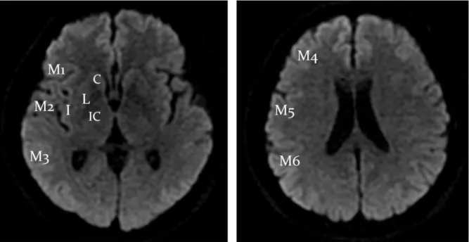
Figure 3 : score ASPECTS (25)
Territoire de l’artère cérébrale moyenne avec chaque item correspondant à 1 point : C = noyau caudé ; L = noyau lenticulaire ; IC = capsule interne ; I = Insula ; M1 à M6 = territoires corticaux de l’artère cérébrale moyenne superficielle
A = territoire de l’artère cérébrale antérieure P = territoire de l’artère cérébrale postérieure
25Barber PA, Demchuk AM, Zhang J, Buchan AM. Validity and reliability of a quantitative computed
tomography score in predicting outcome of hyperacute stroke before thrombolytic therapy. The Lancet. 13 mai 2000;355(9216):1670-4.
IRM de diffusion
La séquence de diffusion est basée sur la physique du mouvement moléculaire de l’eau dans les tissus (26). Elle montre en hypersignal les zones d’ischémie cérébrale au sein desquelles on observe une diminution du mouvement moléculaire, en lien avec l’œdème cytotoxique cellulaire qui est responsable d’un transfert d’eau depuis le secteur extra-cellulaire vers la cellule, et donc de la diminution de l’eau « libre » dans le milieu extra-cellulaire (27).
Cette diminution du mouvement moléculaire peut-être quantifiée à travers le coefficient de diffusion apparent, calculé à partir de l’application de plusieurs gradients de diffusion différents, puis représentée visuellement sur l’image de cartographie ADC. Il s’agit actuellement de la technique de détection de l’infarctus la plus sensible (88-100%) et la plus spécifique (95-(88-100%), même lors de la phase précoce de l’ischémie
(28).
De plus, la zone d’hypersignal en diffusion avec un ADC < 600 ou 620 µm²/s est considérée comme une évaluation fiable de la zone d’ischémie irréversible (29). Le score DWI-ASPECTS est une adaptation du score ASPECTS à l’IRM de diffusion (DWI = Diffusion Weighted Imaging) (30).
26 Vert C, Parra-Fariñas C, Rovira À. MR imaging in hyperacute ischemic stroke. European Journal of
Radiology. nov 2017;96:125‑32
27Kastler B, Vetter D. Comprendre l’IRM, 7th Edition. Editions Elsevier Masson. 28Latchaw RE, Alberts MJ, Lev MH, Connors JJ, Harbaugh RE, Higashida RT, et al.
Recommendations for Imaging of Acute Ischemic Stroke: A Scientific Statement From the American Heart Association. Stroke. nov 2009;40(11):3646‑78.
29Campbell BC, Purushotham A, Christensen S, Desmond PM, Nagakane Y, Parsons MW, et al. The
Infarct Core is Well Represented by the Acute Diffusion Lesion: Sustained Reversal is Infrequent. J Cereb Blood Flow Metab. janv 2012;32(1):50‑6.
30Singer OC, Kurre W, Humpich MC, Lorenz MW, Kastrup A, Liebeskind DS, et al. Risk Assessment
C’est un marqueur prédictif connu du pronostic fonctionnel dans l’AVC de la circulation antérieure avec ou sans traitement thrombolytique, mais son pouvoir prédictif n’a pas été évalué pour la thrombectomie mécanique (31, 32, 33).
Sa cotation est identique à celle du score ASPECTS scanographique (Figure 3), réalisé habituellement à partir de la séquence de diffusion b1000 comme représenté dans la figure 4.
Figure 4 : Concordance anatomique du score DWI-ASPECTS sur une séquence IRM de diffusion (b1000) (34)
C = noyau caudé ; L = noyau lenticulaire ; IC = capsule interne ; I = Insula ; M1 à M6 = territoires corticaux de l’artère cérébrale moyenne superficielle
2009;40(8):2743‑8.
31 Tei H, Uchiyama S, Usui T, Ohara K. Diffusion-weighted ASPECTS as an independent marker for
predicting functional outcome. Journal of Neurology. avr 2011;258(4):559‑65.
32Kimura K, Iguchi Y, Shibazaki K, Terasawa Y, Inoue T, Uemura J, et al. Large Ischemic Lesions on
Diffusion-Weighted Imaging Done Before Intravenous Tissue Plasminogen Activator Thrombolysis Predicts a Poor Outcome in Patients With Acute Stroke. Stroke. août 2008;39(8):2388‑91.
33Nezu T, Koga M, Kimura K, Shiokawa Y, Nakagawara J, Furui E, et al. Pretreatment ASPECTS on
DWI predicts 3-month outcome following rt-PA: SAMURAI rt-PA Registry. Neurology. 10 août 2010;75(6):555‑61.
34M. Y. Jung1, S. H. Jung1, et al. Impact of Pretreatment DWI-ASPECTS on the Outcomes of
Mechanical Thrombectomy for Acute Anterior Circulation Stroke. ECR 2013. 10.1594/ecr2013/C-0858
Du fait de la simplicité d’interprétation de l’IRM de Diffusion par rapport au scanner sans injection, le score DWI-ASPECTS semble plus performant qu’en scanner (35). Cependant il reste un score subjectif dont la reproductibilité peut être limitée notamment en centres non expert. En effet, même si la concordance inter et intra-observateur est relativement bonne, elle reste variable selon les études et les opérateurs, avec un score Kappa à environ 0.9 en centre de neuroradiologie diagnostique expert, mais rapporté à environ 0.6 dans une étude multicentrique incluant neurologues et radiologues interventionnels (36, 37).
L’avènement récent des logiciels de post-traitement d’imagerie dédiés à l’AVC a permis la mise à disposition des cliniciens de mesures automatisées, fiables et reproductibles du volume d’ischémie cérébrale en IRM de Diffusion, calculées à partir d’un seuillage de l’ADC (38).
Il existe actuellement deux logiciels spécialisés dans le sujet - iSchemaView Rapid et Olea medical – utilisés et validés dans des essais thérapeutiques puis en routine clinique. (16, 39).
35McTaggart RA, Jovin TG, Lansberg MG, Mlynash M, Jayaraman MV, Choudhri OA, et al. Alberta
Stroke Program Early Computed Tomographic Scoring Performance in a Series of Patients
Undergoing Computed Tomography and MRI: Reader Agreement, Modality Agreement, and Outcome Prediction. Stroke. févr 2015;46(2):407‑12.
36 Kellner E, Reisert M, Kiselev VG, Maurer CJ, Urbach H, Egger K. Comparison of automated and
visual DWI ASPECTS in acute ischemic stroke. Journal of Neuroradiology. sept 2019;46(5):288‑93.
37 Fahed R, Lecler A, Sabben C, Khoury N, Ducroux C, Chalumeau V, et al. DWI-ASPECTS
(Diffusion-Weighted Imaging–Alberta Stroke Program Early Computed Tomography Scores) and DWI-FLAIR (Diffusion-Weighted Imaging–Fluid Attenuated Inversion Recovery) Mismatch in Thrombectomy Candidates: An Intrarater and Interrater Agreement Study. Stroke. janv 2018;49(1):223‑7.
38Gautheron V, Xie Y, Tisserand M, Raoult H, Soize S, Naggara O, et al. Outcome After Reperfusion
Therapies in Patients With Large Baseline Diffusion-Weighted Imaging Stroke Lesions: A THRACE Trial (Mechanical Thrombectomy After Intravenous Alteplase Versus Alteplase Alone After Stroke) Subgroup Analysis. Stroke. 2018;49(3):750‑3.
39Campbell BCV, Mitchell PJ, Kleinig TJ, Dewey HM, Churilov L, Yassi N, et al. Endovascular
Therapy for Ischemic Stroke with Perfusion-Imaging Selection. New England Journal of Medicine. 11 févr 2015;150211090353006.
Leur mode de fonctionnement est similaire : le logiciel se base sur la cartographie ADC reconstruite à partir des séquences de diffusion pour extraire la zone de parenchyme cérébral dont le coefficient de diffusion apparent est inférieur à une valeur seuil prédéfinie correspondant à la zone d’ischémie constituée (600 µm²/s pour Olea et 0.620 µm²/s pour Rapid).
Le logiciel réalise cette opération sur l’ensemble des coupes disponibles, permettant une estimation du volume infarci sur l’ensemble du parenchyme cérébral.
Problématique
Il existe actuellement 2 outils fiables et reproductibles permettant l’évaluation de l’extension de l’infarctus cérébral en phase aigüe avant revascularisation : le score DWI-ASPECTS et la volumétrie automatisée.
Ces deux éléments sont des facteurs prédictifs du pronostic fonctionnel des patients traités par thrombolyse intraveineuse et/ou thrombectomie mécanique, et ont chacun leurs avantages et inconvénients (40, 41, 42).
Le score DWI-ASPECTS est un outil disponible, réalisable facilement sans logiciel supplémentaire, mais donne une évaluation qualitative subjective, dont la reproductibilité est limitée en particulier dans un environnement non spécialisé.
A l’inverse l’évaluation du volume d’infarctus en imagerie par un logiciel dédié apporte une évaluation quantitative standardisée plus fiable et parfaitement reproductible, mais dont les performances ont été essentiellement évaluées en scanner de perfusion et qui nécessite l’implémentation à grande échelle d’un logiciel relativement onéreux. Même s’il existe une bonne corrélation inverse globale entre les valeurs du DWI-ASPECTS et la volumétrie de la zone d’ischémie, surtout mise en évidence pour les valeurs extrêmes, la relation n’est pas linéaire et il persiste des discordances importantes notamment dans les valeurs intermédiaires (43). Cet élément est particulièrement important car c’est justement dans cette zone grise que les décisions
40 Šaňák D, Nosál′ V, Horák D, Bártková A, Zeleňák K, Herzig R, et al. Impact of diffusion-weighted
MRI-measured initial cerebral infarction volume on clinical outcome in acute stroke patients with middle cerebral artery occlusion treated by thrombolysis. Neuroradiology. sept 2006;48(9):632‑9.
41 Xie Y, Oppenheim C, Guillemin F, Gautheron V, Gory B, Raoult H, et al. Pretreatment Lesional
Volume Impacts Clinical Outcome and Thrombectomy Efficacy. Ann Neurol. 3 janv 2018;
42 Campbell BCV, Majoie CBLM, Albers GW, Menon BK, Yassi N, Sharma G, et al. Penumbral
imaging and functional outcome in patients with anterior circulation ischaemic stroke treated with endovascular thrombectomy versus medical therapy: a meta-analysis of individual patient-level data. The Lancet Neurology. janv 2019;18(1):46‑55.
43 De Margerie-Mellon C, Turc G, Tisserand M, Naggara O, Calvet D, Legrand L, et al. Can
de revascularisation en urgence sont difficiles et nécessitent le maximum de données pronostiques fiables, comme illustré dans l’exemple de la figure 5.
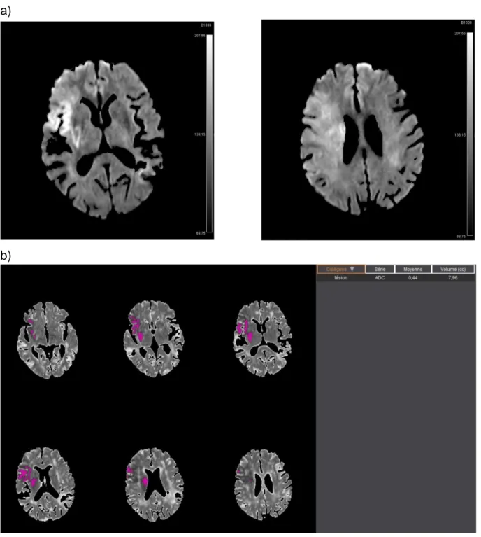
Figure 5 : Exemple de discordance entre le score DWI-ASPECTS et la volumétrie automatisée
a)
b)
a) DWI-ASPECTS = 5 avec hypersignal b1000 des territoires M1, M4, M5, insulaire et lenticulaire b) Volumétrie automatisée selon Olea Sphere 3.0 estimant le volume d’infarctus cérébral à 8 cc
Le score DWI-ASPECTS < 6 ne fait donc pas retenir une indication à un traitement endovasculaire alors que le volume reste faible. Ceci pourrait être expliqué par la précocité de la prise en charge, avec une zone d’ischémie visible en diffusion, sans chute de l’ADC au stade précoce.
Ces discordances entre les mesures pourraient s’expliquer par les limites propres de chacune :
i- le score DWI-ASPECTS est une évaluation qualitative subjective impliquant une variabilité de mesure incompressible et de possibles erreurs d’interprétation, et ii- la variabilité individuelle du délai entre l’apparition de l’hypersignal signal en B1000 et la baisse associée de l’ADC en dessous de la valeur seuil prédéfinie peut être responsable d’une sous-estimation de la lésion par rapport à l’estimation visuelle faite en B1000 (20).
Il apparait donc important d’identifier quel outil est le plus performant pour évaluer le pronostic fonctionnel après thrombectomie mécanique, afin de mieux sélectionner les patients et d’aider à la construction de futurs essais cliniques.
Objectif
L’objectif de cette étude était de comparer les performances du score DWI-ASPECTS et du volume d’infarctus en Diffusion dans la prédiction du pronostic fonctionnel des patients traités par thrombectomie mécanique dans les 8 heures après le début des symptômes d’un AVC ischémique de circulation antérieure.
INTRODUCTION
Baseline infarct extent assessment is key to proper selection of patients with acute ischemic stroke and a large vessel occlusion in anterior circulation (AIS-LVO) for endovascular reperfusion therapy (EVT) 1,2. Common practice, as well as recent guidelines, are widely based on the Alberta Stroke Program Early CT Score (ASPECTS), initially described on non-contrast CT, and secondarily adapted to Diffusion MRI (DWI-ASPECTS) 3. Recent data suggests that DWI-ASPECTS is a good and reliable tool to evaluate the infarct core before EVT 4. However it may suffer from a relatively low inter and intra-rater agreement which may hinder its predictive performance with regards to clinical outcome, especially when used in a non-expert environment, for emergent decision making and for patient selection in the setting of a clinical trial 5,6.
In this context, the automated infarct core volume quantification from Diffusion MRI data has been recently commonly adopted to reliably quantify the infarct lesion before EVT. A recent meta-analysis from the HERMES collaboration showed that the infarct core volume on Diffusion MRI was independently associated with functional outcome. Nonetheless, it requires a uniform implementation of an approved software in the regional stroke network. A recent study suggests that the infarct volume defined on CTP is better than CT-ASPECTS to predict outcomes after EVT7. However, there is a need of data comparing the ability of infract volume and ASPECTS to predict outcomes in patients who underwent MRI as initial imaging strategy before EVT.
We aimed to compare the predictive values of DWI-ASPECTS versus Diffusion infarct volume on functional outcomes in AIS-LVO patients undergoing EVT.
MATERIALS AND METHODS
Study design
We performed a retrospective analysis of our prospective clinical registry of consecutive stroke patients treated by EVT in a single comprehensive stroke center. For the purpose of this study we considered only patients treated between 2015 and 2018.
The ethical committee approved the study on March 9th, 2010 (registration number 10.677). Patients gave informed consent themselves or via a close relative for the follow-up.
Population
We included all consecutive patients with an anterior circulation AIS-LVO treated by EVT. For this study, we considered only patients screened by brain MRI and MRA on baseline imaging. Patients who underwent arterial puncture more than 90 min after imaging (picture to puncture time) were excluded from the main analysis to limit bias, accordingly with other similar studies 7. Patients with wake-up strokes or treated beyond 8hours from onset were excluded. Eligibility criteria for IVT and EVT have been previously published 8. Briefly, IVT was administrated on the basis of modified ECASS-2 (European Cooperative Acute Stroke Study) criteria without any upper age limit and EVT procedure was performed within 8 hours from onset under conscious sedation, using standard techniques (stent-retriever and/or aspiration catheters).
Clinical data
We prospectively collected demographic characteristics and vascular risk factors. The clinical severity was assessed by a senior vascular neurologist using the NIHSS immediately before MT. We recorded times of symptom onset, baseline cerebral imaging, and groin puncture. Symptomatic intracerebral hemorrhage (sICH) was defined according to ECASS-2 criteria 9. Scores on the mRS were assessed at 3-months by a stroke neurologist through formal, structured in-person interviews.
Imaging data
All images were reviewed by experienced interventional neuroradiologists blinded to clinical data. Ischemic lesions were evaluated using the DWI-ASPECT score on the last brain MRI available before EVT. To be considered as abnormal, DWI hyperintense signal had to be confluent. A small hyperintense speck was not enough 10. Inter-rater reliability of DWI-ASPECTS was assessed in a random sample of 50 patients and the weighted Kappa value was excellent (k=0.83; 95%CI, 0.76 to 0.90). Volumes of the ischemic cores were estimated using an automated software (Olea Sphere, Olea Medical SAS, La Ciotat, France) based on a thresholding method of the initial ADC maps with an upper level of 0.6 .10-3 mm2/s.
The occlusion site was assessed on initial angiogram. Recanalization was rated on the basis of findings on final angiogram using the revised TICI scale. Intracerebral hemorrhage (ICH) on day-1 follow-up MRI was analyzed according to the ECASS-2 criteria.
Outcomes
Favorable functional outcome (main outcome) was defined as a mRS score 0-2 or similar to the pre-stroke mRS at 3 months. Secondary outcomes included poor functional outcome (defined as a mRS score 4-6), sICH and parenchymal hematoma (PH type 1 or 2).
Statistical analysis
Quantitative variables are expressed as means (standard deviation, SD) in the case of normal distribution or medians (interquartile range, IQR) otherwise. Categorical variables are expressed as numbers (percentage). Normality of distributions was assessed using histograms and the Shapiro-Wilk test. Correlation between pre-treatment DWI-ASPECTS and infarct volume was assessed by calculating the Spearman’s rank correlation coefficients. We evaluated the predictive values of baseline DWI-ASPECTS and infarct volume with study outcomes (favorable outcome, poor outcome, sICH and parenchymal hematoma) using logistic regression models. As distribution of infarct volume was lognormal, log-transformed values (+1) were used. Log-linearity assumption for both DWI-ASPECTS and DWI infarct volume (log-transformed) was examined using restricted cubic spline functions and no deviation was found. Odds ratio (ORs) per 1 point increase in DWI-ASPECTS and per log increase in infarct volume were calculated as effect sizes with theirs 95% confidence intervals (CIs). We also assessed and compared the predictive abilities of pre-treatment DWI-ASPECTS and infarct volume for each study outcome by calculating the area under receiver operating characteristic curve (or c-statistic) and by using the Delong test. The associations of pre-treatment DWI-ASPECTS and infarct volume with
each study outcomes were further investigated in multivariate logistic regressions models including well known prognostic factors (namely, age, hypertension, diabetes, hypercholesterolemia, pre-treatment NIHSS score, IVT, pre-stroke mRS <2, onset-to-puncture time, picture to onset-to-puncture time and site of occlusion). Our first analyses covered the whole study group. We further investigated the prognostic values of pretreatment DWI-ASPECTS and infarct volumes on functional outcomes according to successful reperfusion status. Heterogeneity in associations across successful reperfusion subgroups was tested by introducing the corresponding multiplicative interaction term into logistic regression models. All analyses were performed in patients treated with a picture to puncture <90 minutes as main analysis and in patients treated with a picture to puncture <150 minutes as sensitivity analysis.
Statistical testing was conducted at the two-tailed α-level of 0.05. Data were analyzed using the SAS software version 9.4 (SAS Institute, Cary, NC).
RESULTS
Amongst 1156 consecutive patients treated by EVT between 2015 and 2018, 625 patients were excluded from the present study for the following reasons: posterior circulation occlusion (n=96), CT-based screening (n=71), isolated extracranial carotid occlusion (n=27), wake-up strokes/unknown time of onset or treated beyond 8 hours from onset (n=243), or picture to puncture time longer than 150 minutes (n=188). Of the 531 included patients, 277 patients with picture to puncture time <90 minutes were included in main analysis (Flow chart supplemental Figure I). Patient’s characteristics and outcomes of the study population are reported in table 1. Median DWI-ASPECTS was of 8 (IQR, 6 to 9) and median Diffusion MRI infarct volume was 11 mL (IQR, 4 to 28). As expected, DWI-ASPECTS and infarct volume were strongly negatively correlated (r=-0.83; p<0.001; supplemental Figure II). EVT led to successful recanalization in 217 patients (78.3%; 95%CI, 73.0 to 83.0%) within a median time from stroke onset of 288 minutes (IQR, 231 to 336). Overall, 25 patients had a parenchymal hematoma (9.1%, 95%CI, 6.0 to 13.1%) on day-1 follow-up MRI and 10 patients had a sICH (3.6%; 95%CI, 1.7 to 6.6%).
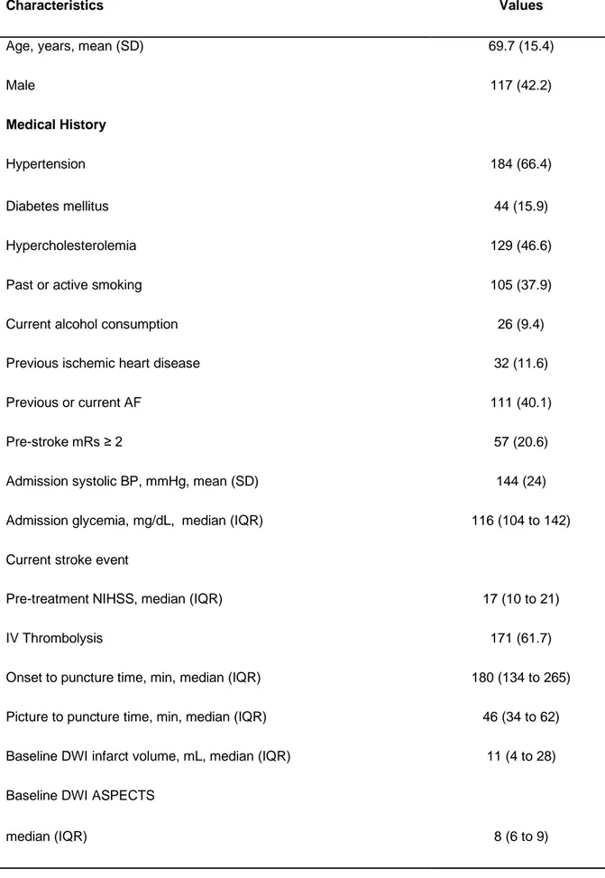
Table 1. Baseline characteristics of AIS patients treated by endovascular reperfusion therapy with picture to puncture time lower than 90 minutes (n=277)
Characteristics Values
Age, years, mean (SD) 69.7 (15.4)
Male 117 (42.2)
Medical History
Hypertension 184 (66.4)
Diabetes mellitus 44 (15.9)
Hypercholesterolemia 129 (46.6)
Past or active smoking 105 (37.9)
Current alcohol consumption 26 (9.4)
Previous ischemic heart disease 32 (11.6)
Previous or current AF 111 (40.1)
Pre-stroke mRs ≥ 2 57 (20.6)
Admission systolic BP, mmHg, mean (SD) 144 (24)
Admission glycemia, mg/dL, median (IQR) 116 (104 to 142)
Current stroke event
Pre-treatment NIHSS, median (IQR) 17 (10 to 21)
IV Thrombolysis 171 (61.7)
Onset to puncture time, min, median (IQR) 180 (134 to 265) Picture to puncture time, min, median (IQR) 46 (34 to 62) Baseline DWI infarct volume, mL, median (IQR) 11 (4 to 28) Baseline DWI ASPECTS
Values expressed as number (%) unless otherwise indicated. Favorable outcome was defined as a 90-day mRS score of 0–2 or equal to pre-stroke mRs. Poor outcome was defined as a 90-90-day mRS score of 4-6.
Abbreviations: AIS=acute ischemic stroke; ASPECTS=Alberta stroke program early computed tomography score; AF=atrial fibrillation; BP=blood pressure; DWI=diffusion-weighted imaging; ECASS II=European Co-operative Acute Stroke Study II; ICA=internal carotid artery; IQR=interquartile range; MCA=middle cerebral artery; mRs=modified Rankin scale; NIHSS=National Institutes of Health Stroke Scale; SD=standard deviation; sICH=symptomatic intracerebral hemorrhage.
≤ 5 60 (21.7)
Site of occlusion on initial angiogram
M1-MCA 131 (47.3) M2-MCA 57 (20.6) Intracranial ICA 49 (17.7) Tandem 40 (14.4) Outcomes Successful reperfusion 217 (78.3) Favorable outcome 144 (52.0) Poor outcome 93 (33.6) Mortality at 3 months 49 (17.7) sICH (ECASS 2) 10 (3.6) Parenchymal hematoma 25 (9.1)
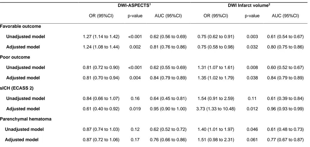
Prognostic values of pre-treatment DWI- ASPECTS and infarct volume on functional outcomes
As shown in table 2, the predictive abilities of DWI-ASPECTS and infarct volume for favorable and poor outcomes were comparable, with an AUC of 0.62 for DWI-ASPECTS and 0.61 for infarct volume (p=0.61 for favorable outcome and p=0.57 for poor outcome). Multivariate analyses found that both DWI-ASPECTS and infarct volume were significant predictors of favorable and poor outcomes; the adjusted OR (95%CI) for favorable outcome was 1.24 (1.08 to 1.44) per point increase in DWI-ASPECTS and 0.75 (95%CI, 0.58 to 0.98) per 1 log increase in infarct volume; for poor outcome the corresponding adjusted ORs (95%CI) were 0.81 (0.70 to 0.94) and 1.35 (95%CI, 1.02 to 1.79), respectively. The predictive abilities of both multivariable models were similar: the AUC for predicting favorable outcome were 0.81 for DWI-ASPECTS and 0.80 for infarct volume (p=0.46), and the AUC for predicting poor outcome were 0.84 for both DWI-ASPECTS and infarct volume (p=0.17).
Table 2. Association of pre-treatment DWI- ASPECTS and infarct volume with functional outcomes and hemorrhagic complications in AIS patients treated by mechanical thrombectomy with picture to puncture time lower than 90 minutes
DWI-ASPECTS1 DWI Infarct volume2
OR (95%CI) p-value AUC (95%CI) OR (95%CI) p-value AUC (95%CI)
Favorable outcome Unadjusted model 1.27 (1.14 to 1.42) <0.001 0.62 (0.56 to 0.69) 0.75 (0.62 to 0.91) 0.003 0.61 (0.54 to 0.67) Adjusted model 1.24 (1.08 to 1.44) 0.002 0.81 (0.76 to 0.86) 0.75 (0.58 to 0.98) 0.032 0.80 (0.75 to 0.86) Poor outcome Unadjusted model 0.81 (0.72 to 0.90) <0.001 0.62 (0.55 to 0.69) 1.31 (1.07 to 1.61) 0.008 0.60 (0.52 to 0.67) Adjusted model 0.81 (0.70 to 0.94) 0.004 0.84 (0.79 to 0.89) 1.35 (1.02 to 1.79) 0.038 0.84 (0.79 to 0.89) sICH (ECASS 2) Unadjusted model 0.84 (0.66 to 1.07) 0.16 0.64 (0.45 to 0.81) 1.54 (0.91 to 2.59) 0.11 0.61 (0.39 to 0.84) Adjusted model 0.61 (0.40 to 0.92) 0.019 0.95 (0.90 to 1.00) 3.73 (1.33 to 10.48) 0.012 0.96 (0.93 to 0.99) Parenchymal hematoma Unadjusted model 0.87 (0.74 to 1.03) 0.12 0.62 (0.52 to 0.72) 1.40 (1.01 to 1.97) 0.046 0.61 (0.48 to 0.73) Adjusted model 0.87 (0.72 to 1.06) 0.17 0.76 (0.66 to 0.86) 1.51 (0.98 to 2.31) 0.061 0.77 (0.67 to 0.87)
1OR calculated per 1-point increase; 2OR calculated per 1 log increase. Adjusted models included the following specified covariates: age, sex, hypertension, diabetes, hypercholesterolemia,
pre-treatment NIHSS, pre stroke mRS <2, site of occlusion, IV thrombolysis, onset to puncture time and picture to puncture time.
Abbreviations: ASPECTS=Alberta stroke program early computed tomography score; AUC=area under the receiver operating characteristic curve (or c-statistic) ; CI=confidence interval; DWI=diffusion-weighted imaging; ECASS II=European Co-operative Acute Stroke Study II; OR=odds ratio; sICH=symptomatic intracerebral hemorrhage.
28 F an tini Ch ris to ph e r
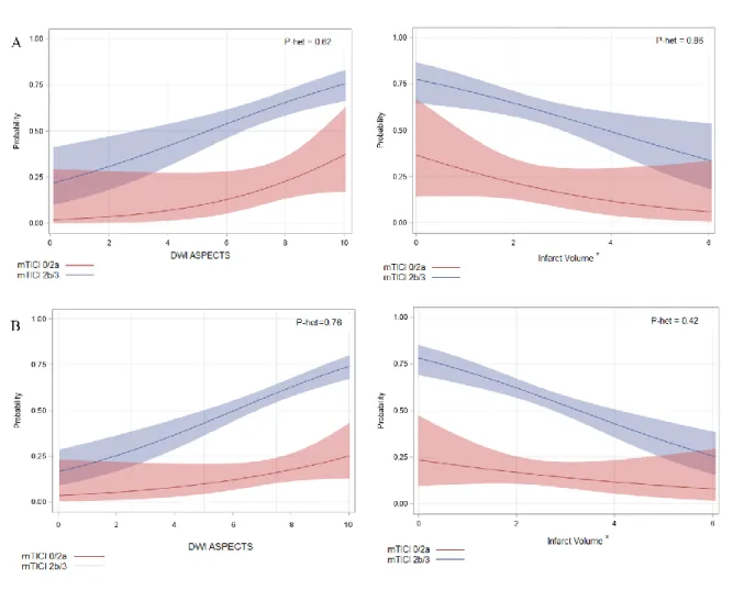
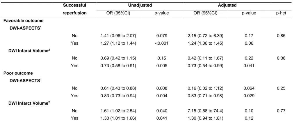
As shown in table 3 and figure 1A, the association of DWI-ASPECTS and infarct volume with favorable and poor outcomes did not differ significantly according to successful reperfusion status after EVT in both unadjusted and pre-specified adjusted analyses (all p for heterogeneity>0.20).
Figure 1. Association of pre-treatment DWI- ASPECTS and infarct volume with favorable outcome according to successful reperfusion status in AIS patients treated by mechanical thrombectomy with picture to puncture time lower than 90 minutes (A) or picture to puncture time lower than 150 minutes (B).
Solid line shows predicted probability of favorable outcome and shaded region show 95%CI of predicted probability calculating using unadjusted logistic regression model including DWI-ASPECTS (or DWI-infarct volume), reperfusion status and the multiplicative interaction term. *log-transformed value (+1). p het indicates the P-values for heterogeneity (interaction) in associations of favorable outcome with DWI-ASPECTS and infarct volume across the reperfusion subgroups
Table 3. Association of pre-treatment DWI- ASPECTS and infarct volume with functional outcomes according to successful reperfusion status in AIS patients treated by mechanical thrombectomy with picture to puncture time lower than 90 minutes.
Successful Unadjusted Adjusted
reperfusion OR (95%CI) p-value OR (95%CI) p-value p-het
Favorable outcome
DWI-ASPECTS1
No 1.41 (0.96 to 2.07) 0.079 2.15 (0.72 to 6.39) 0.17 0.85
Yes 1.27 (1.12 to 1.44) <0.001 1.24 (1.06 to 1.45) 0.06
DWI Infarct Volume2
No 0.69 (0.42 to 1.15) 0.15 0.42 (0.11 to 1.67) 0.22 0.38 Yes 0.73 (0.58 to 0.91) 0.005 0.73 (0.54 to 0.99) 0.041 Poor outcome DWI-ASPECTS1 No 0.61 (0.43 to 0.88) 0.008 0.16 (0.02 to 1.12) 0.064 0.25 Yes 0.83 (0.73 to 0.94) 0.004 0.83 (0.71 to 0.98) 0.029
DWI Infarct Volume2
No 1.61 (1.02 to 2.54) 0.040 7.15 (0.68 to 74.4) 0.10 0.77
Yes 1.30 (1.01 to 1.66) 0.041 1.30 (0.94 to 1.81) 0.12
1OR calculated per 1-point increase; 2OR calculated per 1 log increase. Adjusted models included pre-specified covariates: age, sex, hypertension, diabetes, hypercholesterolemia, pre-treatment
NIHSS, pre stroke mRS <2, site of occlusion, IV thrombolysis, onset to groin puncture time and imaging to puncture time. p-het indicates P-Values for heterogeneity in association of functional outcomes with DWI-ASPECTS and Infarct volume across the reperfusion subgroups
Abbreviations: ASPECTS=Alberta stroke program early computed tomography score; CI=confidence interval; DWI=diffusion-weighted imaging; OR=odds ratio
30 F a n tin i Ch risto p h e r
31 Prognostic values of DWI-ASPECTS and infarct volume on clinically relevant hemorrhagic complications.
Predictive abilities of DWI-ASPECTS and infarct volume for sICH did not differ significantly, with an AUC of 0.64 for DWI-ASPECTS and of 0.61 for infarct volume (p=0.90). Similar results were found for predicting parenchymal hematoma, with an AUC of 0.62 for DWI-ASPECTS and of 0.61 for infarct volume (table 2). In univariate logistic regression models, only the association between parenchymal hematoma and infarct volume reached the statistical significance level. After adjustment, DWI-ASPECTS and infarct volume were significantly associated with sICH only, with an adjusted OR of 0.61 (95%CI, 0.40 to 0.92) per point increase in DWI-ASPECTS and 3.73 (95%CI, 1.33 to 10.48) per 1 log increase in infarct volume. No significant association was found with parenchymal hematoma. Predictive abilities of both multivariable models with DWI-ASPECTS or infarct volume were similar (table 2; p>0.75).
Sensitivity analyses in patients with a picture to puncture time lower than 150 min.
Patient’s characteristics and outcomes of the 531 patients with picture to puncture within 150 minutes are reported in supplemental Table I. Similar results were found about the predictive ability of DWI-ASPECTS and infarct volumes on functional outcomes (favorable and poor outcomes), sICH and parenchymal hematoma. In multivariate analyses, both DWI-ASPECTS and infarct volumes were significantly associated with each outcome (table 4), with similar effect sizes as the main analysis. We did not find any significant heterogeneity in functional outcomes associations (p for heterogeneity>0.19, supplemental table II and figure IIB).
Table 4. Association of pre-treatment DWI- ASPECTS and infarct volume with functional outcomes and hemorrhagic complications in AIS patients treated by mechanical thrombectomy with picture to puncture time lower than 150 minutes.
DWI-ASPECTS1 Infarct volume2
OR (95%CI) p-value AUC (95%CI) OR (95%CI) p-value AUC (95%CI)
Favorable outcome Unadjusted model 1.29 (1.18 to 1.40) <0.001 0.62 (0.58 to 0.67) 0.71 (0.62 to 0.82) <0.001 0.62 (0.57 to 0.67) Adjusted model 1.24 (1.12 to 1.37) <0.001 0.80 (0.76 to 0.84) 0.72 (0.60 to 0.88) <0.001 0.80 (0.77 to 0.84) Poor outcome Unadjusted model 0.81 (0.75 to 0.88) <0.001 0.61 (0.56 to 0.66) 1.31 (1.13 to 1.51) <0.001 0.59 (0.54 to 0.64) Adjusted model 0.80 (0.72 to 0.89) <0.001 0.83 (0.79 to 0.86) 1.44 (1.18 to 1.77) <0.001 0.83 (0.79 to 0.86) sICH (ECASS 2) Unadjusted model 0.76 (0.67 to 0.87) <0.001 0.69 (0.60 to 0.79) 1.68 (1.24 to 2.29) <0.001 0.67 (0.57 to 0.77) Adjusted model 0.70 (0.60 to 0.83) <0.001 0.80 (0.73 to 0.87) 1.99 (1.36 to 2.92) <0.001 0.80 (0.73 to 0.87) Parenchymal hematoma Unadjusted model 0.83 (0.75 to 0.92) <0.001 0.64 (0.57 to 0.70) 1.51 (1.22 to 1.87) <0.001 0.64 (0.58 to 0.71) Adjusted model 0.85 (0.75 to 0.95) 0.006 0.74 (0.68 to 0.80) 1.55 (1.20 to 2.01) 0.001 0.74 (0.69 to 0.80)
1OR calculated per 1-point increase; 2OR calculated per 1 log increase. Adjusted models included the following specified covariates: age, sex, hypertension, diabetes, hypercholesterolemia,
pre-treatment NIHSS, pre stroke mRS <2, site of occlusion, IV thrombolysis, onset to puncture time, and picture to puncture time.
Abbreviations: ASPECTS=Alberta stroke program early computed tomography score; AUC=area under the receiver operating characteristic curve (or c-statistic) ; CI=confidence interval; DWI=diffusion-weighted imaging; ECASS II=European Co-operative Acute Stroke Study II; OR=odds ratio sICH=symptomatic intracerebral hemorrhage.
F a n tin i Ch risto p h e r 32
33
DISCUSSION
In a retrospective analysis, we aimed to evaluate the performance of DWI-ASPECTS and DWI lesion volume to predict 3-months functional outcome and hemorrhagic transformation in stroke patients undergoing EVT within 8 hours from symptom onset. We found that (a) DWI-ASPECTS and DWI lesion volume were independently associated with clinical outcome at 3 months and sICH; (b) the predictive ability of DWI-ASPECTS and DWI lesion volume for each outcome were similar.
Several studies have reported DWI-ASPECTS as a key determinant of clinical outcome and sICH risk after IVT 11, but there is a lack of data on its clinical relevance to select stroke patients for EVT 10. Most Diffusion MRI data come from one trial (THRACE 12). In the HERMES collaboration meta-analysis of individual patient data from five RCTs 13, lower ASPECTS on baseline imaging was associated with lower rates of good clinical outcome and the treatment effect on functional outcome was similar between patients with high baseline ASPECTS (9-10) and those with moderate ASPECTS (6-8). Most patients with low baseline ASPECTS (0-5) were excluded from RCTs and the potential benefit from EVT in this subgroup of patients is being investigated in ongoing trials (LASTE, NCT03811769 and TENSION, NCT03094715). Despite Diffusion MRI being more sensitive than CT on early brain ischemia detection, Fahed et al. found mild inter and intra-rater agreement between physicians scoring DWI-ASPECTS in stroke patients referred for EVT 4. In our study, the inter-rater agreement was higher, which is consistent with findings from Mc Taggart and al. 14. However, DWI-ASPECT were scored by trained physicians that are routinely used to ASPECT scoring on DWI.
34 assessed on DWI before EVT, was independently associated with functional independence, but did not modify the benefit from EVT. A 10mL increase of baseline ischaemic core volume was estimated to reduce the odds of favourable outcome by 20-30% 15. This data supports our findings depicted in Figure 1, suggesting that DWI volume is negatively correlated with the rate of good outcome, and that the benefit from recanalization might persist in selected patients with large volume on baseline imaging. Our study showed strong correlation between DWI-ASPECTS and DWI volume (r=-0.83; p<0.001). Similar finding was described in 2013 by De Margerie-Mellon et al. 16, which suggested DWI-ASPECTS <4 or ≥7 may be used as reliable surrogates of DWI volume >100 or <70 mL, respectively. DWI volume was however estimated using a semiautomated method based on DWI signal intensity thresholding 17, which differs from current FDA-approved automated methods (iSchemaView RAPID or Olea Sphere 3.0), based on apparent diffusion coefficient thresholding. DWI volume estimated by such automated software may overcome the potential slight inter-rater agreement of DWI-ASPECTS, whereas its use in routine might be limited by the software availability in each stroke center.
We identified a probable higher range of lesion volume values in patients with lower DWI-ASPECT scores (supplemental figure II). This discrepancy might be explained by the visualization of early diffusion hyper intensity without drop of ADC under the pre-specified threshold in large patients that may present a “fast progressor” profile 18. Further studies are required to investigate the performances of DWI-ASPECTS and infarct volume in patients with large infarct core.
This study has several limitations. First, this is a retrospective analysis from a prospective registry in a single comprehensive stroke centre. Second, patients with baseline large infarct core were under-represented (20%), which limits interpretation
35 of data in this subgroup of patients. Third, we decided to include in the main analysis only patients treated within a short timeframe from baseline MRI (within 90 min) to limit potential bias and the influence of the imaging-to-reperfusion time. However, the sensitivity analysis performed in a larger number of patients - treated within 150 min from baseline imaging - confirmed the initial findings.
36
SUMMARY
DWI-ASPECTS has similar prognosis performance as infarct volume in AIS-LVO patients undergoing EVT within 8 hours from stroke onset, and may be used as a less resource-intensive tool for routine imaging assessment of infarct size prior to EVT. More data are needed for patients with large pre-treatment volumes.
37
ANNEXES
38
Supplemental Figure II. Scatterplots between pre-treatment DWI-ASPECTS and infarct volume in AIS patients treated by mechanical thrombectomy with picture to puncture time lower than 90 minutes (A) or picture to puncture time lower than 150 minutes (B).
A)
B)
39
Supplemental Table I. Baseline characteristics of AIS patients treated by mechanical thrombectomy with picture to puncture time lower than 150 minutes (n=531)
Characteristics Values
Age, years, mean (SD) 69.5 (15.4)
Male 238 (44.8)
Medical History
Hypertension 355 (66.9)
Diabetes mellitus 90 (16.9)
Hypercholesterolemia 229 (43.1)
Past or active smoking 204 (38.4)
Current alcohol consumption 58 (10.9)
Previous ischemic heart disease 62 (11.7)
Previous or current AF 222 (41.8)
Pre-stroke mRs ≥ 2 122 (23.0)
Admission systolic BP, mmHg, mean (SD) 145 (24)
Admission glycemia, mg/dL, median (IQR) 120 (105 to 150)
Current stroke event
Pre-treatment NIHSS, median (IQR) 17 (11 ; 21)
IV Thrombolysis 356 (67.0)
Onset to puncture time, min, median (IQR) 217 (163 to 270) Picture to puncture time, min, median (IQR) 86 (45 to 125) Pre-treatment DWI Infarct volume, mL, median (IQR) 11 (4 to 30) Pre-treatment DWI-ASPECTS
40
Values expressed as number (%) unless otherwise indicated.
Abbreviations: AIS=acute ischemic stroke; AF=atrial fibrillation; BP=blood pressure; ECASS II=European Cooperative Acute Stroke Study II; ICA=internal carotid artery; IQR=interquartile range; MCA=middle cerebral artery; SD=standard deviation, sICH=symptomatic intracerebral hemorrhage.
≤ 5 124 (23.4)
Site of Occlusion on initial angiogram
M1-MCA 264 (49.7) M2-MCA 91 (17.1) Intracranial ICA 92 (17.3) Tandem 84 (15.8) Outcomes Successful reperfusion 415 (78.2) Favorable outcome 255 (48.0) Poor outcome 206 (38.8) Mortality at 3 months 107 (20.2) sICH (ECASS 2) 35 (6.7) Parenchymal hematoma 73 (13.9)
Supplemental Table 2. Association of pre-treatment DWI- ASPECTS and infarct volume with functional outcomes according to successful reperfusion status in AIS patients treated by mechanical thrombectomy with picture to puncture time lower than 150 minutes .
Successful Unadjusted Adjusted
reperfusion OR (95%CI) p-value OR (95%CI) p-value p-het
Favorable outcome DWI-ASPECTS1 No 1.25 (0.95 to 1.64) 0.11 1.31 (0.82 to 2.08) 0.26 0.44 Yes 1.30 (1.19 to 1.43) <0.001 1.26 (1.13 to 1.42) <0.001 Infarct Volume2 No 0.81 (0.54 to 1.21) 0.31 0.86 (0.42 to 1.74) 0.67 0.19 Yes 0.68 (0.57 to 0.80) <0.001 0.70 (0.57 to 0.87) 0.001 Poor outcome DWI-ASPECTS1 No 0.76 (0.60 to 0.95) 0.017 0.53 (0.31 to 0.91) 0.022 0.81 Yes 0.80 (0.73 to 0.88) <0.001 0.81 (0.72 to 0.91) <0.001 Infarct Volume2 No 1.27 (0.91 to 1.77) 0.17 2.52 (1.11 to 5.70) 0.027 0.67 Yes 1.36 (1.14 to 1.63)) <0.001 1.42 (1.12 to 1.79) 0.004
1OR calculated per 1-point increase; 2OR calculated per 1 log increase. Adjusted models included the following pre-specified covariates: age, sex,
hypertension, diabetes, hypercholesterolemia, pre-treatment NIHSS, pre-stroke mRS <2, site of occlusion, IV thrombolysis, onset to puncture time and picture to puncture time. p-het indicates P-Values for heterogeneity in associations of functional outcomes with DWI-ASPECTS or Infarct volume parameters across reperfusion status
Abbreviations: CI=confidence interval; OR=odds ratio.
41 F a n tin i Ch risto p h e r
42
REFERENCES
1. Albers GW, Marks MP, Kemp S, et al. Thrombectomy for Stroke at 6 to 16 Hours with Selection by Perfusion Imaging. New England Journal of Medicine 2018;378:708–18.
2. Nogueira RG, Jadhav AP, Haussen DC, et al. Thrombectomy 6 to 24 Hours after Stroke with a Mismatch between Deficit and Infarct. New England Journal of Medicine 2018;378:11–21.
3. Barber PA, Hill MD, Eliasziw M, et al. Imaging of the brain in acute ischaemic stroke: comparison of computed tomography and magnetic resonance diffusion-weighted imaging. J Neurol Neurosurg Psychiatry 2005;76:1528–33.
4. Fahed R, Lecler A, Sabben C, et al. DWI-ASPECTS (Diffusion-Weighted Imaging–Alberta Stroke Program Early Computed Tomography Scores) and DWI-FLAIR (Diffusion-Weighted Imaging– Fluid Attenuated Inversion Recovery) Mismatch in Thrombectomy Candidates: An Intrarater and Interrater Agreement Study. Stroke 2018;49:223–7.
5. Schröder J, Thomalla G. A Critical Review of Alberta Stroke Program Early CT Score for Evaluation of Acute Stroke Imaging. Frontiers in Neurology 2017;7.
6. Gupta AC, Schaefer PW, Chaudhry ZA, et al. Interobserver Reliability of Baseline Noncontrast CT Alberta Stroke Program Early CT Score for Intra-Arterial Stroke Treatment Selection. American Journal of Neuroradiology 2012;33:1046–9.
7. Demeestere J, Scheldeman L, Cornelissen SA, et al. Alberta Stroke Program Early CT Score Versus Computed Tomographic Perfusion to Predict Functional Outcome After Successful Reperfusion in Acute Ischemic Stroke. Stroke 2018;49:2361–7.
8. Ferrigno M, Bricout N, Leys D, et al. Intravenous Recombinant Tissue-Type Plasminogen Activator: Influence on Outcome in Anterior Circulation Ischemic Stroke Treated by Mechanical Thrombectomy. Stroke 2018;49:1377–85.
9. Hacke W, Kaste M, Fieschi C, et al. Randomised double-blind placebo-controlled trial of thrombolytic therapy with intravenous alteplase in acute ischaemic stroke (ECASS II). The Lancet 1998;352:1245– 51.
10. Desilles J-P, Consoli A, Redjem H, et al. Successful Reperfusion With Mechanical Thrombectomy Is Associated With Reduced Disability and Mortality in Patients With Pretreatment Diffusion-Weighted Imaging–Alberta Stroke Program Early Computed Tomography Score ≤6. Stroke 2017;48:963–9. 11. Nezu T, Koga M, Kimura K, et al. Pretreatment ASPECTS on DWI predicts 3-month outcome
following rt-PA: SAMURAI rt-PA Registry. Neurology 2010;75:555–61.
12. Bracard S, Ducrocq X, Mas JL, et al. Mechanical thrombectomy after intravenous alteplase versus alteplase alone after stroke (THRACE): a randomised controlled trial. Lancet Neurol 2016;15:1138– 47.
13. Goyal M, Menon BK, Van Zwam WH, et al. Endovascular thrombectomy after large-vessel ischaemic stroke: a meta-analysis of individual patient data from five randomised trials. The Lancet 2016;387:1723–1731.
43 14. McTaggart RA, Jovin TG, Lansberg MG, et al. Alberta Stroke Program Early Computed Tomographic Scoring Performance in a Series of Patients Undergoing Computed Tomography and MRI: Reader Agreement, Modality Agreement, and Outcome Prediction. Stroke 2015;46:407–12.
15. Campbell BCV, Majoie CBLM, Albers GW, et al. Penumbral imaging and functional outcome in patients with anterior circulation ischaemic stroke treated with endovascular thrombectomy versus medical therapy: a meta-analysis of individual patient-level data. The Lancet Neurology 2019;18:46– 55.
16. de Margerie-Mellon C, Turc G, Tisserand M, et al. Can DWI-ASPECTS Substitute for Lesion Volume in Acute Stroke? Stroke 2013;44:3565–7.
17. Labeyrie M-A, Turc G, Hess A, et al. Diffusion Lesion Reversal After Thrombolysis: A MR Correlate of Early Neurological Improvement. Stroke 2012;43:2986–91.
18. Rocha M, Jovin TG. Fast Versus Slow Progressors of Infarct Growth in Large Vessel Occlusion Stroke: Clinical and Research Implications. Stroke 2017;48:2621–7.
44
CONCLUSION GENERALE
Notre étude apporte des précisions sur les performances pronostiques du score DWI-ASPECTS et de la volumétrie automatisée dans la prise en charge initiale des patients présentant un AVC ischémique traités par thrombectomie mécanique.
Dans notre cohorte de patients traités dans les 8 heures, ces deux méthodes d’estimation de l’étendue de l’infarctus cérébral présentent de bonnes performances pronostiques, sans différences statistiquement significatives. Le score DWI-ASPECTS peut donc être utilisé en routine clinique dans les centres ne disposant pas de logiciel dédié à la volumétrie automatique. Des données complémentaires sont cependant nécessaires pour mieux définir les performances de ces outils dans la population des patients avec un large infarctus.
AUTEUR : Nom : FANTINI Prénom : Christopher Date de soutenance : 26/05/2020
Titre de la thèse : DWI-ASPECTS vs volumétrie automatisée de l’infarctus cérébral pour la prédiction du devenir fonctionnel après traitement par thrombectomie mécanique de l’AVC ischémique
Thèse - Médecine - Lille - 2020
Cadre de classement : DES Radiodiagnostic et imagerie médicale
Mots-clés : AVC, DWI-ASPECTS, volumétrie, Thrombectomie Mécanique, devenir fonctionnel, transformation hémorragique
Résumé :
Introduction : Notre objectif était de comparer le score DWI-ASPECTS (Diffusion Weighted Imaging–Alberta Stroke Program Early CT Score) au volume d’infarctus cérébral dans la prédiction du pronostic clinique des patients traités par thrombectomie mécanique dans les 8 heures du début des signes.
Méthodes : Tous les patients consécutifs avec une IRM de Diffusion avant thrombectomie ont été identifiés dans un registre prospectif de 2015 à 2018. Le volume d’infarctus était estimé avec un logiciel de volumétrie automatique dédié. Nous avons étudié les associations entre le score DWI-ASPECTS et le volume avec les pronostics favorable (mRS 0-2) et défavorable (mRS 4-6) à 3 mois en utilisant des modèles de régression logistique.
Résultats : Parmi les 1156 traités par thrombectomie mécanique, 277 respectaient les critères d’inclusion. En analyse univariée, le score DWI-ASPECTS et le volume étaient tous deux corrélés au bon pronostic fonctionnel (p<0.001 et p=0.003 respectivement) ainsi qu’au mauvais pronostic (p<0.001 et p=0.008 respectivement). Les deux modèles d’analyse multivariée étaient équivalents dans la prédiction du bon pronostic des patients, avec des aires sous la courbe comparables (AUC) : 0.81 pour DWI-ASPECTS et 0.80 pour le volume (p=0.46). Les performances étaient également équivalentes dans la prédiction du mauvais pronostic fonctionnel : AUC = 0.84 pour les deux groupes (p=0.17).
Conclusions : Le score DWI-ASPECTS et le volume ont des performances similaires de prédiction du pronostic fonctionnel des patients pris en charge par thrombectomie mécanique dans les 8 heures.
Composition du Jury :
Président : Pr. Jean-Pierre Pruvo
Assesseurs : Pr. Xavier LECLERC, Dr. François PONTANA Directeur de thèse : Dr. Nicolas BRICOUT