Biotechnol. Agron. Soc. Environ. 2002 6 (2), 109–119
1. INTRODUCTION
Major production constraints of the common bean, Phaseolus vulgaris L. in Latin America and Africa regions are Ascochyta blight, Bean Golden Mosaic Virus (BGMV) and Bean Fly. Good sources of resistance have been found mainly in species such as P h a s e o l u s c o c c i n e u s L. and P h a s e o l u s p o l y a n t h u s Greenm. (Baudoin et al., 1992). A critical case study is the introgression of Ascochyta blight resistance from P. polyanthus into the common bean P. vulgaris. In crosses between P. polyanthus and P. vulgaris, the use of P. polyanthus cytoplasm avoids a quick reversal to the recurrent parent P. vulgaris (Baudoin et al., 1992). Some interspecific crosses have been attempted by Lecomte (1997) between P. p o l y a n t h u s ( ) and
P. vulgaris. Although fertilized ovules were obtained, up to 60% of globular embryos failed to develop due to undefined incompatibility barriers between embryo and mother plant. Abortion of the embryo occurred within three to five days, at early heart-shaped or globular stages (Baudoin et al., 1992). Out of more than 3000 crosses, only one hybrid with a low seed production could be obtained by Camarena and Baudoin (1987) using embryo culture. This hybrid is the only one obtained so far when using P. polyanthus as a female.
There are two alternative methods to develop new interspecific hybrids using P. polyanthus as mother parent:
– increasing the number of pollinations and
– rescuing the hybrid embryos at appropriate stage.
Study of the early abortion in reciprocal crosses between
Phaseolus vulgaris L. and Phaseolus polyanthus Greenm.
Pascal Geerts, André Toussaint, Guy Mergeai, Jean-Pierre Baudoin
Unité de Phytotechnie tropicale et d’Horticulture. Faculté universitaire des Sciences agronomiques de Gembloux. Passage des Déportés, 2. B–5030 Gembloux (Belgium). E-mail : toussaint.a@fsagx.ac.be.
Received 25 avril 2001, accepted 18 mars 2002.
The causes of early embryo abortion in the reciprocal crosses between Phaseolus vulgaris L. and P h a s e o l u s polyanthus Greenm. were studied. Methacrylate resin sections, 2 µm thick, of 2 to 6 day-old hybrid seeds were used to examine the stage of embryo development and the state of seed tissues. These observations permitted the determination of the main causes of abortion and the developmental stages at which interspecific embryos should be rescued. Early nutritional barriers in P. polyanthus ( ) x P. vulgaris crosses are related to a deficient endosperm development while in reciprocal crosses, endothelium proliferation and, in some extent, hypertrophy of the vascular elements might be the main causes of early embryo abortion. The importance of the abnormalities observed during embryo development depended to a great extent on the compatibility between the genotypes crossed. Results also suggest that the appropriate time for embryo rescue when P. polyanthus is a female partner is at the early globular stage.
K e y w o r d s . P h a s e o l u s v u l g a r i s L., P h a s e o l u s p o l y a n t h u s Greenm., interspecific hybridization, embryo mortality, incompatibility barriers.
Étude de l’avortement précoce chez les croisements réciproques entre Phaseolus vulgaris L. et Phaseolus polyanthus Greenm. Des coupes semi-fines de 2µm d’épaisseur, réalisées dans des graines âgées de 2 à 6 jours enrobées dans une résine du type méthacrylate, ont permis d’analyser l’origine de l’avortement précoce constaté lors des hybridations réciproques entre Phaseolus vulgaris L. et Phaseolus polyanthus Greenm. Lors du croisement de P. polyanthus ( ) x P. vulgaris, des barrières nutritionnelles s’établissent rapidement en raison d’un développement réduit de l’albumen. Dans le croisement inverse, la prolifération de l’endothélium et, dans une certaine mesure, l’hypertrophie du tissu vasculaire entraînent également l’avortement précoce des embryons. L’importance des défauts constatés au cours du développement embryonnaire est largement liée à la compatibilité entre les génotypes croisés. Lorsque P. polyanthus est utilisé comme parent femelle, les résultats suggèrent qu’un sauvetage des embryons devrait être envisagé dès qu’ils atteignent le stade globulaire.
Mots-clés. Phaseolus vulgaris L., Phaseolus polyanthus Greenm., hybridation interspécifique, mortalité de l’embryon, barrières d’incompatibilité.
The first alternative depends upon the parental combinations and the environmental conditions. The second alternative requires improved in vitro culture techniques and a better knowledge of embryo abortion causes.
Several causes have been suggested as the main factors preventing the production of hybrid seeds. These include the absence of pollen germination, egg f e r t i l i t y, incompatibility, endosperm degeneration, proliferation of endothelium (Chen, Adachi, 1995), growth of nucellus into the embryo sac squeezing the embryo and restricting its development (Singh, 1998), hardening of nucellar tissues, embryological and endosperm imbalances (Johnston e t a l ., 1980; Masuelli, Camadro, 1997), hypertrophy of vascular and nucellar cells, and detachment of nucellar cells from maternal tissue at chalazal end (Lecomte et al., 1998). In the case of P. v u l g a r i s × P h a s e o l u s lunatus L. crosses, Kuboyama et al. (1991) found that pre-fertilization events (inhibition of pollen germination and pollen tube growth) did not constitute a reproductive barrier in P h a s e o l u s. Post-zygotic studies of embryo development of interspecific hybrids within the genus Phaseolus indicated that reduced endosperm development resulted in starvation of developing embryos and finally to their abortion (Hucl, Scoles, 1985). A comparative histological study was carried out by Lecomte et al. (1998) on ovule development. They showed that P. p o l y a n t h u s embryos developed more slowly and had bigger suspensor basal cells than P. vulgaris embryos. These observations suggested a higher nutritive requirement in P. polyanthus than in P. vulgaris. Lecomte et al. (1998) concluded that differences in the absorption rates of nutrients of parental embryos and hybrid embryos do not favor the hybrid P. polyanthus ( )× P. vulgaris.
The aim of this paper is to elucidate causes of embryo abortion in crosses between P. vulgaris ( )× P. polyanthus and in the reciprocal crosses. Events such as primary endosperm nucleus (PEN) development, endosperm degeneration, endothelium proliferation, nucellus ingrowth and cell hypertrophy are analysed.
2. MATERIALAND METHODS
A wild (G 21245) and a cultivated (NI 637) genotype of P. vulgaris and two cultivated genotypes (NI 1015 and G 35348) of P. polyanthus from the Phaseolineae active collection held at the Gembloux Agricultural University and at the Centro Internacional de Agricultura Tropical (CIAT) were used. Accessions were selected on the basis of their good ability to flower in growth chamber conditions.
2.1. Hybridization
The experiment was made from January to December 1999. Hand-pollination was carried out between 8.00 and 10.00 a.m. under growth chamber conditions (24°C/20°C day/night temperature, 70% relative humidity, 580 µE m-2sec-1 light intensity, and 11h30 min day length). Different combinations were made between the selected genotypes using either P. v u l g a r i s or P. polyanthus as female partner. To ensure pollen germination on the stigma, particularly when using P. polyanthus as a female, inflorescences were covered by a humid bag after pollination. Since all pods obtained by crossing P. polyanthus ( ) ×P. vulgaris aborted between 5 to 7 days after pollination (DAP), seeds were collected every day from 1 to 6 DAP, from auto-pollinated flowers and flowers pollinated by either P. v u l g a r i s or P. p o l y a n t h u s. Histological observations were made from these seeds.
2.2. Histological studies
For each cross, five pods of maternal genotypes (P. p o l y a n t h u s or P. v u l g a r i s), containing three or seven seeds respectively, were inspected daily from 2 to 6 DAP. In order to confirm pollen germination, stigmatic surfaces were removed 2 DAP and were immediately incubated in Aniline Blue solution (0.01 % in 0.1 M PO4buffer, pH 9) for 10 min (Ruzin, 1999). Under UV excitation, yellow fluorescence indicates pollen germination.
Seeds from parental genotypes and from interspecific crosses were freshly harvested and eventually nicked with a scalpel to facilitate penetration of fixing and embedding solutions. Samples were fixed in 1.2 % glutaraldehyde in 0.3 M phosphate buffer (pH 6.8) for 48h at 4°C, rinsed in phosphate buffer (pH 6.6), dehydrated in a graded ethanol series and pre-embedded for 1h in a mixed solution (50/50) of absolute ethanol and Technovit 7100 resin (Heraeus Kulzer GmbH). Seeds were then embedded in Technovit 7100 resin for four days at 4°C.
Sections 2 µm thick were cut on a Zeiss HM 360 microtome fitted with a tungsten-carbide knife. They were stained with an adapted Toluidine blue O procedure from Gutmann (1995) and viewed with a Nikon Eclipse E800 fluorescence microscope. Pictures were taken with a JVC 3-CCD color video camera and images were treated with image Archive Plus program of Sony.
2.3. Statistical analysis
For the comparison between the development of tissues in hybrid seeds, 233 ovules from five different
crosses were examined. Data were analyzed using the General Linear Model and the software was Minitab for Windows 95/NT version 12.1. When statistical differences were identified a comparison of means two by two was realized using Tukey test (level of significance: 95%).
3. RESULTS
3.1. Development of fertilized ovules – Parental lines.
In self-pollinated flowers, P. polyanthus seeds were characterized by a lower rate of development of the embryos. Three days after pollination, embryos of P. polyanthus are 3-celled while a 16 to 24 celled embryo is observed in P. v u l g a r i s seeds. Five days after pollination, P. vulgaris embryos are more developed than 7 day-old embryos of P. polyanthus (Figures 1 and 2). At this stage, we noted a well cellularized endosperm in both species, with the endothelium and the nucellus almost completely reabsorbed.
3.2. Development of the hybrid embryos
To determine the causes of seed abortion, histological comparisons were made from seeds collected every day from 2 to 6 DAP. We only considered hybrid seeds if pollen had germinated on the stigmatic surface and pollen tube residues were observed in sections of the micropylar canal. Number of hybrid seeds observed per cross is summarized in table 1.
It was found that embryos aborted at different developmental stages depending on the genotypes used. In more than 20% of the seeds obtained by using P. v u l g a r i s ( G 21245) as a female, a two-celled embryo could be obtained while less than 10% of hybrid embryos reached this stage in the reciprocal crosses (Table 2). When using P. vulgaris as a female, the first division was initiated 3 DAP (Table 3) and embryo developed to an early globular stage within 6 days in 50% of the cases. Mature hybrid seeds were obtained (data not shown). In contrast, when using P. polyanthus as a female, first division was initiated between 4 to 5 DAP and only 4 embryos out of 107 showed more than two cells 6 DAP (Ta b l e 4, Figure 3). Most of them (74.1%) did not divide and remained unicellular. All seeds aborted between 6 to 7 DAP (data not shown).
F i g u re 1 . Median longitudinal section (×480) in a P. v u l g a r i s (G 21245) seed showing a 5 days old embryo. P. v u l g a r i s embryo is already well developed. (End) Endothelium, (CEn) Cellularized endosperm — C o u p e longitudinale médiane (×480) dans un embryon de 5 jours de P. vulgaris (G 21245). L’ e m b ryon est déjà bien développé. ( E n d ) Endothélium, ( C E n ) A l b u m e n c e l l u l a r i s é .
F i g u re 2 . Median longitudinal section (×480) in a P. polyanthus (NI 1015) seed showing a 7 days old embryo. P. p o l y a n t h u s embryo is still not developed. (End) Endothelium, (CEn) Cellularized endosperm — C o u p e longitudinale médiane (×480) dans un embryon de 7 jours de P. polyanthus (NI 1015) encore au stade globulaire. L’embryon est encore peu développé. (End) Endothélium, (CEn) Albumen cellularisé.
Ta b l e 1 . Numbers of hybrid seeds obtained between Phaseolus vulgaris ( )×Phaseolus polyanthus a n d reciprocal crosses — N o m b res de graines obtenues de l’hybridation entre Phaseolus vulgaris ( )×P h a s e o l u s polyanthus et du croisement réciproque.
Female Male G 21245 (v) NI 637 (v) NI 1015 (p) Total G 21245 (v) 123 123 NI 1015 (p) 46 11 57 G 35348 (p) 37 16 53 Total 83 27 123 233
3.3. Endosperm development
Causes of abortion are often related to an endosperm degeneration. Three forms of endosperm degeneration can be observed:
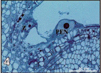
– no division of the primary endosperm nucleus (PEN) which is often compacted to the endothelium, vacuolated with an enlarged nucleoli poorly stained and sometimes occupying the entire volume of the nucleus. No mitotic division of the zygote takes place in this type of degeneration (Figure 4); – only one division of the PEN, indicating a delay in
endosperm development. In general, no apparent degradation is observed but the zygote does not divide (Figure 5);
– development of a multinucleated endosperm characterized by a low cellularization level, multiple small vacuoles spread into the cytoplasm and poorly stained nucleoli (Figure 6).
Our observations in different genotypic combinations show statistical differences according to the parental species or genotypes used in female or male in the crosses (Table 2).
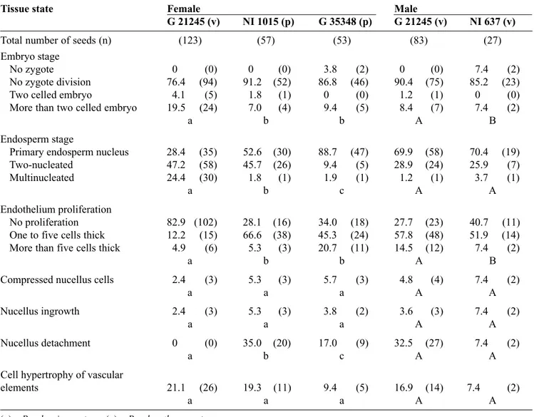
Using P. v u l g a r i s ( G 21245) as a female, coenocytic endosperm developed in 24.4% of the observed seeds while more than 90% of the PEN in reciprocal crosses using P. vulgaris (G 21245 or NI 637) as a male did not divide or divided only once. Table 2. Percentages of hybrid seeds obtained according to the genotypes crossed and to the developmental state of their tissues, 6 days after pollination — Pourcentages de graines hybrides en fonction des génotypes croisés et de l’évolution de leurs différents types de tissu, 6 jours après pollinisation.
Tissue state Female Male
G 21245 (v) NI 1015 (p) G 35348 (p) G 21245 (v) NI 637 (v)
Total number of seeds (n) (123) (57) (53) (83) (27)
Embryo stage
No zygote 0 (0) 0 (0) 3.8 (2) 0 (0) 7.4 (2)
No zygote division 76.4 (94) 91.2 (52) 86.8 (46) 90.4 (75) 85.2 (23)
Two celled embryo 4.1 (5) 1.8 (1) 0 (0) 1.2 (1) 0 (0)
More than two celled embryo 19.5 (24) 7.0 (4) 9.4 (5) 8.4 (7) 7.4 (2)
a b b A B
Endosperm stage
Primary endosperm nucleus 28.4 (35) 52.6 (30) 88.7 (47) 69.9 (58) 70.4 (19)
Two-nucleated 47.2 (58) 45.7 (26) 9.4 (5) 28.9 (24) 25.9 (7)
Multinucleated 24.4 (30) 1.8 (1) 1.9 (1) 1.2 (1) 3.7 (1)
a b c A A
Endothelium proliferation
No proliferation 82.9 (102) 28.1 (16) 34.0 (18) 27.7 (23) 40.7 (11)
One to five cells thick 12.2 (15) 66.6 (38) 45.3 (24) 57.8 (48) 51.9 (14)
More than five cells thick 4.9 (6) 5.3 (3) 20.7 (11) 14.5 (12) 7.4 (2)
a b b A B
Compressed nucellus cells 2.4 (3) 5.3 (3) 5.7 (3) 4.8 (4) 7.4 (2)
a a a A A
Nucellus ingrowth 2.4 (3) 5.3 (3) 3.8 (2) 3.6 (3) 7.4 (2)
a a a A A
Nucellus detachment 0 (0) 35.0 (20) 17.0 (9) 32.5 (27) 7.4 (2)
a b c A A
Cell hypertrophy of vascular
elements 21.1 (26) 19.3 (11) 9.4 (5) 16.9 (14) 7.4 (2)
a a a A A
(v) = P. vulgaris genotype; (p) = P. polyanthus genotype. Number of seeds are indicated between brackets (n).
Data for NI 1015 used as male correspond to those for G 21245 used as female and are thus omitted in this table.
Different small or capital letters indicate a significant difference for the states of the tissues observed when using respectively female or male parents (P<0.05).
F i g u re 3 . Median longitudinal section (×200) in a P. polyanthus ( )×P. vulgaris seed (G 35348 ×G 21245), 6 DAP, showing an early globular hybrid embryo at the micropylar end — Coupe longitudinale médiane (× 200) dans une graine de P. p o l y a n t h u s ( )×P. v u l g a r i s ( G 35348 ×G 21245) montrant, six jours après pollinisation, un proembryon hybride au pôle micropylaire.
Figure 4. Median longitudinal section (× 480) in a P. polyanthus ( )×P. vulgaris seed (NI 1015 ×G 21245), 5 D A P, showing the primary endosperm nucleus (PEN) compacted to the endothelium. No mitotic division of the zygote (Z) has taken place — Coupe longitudinale médiane (×480) dans une graine de P. polyanthus ( )×P. vulgaris (NI 1015 × G 21245) montrant, cinq jours après pollinisation, le noyau primaire de l’albumen ( P E N ) repoussé contre l’endothélium. La pre m i è re division mitotique du zygote (Z) n’a pas encore eu lieu.
F i g u re 5 . Median longitudinal section (×800) in a P. vulgaris ( ) × P. polyanthus seed (G 21245 × NI 1015), 3 DAP, showing the first division of the primary endosperm nucleus giving a coenocytic endosperm (CoEn). No mitotic division of the zygote (Z) has taken place — C o u p e longitudinale médiane (× 800) dans une graine de P. vulgaris ( ) × P. polyanthus (G 21245 × NI 1015) montrant, trois jours après pollinisation, la pre m i è re division du noyau primaire de l’albumen qui évolue en albumen coenocytique ( C o E n ). La pre m i è re division mitotique du zygote (Z) n’a pas encore eu lieu.
Figure 6. Median longitudinal section (×800) in a P. polyanthus ( ) x P. vulgaris seed (NI 1015 ×G 21245), 6 D A P, showing a multinucleated endosperm (CoEn) characterized by low cellularization, multiple small vacuoles spread into the cytoplasm and poorly stained nucleoli (arrowheads). Micropylar end (M) and endothelium (End) are indicated — Coupe longitudinale médiane (×800) dans une graine de P. polyanthus ( )×P. vulgaris (NI 1015 × G 21245) montrant, six jours après pollinisation, un albumen multinucléé (CoEn), caractérisé par une faible cellularisation, de multiples petites vacuoles incluses dans le cytoplasme et une coloration atténuée des nucléoles (flèches). Le pôle micropylaire et l’endothélium sont signalés respectivement par les sigles (M) et (End).
Table 3. Percentages of hybrid seeds obtained over time (in DAP) according to the developmental state of their tissues when P. v u l g a r i s genotypes are used as a female — P o u rcentages de graines hybrides selon l’évolution de leurs différents types de tissu au cours du temps (en nombre de jours après fécondation) lorsque les génotypes de P.vulgaris sont utilisés comme parent femelle.
Tissue state Days after pollination
2 3 4 5 6
Total number of seeds (n) (13) (49) (37) (9) (12)
Embryo stage
No zygote 0 (0) 0 (0) 0 (0) 0 (0) 0 (0)
No zygote division 100.0 (13) 79.6 (39) 75.7 (28) 100.0 (0) 33.3 (4)
Two celled embryo 0 (0) 8.2 (4) 0 (0) 0 (0) 8.4 (1)
More than two celled embryo 0 (0) 12.2 (6) 4.3 (9) 0 (0) 58.3 (7)
Endosperm stage
Primary endosperm nucleus 30.7 (4) 22.5 (11) 43.2 (16) 0 (0) 25.0 (3)
Two-nucleated 30.8 (4) 32.6 (16) 54.1 (20) 100.0 (9) 58.3 (7)
Multinucleated 38.5 (5) 44.9 (22) 2.7 (1) 0 (0) 16.7 (2)
Endothelium proliferation
No proliferation 100.0 (13) 0 (0) 86.5 (32) 100.0 (9) 0 (0)
One to five cells thick 0 (0) 98.0 (48) 13.5 (5) 0 (0) 58.3 (7)
More than five cells thick 0 (0) 2.0 (1) 0 (0) 0 (0) 41.7 (5)
Compressed nucellus cells 0 (0) 0 (0) 0 (0) 0 (0) 25.0 (3)
Nucellus ingrowth 0 (0) 6.1 (3) 0 (0) 0 (0) 0 (0)
Nucellus detachment 0 (0) 0 (0) 0 (0) 0 (0) 0 (0)
Cell hypertrophy of vascular
elements 0 (0) 42.9 (21) 13.5 (5) 0 (0) 0 (0)
Number of seeds are indicated between brackets (n).
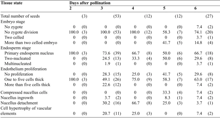
Table 4. Percentages of hybrid seeds obtained over time (in DAP) according to the developmental state of their tissues when P. polyanthus genotypes are used as a female — Pourcentages de graines hybrides selon l’évolution de leurs différents types de tissu au cours du temps (en nombre de jours après fécondation) lorsque les génotypes de P. polyanthus sont utilisés comme parent femelle.
Tissue state Days after pollination
2 3 4 5 6
Total number of seeds (3) (53) (12) (12) (27)
Embryo stage
No zygote 0 (0) 0 (0) 0 (0) 0 (0) 7.4 (2)
No zygote division 100.0 (3) 100.0 (53) 100.0 (12) 58.3 (7) 74.1 (20)
Two celled 0 (0) 0 (0) 0 (0) 0 (0) 3.7 (1)
More than two celled embryo 0 (0) 0 (0) 0 (0) 41.7 (5) 14.8 (4)
Endosperm stage
Primary endosperm nucleus 100.0 (3) 73.6 (39) 66.7 (8) 50.0 (6) 66.7 (18)
Two-nucleated 0 (0) 24.5 (13) 33.3 (4) 50.0 (6) 29.6 (8)
Multinucleated 0 (0) 1.9 (1) 0 (0) 0 (0) 3.7 (1)
Endothelium proliferation
No proliferation 0 (0) 28.3 (15) 25.0 (3) 41.7 (5) 29.6 (8)
One to five cells thick 100.0 (3) 49.1 (26) 75.0 (9) 58.3 (7) 63.0 (17)
More than five cells thick 0 (0) 22.6 (12) 0 (0) 0 (0) 7.4 (2)
Compressed nucellus cells 0 (0) 0 (0) 0 (0) 33.3 (4) 7.4 (2)
Nucellus ingrowth 0 (0) 3.7 (2) 0 (0) 8.3 (1) 7.4 (2)
Nucellus detachment 0 (0) 30.2 (16) 66.7 (8) 25.0 (3) 3.7 (1)
Cell hypertrophy of vascular
elements 0 (0) 20.7 (11) 25.0 (3) 0 (0) 7.4 (2)
Also, when using P. vulgaris (G 21245) as a female, PEN divided once between 2 to 4 DAP in more than 40% of the seeds (Table 3) allowing the development of an embryo. When using P. polyanthus (NI 1015 or G 35348) as a female, first division of PEN was initiated between 3 to 6 DAP but no further development was observed. Only in two seeds, a coenocytic endosperm was observed which represent 1.9% of total seeds.
Moreover, a highly significant difference (P<0.05) between the two P. polyanthus genotypes (NI 1015 or G 35348) used as female parent was observed. Indeed, using NI 1015 accession as female parent, 45.7% of PEN divided once compared to only 9.4% using G 35348 accession (Table 2).
3.4. Proliferation of the endothelium
Among the severe barriers to interspecific hybridization between genotypes, endothelium proliferation was considered as an important cause of embryo abortion. Indeed, a partial endothelial proliferation was often observed at the beginning of the seed development. This tissue was generally one to five cells thick but sometimes continued to grow and then fill up the embryo sac cavity, preventing the embryo from continued division (Figure 7).
In general, our observations showed significant differences (P<0.05) between all crosses (Table 2). Using P. vulgaris genotype (G 21245) as a female, endothelium remained normal in more than 80% of the seeds, while using P. polyanthus as a female, no more than 34% of the seeds contained a normal endothelium. Also, differences were observed between P. vulgaris genotypes (G 21245 and NI 637) when used as a male. Endothelium proliferation was more important when using the wild genotype (G 21245) as male.
If endothelium proliferation was observed within 2 D A P in P. p o l y a n t h u s ( )×P. v u l g a r i s seeds, the thickness of the tissue was very often limited to two to five cells. In P. vulgaris ( )×P. polyanthus seeds, endothelium proliferation was initiated 1 to 2 days later, but could fill up the entire cavity of the embryo sac at 6 DAP.
3.5. Nucellus degeneration
In self-pollinated flowers of P. vulgaris, at anthesis, nucellar tissue forms a U-shaped cap over the embryo sac in the chalazal region. This nucellar tissue degenerates during the first three DAP as the embryo and embryo sac enlarge. By 4-5 DAP, nucellar tissue is detectable only in the chalazal region.
In hybrid seeds, the development of the nucellus has been difficult to interpret. Several types of changes
were observed. In general nucellar cells were vacuolated indicating that the nutritional balance of aborting seeds was disrupted early in the abortion process. Nucellar tissue can be detached from integuments at the chalazal end suggesting that nutrient exchanges were lowered (F i g u re 8) . Significant differences were observed between P. vulgaris ( )×P. polyanthus and reciprocal crosses (Table 2). No disruption was observed when using P. vulgaris genotype (G 21245) as a female, while it is observed between 3 to 4 DAP in 35% of the seeds using P. p o l y a n t h u s as a female, indicating an important difference between the two crosses. Nucellus degeneration disrupts the vascular system, and subsequent embryo and seed/fruit development stops due to failure of nutrient conduction. In some hybrid seeds, embryo development was limited due to the ingrowth of the nucellus into the embryo sac cavity to the micropylar end (Figure 8) or to the abnormal development of cells of the chalazal nucellus, becoming compressed at the micropylar end (Figure 9). Compressed cells were small, collapsed, with dark condensed cytoplasm. In parallel, the endospermic transfer cell layer also lost contact with the chalazal nucellus. Table 2 shows that these last changes were not significant. Moreover, compressed cells were only visible 5 or 6 DAP (Tables 3 and 4). Initial abortion-related changes in maternal tissues, which may result in embryo growth failure, are thus neither related directly to nucellus ingrowth nor to compressed nucellus cells.
3.6. Hypertrophy of vascular elements
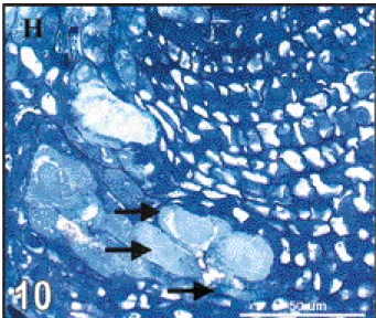
We also observed hypertrophy of vascular elements at the chalazal end. Hypertrophy was mainly located at hypostase level (Figure 10). This direct disruption of nutrient transfer was observed in all crosses (Table 2) without significant differences between them. Table 3 and t a b l e 4 show that hypertrophy of vascular elements was observed mainly 3 to 4 DAP.
4. DISCUSSION
The parental P. p o l y a n t h u s embryo development corresponded to the main features observed in Phaseolus embryony (Yeung, Clutter, 1978). Although development of the different tissues are identical between P. v u l g a r i s and P. p o l y a n t h u s, some differences were shown by Lecomte et al. (1998). In particular, the development of the embryo is slower in P. p o l y a n t h u s than in P. v u l g a r i s seeds. Delayed embryo growth in P. polyanthus seeds was related to suspensor size. Basal cells of the suspensor of P. polyanthus embryos are bigger and contain larger vacuoles. Also, we noted a 2 to 3 days delay in embryo
Figure 7. Median longitudinal section (× 160) in a P. vulgaris ( )×P. polyanthus seed (G 21245 ×NI 1015), 5 DAP, showing endothelium proliferation (PR) filling up embryo sac cavity — Coupe longitudinale médiane (×160) dans une graine de P. vulgaris ( ) × P. polyanthus (G 21245 ×NI 1015) montrant, cinq jours après pollinisation, l’envahissement du sac embryonnaire par une prolifération de l’endothélium (PR).
Figure 8. Median longitudinal section (× 110) in a P. polyanthus ( )×P. vulgaris seed (NI 1015 ×G 21245), 5 DAP, showing: (I) hypertrophic growth of the nucellus into the embryo sac cavity and (D) detachment of the nucellus from the inner integument at the chalazal end — Coupe longitudinale médiane (× 110) dans une graine de P. polyanthus ( ) ×P. vulgaris (NI 1015 ×G 21245) montrant, cinq jours après pollinisation, l’hypertrophie du nucelle au sein du sac embryonnaire (I) et le décollement du nucelle de la paroi interne de l’ovule au pôle chalazien (D).
Figure 9. Median longitudinal section (× 480) in a P. polyanthus ( )×P. vulgaris seed (G 35348 ×G 21245), 5 DAP, showing cells (Cc) of the chalazal nucellus (Nu) compressed at the micropylar end. Cells are small, collapsed, with dark condensed cytoplasm. Endospermic layer lost contact with the chalazal nucellus and degenerates, showing vacuolated cells with poorly stained nucleoli (arrowheads) — Coupe longitudinale médiane (×
480) dans une graine de P. polyanthus ( )×P. vulgaris (G 35348 ×G 21245) montrant, cinq jours après pollinisation, des cellules (Cc) du nucelle provenant du pôle chalazien (Nu) comprimées à l’extrémité micropylaire. Ces cellules sont petites, écrasées, avec un cytoplasme sombre et dense. Les cellules périphériques de l’albumen perdent le contact avec le nucelle et dégénèrent. Ces cellules sont vacuolisées et la coloration du noyau est atténuée (flèches).
Figure 10. Median longitudinal section (× 580) in a P. vulgaris ( )×P. polyanthus seed (G 21245 ×NI 1015), 3 D A P, showing hypertrophy of vascular elements (arrowheads) at the chalazal end (H indicates hypostase) — Coupe longitudinale médiane (×580) dans une graine de P. vulgaris ( ) × P. polyanthus (G 21245 × NI 1015) montrant, trois jours après pollinisation, une hypertrophie des vaisseaux conducteurs (flèches) au pôle chalazien (H indique l’hypostase).
development between the two parental species. Lecomte et al. (1998) indicated that in P. vulgaris, wall thickening between endothelium cells is radial, increasing nutrient exchanges to the micropylar end. In P. polyanthus, these thick radial walls were not observed and seem to be replaced by tangential wall thickening of cells, in continuity between the two integuments. Authors suggested that nutrient exchange could be more rapid in P. v u l g a r i s than in P. polyanthus seeds allowing a faster development of P. v u l g a r i s embryos. In P. p o l y a n t h u s ( )×P. vulgaris crosses, nutrient requirements of the hybrid embryo could be too high leading to under-nutrition at the early stages of development and, consequently, to embryo abortion.
In order to have a better knowledge of the stages of embryo abortion, we compared the embryo development in P. vulgaris ( )×P. polyanthus and in reciprocal crosses during the first sixth DAP. In P. vulgaris ( )×P. polyanthus hybrid seeds, zygote division was initiated between 2 to 3 DAP and, in more than 50% of the seeds, embryo continued to develop to reach a globular stage 6 DAP. Some mature seeds could also be obtained 40 D A P (data not shown). In this case embryo development was accompanied by an early initiation of PEN division. Three days after pollination, multinucleated endosperm was observed in 44.9% of the seeds which corresponded with the number of embryos reaching a globular stage at 6 D A P. When hybrid globular embryos could be obtained, proliferation of the endothelium was limited to one to five cells in thickness and no hypertrophy was observed. When an embryo aborted at a pre-globular stage, endothelium proliferation was initiated 3 DAP and reached more than 5 cells thick in 40% of the seeds. Therefore, at 4 and 5 DAP, multinucleated endosperm was difficult to distinguish from endothelium proliferation. In parallel, hypertrophy of vascular elements was observed 4 DAP in 42.9% of the early aborting seeds. Therefore, in P. vulgaris ( )×P. polyanthus crosses, early abortion is mainly caused by endothelium proliferation. Later, hybrid embryo abortion is more related to the abnormal development of its suspensor, which is detached from the growing embryo (data not shown). Considering the widely assumed haustorial function of the suspensor (Chamberlin et al., 1993; Yeung, Meinke, 1993), problems in suspensor development result in nutrient supply perturbation for the embryo and affect, consequently, the further embryo development. Nesling and Morris (1979) and Singh (1998) showed that abortion in the hybrids may be associated with failure by the endosperm to provide cytokinins to the embryo and this may lead to abnormalities in protein synthesis in the suspensor cells and eventually to abortion of the embryo. In this
case, an in vivo application of cytokinin at regular intervals may help to sustain the development of the endosperm and consequently of the embryo. Whether an exogenous application of cytokinin in these crosses can prevent degeneration of the embryos needs to be investigated.
In P. polyanthus ( )×P. vulgaris hybrid seeds, first division of the zygote was initiated between 4 and 5 DAP. Only 4 embryos out of 107 could reach a 16 to 24 celled embryo 6 DAP before dying, 7 DAP. No mature seed could be obtained. This low rate of embryo development was related to a weak endosperm development. Indeed, only two-celled coenocytic endosperm was generally obtained 5 DAP and further division was rare. At the same time, proliferation of the endothelium was initiated 2 DAP but was always limited to one to five cells in thickness. We identify two other main causes of abortion in seeds 3 to 6 DAP. The first observed between 3 to 4 DAP, in 45.7% of the seeds, involves some hypertrophy of vascular elements suggesting an early reduction in nutrient transport from maternal tissue to the embryo sac. The second observed at 5 DAP, in 58.8% of the seeds, involves degeneration of the nucellus with a high percentage of compressed cells and detachment from integuments at the chalazal end. Degradation of the nucellus was similar as in Alstroemeria sp. (De Jeu, Calderé, 1997). Cells of the chalazal nucellus became compressed 6 DAP, showing small collapsed cells with dark cell thickenings. Therefore, in P. polyanthus ( )×P. v u l g a r i s seeds, the main cause of early abortion is a weak endosperm development. This observation is coherent with several other works (Van Tuyl et al., 1990, Zenkteler, 1991) stating that embryo abortion is the result of poor coordination of simultaneous development of embryo, endosperm and sporophytic surrounding tissue. Normal development of the endosperm is considered to be the most important factor in almost all species (Brink, Cooper, 1947) as demonstrated for Lupinus sp. (Busmann-Loock et al., 1992), Cyclamen sp. (Ishizaka, Uematsu, 1992) and Alstroemeria sp. (De Jeu, Calderé, 1997). The Endosperm Balanced Number (EBN) (Johnston e t a l ., 1980), the Polar-Nuclei Activation Index hypothesis (Nishiyama, Yabuno, 1978), and other mechanisms related to ploidy might be operative soon after fertilization. Using EBN ratios, Masuelli and Camadro (1997) pointed out, among wild potato species, differences between interspecific intra-EBN and interspecific inter-EBN. Such differences between crosses were also observed in other species: Sangduen et al. (1983) in Medicago sp., Abbo and Ladizinsky (1991) in L e n s sp., Chen and Adachi (1995) in Lycopersicon sp. and Singh (1998) in Arachis sp.
As reported by previous researchers studying crosses P. vulgaris ( )×P. lunatus (Mok et al., 1978;
Leonard et al., 1987), the choice of maternal parent seems especially important. In our study, maternal parent also had an important effect on the development of PEN, and, consequently, on embryo development. In addition, we also pointed out some genotypic differences. For example, the use of NI 637 instead of G 21245 as P. v u l g a r i s paternal parent decreased the rate of endothelium proliferation and nucellus detachment. The use of NI 1015 instead of G 35348 as P. p o l y a n t h u s maternal parent also increased nucellus degeneration but decreased endothelium proliferation.
5. CONCLUSION
D i fferences between early embryo abortion in reciprocal crosses are mainly related to the endosperm development. While a rapid division of PEN is observed in P. v u l g a r i s ( )×P. p o l y a n t h u s s e e d s , allowing the further development of the embryo which is initiated 2 to 3 DAP, PEN stay uninucleated in P. polyanthus ( )×P. vulgaris seeds during the first four DAP, limiting nutrient exchange between maternal tissue and zygote. Moreover, our results showed that zygotes of P. polyanthus ( )×P. vulgaris seeds were still able to divide 5 DAP when PEN had divided at least once. This suggests that embryo abortion in P. polyanthus ( )×P. vulgaris seeds could be related to a decrease in nutrient exchange at the beginning of its development, increasing the time at which first division can occur rather than incompatibilities between hybrid embryo and endosperm. This hypothesis is supported by the observations of Lecomte et al. (1998) describing wall thickening of the endothelial cells in P. polyanthus seeds that are tangential while they are radial in P. vulgaris seeds. Histological differences between maternal tissues in reciprocal crosses could thus be a key factor in the abortion processes.
Later in the hybrid embryo development, the proliferation of the endothelium was clearly described as the main factor of embryo abortion. Differences in the developmental rate of this endothelium proliferation between reciprocal crosses could be attributed to genetic factors or to the rate of endosperm development. In P. polyanthus ( )×P. vulgaris seeds proliferation could be limited to one to five cells in thickness due to the poor endosperm development, while in P. v u l g a r i s ( )×P. p o l y a n t h u s seeds the development of multinucleated endosperm could lead to a greater endothelial cell proliferation and subsequent later embryo abortion.
On the basis of these results, rescue of P. p o l y a n t h u s ( )×P. v u l g a r i s embryos could be facilitated by the development of an in vitro technique permitting the culture of early globular embryo. A pod
culture technique was described by Geerts e t a l . (2000) allowing the development of two day-old P. vulgaris embryos. This technique could be applied to hybrid pods in order to rescue P. polyanthus ( ) ×P. vulgaris embryos.
Acknowledgement
This work was funded by the Belgian Administration to Development Cooperation (BADC) within the framework of a research project carried out in collaboration with the Centro Internacional de Agricultura Tropical (CIAT). We thank the Walloon Ministry for Employment and Training for its contribution to this research by PRIME projects.
Bibliography
A b b o S., Ladizinsky G. (1991). Anatomical aspects of hybrid embryo abortion in the genus Lens L. Bot. Gaz. 152, p. 316–320.
B a u d o i n J P., Camarena FM., Schmit V. (1992). Contribution à une meilleure connaissance de la position phylétique de la légumineuse alimentaire Phaseolus polyanthus Greenm. Bull. Rech. A g ron. Gembloux 2 7 ( 2 ) , p. 167–198.
Brink RA., Cooper DC. (1947). The endosperm in seed development. Bot. Rev. XIII, p. 423–465.
B u s m a n n - L o o c k A., Dambroth M., Menge-Hartmann U . (1992). Histological observations on interspecific crosses in the genus L u p i n u s. Plant Bre e d . 1 0 9, p. 82–85.
Camarena F., Baudoin JP. (1987). Obtention des premiers hybrides interspécifiques entre Phaseolus vulgaris et Phaseolus polyanthus avec le cytoplasme de cette dernière forme. Bull. Rech. Agron. Gembloux 22 (1), p. 43–55.
C h a m b e r l i n MA., Horner H T., Palmer RG. (1993). Nutrition of ovule, embryo sac, and young embryo in soybean: an anatomical and autoradiographic study. Can. J. Bot. 71, p. 1153–1168.
C h e n L., A d a c h i T. (1995). Mechanism of abortion of postfertilization hybrid embryo in interspecific backcross, Lycopersicon esculentum x (L. esculentum x L. peruvianum). Cytologia 60, p. 121–131.
De Jeu MJ., Calderé FG. (1997). Retarded embryo growth and early degeneration of sporophytic tissue are associated with embryo abortion in the interspecific cross Alstroemeria pelegrina x Alstroemeria aurea. Can. J. Bot. 75, p. 916–924.
G e e r t s P., Khaled S., Merg e a i G., Baudoin J P. (2000). Development of an in vitro pod culture technique for young pods of Phaseolus vulgaris L. In Vitro Cell. Dev. Biol. – Plant 36 (6), p. 481–487.
photographic documentation of phenolic deposits in semithin sections of plant tissue. J. Microsc. 179 (3), p. 277–281.
Hucl P., Scoles GJ. (1985). Interspecific hybridization in the common bean: a review. H o rt s c i e n c e 2 0, p. 352–356.
Ishizaka H., Uematsu J. (1992). Production of interspecific hybrids of Cyclamen persicum Mill and C. hederifolium Aiton. by ovule culture. Jn J. Breed. 42, p. 353–366. Johnston SA., den Nijs TPM., Peloquin SJ., Hanneman RE.
(1980). The significance of genic balance to endosperm development in interspecific crosses. T h e o r. A p p l . Genet. 57, p. 5–9.
K u b o y a m a T., Shintaku Y., Ta k e d a G. (1991). Hybrid plants of Phaseolus vulgaris L. and Phaseolus lunatus L. obtained by means of embryo rescue and confirmed by restriction endonuclease analysis of rDNA. Euphytica 54, p. 177–182.
Lecomte B. (1997). Étude du développement embryonnaire in vivo et in vitro dans le genre Phaseolus L. Thèse doct. sci. agron., Fac. Univ. Sci. Agron., Gembloux (Belgium), 186 p.
L e c o m t e B., Longly B., Crabbe J., Baudoin J P. (1998). Étude comparative du développement de l’ovule chez deux espèces de Phaseolus : P. polyanthus et P. vulgaris. Développement de l’ovule dans le genre Phaseolus. Biotechnol. Agron. Soc. Environ. 2 (1), p. 77–84. Leonard MF., Stephen LC., Summers WL. (1987). Effect of
maternal genotype on development of P h a s e o l u s vulgaris L. x P. lunatus L. interspecific hybrid embryos. Euphytica 36, p. 327–332.
M a s u e l l i RW., Camadro EL. (1997). Crossability relationships among wild potato species with different ploidies and endosperm balance numbers (EBN). Euphytica 94, p. 227–235.
M o k DWS., Mok MC., Rabakoarihanta A. (1978). Interspecific hybridization of Phaseolus vulgaris with
Phaseolus lunatus and Phaseolus acutifolius. Theor. Appl. Genet. 52, p. 209–215.
Nesling FAV., Morris DA. (1979). Cytokinin levels and embryo abortion in interspecific Phaseolus crosses. Z. Pflanzenphysiol. 91, p. 345–358.
N i s h i y a m a I., Ya b u n o T. (1978). Causal relationship between the polar nuclei in double fertilization and interspecific cross-incompatibility. C y t o l o g i a 4 3, p. 453–466.
Ruzin SE. (1999). Plant microtechnique and microscopy. New York: Oxford University Press. 322 p. ISBN 0-19-508956-1.
Sangduen N., Kreitner GL., Sorensen EL. (1983). Light and electron microscopy of embryo development in perennial and annual Medicago species. Can. J. Bot. 61, p. 837–949.
S i n g h AK. (1998). Hybridization barriers among the species of Arachis L. namely of the sections Arachis (including the groundnut) and E re c t o i d e s. G e n e t . Resour. Crop Evol. 45, p. 41–45.
Van Tu y l JM., Van de Sande K., Van Diën M J . , Straathof D., Van Holsteijn HMC. (1990). Overcoming interspecific crossing barriers in Lilium by ovary and embryo culture. Acta Hortic. 266, p. 317–322.
Yeung EC., Clutter ME. (1978). Embryogeny of Phaseolus coccineus: growth and microanatomy. Protoplasma 94, p. 19–40.
Ye u n g EC., Meinke D W. (1993). Embryogenesis in angiosperms: development of the suspensor. Plant Cell 5, p. 1371–1381.
Z e n k t e l e r M. (1991). Ovule culture and test tube fertilization. Med. Fac. Landbouwwet. Rijksuniv. Gent. 56, p. 1403–1410.
Les Presses agronomiques de Gembloux
Passage des Déportés, 2. B–5030 Gembloux
E-mail: pressesagro@fsagx.ac.be — tél/fax : 32 (0) 81 62 22 42 — URL: http://www.bib.fsagx.ac.be/presses/
Willy Delvingt
La forêt des hommes
Terroirs villageois en forêt tropicale africaine
2001, 288 p., 39 ill. ISBN 2-87016-064-X BEL : 26,50 ¤ ; ETR : 30 ¤
L’exploitation forestière industrielle est responsable de la disparition des forêts tropicales. L’installation de "villes en forêt" modifie les pratiques traditionnelles de gestion et d’exploitation des ressources de la forêt amenant à une surexploitation de la faune et la flore, rompant l’équilibre ancien. Comme l’exportation du bois contribue au PIB des pays concernés, ceux-ci n’envisagent pas de réduire le rythme d’extraction. On ne peut donc que tenter de limiter les effets indirects de l’exploitation industrielle des forêts, ceci dans un double objectif d’améliorer le revenu des populations locales qui ont toujours vécu de la forêt et ne retirent guère de bénéfice de son exploitation intensive et de sauvegarder cet irremplaçable réservoir de biodiversité. À la recherche de solutions pragmatiques, les onze auteurs ont concentré leurs efforts sur un territoire délimité en forêt dense humide tropicale : le plateau méridional camerounais au nord de la Réserve de faune du Dja, occupé depuis deux à trois cents ans par le peuple Badjoué. Les uns ont étudié le système de production des Badjoué : agriculture itinérante sur brûlis, pêche, chasse, récolte du vin de palme. D’autres ont expérimenté l’exploitation, à des fins monétaires, dans le cadre des "forêts communautaires", de produits tels que bois sciés et fruits. D’autres enfin ont analysé les interactions du système traditionnel de gestion de ressources avec la politique forestière gouvernementale (réserves de faune, unités forestières d’aménagement, forêts communautaires). À travers ce cas particulier du Cameroun, c’est la problématique du développement – ou de la simple survie – des communautés villageoises en forêt tropicale qui est abordée.
Albert Ledent, Philippe Burny La politique agricole commune
des origines au 3emillénaire
2002, 600 p., 4 ill., 50 tab. ISBN 2-87016-066-6 BEL : 30 ¤ ; ETR : 35,50 ¤
Dès l’entrée en vigueur du traité CEE, en 1958, l’agriculture n'a cessé de susciter d’âpres débats, renfor-cés et aggravés par les élargissements successifs. La politique agricole commune est restée longtemps la principale construction mise en place par les partenaires. Elle est toujours un élément essentiel de l’UE, à cause de l’importance économique et sociale du secteur agroalimentaire, de la diversité de l’agriculture, de son rôle en matière spatiale. L’ouvrage retrace l’évolution de la politique agricole. Il donne une vue d’ensemble de la PAC ; il la place dans le cadre de la construction européenne et permet de comprendre ses incontestables succès mais aussi ses échecs. Il relève de nombreux défis auxquels la politique agricole a dû et devra faire face, parmi lesquels la libération des échanges, les négociations commerciales multilatérales à l’OMC, l’élargissement de l’Union européenne, la sécurité et la qualité des produits, le développement rural, la situation mondiale de l’agriculture et de
l’alimentation. Enfin, il trace des pistes de réflexion pour garantir la pérennité de l’agriculture européenne dans un monde sans cesse changeant. Cet ouvrage est le fruit de l'expérience d’A. Ledent, président du Conseil supérieur Wallon de l’agriculture, l’agroalimentaire et l’alimentation, recteur honoraire de la Faculté universitaire des Sciences agronomiques de Gembloux où il a enseigné l’économie rurale durant 40 ans, ancien directeur de l’Office belge chargé de l’intervention sur les marchés agricoles et de P. Burny, son ancien élève, chercheur qualifié, maître de conférence à la même faculté et attaché au cabinet du Ministre Wallon de l’agriculture et de la ruralité.