*EC and MH contributed equally to this work.
Funding: MH is Télévie Research Fellow, SHB Postdoctoral Researcher, FB Senior Research Associate, and VG Research Director at the National Fund for Scientific Research (FNRS) Belgium. The study was in part supported by funds from the FNRS, the Belgian Foundation against Cancer (FBC), the anti-cancer foundation from the ULg, the CHU of Liège, the Terry Fox foundation, and by grants CA 78902, CA 18029 and HL 36444 from the National Institutes of Health, Bethesda, MD.
Acknowledgments: we thank Y Henrotin and M Mathy for helpful discussions about PCR analyses, V Dhennin from the Genotranscriptomics Platform of the GIGA, for help with T-cell spectratyping, and S Ormenese from the Imaging and Flow Cytometry Platform of the GIGA for help with flow cytometry analyses. The authors are also grateful to O Dengis and C Daulne for excellent technical assistance, to physicians and clinical staff for their dedicated care of the patients, and to N Schaaf-Lafontaine from the laboratory of Hematology. Manuscript received on June 29, 2010. Revised version arrived on September 15, 2010. Manuscript accepted on October 5, 2010.
Correspondence:
Frédéric Baron, University of Liège, department of Hematology, CHU Sart-Tilman, 4000 Liège, Belgium. Phone: international +32.4.3667201. Fax international +32.4.3668855. E-mail: f.baron@ulg.ac.be The online version of this article has a supplementary Appendix. Background
Long-term immune recovery in older patients given hematopoietic cell transplantation after non-myeloablative conditioning remains poorly understood. This prompted us to investigate long-term lymphocyte reconstitution and thymic function in 80 patients given allogeneic peripheral blood stem cells after non-myeloablative conditioning.
Design and Methods
Median age at transplant was 57 years (range 10-71). Conditioning regimen consisted of 2 Gy total body irradiation (TBI) with (n=46) or without (n=20) added fludarabine, 4 Gy TBI with fludarabine (n=6), or cyclophosphamide plus fludarabine (n=8). Stem cell sources were unma-nipulated (n=56), CD8-depleted (n=19), or CD34-selected (n=5) peripheral blood stem cells. Immune recovery was assessed by signal-joint T-cell receptor excision circle quantification and flow cytometry.
Results
Signal-joint T-cell receptor excision circle levels increased from day 100 to one and two years after transplantation in patients under 50 years of age (n=23; P=0.02 and P=0.04, respectively), and in those aged 51-60 years (n=35; P=0.17 and P=0.06, respectively), but not in patients aged over 60 (n=22; P=0.3 and P=0.3, respectively). Similarly, CD4+CD45RA+(naïve) T-cell counts
increased from day 100 to one and two years after transplantation in patients aged 50 years and under 50 (P=0.002 and P=0.02, respectively), and in those aged 51-60 (P=0.4 and P=0.001, respectively), but less so in patients aged over 60 (P=0.3 and P=0.06, respectively). In multivari-ate analyses, older patient age (P<0.001), extensive chronic GVHD (P<0.001), and prior (resolved) extensive chronic graft-versus-host disease (P=0.008) were associated with low sig-nal-joint T-cell receptor excision circle levels one year or more after HCT.
Conclusions
In summary, our data suggest that thymic neo-generation of T cells occurred from day 100 onwards in patients under 60 while signal-joint T-cell receptor excision circle levels remained low for patients aged over 60. Further, chronic graft-versus-host disease had a dramatic impact on thymic function, as observed previously in patients given grafts after myeloablative condi-tioning.
Key words: thymus, hematopoietic cell transplantation, non-myeloablative, graft-versus-host disease, immunity, age.
Citation: Castermans E, Hannon M, Dutrieux J, Humblet-Baron S, Seidel L, Cheynier R, Willems E, Gothot A, Vanbellinghen J-F, Geenen V, Sandmaier BM, Storb R, Beguin Y, and Baron F. Thymic recovery after allogeneic hematopoietic cell transplantation with non-myeloablative conditioning is limited to patients younger than 60 years of age. Haematologica 2011;96(2):298-306.
doi:10.3324/haematol.2010.029702
©2011 Ferrata Storti Foundation. This is an open-access paper.
Thymic recovery after allogeneic hematopoietic cell transplantation
with non-myeloablative conditioning is limited to patients younger
than 60 years of age
Emilie Castermans,1,2* Muriel Hannon,1,2* Jacques Dutrieux,3Stéphanie Humblet-Baron,2 Laurence Seidel,4
Rémi Cheynier,3Evelyne Willems,1,2André Gothot,5Jean-François Vanbellinghen,6Vincent Geenen,7
Brenda M. Sandmaier,8,9Rainer Storb,8,9Yves Beguin,1,2and Frédéric Baron1,2
1Department of Medicine, Division of Hematology, University and CHU of Liège, Liège, Belgium; 2GIGA Research-Hematology,
University of Liège, Liège, Belgium; 3Department of Virology, Institut Pasteur, Paris, France; 4Department of Statistics, University of
Liège, Liège, Belgium; 5Department of Laboratory Medicine, Division of Laboratory Hematology and Immuno-Hematology, University
of Liège, Liège, Belgium; 6,7Department of Genetics, CHU of Liège, Liège, Belgium; Center of Immunology, University of Liège, Liège,
Belgium; 8Fred Hutchinson Cancer Research Center, Seattle, WA, USA, and 9University of Washington, Seattle, WA, USA
Introduction
It is well established that the thymus undergoes a phys-iological involution with aging that affects its structure
and cytokine environment.1,2 However, studies by
Steinman et al. in the 1980s documented lymphocytic thymic tissue in adults up to 107 years of age.1Further,
several studies have suggested that the human thymus could continue to mature new T cells throughout life, exporting signal-joint T-cell receptor excision circle (sjTREC) bearing T cells into the peripheral blood even in elderly patients.3-5
Non-myeloablative conditioning regimens followed by allogeneic hematopoietic cell transplantation (HCT) have opened the way to performing allogeneic HCT in patients with hematologic malignancies aged up to 70-75 years.6-9
This approach has relied on optimization of pre- and post-transplant immunosuppression to overcome
graft-versus-host disease (GVHD) reactions,10 thereby allowing
engraftment and eradication of tumors nearly exclusively via immune-mediated graft-versus-tumor effects.11-13
T-cell recovery after allogeneic HCT following high-dose (myeloablative) conditioning depends on both peripheral expansion of mature T cells contained in the graft (thymo-independent pathway), and T-cell neo-pro-duction from donor hematopoietic stem cells (thymo-dependent pathway).14-17In young patients given
myeloab-lative allogeneic HCT, most circulating T cells during the first 3-6 months following HCT are the progeny of T cells infused with the grafts,18while neogeneration of T cells by
the thymus plays an important role in reconstituting the T-cell pool beyond day 100 after HCT.19-24
We recently analyzed immune recovery after non-mye-loablative conditioning the first year after non-myeloabla-tive conditioning.25,26Main observations were that
unrelat-ed donor status and donor age affectunrelat-ed early immune recovery,25 while sjTREC levels significantly increased
from day 100 to day 365 in a cohort of 16 patients,26
sug-gesting that the thymic pathway might play a role in immune recovery in these older patients given non-mye-loablative conditioning. Here, we analyzed long-term (one year or more) immune recovery and thymic function in 80 patients given non-myeloablative conditioning regimen.
Design and Methods
Patients and donors
Data from 80 patients transplanted between March 2000 and April 2008 at the University of Liège were included in the study. Results were analyzed as of July 29, 2009. Patients’ characteristics are summarized in the Online Supplementary Table S1. Median patient age was 57 (range 10-71) years. Thirty-three of the 80 patients received grafts from HLA-matched related donors, 22 from HLA-matched unrelated donors, and 25 from HLA-mis-matched related or unrelated donors. Stem cell sources were unmanipulated (n=56), CD8-depleted (n=19), or CD34-selected (n=5) peripheral blood stem cells (PBSC). CD8-depletion and CD34-selection of PBSC were carried out as previously report-ed.26,27Nine patients given grafts from HLA-mismatched donors
were enrolled in a trial of mesenchymal stem cell co-transplanta-tion as a potential way to prevent severe GVHD28. Immune
recov-ery in 35 of the 80 patients for the first six (n=14)27or 12 (n=21)26
months after HCT has been reported previously. Written informed consent was obtained from each patient to undergo
non-myeloab-lative HCT and to collect, store and analyze blood samples for research purposes. The Ethics Committee of the University of Liège approved the consent form as well as the current research study protocol.
Treatment and evaluation
The non-myeloablative conditioning regimens consisted of 2 Gy total body irradiation (TBI) alone (n=20; patients with low risk of graft rejection given PBSC from HLA-identical siblings), 2 Gy TBI with 90 mg/m2fludarabine (n=46; standard regimen), 4 Gy
TBI with 90 mg/m2fludarabine (n=6; patients at high risk of early
disease progression and/or graft rejection), or fludarabine 90 mg/m2with cyclophosphamide 3 g/m2(n=8, patients with
previ-ous irradiation precluding the use of TBI). Post-grafting immuno-suppression combined mycophenolate mofetil (MMF) with a cal-cineurin inhibitor for all patients, as previously described.26,27
Clinical management
Twenty-four patients received at least one unmanipulated (n=18) or CD8-depleted (n=6) pre-emptive donor lymphocyte infusion (DLI) the first year after HCT (including the 5 patients given CD34-selected PBSC who received CD8-depleted pre-emptive DLI). G-CSF (5 μg/kg/d) was administered when the granulocyte count declined below 1.0¥109/L. The diagnosis,
clin-ical grading, and treatment of acute GVHD were performed according to established criteria.29Treatment was usually given
for grade II-IV acute GVHD and for extensive chronic GVHD. Initial treatment of acute GVHD usually consisted of pred-nisolone, 2 mg/kg/day, with tapering initiated within 14 days. In addition, the calcineurin inhibitor was usually resumed at full doses. Steroid-refractory acute GVHD was treated as per avail-able investigational protocols or standard practice. Treatment of chronic GVHD consisted of methylprednisolone (1 mg/kg) with alternate-day calcineurin inhibitor. Steroid-refractory chronic GVHD was generally treated with rapamycin, mycophenolate mofetil, or photophoresis.
Infection prophylaxis generally consisted of acyclovir (400 mg t.i.d. orally), oral itraconazole solution (200 mg b.i.d.) or oral flu-conazole (200 mg b.i.d.), and trimethoprim sulfamethoxazole or aerosolized pentamidine. Polymerase chain reaction (PCR) for cytomegalovirus (CMV) was performed weekly until day 100 and every 2-4 weeks thereafter. Patients with a positive PCR received preemptive i.v. ganciclovir.
Disease evaluations were routinely carried out on days 40, 100, 180 and 365 after HCT.
Chimerism
Chimerism levels among peripheral T cells were assessed on days 28, 40, 100, 180 and 365 after HCT using fluorescence in situ hybridization to detect X and Y chromosomes for recipients of sex-mismatched transplants and PCR-based analysis of polymor-phic microsatellite regions (AmpFlSTR® Identifiler®, Applied
Biosystems, Lennik, Belgium) for recipients of sex-matched trans-plants.26,27 CD3 (T cell) selection was carried out with the
RosetteSepRhuman T-cell enrichment kit (StemCell Technologies,
Vancouver, Canada). Graft rejection was defined as the occurrence of less than 5% T cells of donor origin after HCT, as previously described.30,31
Immune recovery
Immune recovery was prospectively assessed as previously described.27Briefly, patients’ peripheral white blood cells were
phenotyped on days 28, 42, 60, 80, 100, 120, 180, 365, 540, 730 and yearly thereafter using 4 color flow cytometry after treatment with a red blood cell lyzing solution. The analyzed cell subsets
were T cells (CD3+), CD4+T cells (CD3+CD4+lymphocytes), CD8+
T cells (CD3+CD8+ lymphocytes), naïve CD4+ T cells
(CD4+CD45RAhighdouble positive lymphocytes), memory CD4+T
cells (CD4+CD45RO+ double positive lymphocytes), NK cells
(CD3-CD56+ lymphocytes) as well as B cells (CD19+
lympho-cytes). The percentage of positive cells was measured relative to total nucleated cells, after subtraction of non-specific staining. Absolute counts were obtained by multiplying the percentages of positive cells by the white blood cell counts (Advia 120 hematol-ogy analyzer, Bayer Technicon, Tarrytown, USA). Lower and higher limits of normal values for each cell subset were defined respectively as 5 and 95 percentiles of values obtained in 47 age-matched healthy volunteer donors.
More detailed T- and B-cell phenotyping was retrospectively performed using cryopreserved PBMC from 33 patients obtained two years after transplantation using 6 color flow cytometry. The different populations were defined as follows: naïve CD4+T cells,
CD4+CD45RA+CCR7+CD27+lymphocytes; central memory CD4+
T cells, CD4+CD45RA-CCR7+CD27+lymphocytes; effector
mem-ory CD4+T cells, CD4+CD45RA-CCR7-CD27+lymphocytes; late
differentiation effector memory CD4+ T cells, CD4+CD45RA
-CCR7-CD27- lymphocytes; naïve CD8+ T cells,
CD8+CD45RA+CCR7+ lymphocytes; central memory CD8+ T
cells, CD8+CD45RA-CCR7+lymphocytes; effector memory CD8+
T cells, CD8+CD45RA-CCR7-lymphocytes; effector memory RA+
CD8+T cells, CD8+CD45RA+CCR7-lymphocytes; naïve B cells,
CD19+CD27- lymphocytes; memory B cells, CD19+CD27+
lym-phocytes; IgM+IgD+memory B cells, CD19+CD27+IgM+IgD+
lym-phocytes; switched memory B cells, CD19+CD27+IgM-IgD-
lym-phocytes. Absolute counts were calculated by multiplying the per-centage of positive cells in the lymphoid gate by the absolute lym-phocyte count of the patient the day of PBMC collection.
CDR3 spectratyping (immunoscope)
T-cell receptor beta chain (TCRB) CDR3 spectratyping analyses were performed between two and nine (median three) years after HCT in 21 patients. RNA was extracted from 10¥106 cells by
Tripure (Roche) according to the manufacturer’s protocol. The quality of the extracted RNA was controlled using the Experion RNA StdSens Starter Kit (Bio-Rad, Nazareth Eke, Belgium) in a gel electrophoresis experiment (Experion Automated Electrophoresis System, Bio-Rad). RNA optical density measurements were per-formed on a Nanodrop ND1000 (Isogen, Sint-Pieters Leeuw, Belgium). First-strand cDNA was generated from 1 μg total RNA using Transcriptor 1ststrand cDNA synthesis kit (Roche) according
to the manufacturer’s protocol. Each TCRB segment was ampli-fied with one of the 24 TCRBV family-specific primers (Vb1-Vb24) and a TCRBC primer conjugated to fluorescent dye 6-FAM (Applied Biosystems, Lennik, Belgium) for CDR3 analysis. The size distribution of each fluorescent PCR product was determined by electrophoresis on an ABI 3730 automatic capillary sequencer (Applied Biosystems, Foster City, CA, USA) and data were ana-lyzed by Genemapper v4.0 (Perkin Elmer Cetus Instruments, Emeryville, CA, USA). The overall complexity within a T-cell receptor Vbeta subfamily was determined by counting the num-ber of peaks (intervals of 3 nucleotides) per subfamily. The overall spectratyping complexity (T-cell receptor Vbeta score) was calcu-lated as the sum of the numbers of peaks in the 24 subfamilies. The median T-cell receptor Vbeta score in 20 age-matched volun-teer blood donors was 239 (range 179-251) peaks. Further, in order to measure the deviation from normality of a repertoire, we meas-ured its quadratic distance to the average of 2 normalized reper-toires that were each artificially created by mixing in comparable amounts the cDNAs of the PBMC of 6 age-matched volunteer blood donors, as previously reported.32
T-cell receptor excision circles (TREC) assay
Blood samples were collected on days 40, 100, 180, 365, 540 and then yearly after HCT. TREC assays were performed on samples collected on days 100, 365, 540 and then yearly after HCT as previously described. Briefly, peripheral blood mononu-clear cells (PBMC) were isolated using Ficoll-Hypaque (Amersham Pharmacia Biotech, Uppsala, Sweden) gradient cen-trifugation and then cryopreserved. SjTREC were quantified for each sample by nested real time PCR, as previously described.26,33Briefly, thawed PBMC were lysed 30 min at 56°C
with Tween-20 (0.05%), NP-40 (0.05%) and Proteinase K (100 μg/mL). Cell lysis was stopped by incubating 15 min at 99°C. Multiplex PCR amplification was achieved for sjTREC together with the CD3gamma/chain, used as a housekeeping gene, with specific 3’/5’ outer primers for each amplicon. Cycle conditions and primer/probe sequences have been reported elsewhere.33-35
PCR products were 10-fold diluted prior to PCR quantification using the Lightcycler™technology. Quantitative PCR conditions
were: 5 min initial denaturation at 95°C followed by 40 cycles of amplification (5 second at 95°C, 15 seconds at 60°C, 10 seconds at 72°C). Fluorescence emissions were assessed after the hybridization steps. Each PCR product was run for both sjTREC and CD3/gamma chain in 2 separate Lightcycler experiments. Every sample was run in at least 2 independent experiments. The results were first calculated as absolute numbers of sjTREC per 105 PBMC; because each PBMC contains 2 CD3/gamma
chain copies, sjTREC/105PBMC=(sjTREC/CD3gamma) x 2 105.
Because sjTREC are only present in lymphocytes, and because PBMC composition can be variable, sjTREC concentration in peripheral blood was computed using the formula: (sjTREC/105
PBMC x PBMC/μL) /100 where PBMC/μL=(white blood cells /μL x (%lympho + %mono))/100. Similar adjustments for calcu-lating absolute T-cell subset counts from their frequencies in PBMC have been made previously by other groups of investiga-tors.36The nested character of this quantitative PCR allows high
sensitivity (detection of one copy of sjTREC per PCR reaction). SjTRECs were also measured in 47 age-matched healthy volun-teers.
Statistical analyses
Wilcoxon’s matched pair test was used to compare sjTRECs concentrations and CD4+CD45RA+T cells at day 100 after HCT
with values obtained in the same patients on days 365 and 730 after HCT. Spearman’s correlation coefficient was used to analyze potential associations between lymphocyte subset counts and sjTRECs levels after HCT. The Mann-Whitney test was used to compare lymphocyte subset counts after HCT in patients under 57 years of age or 57 years and over at HCT. To determine factors affecting the counts of CD3+T cells, CD4+T cells, CD4+CD45RA+
(naïve) T cells, CD8+T cells, B cells, and sjTREC levels at one year
or more after HCT, multivariable linear regression models for the different MNC counts one year or more year after HCT were fit-ted using stepwise selection. Potential factors examined were: patient age, donor age, donor type (related vs. unrelated), HLA-compatibility (10/10 HLA-matched vs. other), number of CD3+T
cells transplanted, day post-HCT, extensive chronic GVHD at the time of analysis, limited chronic GVHD at the time of analysis, and prior (resolved, defined as discontinuation of systemic immunosuppressive therapy) extensive chronic GVHD. Logarithmic transformation of the responses was used for all mod-els. Patients were censored at the time of graft rejection and/or dis-ease progression. The comparison of the probability of chronic GVHD in patients with day 100 sjTREC levels above or below the median was made with the log rank test. The probability of infec-tion from one to two years after HCT according to one year
sjTREC levels was calculated using the cumulative incidence method, using death, and graft rejection and progression as com-petitive risks. Cox’s regression models were applied to fit risk of dying from infection. Results were significant at the 5% critical level (P<0.05). Statistical analyses were carried out with Graphpad Prism (Graphpad Software, San Diego, CA, USA) and SAS version 9.1 for Windows (SAS Institute, Cary, NC, USA).
Results
Reconstitution of thymic function (sjTRECs)
Confirming previous observations by our group in a cohort of 16 patients,26 thymic function assessed by
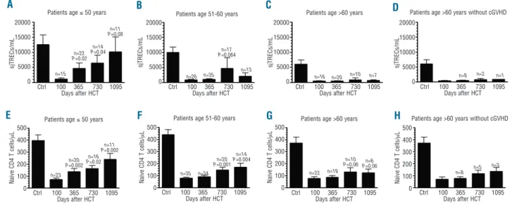
sjTREC/mL of blood increased from day 100 to one and two years after transplantation. Specifically, sjTREC con-centrations were 786±1,207 sjTREC/mL on day 100, 1,890±4,784 sjTREC/mL one year after HCT (P=0.0066 in comparison to day 100), and 4,333±1,1131 sjTREC/mL two years after HCT (P=0.0015 in comparison to day 100 and P=0.0422 in comparison to one year after HCT), respectively. However, as shown in Figure 1A-D, thymic recovery did not occur in patients over 60 years of age. Specifically, sjTREC concentrations increased from day 100 to one and two years after transplantation in patients aged 50 years and under (P=0.0245 and P=0.0371, respec-tively), and in those 51-60 years of age (P=0.17 and
P=0.064, respectively), but not in patients over 60 years
old (P=0.3 and P=0.3, respectively), even censored at onset of chronic GVHD. Similar results were observed when taking into consideration only patients for whom we had data on each day 100, one year, two years and three years after transplantation (Online Supplementary Figure 1A).
An inverse correlation was observed between sjTREC concentrations one year or more after HCT and recipient (R=-0.41, P<0.0001) and donor (R=-0.25, P=0.0002) ages (Figure 2A). In addition, chronic GVHD had a profound negative impact on sjTREC concentrations one year or more year after HCT. Specifically, sjTREC concentrations were 5,018±9,331 sjTREC/mL in patients without chronic GVHD, 2,493±7,383 sjTREC/mL (P=0.0078) in patients with limited chronic GVHD, 1,209±5,029 sjTREC/mL (P<0.0001) in patients with extensive chronic GVHD, and 4,166±12,436 sjTREC/mL (P=0.0021) in patients with resolved extensive chronic GVHD, respectively. The same results were observed when the impact of chronic GVHD presence / severity was assessed separately one year, two years and three years after transplantation (Figure 2B). As shown in the Online Supplementary Figure 1C-E, sjTREC concentrations one year after transplantation were not sta-tistically affected by PBSC manipulation, donor lympho-cyte infusion, or by the type of the non-myeloablative conditioning regimen administered. Further, mean±SD sjTREC concentration one year after HCT were 2,396±5,963 sjTREC/mL in patients given unmanipulated PBSC and no MSC, versus 1,303±2,860 sjTREC/mL (P=0.5) in those given unmanipulated PBSC and MSC.
We then performed a multivariate analysis of factors affecting long-term thymic function, taking together sjTREC data from each patient at any time point one year or more after HCT (Table 1). In multivariate analysis, high patient age (P<0.0001), extensive chronic GVHD (P<0.0001), and resolved extensive chronic GVHD (P=0.0075) were independently associated with low sjTREC concentrations (Table 1).
Reconstitution of peripheral lymphocyte subsets
T cellsCD4+T-cell counts remained below normal values
dur-ing the first 540 days after HCT, then reached the 5th
per-centile of normal values on day 540 after HCT. This was mainly due to recovery of memory CD4+ T cells that
reached the 5th percentile of normal values on day 365
after HCT, while CD4+CD45RA+(naïve) T cells remained
below normal values for the first three years after HCT (Figure 3). Recovery of lymphocyte subset counts were similar in patients under 57 years of age or 57 years and over, except for CD4+CD45RA+ (naïve) T-cell recovery
that was lower in older than in younger recipients. Specifically, CD4+CD45RA+T-cell counts increased from
day 100 to one and two years after transplantation in patients aged 50 years and under (P=0.002 and P=0.02, respectively), and in those 51-60 years of age (P=0.4 and
P=0.001, respectively), but less so in patients over 60
years old (P=0.3 and P=0.06, respectively), even when censored at onset of chronic GVHD (Figure 1E-H). Similar results were observed when taking into consideration only patients for whom we had data on each day 100, one year, two years and three years after transplantation (Online Supplementary Figure 1B).
As reported by other investigators,37 we observed a
close correlation between sjTREC concentrations and CD4+CD45RA+T-cell counts (R=0.47, P<0.0001) one year
or more after HCT. In contrast to that observed in the
CD4+ T-cell compartment, CD8+ T cells normalized
quickly, reaching the 5th percentile of normal values as
soon as two months after HCT. This indicates that peripheral expansion of mature T cells contained in the graft was more efficient for CD8+T cells than for CD4+T
cells, as previously reported by other groups of investiga-tors.15As shown in the Online Supplementary Figure 1C-E,
CD3+ T cell and CD4+CD45RA+ T-cell counts one year
after transplantation were not statistically affected by PBSC manipulation, donor lymphocyte infusion, or by the type of the non-myeloablative conditioning regimen
administered. Furthermore, mean ± SD CD3+ T-cell
counts one year after HCT were 1,350±1,409 cells/μL in patients given unmanipulated PBSC and no MSC versus 1,867±1,544 cells/μL (P=0.3) in those given unmanipulat-ed PBSC and MSC.
B and NK cells
CD19+ B-cell counts reached normal values at one year after HCT, while the counts of NK cells reached normal values around six months after HCT.
Multivariate analysis
We then performed a multivariate analysis of factors affecting long term T- and B-cell immune recovery, taking together data from each patient at any time point one year or more after HCT (Table 1). Extensive chronic GVHD was associated with lower counts of T cells (P<0.001), CD8+T
cells (P<0.001), CD4+T cells (P<0.001), CD4+CD45RA+T
cells (P<0.001), and B cells (P=0.003); higher patient age was associated with lower counts of CD4+CD45RA+ T
cells (P<0.001) and B cells (P=0.002) while longer delay after transplantation was associated with higher counts of CD4+ T cells (P<0.001), CD4+CD45RA+T cells (P=0.016),
and B cells (P<0.001). Further, patients given grafts contain-ing less than 1¥106 T cells/kg had lower CD8+ T-cell
Correlation between thymic function and T- and B-cell
phenotypes two years after HCT
We then analyzed the correlation between sjTREC lev-els two years after HCT and detailed T- and B-cell pheno-types two years after HCT in a cohort of 33 patients for whom we had cryopreserved PBMC at that time point. There was a statistically significant correlation between 2-year sjTREC levels and patient age (inverse correlation, R=-0.54, P=0.001) as well as with 2-year counts of naïve
CD4+CD45RA+CCR7+ T cells (positive correlation,
R=0.43, P=0.012), central memory CD4+T cells (positive
correlation, R=0.38, P=0.028), naïve CD8+T cells (positive
correlation, R=0.59, P<0.001), central memory CD8+ T
cells (positive correlation, R=0.55, P=0.001), effector mem-ory CD8+ T cells (positive correlation, R=0.39, P=0.025),
naïve B cells (positive correlation, R=0.53, P=0.001), mem-ory B cells (positive correlation, R=0.45, P=0.008), IgM+IgD+ memory B cells (positive correlation, R=0.43, P=0.012) and switched memory B cells (positive
correla-tion, R=0.45, P=0.008) (Figure 4A-C).
We also analyzed the impact of patient age and exten-sive chronic GVHD on T- and B-cell phenotypes. Patients aged 57 years and over (n=15) had lower counts of naïve CD8+ T cells (P=0.06), naïve B cells (P=0.02), memory B
cells (P=0.06) and switched memory B cells (P=0.006) than those under 57 years of age at HCT (n=18) (Figure 4D-F). Finally, patients who had extensive chronic GVHD two years after HCT had lower counts of effector memory CD8+T cells (P=0.04), effector memory RA+CD8+T cells
(P=0.04), naïve B cells (P=0.002), memory B cells (P=0.04), and switched memory B cells (P=0.07) than those who did not (Figure 4G-I).
Correlation between thymic function and T-cell Vbeta
repertoire diversity
In order to assess the impact of thymic function on Vbeta repertoire diversity, T-cell receptor Vbeta repertoire CDR3 spectratyping analyses were performed between two and nine (median three) years after HCT in 21 patients with low (<500 sjTREC/mL; n=9) or high (>500
sjTREC/mL; n=12) sjTREC levels, measured on the same day as the spectratyping analysis. As illustrated in the
Online Supplementary Figure 2, the Vbeta repertoire was
rel-atively complex even in patients with low thymic func-tion, probably reflecting the fact that peripheral expansion of T cells contained in the graft eventually allowed a rela-tively diverse T-cell receptor Vbeta repertoire, although some oligoclonal T-cell receptor Vbeta families were observed, and particularly so in patients with low sjTREC levels. Specifically, median T-cell receptor Vbeta score was 239 (range 179-251) peaks in age-matched controls, 207
Table 1.Multivariable analyses of factors affecting immune recovery one year or more after non-myeloablative conditioning (n=242)*.
Cell subset Factor(s) associated with lower levels /
cell subset counts
sjTREC concentration - high patient age†(P<0.0001).
- extensive chronic GVHD (P<0.0001). - antecedent of extensive chronic GVHD
(P=0.0075).
n. T cells - extensive chronic GVHD (P<0.0001). - related donor (P=0.0353). n. CD4+T cells - extensive chronic GVHD (P=0.0007).
- shorter delay after HCT†(P<0.0001).
n. naïve CD4+T cells - extensive chronic GVHD (P<0.0001).
- high patient age†(P=0.0004).
- shorter delay after HCT †(P=0.016).
n. CD8+T cells - extensive chronic GVHD (P=0.0001).
- n. CD3+T cells transplanted < 1¥106/kg
(P=0.0018).
- absence of limited chronic GVHD (P=0.008). n. B cells - extensive chronic GVHD (P=0.003).
- high patient age†(P=0.0017).
- shorter delay after HCT†(P=0.0006).
*Other factors assessed were donor age, HLA-mismatch between donor and recipient or not; †modeled as a continuous linear variable; GVHD, graft-versus-host disease.
Figure 1.Impact of patient age (at HCT) on sjTREC (A-D) and naïve CD4 T cell (E-H) recovery. (Dand H) Patients were censured at time of occurrence of chronic GVHD. P values are given for comparison with day 100 values.
Ctrl 100 365 730 1095 Days after HCT Ctrl 100 365 730 1095 Days after HCT Ctrl 100 365 730 1095 Days after HCT Ctrl 100 365 730 1095 Days after HCT Ctrl 100 365 730 1095 Days after HCT Ctrl 100 365 730 1095 Days after HCT Ctrl 100 365 730 1095 Days after HCT Ctrl 100 365 730 1095 Days after HCT Patients age ≤ 50 years
Patients age ≤ 50 years 20000 15000 10000 5000 0 500 400 300 200 100 0 20000 15000 10000 5000 0 20000 15000 10000 5000 0 20000 15000 10000 5000 0 n=15 n=23 n=35 n=22 n=8 n=5 n=3 n=19 n=34 n=26 n=35 n=13 n=16 n=20 n=10 n=7 n=8 n=2 n=1 n=22 P=0.02 n=20 P=0.002 n=16 P=0.02 n=20 P=0.001 n=10 P=0.06 P=0.06n=6 n=14 P=0.004 n=11 P=0.002 n=14 P=0.04 n=17 P=0.064 n=11 P=0.08
Patients age 51-60 years
Patients age 51-60 years
Patients age >60 years
Patients age >60 years
Patients age >60 years without cGVHD
Patients age >60 years without cGVHD
s jT R E C s /m L s jT R E C s /m L s jT R E C s /m L s jT R E C s /m L N a iv e C D 4 T c e ll s /μ L 500 400 300 200 100 0 N a iv e C D 4 T c e ll s /μ L 500 400 300 200 100 0 N a iv e C D 4 T c e ll s /μ L 500 400 300 200 100 0 N a iv e C D 4 T c e ll s /μ L A B C D H G F E
(range 173-246) peaks in patients with low sjTREC levels (P=0.038 in comparison to controls), and 218 (range 182-252) in patients with high sjTREC levels (NS in compari-son to controls and to patients with low sjTREC). Furthermore, we quantified the deviation from normality of each repertoire by measuring its quadratic distance to an average repertoire measured in healthy donors (see
Design and Methods section).32The quadratic distance was
4.5 (range 3.2-6.5) in age-matched controls, 8.1 (range 3.8-11.3) in patients with low sjTREC levels (P=0.003 in com-parison to controls), and 5.3 (range 4.5-9.1) in patients with high sjTREC levels (P=0.016 in comparison to con-trols, P=0.22 in comparison to patients with low sjTREC).
Correlation between thymic function and clinical
events
Day 100 sjTREC levels were similar in patients who had grade II-IV acute GVHD before day 100, as compared to those who did not (687±947 vs. 824±1,303 sjTREC/mL,
P=0.5). Day 100 sjTREC levels was not associated with
the occurrence of chronic GVHD: the 1-year probability of chronic GVHD was 57% in patients with day 100 sjTREC levels below the median (323 sjTREC/mL) versus 52% in those with higher sjTREC levels on day 100 (P=0.5).
We then studied infections occurring between day 365 to 730 based on day 365 sjTREC levels. The probabilities of bacterial, fungal and viral infections were 40%, 9%, and 13%, respectively, in patients with day 365 sjTREC levels below the median (523 sjTREC/mL) versus 31% (P=0.3), 5% (P=0.6) and 17% (P=0.6), respectively, in those with higher levels. The 7-year probability of dying from infec-tion was 40% in patients with day 365 sjTREC levels below the median versus 0% in those with higher values (P=0.01, Figure 2C). However, 6 of 7 patients who died from infections had extensive chronic GVHD at the time of death. Further, the association between 1-year sjTREC levels and the subsequent risk of dying from infection was no longer statistically significant (P=0.17) after adjustment for the presence of extensive chronic GVHD one year after HCT in a Cox’s model where sjTREC levels were modeled as a continuous linear variable.
Discussion
Late infections (with or without chronic GVHD) are the first causes of non-relapse death beyond one year after HCT in young patients given myeloablative
condition-0 25 50 75 100 Age at transplantation 0 365 730 1095 1460 1825 Days after HCT 0 365 730 1095 1460 1825 Days after HCT 0 365 730 1095 1460 1825 Days after HCT 0 365 730 1095 1460 1825 Days after HCT 0 365 730 1095 1460 1825 Days after HCT 0 365 730 1095 1460 1825 Days after HCT CD3+CD8+ CD56+ CD19+ CD4+CD45RO+ 100 500 0 750 500 250 0 1500 1000 500 0 2000 1000 0 1000 500 0 500 250 0 CD4+CD45RA+ C ells/ m L C ells/ m L C ells/ m L C ells/ m L C ells/ m L C ells/ m L CD3+CD4+ 0 1 2 3 4 5 6 7 8 9
Years after transplantation 1.0 0.8 0.6 0.4 0.2 0.0 1 year 2 years 3 years
r=-0.41 P<0.0001 100000 10000 1000 100 10 1 0.1 sjTR EC s/ m L 100000 10000 1000 100 sjTR EC s/ m L C l of deat h fr om inf ect ion
Figure 2.(A) Correlation between patient age at HCT and sjTREC levels one year or more after HCT. (B) Impact of chronic GVHD on sjTREC concentration (mean ± standard deviation) 1-3 years after HCT. Black bars, no chronic GVHD; white bars; limited chronic GVHD, black and white bars, extensive chronic GVHD; gray bars, resolved extensive chronic GVHD. P values (obtained with the Mann-Whitney test) are given in comparison to patients without chronic GVHD. (C) Cumulative incidence of dying from infection in patients with sjTREC levels on day 365 below (continuous line) or above (broken line) the median.
Figure 3.Mean MNC-subset cell counts after nonmyeloabla-tive conditioning in patients under 57 years of age or 57 years and over at HCT. Horizontal lines show the 5thand 95th
percentiles in 47 age-matched healthy volunteer donors. *,P<0.05. A B C A B C D E F
ing.38,39 They have been attributed to slow recovery of
CD4+T cells.38,40Nevertheless, despite the well established
association between higher patient age and impaired
immune recovery following allogeneic HCT,14HCT after
non-myeloablative conditioning has been successful treat-ment for many patients aged up to between 60 and 70 years of age, most of whom were without evidence of severe infection once systemic immunosuppression was discontinued.31,41 This apparent paradox prompted us to
assess long-term immune recovery after non-myeloabla-tive conditioning. Several observations were made.
First and most importantly, while patients below 50 years of age at the time of HCT had evidence of thymic recovery from day 100 to three years after HCT, thymic recovery was much slower in patients aged 50 to 60 years, and was virtually absent in patients over 60, even in those without chronic GVHD. These results mirror those report-ed by Hakim et al. in patients given autologous HCT where the authors observed a significant thymic rebound in approximately 80% of patients younger than 40 years, 45% of those aged 40 to 50 years, but in less than 15% of those aged 50 years and older.42
Recovery of naïve CD4 T cells after HCT in current patients somewhat mirrored recovery of thymic function assessed by sjTREC levels. Specifically, recovery of naïve T cells was observed by day 365 in patients below 50 years of age, but only at two years after HCT (and to a lesser extent) in patients above 50-60 years of age. Further,
sjTREC levels correlated with those of CD4+CD45RA+T
cells one year and over after HCT, and we observed a strong correlation between sjTREC levels and naïve CD4+CD45RA+CCR7+T-cell and naïve CD8+T-cell counts
two years after HCT. These observations are in agreement with previous reports showing that new naïve T cells orig-inate mainly from the thymic pathway after HCT.43,44
Given the slow or absent thymic recovery in patients
above 50 or 60 years, respectively,
immune recovery in those patients should have depended mainly on peripheral expansion of mature T cells con-tained in the graft. Consistent with this possibility, recov-ery of CD8+T cells occurred earlier than recovery of CD4+
T cells, and T-cell recovery was derived from memory rather than naïve T cells.15Interestingly, peripheral
expan-sion was sufficient to produce an efficient immune system in the majority of patients without GVHD. Indeed, TCRB repertoires were relatively complex both in patients with low compared to high sjTREC levels, and incidences of viral, bacterial and fungal infections were similar in patients with low or high sjTREC levels. The risk of death from infection was significantly higher in patients with low sjTREC levels, although all but one patient who died from infection had extensive severe chronic GVHD at the time of death suggesting that GVHD or GVHD treatment might also have played an important role in the risk of mortality from infection in the current study. The observa-tion that late immunity in older patients mainly depends on mature T cells contained in the graft has important implications since it suggests that long-term immune func-tion after HCT might be affected by techniques of in vitro or in vivo T-cell depletion of the graft in these patients. Analysis of long-term immune recovery in older patients given PBSC after alemtuzumab-based reduced-intensity conditioning45would be particularly interesting to confirm
this hypothesis.
Another observation of our study was the dramatic impact of chronic GVHD on immune recovery. Specifically, extensive chronic GVHD was significantly associated with low sjTREC levels, as well as low counts of CD4+ T cells, CD4+CD45RA+ (naïve) T cells, CD8+ T
cells and B cells one year and over after HCT, and was sig-nificantly associated with lower counts of effector memo-ry CD8+T cells, effector memory RA+CD8+T cells, naïve Figure 4. CD4+ T-cell
(A), CD8+T-cell (B) and
B-cell (C) phenotypes two years after HCT in patients with sjTREC levels below (white bars) or above (black bars) median (1,000 sjTREC/μL) at that time (n=33). CD4+T-cell (D),
CD8+ T-cell (E) and
B-cell (F) subtype pheno-types two years after HCT in patients (white bars) or (black bars) under 57 years of age or 57 years and over at HCT. CD4+ T-cell (G),
CD8+ T-cell (H) and
B-cell (I) subtype pheno-types two years after HCT in patients without (white bars) or with (black bars) extensive chronic GVHD at that time. TCM, central memory T cells; TEM, effector memory T cells; TEM RA+, TEM RA+
CD8+T cells; M.,
memo-ry; M.IgM+IgD+IgM+IgD+
memory B cells; M.Switched, switched memory B cells.
Naive TCM TEM Late TEM
Naive TCM TEM Late TEM
Naive TCM TEM Late TEM Naive TCM TEM TEM RA+
Naive TCM TEM TEM RA+
Naive TCM TEM TEM RA+ Naive M. M.IgM+IgD+M.Switched
Naive M. M.IgM+IgD+M.Switched
Naive M. M.IgM+IgD+M.Switched
CD4+T cells CD8+T cells B cells
CD4+T cells Age cGVHD sjTREC CD8+T cells P=0.04 P=0.02 P=0.002 P=0.03 P=0.002 P=0.009 P=0.02 P=0.06 P=0.07 P=0.06 P=0.006 P=0.07 P=0.03 P=0.002 P=0.04 P=0.04 P=0.01 P=0.02 P<0.001 B cells
CD4+T cells CD8+T cells B cells
600 500 400 300 200 100 0 600 500 400 300 200 100 0 600 500 400 300 200 100 0 500 400 300 200 100 0 1000 800 600 400 200 0 1000 800 600 400 200 0 1000 800 600 400 200 0 C e ll s /μ L C e ll s /μ L C e ll s /μ L 500 400 300 200 100 0 500 400 300 200 100 0 C e ll s /μ L C e ll s /μ L C e ll s /μ L C e ll s /μ L C e ll s /μ L C e ll s /μ L A D G H I E F B C
B cell and memory B cells two years after HCT. The neg-ative impact of chronic GVHD on immune recovery is probably multi-factorial.46 First, the negative impact of
chronic GVHD on thymus architecture46and function47-52
has been well demonstrated, and has been attributed to direct donor T-cell alloreactivity towards recipient thy-mus,53 and to a negative impact of immunosuppressive
drugs (and particularly of glucocorticosteroids) on the thy-mus.54,55 Secondly, chronic GVHD has been shown to
impair peripheral expansion of mature T cells, and the same is obviously true for immunosuppressive drugs given in the treatment of patients with chronic GVHD. Thirdly, chronic GVHD has been associated with reduced B-cell lymphopoiesis perhaps because of B-cell inhibitory cytokines produced by activated T cells in chronic GVHD patients or because of GVHD treatments.56
In summary, our data suggest that thymic neo-genera-tion of T cells occurs from day 100 onwards in patients
under 60 years of age. In contrast, the levels of sjTREC remain low for patients over 60 who depended nearly exclusively on peripheral expansion of mature donor T cells contained in the graft to reconstitute their T-cell com-partment. This suggests that long-term immune function after HCT might be negatively impacted by techniques of
in vitro or in vivo T-cell depletion of the graft in older
patients given allogeneic HCT.
Authorship and Disclosures
The information provided by the authors about contributions from persons listed as authors and in acknowledgments is avail-able with the full text of this paper at www.haematologica.org.
Financial and other disclosures provided by the authors using the ICMJE (www.icmje.org) Uniform Format for Disclosure of Competing Interests are also available at www.haematologica.org.
References
1. Steinmann GG, Klaus B, Muller-Hermelink HK. The involution of the ageing human thymic epithelium is independent of puber-ty. A morphometric study. Scand J Immunol. 1985;22(5):563-75.
2. Fry TJ, Mackall CL. Current concepts of thymic aging. Springer Semin Immunopathol. 2002;24(1):7-22.
3. Douek DC, McFarland RD, Keiser PH, Gage EA, Massey JM, Haynes BF, et al. Changes in thymic function with age and during the treatment of HIV infection. Nature. 1998;396(6712):690-5.
4. Jamieson BD, Douek DC, Killian S, Hultin LE, Scripture-Adams DD, Giorgi JV, et al. Generation of functional thymocytes in the human adult. Immunity. 1999;10(5):569-75. 5. Haynes BF, Markert ML, Sempowski GD, Patel DD, Hale LP. The role of the thymus in immune reconstitution in aging, bone marrow transplantation, and HIV-1 infec-tion (Review). Annu Rev Immunol. 2000; 18:529-60.
6. Sandmaier BM, Mackinnon S, Childs RW. Reduced intensity conditioning for allo-geneic hematopoietic cell transplantation: current perspectives. Biol Blood Marrow Transplant. 2007;13(1 Suppl 1):87-97. 7. Baron F, Storb R. Allogeneic hematopoietic
cell transplantation following nonmyeloab-lative conditioning as treatment for hema-tologic malignancies and inherited blood disorders (Review). Molecular Therapy. 2006;13(1):26-41.
8. Barrett AJ, Savani BN. Stem cell transplan-tation with reduced-intensity conditioning regimens: a review of ten years experience with new transplant concepts and new therapeutic agents. Leukemia. 2006;20(10): 1661-72.
9. Baron F, Storb R, Storer BE, Maris MB, Niederwieser D, Shizuru JA, et al. Factors associated with outcomes in allogeneic hematopoietic cell transplantation with non-myeloablative conditioning after failed mye-loablative hematopoietic cell transplanta-tion. J Clin Oncol. 2006;24(25):4150-7. 10. Storb R, Yu C, Wagner JL, Deeg HJ, Nash
RA, Kiem H-P, et al. Stable mixed hematopoietic chimerism in DLA-identical
littermate dogs given sublethal total body irradiation before and pharmacological immunosuppression after marrow trans-plantation. Blood. 1997;89(8):3048-54. 11. McSweeney PA, Niederwieser D, Shizuru
JA, Sandmaier BM, Molina AJ, Maloney DG, et al. Hematopoietic cell transplanta-tion in older patients with hematologic malignancies: replacing high-dose cytotox-ic therapy with graft-versus-tumor effects. Blood. 2001;97(11):3390-400.
12. Baron F, Maris MB, Sandmaier BM, Storer BE, Sorror M, Diaconescu R, et al. Graft-versus-tumor effects after allogeneic hematopoietic cell transplantation with nonmyeloablative conditioning. J Clin Oncol. 2005;23(9):1993-2003.
13. Baron F, Petersdorf EW, Gooley T, Sandmaier BM, Malkki M, Chauncey TR, et al. What is the role for donor natural killer cells after nonmyeloablative condi-tioning? Biol Blood Marrow Transplant. 2009;15(5):580-8.
14. Storek J, Witherspoon RP. Immunological reconstitution after hemopoietic stem cell transplantation. In: Atkinson K, Champlin R, Ritz J, Fibbe WE, Ljungman P, Brenner MK, editors. Clinical Bone Marrow and Blood Stem Cell Transplantation. Cambridge, UK: Cambridge University Press, 2004:194-226.
15. Crooks GM, Weinberg K, Mackall C. Immune reconstitution: from stem cells to lymphocytes. Biol Blood Marrow Transplant. 2006;12(1 Suppl. 1):42-6. 16. Peggs KS. Reconstitution of adaptive and
innate immunity following allogeneic hematopoietic stem cell transplantation in humans. Cytotherapy. 2006;8(5):427-36. 17. Gress RE, Emerson SG, Drobyski WR.
Immune reconstitution: how it should work, what’s broken, and why it matters. Biol Blood Marrow Transplant. 2010;16(1 Suppl):S133-7.
18. Storek J, Dawson MA, Maloney DG. Correlation between the numbers of naive T cells infused with blood stem cell allo-grafts and the counts of naive T cells after transplantation. Biol Blood Marrow Transplant. 2003;9(12):781-4.
19. Douek DC, Vescio RA, Betts MR, Brenchley JM, Hill BJ, Zhang L, et al. Assessment of thymic output in adults after
haematopoietic stem-cell transplantation and prediction of T-cell reconstitution. Lancet. 2000;355(9218):1875-81.
20. Weinberg K, Blazar BR, Wagner JE, Agura E, Hill BJ, Smogorzewska M, et al. Factors affecting thymic function after allogeneic hematopoietic stem cell transplantation. Blood. 2001;97(5):1458-66.
21. Hochberg EP, Chillemi AC, Wu CJ, Neuberg D, Canning C, Hartman K, et al. Quantitation of T-cell neogenesis in vivo after allogeneic bone marrow transplanta-tion in adults. Blood. 2001;98(4):1116-21. 22. Lewin SR, Heller G, Zhang L, Rodrigues E,
Skulsky E, van den Brink MR, et al. Direct evidence for new T-cell generation by patients after either T-cell-depleted or unmodified allogeneic hematopoietic stem cell transplantations. Blood. 2002;100(6): 2235-42.
23. Jimenez M, Martinez C, Ercilla G, Carreras E, Urbano-Ispizua A, Aymerich M, et al. Clinical factors influencing T-cell receptor excision circle (TRECs) counts following allogeneic stem cell transplantation in adults. Transpl Immunol. 2006;16(1):52-9. 24. Hazenberg MD, Otto SA, de Pauw ES,
Roelofs H, Fibbe WE, Hamann D, et al. T-cell receptor excision circle and T-T-cell dynamics after allogeneic stem cell trans-plantation are related to clinical events. Blood. 2002;99(9):3449-53.
25. Baron F, Storer B, Maris MB, Storek J, Piette F, Metcalf M, et al. Unrelated donor status and high donor age independently affect immunologic recovery after nonmyeloabla-tive conditioning. Biol Blood Marrow Transplant. 2006;12(11):1176-87.
26. Castermans E, Baron F, Willems E, Schaaf-Lafontaine N, Meuris N, Gothot A, et al. Evidence for neo-generation of T cells by the thymus after non-myeloablative condi-tioning. Haematologica 2008;93(2):240-7. 27. Baron F, Schaaf-Lafontaine N,
Humblet-Baron S, Meuris N, Castermans E, Baudoux E, et al. T-cell reconstitution after unmanip-ulated, CD8-depleted or CD34-selected nonmyeloablative peripheral blood stem-cell transplantation. Transplantation. 2003;76(12):1705-13.
28. Baron F, Lechanteur C, Willems E, Bruck F, Baudoux E, Seidel L, et al. Cotransplantation of mesenchymal stem cells might prevent
death from graft-versus-host disease (GVHD) without abrogating graft-versus-tumor effects after HLA-mismatched allo-geneic transplantation following nonmye-loablative conditioning. Biol Blood Marrow Transplant. 2010;16(6):838-47.
29. Sullivan KM. Graft-vs.-host disease. In: Blume KG, Forman SJ, Appelbaum FR, edi-tors. Thomas' Hematopoietic Cell Transplantation. Oxford, UK: Blackwell Publishing Ltd., 2004: 635-64.
30. Baron F, Sandmaier BM. Chimerism and outcomes after allogeneic hematopoietic cell transplantation following nonmyeloab-lative conditioning. Leukemia. 2006;20(10): 1690-700.
31. Willems E, Baron F, Baudoux E, Wanten N, Seidel L, Vanbellinghen JF, et al. Non-mye-loablative transplantation with CD8-depleted or unmanipulated peripheral blood stem cells: a phase II randomized trial. Leukemia. 2009;23(3):608-10. 32. Baron V, Bouneaud C, Cumano A, Lim A,
Arstila TP, Kourilsky P, et al. The repertoires of circulating human CD8(+) central and effector memory T cell subsets are largely distinct. Immunity. 2003;18(2):193-204. 33. Dion ML, Poulin JF, Bordi R, Sylvestre M,
Corsini R, Kettaf N, et al. HIV infection rap-idly induces and maintains a substantial suppression of thymocyte proliferation. Immunity. 2004;21(6):757-68.
34. Poulin JF, Sylvestre M, Champagne P, Dion ML, Kettaf N, Dumont A, et al. Evidence for adequate thymic function but impaired naive T-cell survival following allogeneic hematopoietic stem cell transplantation in the absence of chronic graft-versus-host disease. Blood. 2003;102(13):4600-7. 35. Morrhaye G, Kermani H, Legros JJ, Baron F,
Beguin Y, Moutschen M, et al. Impact of growth hormone (GH) deficiency and GH replacement upon thymus function in adult patients. PLoS One 2009;4(5):e5668. 36. Storek J, Dawson MA, Storer B,
Stevens-Ayers T, Maloney DG, Marr KA, et al. Immune reconstitution after allogeneic marrow transplantation compared with blood stem cell transplantation. Blood. 2001;97(11):3380-9.
37. Cavazzana-Calvo M, Carlier F, Le Deist F, Morillon E, Taupin P, Gautier D, et al. Long-term T-cell reconstitution after hematopoi-etic stem-cell transplantation in primary T-cell-immunodeficient patients is associated with myeloid chimerism and possibly the primary disease phenotype. Blood. 2007; 109(10):4575-81.
38. Storek J, Gooley T, Witherspoon RP, Sullivan KM, Storb R. Infectious morbidity in long-term survivors of allogeneic mar-row transplantation is associated with low CD4 T cell counts. Am J Hematol. 1997; 54:131-8.
39. Robin M, Porcher R, De Castro AR, de Latour RP, Devergie A, Rocha V, et al. Risk factors for late infections after allogeneic hematopoietic stem cell transplantation from a matched related donor. Biol Blood Marrow Transplant. 2007;13(11):1304-12. 40. Roux E, Dumont-Girard F, Starobinski M,
Siegrist CA, Helg C, Chapuis B, et al. Recovery of immune reactivity after T-cell-depleted bone marrow transplantation depends on thymic activity. Blood. 2000; 96(6):2299-303.
41. Maris MB, Sandmaier BM, Storer BE, Maloney DG, Shizuru JA, Agura E, et al. Unrelated donor granulocyte colony-stimu-lating factor-mobilized peripheral blood mononuclear cell transplantation after non-myeloablative conditioning: the effect of postgrafting mycophenolate mofetil dos-ing. Biol Blood Marrow Transplant. 2006; 12(4):454-65.
42. Hakim FT, Memon SA, Cepeda R, Jones EC, Chow CK, Kasten-Sportes C, et al. Age-dependent incidence, time course, and consequences of thymic renewal in adults. J Clin Invest. 2005;115(4):930-9.
43. Heitger A, Neu N, Kern H, Panzer-Grumayer ER, Greinix H, Nachbaur D, et al. Essential role of the thymus to reconsti-tute naive (CD45RA+) T-helper cells after human allogeneic bone marrow transplan-tation. Blood. 1997;90(2):850-7.
44. Mackall CL, Fleisher TA, Brown MR, Andrich MP, Chen CC, Feuerstein IM, et al. Age, thymopoiesis, and CD4+ T-lympho-cyte regeneration after intensive chemotherapy. N Engl J Med. 1995;332(3): 143-9.
45. D'Sa S, Peggs K, Pizzey A, Verfuerth S, Thuraisundaram D, Watts M, et al. T- and B-cell immune reconstitution and clinical out-come in patients with multiple myeloma receiving T-cell-depleted, reduced-intensity allogeneic stem cell transplantation with an alemtuzumab-containing conditioning regi-men followed by escalated donor lympho-cyte infusions. Br J Haematol. 2003;123(2): 309-22.
46. Krenger W, Hollander GA. The immunopathology of thymic GVHD. Semin Immunopathol. 2008;30(4):439-56. 47. Noel DR, Witherspoon RP, Storb R,
Atkinson K, Doney K, Mickelson EM, et al. Does graft-versus-host disease influence the tempo of immunologic recovery after allogeneic human marrow transplantation? An observation on 56 long-term survivors. Blood. 1978;51:1087-105.
48. Atkinson K, Incefy GS, Storb R, Sullivan KM, Iwata T, Dardenne M, et al. Low serum thymic hormone levels in patients with chronic graft-versus-host disease. Blood. 1982;59(5):1073-7.
49. Dulude G, Roy DC, Perreault C. The effect of graft-versus-host disease on T cell pro-duction and homeostasis. J Exp Med. 1999; 189(8):1329-42.
50. Clave E, Busson M, Douay C, Peffault de Latour R, Berrou J, Rabian C, et al. Acute graft-versus-host disease transiently impairs thymic output in young patients after allo-geneic hematopoietic stem cell transplanta-tion. Blood. 2009;113(25):6477-84. 51. Bahceci E, Epperson D, Douek DC,
Melenhorst JJ, Childs RC, Barrett AJ. Early reconstitution of the T-cell repertoire after non-myeloablative peripheral blood stem cell transplantation is from post-thymic T-cell expansion and is unaffected by graft-versus-host disease or mixed chimaerism. Br J Haematol. 2003;122(6):934-43. 52. Fallen PR, McGreavey L, Madrigal JA,
Potter M, Ethell M, Prentice HG, et al. Factors affecting reconstitution of the T cell compartment in allogeneic haematopoietic cell transplant recipients. Bone Marrow Transplant. 2003;32(10):1001-14.
53. Hauri-Hohl MM, Keller MP, Gill J, Hafen K, Pachlatko E, Boulay T, et al. Donor T-cell alloreactivity against host thymic epitheli-um limits T-cell development after bone marrow transplantation. Blood. 2007;109 (9):4080-8.
54. Yamada K, Gianello PR, Ierino FL, Fishbein J, Lorf T, Shimizu A, et al. Role of the thy-mus in transplantation tolerance in minia-ture swine: II. Effect of steroids and age on the induction of tolerance to class I mis-matched renal allografts. Transplantation. 1999;67(3):458-67.
55. Hattori A, Kunz HW, Gill TJ 3rd, Shinozuka H. Thymic and lymphoid changes and serum immunoglobulin abnor-malities in mice receiving cyclosporine. Am J Pathol. 1987;128(1):111-20.
56. Storek J, Wells D, Dawson MA, Storer B, Maloney DG. Factors influencing B-lym-phopoiesis after allogeneic hematopoietic cell transplantation (Brief Report). Blood. 2001;98(2):489-91.