R E S E A R C H A R T I C L E
Open Access
Arterial dP/dt
max
accurately reflects left
ventricular contractility during shock when
adequate vascular filling is achieved
Philippe Morimont
1*, Bernard Lambermont
1, Thomas Desaive
2, Nathalie Janssen
3, Geoffrey Chase
4and
Vincent D
’Orio
3Abstract
Background: Peak first derivative of femoral artery pressure (arterial dP/dtmax) derived from fluid-filled catheter
remains questionable to assess left ventricular (LV) contractility during shock. The aim of this study was to test if
arterial dP/dtmaxis reliable for assessing LV contractility during various hemodynamic conditions such as
endotoxin-induced shock and catecholamine infusion.
Methods: Ventricular pressure-volume data obtained with a conductance catheter and invasive arterial pressure obtained with a fluid-filled catheter were continuously recorded in 6 anaesthetized and mechanically ventilated pigs. After a stabilization period, endotoxin was infused to induce shock. Catecholamines were transiently
administrated during shock. Arterial dP/dtmaxwas compared to end-systolic elastance (Ees), the gold standard
method for assessing LV contractility.
Results: Endotoxin-induced shock and catecholamine infusion lead to significant variations in LV contractility. Overall, significant correlation (r = 0.51; p < 0.001) but low agreement between the two methods were observed. However, a far better correlation with a good agreement were observed when positive-pressure ventilation
induced an arterial pulse pressure variation (PPV)≤ 11% (r = 0.77; p < 0.001).
Conclusion: While arterial dP/dtmaxand Ees were significantly correlated during various hemodynamic conditions,
arterial dP/dtmaxwas more accurate for assessing LV contractility when adequate vascular filling, defined as PPV≤
11%, was achieved.
Keywords: Left ventricular function, Aortic pressure, Septic cardiomyopathy, Preload responsiveness, Endotoxin-induced shock
Background
Sepsis-induced myocardial dysfunction occurs more fre-quently than expected while its severity is often under-estimated [1]. Indeed, quantification of left ventricular (LV) inotropic function during sepsis is an ongoing pre-occupation of clinicians. The theoretical gold standard for assessment of LV contractility is the end-systolic pressure volume relation (ESPVR). The slope of the rela-tion defines the maximum elastance, also called end-sys-tolic elastance (Ees), a load-independent index of LV contractility [2]. However, this method is difficult to
apply in clinical practice because requiring preload reduction and catheterization of the left ventricle with a high-fidelity pressure catheter. Several other contractility indices have been proposed but most of them are influ-enced by cardiac loading conditions [3-8]. A relative exception to this is the peak first derivative of LV
pres-sure (LV dP/dtmax) which is relatively afterload
indepen-dent [9]. Most critically ill patients with hemodynamic instability are instrumented with a femoral fluid-filled catheter for accurate arterial pressure monitoring. dP/
dtmax can be derived from the arterial pressure curve
(the maximal ascending slope of the peripheral arterial
pressure curve). However, the use of arterial dP/dtmaxas
an index of LV contractility remains questionable * Correspondence: [email protected]
1Medical Intensive Care Unit, University Hospital of Liège, Liège, Belgium
Full list of author information is available at the end of the article
© 2012 Morimont et al; licensee BioMed Central Ltd. This is an Open Access article distributed under the terms of the Creative Commons Attribution License (http://creativecommons.org/licenses/by/2.0), which permits unrestricted use, distribution, and reproduction in any medium, provided the original work is properly cited.
because dP/dtmax is derived from arterial pressure
obtained with a fluid-filled catheter and is influenced by preload and vascular filling conditions [4,10]. Vascular filling and fluid requirement in critically ill patients are usually assessed by positive-pressure ventilation-induced arterial pulse pressure variation (PPV), which is a sensi-tive and specific predictor of preload responsiveness. PPV is measured over a single respiratory cycle and defined as the maximal pulse pressure (systolic-diastolic pressure) less the minimal pulse pressure divided by the average of these two pressures. In hypovolemic states, PPV due to cycling pressure gradient from mechanical ventilation is high. However, when adequate vascular filling is obtained, PPV is low [11]. The purpose of this
study was to investigate whether arterial dP/dtmax
derived from fluid-filled femoral artery catheter can be used to assess LV contractility in different hemodynamic
conditions. To test this hypothesis, arterial dP/dtmax, LV
dP/dtmaxand Ees were compared during various
altera-tions in LV contractile function resulting from endo-toxin-induced shock and catecholamine infusion. PPV was continuously monitored to assess the vascular filling.
Methods
All experimental procedures and protocols used in this investigation were reviewed and approved by the ethical committee of the Medical Faculty of the University of Liege and conformed to the Guide for the Care and Use of Laboratory Animals published by the US National Institutes of Health (NIH Publication No. 85-23, revised 1996).
Experimental model
Experiments were performed on 6 healthy pure Pietran pigs of either sex weighing from 16 to 28 kg. The ani-mals were premedicated with intramuscular administra-tion of tiletamine (250 mg) and zolazepam (250 mg). Anaesthesia was then induced and maintained by a
con-tinuous infusion of sufentanil (0.5 μg/kg/h) and
pento-barbital (5 mg/kg/h). Spontaneous movements were prevented by cisatracurium (0.1 mg/kg/h). After endo-tracheal intubation via a cervical tracheostomy, the pigs were connected to a volume-cycled ventilator (Datex Ohmeda, Engström Carestation, General Electric, USA) set to deliver a tidal volume of 10 ml/kg at a respiratory
rate of 20/min with a FiO2 of 0.4 and a PEEP of 5 cm
H2O. A 7F, multi-electrode (9-mm interelectrode dis-tance) conductance micromanometer-tipped catheter (Scisense, Canada) was inserted through the left carotid artery into the left ventricle and positioned so that all electrodes remained in the LV cavity. A central venous line was inserted into the right jugular vein and placed inside the superior vena cava. Arterial blood pressure
was monitored via a 4F fluid-filled catheter (Pulsiocath, Pulsion Medical System, Germany) inserted into the right femoral artery. A 6F Fogarty balloon catheter (Bax-ter Healthcare Corp., Oakland, CA, USA) was advanced into the inferior vena cava through a right femoral venotomy. Inflation of this balloon produced a gradual preload reduction.
Experimental protocol
After surgical preparation, the animals were allowed to
stabilize for 30 min (’basal’ period). Hemodynamic data
including mean arterial blood pressure, heart rate (HR), cardiac output (CO), LV volume and pressure, were continuously recorded. Then, the animals had a 0.5 mg/ kg intravenous infusion of a freshly prepared endotoxin solution (lipopolysaccharide from E.coli serotype 0127: B8, Sigma, St Louis, MO, USA) over 30 min (’endo’ per-iod). When systolic arterial pressure significantly dropped, dobutamine (5 mcg/kg/min) and norepinephr-ine (0.05 mcg/kg/min) were administrated during 60
minutes (’catechol’ period) and then stopped (’shock’
period). Fluid administration with Hartmann’s solution
was continuously controlled by preload responsiveness.
When PPV was≤ 11%, animals were considered as
ade-quately filled.
Data collection and analysis
All analog signals were continuously digitalized and recorded (Notocord-hem evolution, Notocord, Paris, France). During each period of measurement, three tran-sient occlusions of the inferior vena cava using the Fogarty balloon were performed during apnea. Analysis
of the signals was performed offline. Arterial dP/dtmax
and LV_dP/dtmax were calculated on 6 steady-state
cycles just before occlusion of the vena cava. These indices were compared with the gold-standard Ees, cal-culated during transient preload reduction.
Statistical analysis
Arterial dP/dtmax, LV dP/dtmaxand Ees were compared
using linear regressions. A normalized Bland-Altman test (Statistica version 7, StatSoft) was also performed. Changes in hemodynamic parameters were evaluated by a repeated-measures analysis of variance. Data were expressed as mean ± standard deviation (SD).
Results
The effects of endotoxin infusion and catecholamine administration on arterial pressure, HR, ejection fraction (EF) and cardiac output (CO) are summarized in Table 1. The evolution of LV contractility assessed by both
Ees and arterial dP/dtmaxis shown in Figure 1. Ees
sig-nificantly decreased from 1.63 ± 0.4 to 1.18 ± 0.55 mm Hg/mL during the state of shock. Arterial and LV dP/
dtmaxsignificantly decreased from 1004 ± 41 and 2414 ±
514 to 795 ± 305 and 1235 ± 224 mm Hg/sec, respec-tively. However, during catecholamine infusion, Ees sig-nificantly increased to 2.5 ± 0.77 mm Hg/mL and
arterial and LV dP/dtmaxsignificantly increased to 1872
± 491 and 3181 ± 485 mm Hg/sec, respectively.
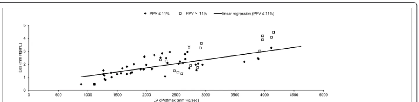
Overall, arterial dP/dtmax and Ees were significantly
correlated (r = 0.51,p < 0.001) but there was low
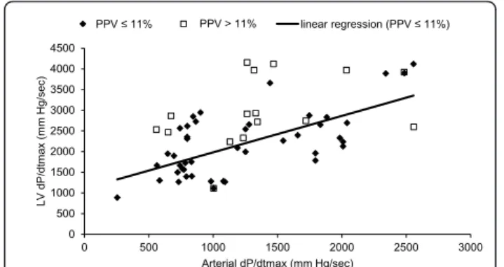
agree-ment (Figures 2 and 3). LV dP/dtmax and arterial dP/
dtmaxwere significantly correlated (r = 0.58,p < 0.001)
but arterial dP/dtmax systematically underestimated LV
dP/dtmax. (bias = 1018 ± 364 mmHg/sec). LV dP/dtmax
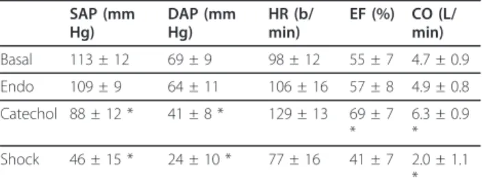
and Ees were significantly correlated (r = 0.78, p <
0.001) (Figure 4).
When adequate filling (PPV ≤ 11%) was obtained, a
far better correlation between arterial dP/dtmaxand Ees
was found (r = 0.77, p < 0.001) (Figure 2). In that case,
normalized Bland-Altman analysis revealed a good agreement between the two methods (Figure 3).
Correla-tion between LV dP/dtmaxand arterial dP/dtmax was also
improved when adequate filling was achieved (r = 0.66,
p < 0.001) while correlation between LV dP/dtmax and
Ees did not significantly changed (r = 0.76,p < 0.001)
(Figure 5). Discussion
Determination of LV contractility is a cornerstone in clinical practice [6,12]. Numerous methods for assessing LV contractility have been reported but none have been adequately validated in clinical practice, or require the presence of an intraventricular pressure catheter, prohi-biting routine use in clinical practice [13,14]. The gold standard method, Ees, requires both ventricular pressure and volume measurements on a beat to beat basis with preload variation [2]. Single beat analysis has been developed, but requires the whole ventricular pressure
waveform [15]. dP/dtmax can be easily calculated in
clini-cal practice but is sensitive to both LV contractility and preload [9]. In the present study, we tested whether
arterial dP/dtmax, derived from femoral fluid-filled
cathe-ter, was accurate for assessing LV contractility. While
LV dP/dtmax is considered as a good index of LV
con-tractility despite its preload dependence, little is known
about arterial dP/dtmax[16,17]. One study performed in
perioperative patients found that arterial dP/dtmax and
LV dP/dtmaxwere significantly correlated and concluded
that changes in arterial dP/dtmax were accurate for
assessing changes LV contractility [18]. To the best of our knowledge, despite its wide use in critically ill
patients, arterial dP/dtmaxhas never been directly
com-pared with Ees during changes in LV function, at differ-ent levels of vascular filling. Our results demonstrated that there was significant correlation between arterial
dP/dtmaxand Ees. Furthermore, a far better correlation
with a good agreement between arterial dP/dtmax and
Ees were observed when adequate vascular filling was achieved. Similar improvement was observed between
arterial and LV dP/dtmax. However, correlation between
LV dP/dtmaxand Ees did not significantly change when
adequate vascular filling was achieved. Arterial dP/dtmax
is an ejection phase index depending on arterial compli-ance and waves reflections from periphery to aorta. All factors that may affect arterial compliance and waves reflections (vascular filling conditions, vasoactive agents)
may also affect arterial dP/dtmax independently of LV
contractile function. As a result, the combination of fluid responsiveness and changes in arterial compliance and waves reflections due to endotoxin and/or catecho-lamines could enhance discrepancies between arterial
and LV dP/dtmaxand consequently between arterial dP/
dtmaxand the reference method, Ees [19]. In this study,
fluid administration was directed by PPV.
Adequate vascular filling was defined as PPV≤ 11%.
On the basis of clinical settings, this PPV threshold
Table 1 Hemodynamic data
SAP (mm Hg) DAP (mm Hg) HR (b/ min) EF (%) CO (L/ min) Basal 113 ± 12 69 ± 9 98 ± 12 55 ± 7 4.7 ± 0.9 Endo 109 ± 9 64 ± 11 106 ± 16 57 ± 8 4.9 ± 0.8 Catechol 88 ± 12 * 41 ± 8 * 129 ± 13 69 ± 7 * 6.3 ± 0.9 * Shock 46 ± 15 * 24 ± 10 * 77 ± 16 41 ± 7 2.0 ± 1.1 * SAP = systolic arterial pressure, DAP = diastolic arterial pressure, HR = heart rate, EF = left ventricular ejection fraction, CO = cardiac output, *P < 0.01 compared to basal
arterial dP/dtmax Ees
Basal Endo Catechol Shock -500 0 500 1000 1500 2000 2500 arteri al dP /dtmax ( mmH g/ s) 0 1 2 3 4 Ee s (mmH g /m L )
Figure 1 LV contractility assessed by both Ees and arterial dP/ dtmax. Basal conditions (’basal’), immediately after endotoxin
infusion (’endo’), during shock with and without catecholamine infusion (’catechol’ and ‘shock’ respectively). Values are given as mean ± SD. All directional changes in contractility were significant (p < 0.05) for each challenge, except between‘basal’ and ‘endo’.
value allows the best discrimination between responders and nonresponders to intravascular fluid administration [20-23]. In perioperative patients, De Hert et al. showed
that changes in femoral dP/dtmax accurately reflected
changes in LV dP/dtmax during various interventions.
However, absolute values of LV contractility are required for potential ventriculo-arterial interaction ana-lysis [24]. These authors also found that leg elevation
induced significant increase in central venous pressure and LV end-diastolic pressure, but arterial and LV dP/
dtmaxremained unaltered [18]. However, it is well
recog-nized that static indices (like central venous pressure or LV end-diastolic pressure) are poor indicators of vascu-lar filling and preload responsiveness [11]. Masutani et
al. showed that LV dP/dtmaxcan be predicted from
aor-tic dP/dtmaxbut their method requires aortic impedance
which is difficult to calculate in clinical practice [25]. Therefore, assessing LV contractility from arterial dP/
dtmax, when adequate vascular filling is achieved, could
be a simple and accurate method with the potential for ventriculo-arterial interaction analysis.
Other methodological issues should be taken into account. First, the use of a fluid-filled catheter could be
another source of discrepancy between arterial dP/dtmax
and Ees. As pointed out by numerous authors, pressure waves measured with a fluid-filled catheter have to be interpreted cautiously, because the pressure waveform may be distorted by the dynamic response of the cathe-ter. By taking care of a properly flushed catheter system and by filtering out artifacts, the catheter response were optimized in our study [26]. Secondly, arterial and LV
dP/dtmax could also be influenced by heart rate. Heart
rate variability was not significant enough to analyze its
0 1 2 3 4 5 0 500 1000 1500 2000 2500 3000 Ees (m m Hg/m L)
Arterial dP/dtmax (mm Hg/sec)
PPV 11% PPV > 11% linear regression (PPV 11%)
Figure 2 Linear regression between arterial dP/dtmaxand Ees.
-4 -2 0 2 4 -3 -2 -1 0 1 2 3 di ff erence average PPV ч 11% PPV > 11%
Figure 3 Normalized Bland-Altman plot of the agreement between Ees and arterial dP/dtmax. Average = (Ees° + arterial dP/
dtmax°)/2 and difference = Ees° - arterial dP/dtmax° where X° is the
normalized value of X [X° = (value of X - mean of X)/standard deviation of X]. Lines represent mean difference (solid lines) and 95% confidence interval (light dashed line) (PPV≤ 11%).
0 1 2 3 4 5 0 500 1000 1500 2000 2500 3000 3500 4000 4500 5000 Ees (m m Hg/m L) LV dP/dtmax (mm Hg/sec) PPV ≤ 11% PPV > 11% linear regression (PPV ≤ 11%)
influence on arterial dP/dtmax in the present
observations. Conclusions
Arterial dP/dtmax, the minimally invasive method
derived from femoral artery fluid-filled pressure catheter and Ees, the reference method for assessing LV contrac-tility, derived from intraventricular conductance micro-manometer-tipped catheter, were significantly correlated over a wide range of hemodynamic conditions resulting from endotoxin-induced shock and catecholamine
infu-sion. However, arterial dP/dtmaxwas more accurate for
assessing LV contractility when adequate vascular filling,
defined as PPV ≤ 11%, was achieved. Using dynamic
indices to ensure adequate vascular filling, LV
contracti-lity could be accurately predicted by arterial dP/dtmax
derived from femoral artery fluid-filled pressure catheter in critically ill patients.
Acknowledgements
This study was supported by a grant from the Leon Fredericq Foundation of the University of Liege.
Author details
1Medical Intensive Care Unit, University Hospital of Liège, Liège, Belgium. 2
Faculty of Sciences, University of Liège, Liège, Belgium.3Emergency Department, University Hospital of Liège, Liège, Belgium.4Mechanical
Engineering Department, University of Canterbury, Christchurch, New Zealand.
Authors’ contributions
PM conceived the study, participated in the experiments, analyzed the data and drafted the manuscript. BL and TD participated in the experiments, analyzed the data and helped to draft the manuscript. NJ participated in the experiments. GC participated in the design of the study and helped to draft the manuscript. VD participated in the design and coordination of the study and helped to draft the manuscript. All authors read and approved the final version of the manuscript.
Received: 1 September 2011 Accepted: 1 March 2012 Published: 1 March 2012
References
1. Levy RJ, Piel DA, Acton PD, Zhou R, Ferrari VA, Karp JS, Deutschman CS: Evidence of myocardial hibernation in the septic heart. Crit Care Med 2005, 33:2752-2756.
2. Sagawa K, Maughan WL, Suga H, Sunagawa K: Cardiac contraction and the pressure-volume relationship New York & Oxford: Oxford Univ Press; 1988. 3. Drake-Holland AJ, Mills CJ, Noble MI, Pugh S: Responses to changes in
filling and contractility of indices of human left ventricular mechanical performance. J Physiol 1990, 422:29-39.
4. Kass DA, Maughan WL, Guo ZM, Kono A, Sunagawa K, Sagawa K: Comparative influence of load versus inotropic states on indexes of ventricular contractility: experimental and theoretical analysis based on pressure-volume relationships. Circulation 1987, 76:1422-1436.
5. Lambert CR Jr, Nichols WW, Pepine CJ: Indices of ventricular contractile state: comparative sensitivity and specificity. Am Heart J 1983, 106:136-144.
6. Robotham JL, Takata M, Berman M, Harasawa Y: Ejection fraction revisited. Anesthesiology 1991, 74:172-183.
7. Zhong L, Tan RS, Ghista DN, Ng EY, Chua LP, Kassab GS: Validation of a novel noninvasive cardiac index of left ventricular contractility in patients. Am J Physiol Heart Circ Physiol 2007, 292:H2764-H2772. 8. Quinones MA, Gaasch WH, Alexander JK: Influence of acute changes in
preload, afterload, contractile state and heart rate on ejection and isovolumic indices of myocardial contractility in man. Circulation 1976, 53:293-302.
9. Little WC: The left ventricular dP/dtmax-end-diastolic volume relation in closed-chest dogs. Circ Res 1985, 56:808-815.
10. Gillebert TC, Leite-Moreira AF, De Hert SG: Relaxation-systolic pressure relation. A load-independent assessment of left ventricular contractility. Circulation 1997, 95:745-752.
11. Michard F, Teboul JL: Predicting fluid responsiveness in ICU patients: a critical analysis of the evidence. Chest 2002, 121:2000-2008.
12. Schiller NB: Ejection fraction by echocardiography: the full monty or just a peep show? Am Heart J 2003, 146:380-382.
13. Bargiggia GS, Bertucci C, Recusani F, Raisaro A, de Servi S, Valdes-Cruz LM, Sahn DJ, Tronconi L: A new method for estimating left ventricular dP/dt by continuous wave Doppler-echocardiography. Validation studies at cardiac catheterization. Circulation 1989, 80:1287-1292.
14. Bendjelid K, Romand JA: Fluid responsiveness in mechanically ventilated patients: a review of indices used in intensive care. Intensive Care Med 2003, 29:352-360.
15. Takeuchi M, Igarashi Y, Tomimoto S, Odake M, Hayashi T, Tsukamoto T, Hata K, Takaoka H, Fukuzaki H: Single-beat estimation of the slope of the end-systolic pressure-volume relation in the human left ventricle. Circulation 1991, 83:202-212.
16. Mason DT: Usefulness and limitations of the rate of rise of intraventricular pressure (dp-dt) in the evaluation of myocardial contractility in man. Am J Cardiol 1969, 23:516-527.
17. Mason DT, Braunwald E, Covell JW, Sonnenblick EH, Ross J Jr: Assessment of cardiac contractility. The relation between the rate of pressure rise and ventricular pressure during isovolumic systole. Circulation 1971, 44:47-58.
18. De Hert SG, Robert D, Cromheecke S, Michard F, Nijs J, Rodrigus IE: Evaluation of left ventricular function in anesthetized patients using femoral artery dP/dt(max). J Cardiothorac Vasc Anesth 2006, 20:325-330. 19. Perel A: Automated assessment of fluid responsiveness in mechanically
ventilated patients. Anesth Analg 2008, 106:1031-1033.
20. Kramer A, Zygun D, Hawes H, Easton P, Ferland A: Pulse pressure variation predicts fluid responsiveness following coronary artery bypass surgery. Chest 2004, 126:1563-1568.
21. Auler JO Jr, Galas F, Hajjar L, Santos L, Carvalho T, Michard F: Online monitoring of pulse pressure variation to guide fluid therapy after cardiac surgery. Anesth Analg 2008, 106:1201-1206, table..
22. Biais M, Stecken L, Ottolenghi L, Roullet S, Quinart A, Masson F, Sztark F: The ability of pulse pressure variations obtained with CNAP device to predict fluid responsiveness in the operating room. Anesth Analg 2011, 113:523-528.
23. Feissel M, Badie J, Merlani PG, Faller JP, Bendjelid K: Pre-ejection period variations predict the fluid responsiveness of septic ventilated patients. Crit Care Med 2005, 33:2534-2539.
0 500 1000 1500 2000 2500 3000 3500 4000 4500 0 500 1000 1500 2000 2500 3000 LV dP/dtm ax (m m Hg/s ec )
Arterial dP/dtmax (mm Hg/sec)
PPV 11% PPV > 11% linear regression (PPV 11%)
Figure 5 Linear regression between left ventricular (LV) dP/ dtmaxand arterial dP/dtmax.
24. Kass DA, Kelly RP: Ventriculo-arterial coupling: concepts, assumptions, and applications. Ann Biomed Eng 1992, 20:41-62.
25. Masutani S, Iwamoto Y, Ishido H, Senzaki H: Relationship of maximum rate of pressure rise between aorta and left ventricle in pediatric patients. Implication for ventricular-vascular interaction with the potential for noninvasive determination of left ventricular contractility. Circ J 2009, 73:1698-1704.
26. Lambermont B, Gerard P, Detry O, Kolh P, Potty P, D’Orio V, Marcelle R: Correction of pressure waveforms recorded by fluid-filled catheter recording systems: a new method using a transfer equation. Acta Anaesthesiol Scand 1998, 42:717-720.
Pre-publication history
The pre-publication history for this paper can be accessed here: http://www.biomedcentral.com/1471-2261/12/13/prepub
doi:10.1186/1471-2261-12-13
Cite this article as: Morimont et al.: Arterial dP/dtmaxaccurately reflects
left ventricular contractility during shock when adequate vascular filling is achieved. BMC Cardiovascular Disorders 2012 12:13.
Submit your next manuscript to BioMed Central and take full advantage of:
• Convenient online submission
• Thorough peer review
• No space constraints or color figure charges
• Immediate publication on acceptance
• Inclusion in PubMed, CAS, Scopus and Google Scholar
• Research which is freely available for redistribution
Submit your manuscript at www.biomedcentral.com/submit