P J M UNIVERSITY DE
10 SHERBROOKE
Faculte de genie
Departement de genie chimique et de genie biotechnologique
DEVELOPPEMENT D'UN SYSTEME
TRIDIMENSIONNEL DE CULTURE CELLULAIRE
POUR ORIENTER LA FORMATION DE
MICROVAISSEAUX
DEVELOPMENT OF A TRIDIMENSIONAL
CELL CULTURE SYSTEM TO ORIENT
MICRO VESSEL FORMATION
These de doctorat Speciality : genie chimique
Irza SUKMANA
Jury : Professeur Patrick VERMETTE (Directeur) Professeur Pierre PROULX (Rapporteur) Professeur Jean-Francois BEAULIEU Professeur Elizabeth JONES
1*1
Library and Archives Canada Published Heritage Branch 395 Wellington Street OttawaONK1A0N4 Canada Bibliotheque et Archives Canada Direction du Patrimoine de I'edition 395, rue Wellington Ottawa ON K1A 0N4 CanadaYour file Votre reference ISBN: 978-0-494-70629-9 Our file Notre reference ISBN: 978-0-494-70629-9
NOTICE: AVIS:
The author has granted a
non-exclusive license allowing Library and Archives Canada to reproduce, publish, archive, preserve, conserve, communicate to the public by
telecommunication or on the Internet, loan, distribute and sell theses
worldwide, for commercial or non-commercial purposes, in microform, paper, electronic and/or any other formats.
L'auteur a accorde une licence non exclusive permettant a la Bibliotheque et Archives Canada de reproduire, publier, archiver, sauvegarder, conserver, transmettre au public par telecommunication ou par I'lnternet, prefer, distribuer et vendre des theses partout dans le monde, a des fins commerciales ou autres, sur support microforme, papier, electronique et/ou autres formats.
The author retains copyright ownership and moral rights in this thesis. Neither the thesis nor substantial extracts from it may be printed or otherwise reproduced without the author's permission.
L'auteur conserve la propriete du droit d'auteur et des droits moraux qui protege cette these. Ni la these ni des extraits substantiels de celle-ci ne doivent etre imprimes ou autrement
reproduits sans son autorisation.
In compliance with the Canadian Privacy Act some supporting forms may have been removed from this thesis.
Conformement a la loi canadienne sur la protection de la vie privee, quelques
formulaires secondares ont ete enleves de cette these.
While these forms may be included in the document page count, their removal does not represent any loss of content from the thesis.
Bien que ces formulaires aient inclus dans la pagination, il n'y aura aucun contenu manquant.
1+1
As a milestone of our 13 anniversary I dedicate this work to my wife,
the other half of my soul Ika Nury Wahidah.
To my mother and the memory of my late father (Almarhum) To my beloved son and daughters
RESUME
Le developpement de micro-vaisseaux fonctionnels dans la culture de substituts tissulaires
constitue un enorme defi du genie tissulaire et de la medecine regeneratrice. Celui-ci est
justifie par la necessite de permettre une meilleure alimentation en nutriments et oxygene des
tissus cultives et une bonne elimination des dechets a l'interieur de ceux-ci. C'est la raison
pour laquelle seulement quelques substituts de tissus sont disponibles pour des applications
medicales. Par consequent, de nombreuses etudes de recherche dans le domaine de l'ingenierie
tissulaire ont mis l'accent sur la culture de micro-vaisseaux a l'interieur de matrices tissulaires
et ce avant leur implantation, tel que presente dans le Chapitre 1 de cette these. Le Chapitre 2
est consacre a l'etude bibliographique presentant revolution recente et les defis futurs lies au
processus de micro-vascularisation de substituts tissulaires. Ce processus est souvent appele
pre-vascularisation.
Le travail experimental presente dans cette these est base sur l'hypothese que des
mono-filaments polymeriques separes les uns des autres d'une facon precise peuvent etre utilises
pour guider des cellules endotheliales dispersees dans un gel de fibrine et ainsi induire des
structures tubulaires de facon directionnelle.
Le Chapitre 3 de cette these presente la validation de la formation de micro-vaisseaux dans un
environnement tridimensionnel. La methode de culture tissulaire presentee ici utilise des fibres
de polymere pour guider des cellules endotheliales et ce afin d'orienter la formation des
micro-vaisseaux. La methode d'attachement cellulaire decrite au Chapitre 3 a permis
l'adherence et la migration de cellules endotheliales humaines de la veine ombilicale
(HUVEC) sur des fibres de poly(ethylene terephtalate) (PET) non-modifiees. Les fibres
recouvertes de HUVEC ont ete « sandwichees » entre deux gels de fibrine pour permettre le
developpement des micro-vaisseaux. Le developpement de l'angiogenese a ensuite ete
caracterise a l'aide d'un certain nombre de techniques d'imagerie, y compris la microscopie par
fluorescence et la microscopie confocale. Apres 4 jours de culture, les micro-vaisseaux
formaient de grandes structures en forme de tube (diametre d'environ 100|im) entre les fibres
adjacentes distancees de 0.1mm.
Le Chapitre 4 presente d'un cote l'etude de l'effet des fibroblastes humains et, d'un autre cote, l'influence de deux facteurs de croissance angiogeniques (i.e., Vascular Endothelial Growth
Factor (VEGF) et le basic Fibroblast Growth Factor (bFGF)) sur les reponses des HUVEC
recouvrant les fibres de PET cultivees en sandwich dans des gels de fibrine. Le developpement et la maturation des micro-vaisseaux ainsi que la formation des lumens ont ete etudies par microscopie confocale en utilisant une matrice de fibrine fluorescente et par histologic L'histologie et les experiences menees avec la fibrine fluorescente ont confirme que ces pseudo-micro-vaisseaux formes entre les fibres de polymere incorporees dans la fibrine avaient un lumen. Les effets du VEGF et du bFGF etaient fonction de la concentration de ces molecules. L'utilisation de fibroblastes a considerablement ameliore la maturation des micro-vaisseaux comparativement aux echantillons controles et ceux cultives avec le VEGF et le bFGF.
La conclusion et les propositions pour des travaux fiiturs constituent le Chapitre 5.
Les Annexe A et B presentent, respectivement, les protocoles detailles utilises dans cette etude pour marquer les cellules et des informations generates sur le cytosquelette.
Mots-cles: polyethylene terephtalate; orientation cellulaire; angiogenese directionnelle;
ABSTRACT
The development of functional and oriented microvessels within tissue constructs is a tremendous challenge in tissue engineering and regenerative medicine. It is justified by the need to allow better nutrient and oxygen supply and waste removal within the core of tissue constructs. It is one of the reasons why only few tissue substitutes are available to clinicians. Therefore, the focus of many research studies in the field of tissue engineering has been placed on promoting microvessel ingrowth inside tissue constructs prior to their implantation, as presented in Chapter 1. This process is often referred to as pre-vascularization. As such, Chapter 2 of this thesis is devoted to review the recent development and future challenges related to the micro vascularization process of tissue constructs.
The experimental work in this thesis is based on the hypothesis that polymer monofilaments precisely distanced from each others can be used to guide endothelial cells when dispersed in a (fibrin) gel and to induce tube-like structures in a directional fashion.
In Chapter 3 of this thesis, the use of polymer fibres as contact guidance of endothelial cells to orient microvessel formation and development in a three-dimensional environment was validated. The novel cell attachment method described in Chapter 3 has resulted in an increase in Human Umbilical Vein Endothelial Cell (HUVEC) adhesion and spreading on bare poly(ethylene terephthalate) (PET) fibres. Furthermore, HUVEC-covered fibres were sandwiched between fibrin gels to allow the development of microvessels. Angiogenesis development was characterized and evaluated using a number of imaging techniques, including fluorescence microscopy and confocal microscopy. After 4 days of culture, microvessels formed large tube-like structures (diameter of approximately 100 um) between adjacent fibres distanced by 0.1mm. This proof-of-concept opens the door to other experiments and to possibility of scaling the system to bioreactor cultures.
In Chapter 4, the effect of human fibroblasts and in another set of experiments, the influence of two angiogenic growth factors (i.e., Vascular Endothelial Growth Factor (VEGF) and basic Fibroblast Growth Factor (bFGF)) over the responses of the HUVEC-covered fibres
sandwiched in fibrin were investigated. The development and maturation of microvessels as well as the assessment of lumen formation were evaluated using a fluorescent fibrin matrix, confocal microscopy and histology. Histology and fluorescent fibrin experiments confirmed that these microvessel-like structures formed between polymer monofilaments embedded in fibrin contained a lumen. The effects of VEGF and bFGF were dose dependent. Furthermore, the use of fibroblasts significantly improved the maturation of the microvessels compared to control and to samples cultured with VEGF and bFGF.
Conclusions and suggested future work are presented in Chapter 5.
Appendices A and B present the detailed protocols used to label cells in this study and general information about cell cytoskeleton, respectively.
Keywords: Polyethylene terephthalate; Contact guidance; Cell patterning; Directional
Acknowledgements
First of all, I would like to express my sincere gratitude to my supervisor Professor Patrick Vermette, whose enthusiastic support and good advices helped me to go through my challenging project.
I am very honoured to have Professor Pierre Proulx and Professor Jean-Francois Beaulieu for their guidance and for being my pre-Doctoral examiners.
A very special thanks to Mr. Yannick Laplante and Dr. Aftab Ahamed, former members of the Laboratoire de Bioingenierie et de Biophysique de l'Universite de Sherbrooke, for their precious help when I started my Ph.D. I thank them also for their hints on general technical and scientific aspects.
I also want to thank all my friends in the group for sharing their ideas and for scientific discussions on daily basis during this course.
I should thank all the technicians in the Department of Chemical and Biotechnological Engineering, especially Mr. Mark Couture for his help in designing and producing frames and culture chambers for my research. Also, I want to thank Mr. Gilles Grondin and Dr. Leonid Volkov for their contribution in confocal microscopy and Mrs. Johanne Dussault and Dr. Sameh Geha who helped me with histology.
I am particularly thankful to my sponsor, the Islamic Development Bank (IDB), for all financial support and living guidance for me and my family.
I am also very grateful to the Muslim community in Sherbrooke who has given me an invaluable support and help in countless ways. I will never forget the support provided by dear brothers: Hendra Hermawan, Abdelfettah and Rachid Bannari, Brahim Selma, Mbark El
Morsli, Rachid Kadouche, Taha Abd-El-Rahman, Ahmad Kamal, Hamdy and Habibie, Deyab
El-Gamal, Ahmed Omran, Said and Hoccain and others, "Jazakumullahu khairan katsiran".
Finally, my most acknowledgment goes to my adorable wife, adik Ika Nury Wahidah, and our
lovely kids: Muhammad, Wafa and Sky. The word "Thanks" is never enough for their love,
patience and encouragement. May Allah SWT give you all the best rewards. Amen.
TABLE OF CONTENT
ABSTRACT ii RESUME iv Acknowledgements vi
Table of Content viii List of Figures xi List of Tables xii List of Abbreviations xiii
CHAPTER 1 Introduction 1
1.1 Thesis Objectives 1 1.2 Thesis Structure 1 1.3 Contributions 3
CHAPTER 2 Vascularization strategies to engineer tissue substitutes: a challenge to support
blood microvessel development 5
2.1 Abstract 6 2.2 Introduction 6 2.3 Scaffolds in tissue engineering 8
2.3.1 Synthetic polymers 10 2.3.2 Natural polymers 11 2.4 Material properties 21 2.4.1 Scaffold porosity 21 2.4.2 Matrix stiffness 23 2.5 Scaffold vascularisation 24
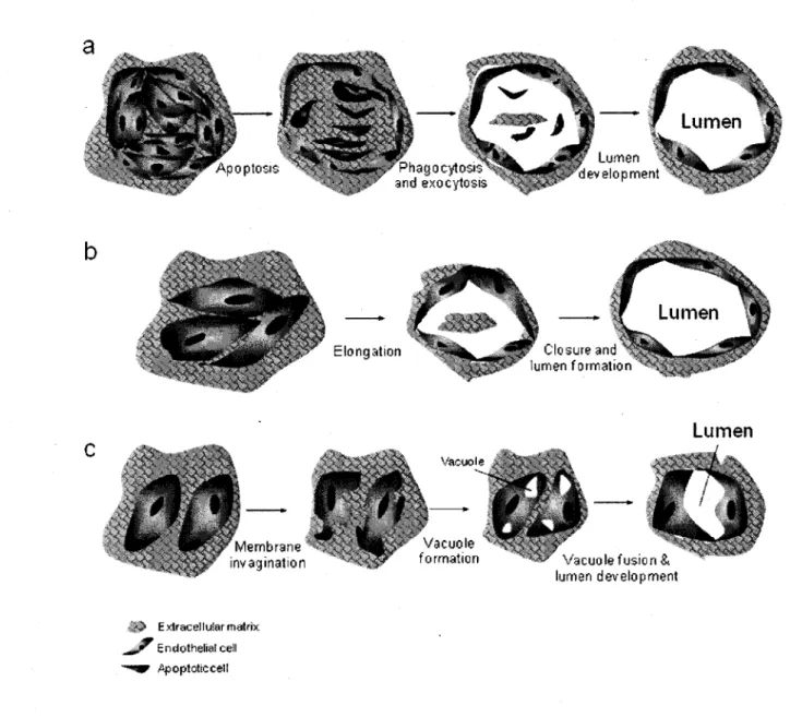
2.5.1 Vasculogenesis and angiogenesis 28 2.5.1.1 Model 1: Cell death and phagocytosis 29 2.5.1.2 Model 2: Wrapping spaces around ECM 29 2.5.1.3 Model 3: Vacuole formation from the coalescence of intracellular vacuoles 31
2.5.2 Promoting angiogenesis in three-dimensional constructs 31
2.5.2.2 Surface modification 33 2.5.2.3 ECM modification 34 2.5.3 Co-culture systems and microvessel maturation 35
2.6 Bioreactor to support vascularization 37
2.7 Concluding remarks 39
CHAPTER 3 Polymer fibres as contact guidance to orient microvascularization in a 3D
Environment 57 3.1 Abstract 58 3.2 Introduction 58 3.3 Materials and methods 59
3.3.1 Materials 59 3.3.2 Cell culture : 60
3.3.3 PET fibres and fibrin gel preparation 60
3.3.4 Cell adhesion assay 61 3.3.5 Cell culture in 3D environment 61
3.3.6 Visualization of microvessels 62 3.3.7 Images and image processing 62
3.3.8 Statistical analysis 63 3.3.9 Confocal microscopy imaging of microvessels 63
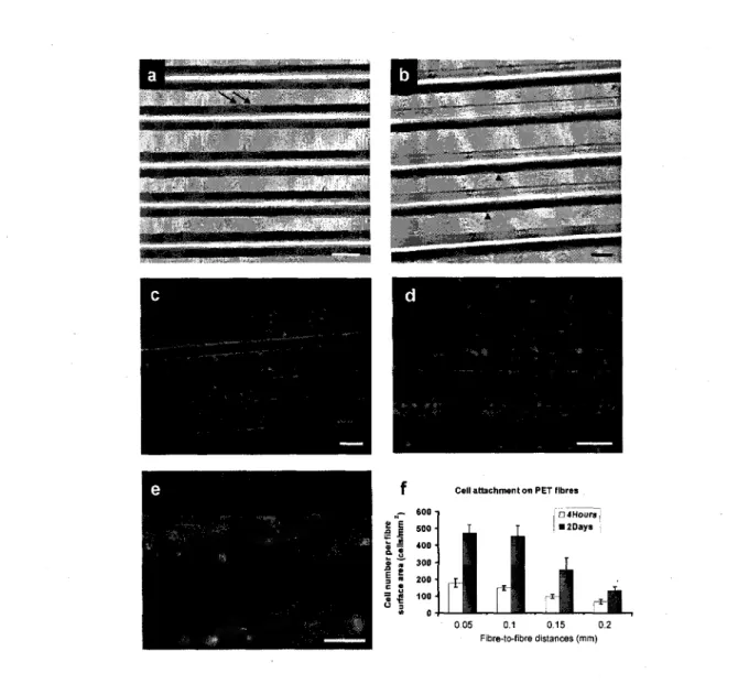
3.4 Results 64 3.4.1 Cell adhesion on untreated PET fibres 64
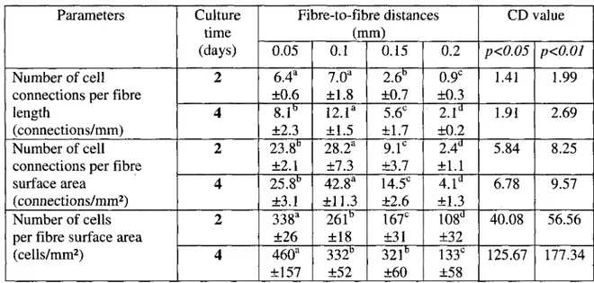
3.4.2 Effect of fibre-to-fibre distance over cell connections and microvessel formation 65
3.4.3 Microvessel formation 66
3.5 Discussion 67 3.6 Conclusions 69
CHAPTER 4 The effects of co-culture with fibroblasts and angiogenic growth factors on
microvascular maturation and multi-cellular lumen formation in HUVEC-oriented
polymer fibre constructs 82
4.1 Abstract.... 83 4.2 Introduction 83 4.3 Materials and methods 85
4.3.1 Materials 85 4.3.2 Cell culture 85 4.3.3 Culture system and fibrin preparation 86
4.3.4 Cell adhesion and angiogenesis assay 86 4.3.5 Visualization of microvessel and lumen formation 87
4.3.6 Histological section 88 4.3.7 Imaging microvessels by confocal microscopy 88
4.3.8 Statistical analysis 89 4.4 Results and discussion 89
4.5 Conclusions 93
CHAPTER 5 Conclusions and suggestions 107
5.1 Conclusions 107 5.2 Conclusion (en francais) 108
5.3 Suggestions 109 5.3.1 Further studies of the angiogenesis assessment 109
5.3.2 The effect of dynamic culture system over microvessel development and proof of
microvessel functionality 111 APPENDIX A Protocol for cell staining 116
LIST OF FIGURES
Figure 2.1: Fibrinogen structure and fibrin clot process 16 Figure 2.2: Models of the development of tube-like structures during vasculogenesis 30
Figure 2.3: Blood vessel structure and development 36 Figure 3.1: Schematic illustration of the in vitro method designed to guide and orient
microvessel formation 75 Figure 3.2: HUVEC attachment on PET fibres 76
Figure 3.3: Effect of fibre-to-fibre distance over cell connections and microvessel
development 77 Figure 3.4: Confocal and inverted microscopy image 78
Figure 3.5: Schematic of the microvascularization process 79 Supplementary Figure 3.i: Scanning electron microscopy (SEM) image of a PET fibre 80
Supplementary Figure 3.ii: Progression of cell connections and microvessel reorganization
along and in between adjacent fibres sandwiched between two fibrin gels 81
Figure 4.1: Effect of fibroblasts over microvessel development 100 Figure 4.2: Immunofluorescence picture illustrating the effect of fibroblasts on microvessel
development 101 Figure 4.3: Immunofluorescence picture (F-actin and nuclei staining) illustrating the effect of
VEGF and bFGF on microvessel development 102 Figure 4.4: Illustration of multi-cellular lumen formation for cultures carried out for a period
of 4 days with fibroblasts 103 Supplementary Figure 4.i: Comparison between systems using cell-free fibres (left) and
HUVEC-covered fibres (right) over cell-to-cell connections & microvessel
development 104 Supplementary Figure 4.ii: Immunofluorescence confocal pictures of a sample cultured with
no fibroblasts 105 Supplementary Figure 4.iii: Fibroblasts stained with the CFSE CellTracer 106
LIST OF TABLES
Table 2.1: Integrins and non-integrin receptors which can bind to fibrin(ogen) 18 Table 2.2: The use of synthetic and natural polymers to support tissue vascularisation 19
Table 3.1: Effects of fibre-to-fibre distance on cells 74 Table 4.1: Effects of VEGF and bFGF concentrations over the number of cells and cell-cell
connections following 2 days of culture 98 Table 4.2: Comparison of the effects of fibroblasts, VEGF, and bFGF over the number of
cells and cell-cell connections 98 Table 4.3: Review of some studies on the effects of VEGF and bFGF on endothelial cells and
LIST OF ABBREVIATIONS
3D Agp-1 ANOVA BAEC bFGF BSA CAM CLS CSTR DLLA EC ECGS ECM ePTFE ERG ESAM FBS FDA FGF GAG HA HBSS HDMEC HUVEC HVEC ICAM-1 MC three-dimensional or tridimensional angiopoietin-1 analysis of variancebovine aortic endothelial cell basic fibroblast growth factor
bovine serum albumin chorioallontoic membrane capillary-like structure continuous stirred-tank reactor (D,L-lactic acid) endothelial cell
endothelial cell growth supplement
extracellular matrix expanded
poly(tetrafluoroethylene) early response genes
endothelial cell selective adhesion molecule foetal bovine serum
Food and drug administration fibroblast growth factor
glycosaminoglycan hyaluronic acid
hanks balances salt saline human dermal endothelial cell
human umbilical vein endothelial cell
human vascular endothelial cell inter-cellular adhesion molecule-1 messenchymal cell o-HA PBS PDGF-BB PDLA PECAM-1 PEG PET PGA PLA PLG PLGA PLLA RAOEC RGD RHAMM SEM SF SMC SM-MHC SMocA STLV TMC VE-cadherin VEGF VSMC vWF oligosaccharides of HA
phosphate buffer saline platelet-derived growth factor-BB
poly(D,L-lactic acid) platelet endothelial cell adhesion molecule-1 poly(ethylene glycol) poly(ethylene terephthalate) poly(glycolic acid) poly(lactic acid) poly(lactic glycolic) poly(lactic-co-glycolic acid) poly(L-lactic acid)
rat aortic endothelial cell arginine-glycine-aspartic acid peptide
receptor for HA mediated motility
scanning electron microscopy skin fibroblast smooth muscle cell
smooth muscle myosin heavy chain
smooth mucle a-actin slow turning lateral vessel trimethylene carbonate vascular endothelial cadherin
vascular endothelial growth factor
vascular smooth muscle cell
CHAPTER 1
Introduction
The development of functional and oriented microvessels within engineered human tissue substitutes constitutes a promising hope in tissue engineering and regenerative medicine [Stock and Vacanti 2001; Eichmann et al. 2005]. To date, the development of thick tissues, such as pancreas, liver, heart, and kidneys is problematic due to the lack of construct vascularisation, resulting in cell and tissue death [Atala 2004; Jain et al. 2005; Kannan et al. 2005]. The focus of current research efforts in tissue engineering has been mainly on developing strategies to study and promote microvascularization within tissue constructs [Kannan et al. 2005; Neuman et al. 2003].
1.1 Thesis objectives
This project was designed to constitute the initial part of a larger research program aiming to create an oriented functional blood microvessel network within three-dimensional tissue scaffolds. One of the aims of this research program is to scale-up tissue engineering methods by the use of dynamic culture conditions using an automated bioreactor system. The principal objective of this thesis was to design and validate static cell culture processes to guide endothelial cell responses and the subsequent microvessel development and orientation.
1.2 Thesis Structure
This thesis has been written in a manner that chapters correspond to scientific articles resulting from this work. At the time of submission, two articles were published and one under preparation for submission. Below is a brief description of their content.
Chapter 1: Introduction. This section contains thesis objectives and structure, and
Chapter 2: Vascularization strategies to engineer tissue substitutes: a challenge to support blood microvessel development. This chapter is a literature review on advancements
in the field of three-dimensional tissue engineering with an emphasis on cellular and angiogenesis guidance as well as on blood microvessel development. Moreover, the effects of cell types, co-cultures, and growth factors on microvessel development are discussed. The use of bioreactors to culture tissue substitutes is also briefly addressed.
Chapter 3: Polymer fibers as contact guidance to orient microvascularization in a 3D environment. This part of the thesis presents a novel culture system for angiogenesis
guidance using poly(ethylene terephthalate) (PET) fibres. Briefly, in an effort to increase the bioactivity of PET fibres, cells are first allowed to attach and cover the fibres using a novel culture method. Then, HUVEC-covered fibres are sandwiched in fibrin and microvessel structures are examined by confocal microscopy. In addition, the effect of fibre-to-fibre distance over angiogenesis development is compared. Results from this chapter reveal that this culture system can be used to orient microvessel structures. The optimum fibre-to-fibre distance found here is further used in the subsequent part of this thesis.
Chapter 4: The effect of co-culture with fibroblasts and angiogenic growth factors on microvascular maturation and multi-cellular lumen formation in HUVEC-oriented polymer fibre constructs. This chapter further validates the use of the culture system
developed in Chapter 3 using a co-culture method with human skin fibroblasts or with angiogenic growth factors (i.e., Vascular Endothelial Growth Factor (VEGF) and Basic Fibroblast Growth Factor (bFGF)). The effects of these two parameters over microvessel development and maturation are investigated. This study is also aiming to determine whether or not these microvessels contained multi-cellular lumen. VEGF and bFGF are shown to enhance HUVEC proliferation and migration, thus improving the development of cell-cell connections and subsequent microvessel development. The use of fibroblasts results in more mature microvessels containing more cell-cell connections, which also contain lumens.
Chapter 5: Conclusions and Suggestions. This part presents a general conclusion and
1.3 Contributions
During the course of this thesis, I have had important help from several people, to whom I am very grateful. Firstly, former members of the Laboratoire de Bioingenierie et de Biophysique de l'Universite de Sherbrooke, Mr. Yannick Laplante, Dr. Aftab Ahamed, and Dr. Mbark El Morsli, have helped me to gain technical know-how and scientific knowledge.
Mr. Yannick Laplante has taught me how to prepare protocols and techniques to carry out cell culture. Furthermore, Mr. Laplante has assisted me in the preparation and evaluation of three-dimensional culture systems. Dr. Aftab Ahamed has tough me statistics and how to prepare a scientific article. Dr. Mbark El Morsli has shared his knowledge in advance mathematics and chemical engineering.
The confocal microscopy was carried out at the Department of Biology (Universite de Sherbrooke) and at the Centre de recherche clinique Etienne-Le Bel (Centre hospitalier universitaire de Sherbrooke (CHUS)), respectively, where Mr. Gilles Grondin and Dr. Leonid Volkov have shared knowledge and helped me to obtain good quality pictures for my publications. Mrs Johanne Dussault and Dr. Sameh Geha have assisted me with histology. After fixation, all my histology samples were sent to the Pathology Department (CHUS). The sectioning and staining have been done by Mrs Johanne Dussault, while further expert interpretation have been done by Dr. Sameh Geha.
References
Atala, A. (2004) Tissue engineering and regenerative medicine: concepts for clinical application. Rejuvenation Res. 7, p. 15-31.
Eichmann, A., Noble, F. L., Autiero, M. and Carmeliet, P. (2005) Guidance of vascular and neural network formation. Current Opinion in Neurobiology 15, p. 108-15.
Jain, R. K., Au, P., Tarn, J., Duda, D. G. and Fukumura, D. (2005) Engineering vascularized tissue. Nat. Biotechnol. 23, p. 821-23.
Kannan, R. Y., Salacinski, H. J., Sales, K., Butler, P. and Seifalian, A. M. (2005) The roles of tissue engineering and vascularisation in the development of micro-vascular networks: a review. Biomaterials 26, p. 1857-75.
Neuman, T., Nicholson, B. S. and Sanders, J. E. (2003) Tissue engineering of perfused microvessels. Microvasc. Res. 66, p. 59-67.
Stock, U. and Vacanti, J. P. (2001) Tissue engineering: current state and prospects. Annu. Rev.
CHAPTER 2
Vascularization strategies to engineer tissue substitutes:
a challenge to support blood microvessel development
Irza Sukmana
1'
21. Laboratoire de Bioingenierie et de Biophysique de l'Universite de Sherbrooke, Department of Chemical and Biotechnological Engineering, Universite de Sherbrooke, 2500, blvd de l'Universite, Sherbrooke, QC, Canada, J1K 2R1.
2. Research Centre on Aging, Institut universitaire de geriatrie de Sherbrooke, 1036, rue Belvedere Sud, Sherbrooke, QC, Canada, J1H 4C4.
2.1 Abstract
The guidance of endothelial cell organization into a capillary network has been a long standing challenge in tissue engineering. Some research efforts have been made to develop methods to promote blood capillary networks inside engineered tissue constructs. Mature microvessels that would mimic blood microvessel function can be used to subsequently facilitate oxygen and nutrient transfer as well as waste removal. Microvascularization of engineered tissue-scaffold appears to be one of the most favorable approaches to overpass nutrient and oxygen supply limitation, which is often the major hurdle in developing thick and complex tissue or organ substitutes. This review chapter addresses recent advances and future challenges in developing and using bioactive scaffolds and 3D culture systems to promote tissue construct vascularization allowing mimicking blood microvessel development and function encountered
in vivo. Vascularization can be achieved through strategies in which endothelial cells are
cultured in bioactive scaffolds and/or extracellular matrix (ECM)-like environments, with or without angiogenic growth factors as well as using cell co-cultures and combinations of those. In the near future, bioreactors will be used to create fully vascularized functional tissue and organ substitutes.
2.2 Introduction
Tissue engineering is an emerging field. In 1987, the National Science Foundation defined tissue engineering as "an interdisciplinary field that applies the principles of engineering and the life sciences towards the development of biological substitutes that restore, maintain or improve tissue function" [Langer and Vacanti 2003]. Tissue engineering involve the repair and restoration of various tissue and organ functions, while limiting host rejection and side effects for the patient by delivering cells and/or biomolecules through the use of 3D scaffolds [Fuchs et al. 2001; Mooney et al. 1997].
Although there have been some successes to replace and restore some tissues using tissue engineering approaches, these have been mostly limited to thin or avascular tissues, such as cartilage, skin or bladder [Mooney and Mikos 1999]. However, for thick and vascular tissues and for most organs, the lack of a sufficient supply of nutrients and oxygen to growing tissues,
as well as the waste removal represent two important factors that limit the successful development and implantation of engineered tissue constructs [Arthur et al. 1998; Neumann et al. 2003]. These issues of poor mass transport and mass transfer have often led to the failure of the culture process or even to that of implants [Arthur et al. 1998; Sipe 2002].
One possible strategy for creating thick engineered tissue substitutes in vitro is to use a bioactive scaffold that allows the guidance of endothelial cells promoting microvessel development in a directional fashion in order to vascularized the tissue construct [Godbey and Atala 2002]. It is quite well accepted among the scientific community that pre-vascularization of tissue construct appears to be one of the most favorable and efficient approaches to address the problem of tissue survival due to a lack of oxygen and nutrient supply [Atala 2004; Mikos et al. 1993; Montano et al. 2009; Nomi et al. 2002]. The concept of pre-vascularization mainly involves the incorporation of endothelial cells into a bioactive scaffold to form a capillary network inside the structure prior to its implantation [Atala 2004; Langer et al. 1995]. This could accelerate the formation of functional microvessels within the core of an implant [Godbey and Atala 2002; Neumann et al. 2003].
The idea of using pre-vascularized engineered tissue substitutes was first suggested by Mikos et al. (1993) when comparing the performance of pre-vascularized tissues to non-vascularized ones. Later, Sakakibara et al. (2002) concluded that pre-vascularization enhanced the benefits of cardiomyocyte transplantation. More recently, Levenberg et al. (2005) demonstrated that pre-vascularization improved the performance of skeletal muscle tissue constructs when implanted in mice. Furthermore, studies aiming to induce and control vascularization may also advance general knowledge on the development of. therapeutics targeting angiogenesis. Numerous pathological conditions are associated with insufficient oxygen and blood supply
[Atala 2004; Carmeliet 2005; Neumann et al. 2003]. Also, uncontrolled angiogenesis is associated with many diseases, including rheumatoid arthritis, macular degeneration, and tumour growth [Carmeliet and Conway 2001].
Advances in tissue engineering have brought significant knowledge on the mechanisms and parameters related to vascularization and angiogenesis development and blood microvessel
network formation [Levenberg and Langer 2004; Nerem 2006]. This review will report status of the current research and development related to bioactive scaffolds and culture systems designed to promote angiogenesis and vascularization. It is necessary to improve our understanding of the mechanisms behind angiogenesis and to apply that knowledge to guide microvessel growth in a regulated manner. Other aspects including the challenges in angiogenesis guidance, assessment of angiogenesis and lumen formation and the use of bioreactor system to culture vascularized tissue constructs will also be outlined.
2.3 Scaffolds in tissue engineering
The needs for engineered tissue substitutes are important. Currently, the demand for organ transplants is higher than the supply. In the United States alone, there were 79,512 patients on the transplantation waiting list in 2002, and only 24,422 received organs while 6,297 died while waiting [Nerem 2006]. In addition, although organ transplantation is one of the less expensive therapies in regenerative medicine, tissue engineering offers hope for more consistent and rapid treatment of those in need [Lanza and Chick 1997].
As an interdisciplinary approach between engineering and life science, tissue engineering seeks an opportunity to develop suitable biomaterial-cell hybrid constructs to support the regeneration and restoration of tissue structure and function. The critical challenge facing tissue engineering today relies on our knowledge and ability to fabricate tissue and organ replacements that would fulfill physiological function [Koh et al. 2009; Nerem 2006]. Also, the success of tissue engineering methods relies on the ability of the construct to integrate at the implantation site with the native tissue. Tissue engineering is facing important clinical and practical problems, such as cell sourcing, rejection, healing, and cell/tissue death [Conway and Carmeliet 2004; Gimble and Guilak 2003].
Various key concepts in tissue engineering and regenerative medicine have been pursued to overcome those problems and these concepts include injection of tissue-specific viable cells directly into a damaged tissue (for example brain cells in the case of Parkinson or Alzheimer), encapsulation of specific cell types within synthetic permeable matrices that allow release of therapeutics (e.g., the release of insulin or dopamine from pancreatic islets in the treatment of
diabetes), and to seed scaffolds with living cells in vitro, allowing their maturation before being implanted [Lanza et al. 2000; Yang et al. 2001a]. In the present thesis, we are mainly interested with the last concept.
If an isolated cell population can be expanded in vitro using cell culture and bioreactor techniques, in theory, only a very small number of cells from donors would be necessary to prepare such biological implants. Since the isolated cells cannot form new tissue by themselves, a (temporary) template is needed, which we refer here to as scaffold. Scaffolds are expected to provide a control over tissue architecture and mechanical properties. They can allow cells to adhere, proliferate, and migrate [Howe et al. 1998; Huttenlocher et al. 1995] in order to form a required structure and to synthesize their own extracellular matrix (ECM) molecules [Chang and Werb 2001], thus hopefully allowing tissue regeneration or repair [Nerem 2007; Yang et al. 2001b].
It is believed that the success to develop tissue constructs depends on many aspects, such as cell sourcing, type of biomaterials used to make scaffolds, tissue culture methods, only to name a few. For example, using cells from other species, such as pig, remains a debate since there is a risk of transferring diseases from animals to humans [Bach 1998]. Using cells from the same genotype or close relatives of a patient could avoid problems associated with immune rejection, which can result in tissue death [Bach 1998; Lee et al. 2005; Ye et al. 2000b].
The behaviour of individual cells and the dynamic state of multi-cellular tissues are regulated by the interaction between cells and their surrounding matrix. Therefore, the design of scaffolds from the macroscopic scale (e.g., pore structure) to the nanoscopic level (e.g., surface properties) is very important. Firstly, decision of using either natural or synthetic scaffolds should be based on their capability to provide a specific microenvironment that mimics the natural environment of the targeted anatomical site [Garcia et al. 2005]. Secondly, the three-dimensional scaffold should fulfill some requirements with respect to: biocompatibility, degradation rate, porosity, mechanical properties (e.g., stiffness), and chemistry (e.g., surface chemical/protein composition) [Chaikof et al. 2002].
In the in vivo environment, cells interact with their ECM in a dynamic manner. The concept of dynamic "communication" between cells and their matrix has opened a wide exploration of the ECM molecules and scaffold materials that can be used in tissue engineering. Scaffolds can be made from synthetic polymers, naturally occurring materials, or a combination of both.
2.3.1 Synthetic polymers
Synthetic polymers have been investigated and used to make scaffolds in tissue engineering for a variety of possible applications. The principal advantage of using synthetic polymers is that their properties (e.g., biodegradation, physicochemistry and mechanical stiffness) can be controlled by manipulating their molecular weight and compositions, for example [Chaikof et al. 2002]. Among them, synthetic degradable polymers from the poly(a-esters) family, such as poly(lactic acid) (PLA), poly(glycolic acid) (PGA) and their co-polymers, have been extensively investigated in biomaterials and tissue engineering.
Synthetic polymers from the poly(a-esters) group are degraded mainly through chemical hydrolysis and are mostly insensitive to enzymatic attack [Larson and Chu 2006], and often, the degradation profile does not vary between patients [Mooney et al. 1997]. For example, it was recognized that the constituting monomers of PLA and PGA are nontoxic and are metabolized in the body [Tabata 2006; Tabata 2009]. Therefore, PLA, PGA and their copolymers (e.g., poly(lactic-co-glycolic acid) (PLGA)) are FDA-approved and they can be produced to form a variety of implants ranging from screws, meshes, and sutures to porous scaffolds [Larson and Chu 2006; Lee et al, 2008; Tabata 2006].
PGA is an inelastic polyester with a high crystallinity (46-50%) and is degraded by water (through hydrolysis) to form glycolic acid [Kikuchi et al. 1997; Vert et al. 1994]. PLA is less crystalline, more hydrophobic, and less susceptible to hydrolysis than PGA [Larson III and Chu 2006; Vert et al. 1994]. For tissue engineering applications, their copolymers, such as PLLA (poly(L-lactic acid)), PLGA (poly(lactic-co-glycolic acid)) and PDLLA (poly(D,L-lactic acid) are more popular, since their properties can be tailored [Vert et al. 1994]. For example, the degradation time of PLLA has been reported to be very slow, while PDLLA hydrolysed in a matter of weeks [Yang et al. 2001a]. Even a small amount of D,L-LA in the polymer chain of
PLA can accelerate the degradation time dramatically [Bramfeld et al. 2007; Grizzi et al. 1995; Tabata 2006].
Several investigators have explored the potential use of polymers from the PLA and PGA family to fabricate scaffolds for the promotion of microvascularization and angiogenesis. For example, Grizzi et al. (1995) have produced poly(D,L-lactic-co-glicolic acid) scaffolds to engineer tubular tissues. Furthermore, when endothelial cells were seeded in these constructs, they were able to generate a capillary network inside the scaffold [Mooney et al. 1994; Mooney et al. 1996]. Also, in a more recent study, Levenberg et al. (2005) have successfully pre-vascularized PLLA/PLGA sponges with pore size ranging from 225 to 500um. Then, they implanted the pre-vascularized scaffold in skeletal muscles of mice and found that the method was able to promote angiogenesis in the implant.
In another application, D,L-lactic acid (DLLA) was combined with 1,3-trimethylene carbonate (TMC) at a specific molecular weight ratio of 81:19 (DLLA:TMC) in order to produce a TMC-DLLA copolymer. This copolymer was processed to make a scaffold with lOOum average pore size having a high interconnectivity. This scaffold was tested in vitro and shown to support cardiomyocyte survival [Pego et al. 2003b]. When this scaffold seeded with cells was subcutaneously implanted in rats, it elicited an acute inflammatory reaction [Pego et al. 2003a].
2.3.2 Natural polymers
Comparing to synthetic polymers, natural polymers have longer history and have been broadly used in many applications in the biomedical, pharmaceutical, and tissue engineering fields. While synthetic scaffolds offer good mechanical properties and less product variability with a high level of control, natural scaffolds provide a better environment for cell attachment and signalling, resulting in more efficient regulation of cell structures and functions [Chiu et al. 2009; Grizzi et al. 1995]. Natural polymers can be made from proteins (e.g., collagens, gelatin, albumin, and fibrinogen), polysaccharides (e.g., chitosan, hyaluronic acid, alginate, cellulose and dextran), and their chemical derivatives.
ECM-derived polymers are attractive materials to make bioactive scaffolds, since they can provide cells with an environment more similar to the cell native ECM [Howe et al. 1998; Larson III and Chu 2006]. The ECM can be defined as a complex protein structure outside the cells, which mainly consists of collagens and proteoglycans. The primary function of the ECM is to support the cellular structure. Some ECM components regulate cellular processes, such as cell proliferation, motility, differentiation, migration and adhesion [Chiu et al. 2009].
Each tissue has a unique ECM composition and environment. Therefore, the design of ECM-derived scaffold should mimic certain features and functions of the ECM for the targeted end use. For example, in the case of scaffold vascularization, the matrix should provide an environment such as in the connective tissue for the endothelial cells to adhere and proliferate as well as to form and remodel vascular structures [Becker et al. 2009]. Furthermore, as endothelial cells are lining the innermost layer of blood vessels and capillary microvessels, their interaction with the underlying ECM is essential to maintain cellular integrity and functional activity for the development of functional and mature blood vessels [Hutchings 2003; Jain 2003]. To date, natural polymers such as hyaluronic acid, chitosan, alginate, collagens, and fibrin are the most important biodegradable materials to fabricate scaffolds.
Hyaluronic acid (HA), also known as hyaluronan, is a glycosaminoglycan (GAG) that has a linear polysaccharide branch (glucuronic acid N-acetyl D-glucosamine). Hyaluronic acid is the embryo's first ECM material and is present in nearly all adult mammalian tissues [Brekke et al. 2006; Toole 2004]. Hyaluronic acid, with high molecular mass (ranging between 10 to 1,000 kDa), has a unique characteristic [Brekke et al. 2006; Slevin et al. 2002]. Indeed, HA shows poor cell adherence and inhibits endothelial cell proliferation, while its degradation products (e.g., oligosaccharides of HA, o-HA) are pro-angiogenic and, through chemotaxis, can stimulate cell migration, differentiation and the overall angiogenesis process [Rao et al. 1997; Slevin et al. 2002]. For example, West et al. (1985) have demonstrated that o-HA (molecular mass < 10 kDa) induced angiogenesis with human umbilical endothelial cells (HUVEC) in the chorioallontoic membrane (CAM) assay. Furthermore, the CD44 receptors in endothelial cells was found to bind o-HA and to initiate the expression of early response genes (ERG), resulting in cell proliferation and migration [Lesley et al. 2000; Miletti-Gonzalez et al.
2005]. More recently, o-HA was reported to stimulate angiogenesis, either in vitro or in vivo, with vascular endothelial cell through both CD44 and RHAMM (receptor for HA mediated motility) [Lokeshwar and Selzer 2000; Miletti-Gonzalez et al. 2005; Slevin et al. 2002]. In addition, added Fibroblast Growth Factor (FGF) in the HA matrix improved the neovascularization of the construct [Rauh et al. 2006].
Other polysaccharides, such as chitosan and alginate, have also been investigated for various tissue engineering and biomedical applications. Chitosan is a linear polysaccharide, composed of iV-acetyl and D-glucosamine, and has been investigated for some applications, such as to make contact lens [Park et al. 2003], matrix to encapsulate cells [Zielinski and Aebischer 1994], drug release device, and to engineer cartilage and bone substitutes [Rauh et al. 2006]. Chitosan is an attractive biomaterial to fabricate scaffolds [Park et al. 2003]. For example, Black et al. (1999) and Slevin and Kumar (2002) have produced a scaffold made of chitosan cross-linked with HA. This scaffold improved endothelial cell proliferation and induced capillary network and angiogenesis development inside the construct [Black et al. 1999; Slevin et al. 2002]. Alginate gel is a hydrogel that is not affected by temperature changes [Draget et al. 1997]. It has been tested for drug delivery and cell transplantation [Draget et al. 1997; Drury and Mooney 2003; Smidsrod and Skjak-Braek 1990]. Furthermore, incorporating vascular endothelial growth factor (VEGF) in alginate gels promoted neovascularization in the matrix [Chang et al. 2010; Drury and Mooney 2003]. This system has been suggested as a promising approach for clinical applications [Chang et al. 2010].
Collagens are the most abundant proteins, being found in nearly all tissues in mammalians [Becker et al. 2009]. Type I, II, III, and IV are the most abundant forms and make up approximately 90% of the collagens in the human body [Becker et al. 2009; Haarer and Dee 2006]. To date, over 25 types of collagens have been identified and have been processed into various forms, including films, sponges, fibres, and gels [Haarer and Dee 2006]. Collagen type I, II, III, V and XI can self-assemble into fibrils. Other collagens (e.g., type IV, VIII, and X) form network and are found in the basement membrane [Becker et al. 2009; Chen et al. 1995].
In tissue engineering, physical and chemical cross-linking is often used to increase the mechanical strength and to avoid rapid degradation of collagens [Chen et al. 1995]. Photo-oxidation, de-hydrothermal treatment, and ultraviolet irradiation are examples of physical cross-linking methods, while chemical methods include treatment with carbodiimides, glutaraldehydes, and poly(glycidyl ether) [Ma et al. 2004]. Often, chemical methods result in higher cross-linking degree; they are therefore more common than physical methods. Also, chemical treatment parameters (e.g., time and temperature) as well as catalyst concentration can be adapted to vary mechanical and degradation properties of collagen scaffolds [Angele et al. 2004; Ma et al. 2004]. On the other hand, chemical methods could leave some potentially toxic chemical residues [Ma et al. 2004].
Collagens can be purified from animal and human sources, but the concern of the immunologic and disease transmissions, especially for animal collagens, still remains. To avoid those risks, Toman et al. (1999) have suggested a method to produce recombinant collagens. Recombinant human collagen types I and II are commercially available (e.g., FibroGen Inc, San Francisco, CA) [Becker et al. 2009; Toman et al. 1999]. Engineered tissue substitutes for skin replacement called Apligraft™ (Organogenesis Inc, Canton, Massachusetts, USA) is made from collagen type I and was the first commercialized man-made tissue substitute [Zisch et al. 2003]. Collagen can also be extracted from tilapia
(Oreochromis niloticas). Indeed, Sugiura et al. (2009) have produced collagen sponge from
tilapia. In vivo implantation of the scaffold into rabbit muscle revealed that tilapia collagen caused less inflammatory responses, when compared to porcine collagen [Sugiura et al. 2009].
Other studies have reported the use of type I and IV collagens to carry out angiogenesis and vasculogenesis assays [Soker et al. 2000]. In vitro culture of endothelial cells in 3D matrix made of type I collagen resulted in an increased number of tube-like structures and supported angiogenesis development [Francis et al. 2008]. Also, with FGF, collagen type IV scaffold supported endothelial cell growth and differentiation, thus regulating capillary development [Ingber and Folkman 1989]. Xu et al. (2001) concluded that denaturation of collagen type IV can promote a specific angiogenic cryptic epitope (i.e., HUIV26), which can bind to the cellular integrin ctvP3. To date, at least four different collagen binding integrins on endothelial
cells are known and these include ocipi, oc2pl, al0(3l, and a l i p i [Davis and Senger 2005; Gulberg and Lundgren-Akerlund 2002; Senger et al. 1997; Xu et al. 2001].
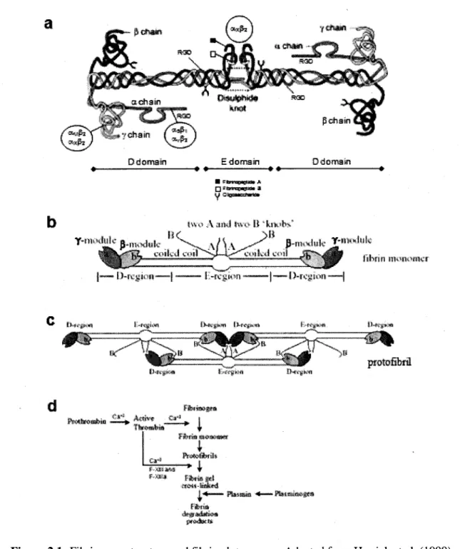
Human fibrinogen is a large, complex and fibrous glycoprotein with a molecular weight of 340 kDa. It is 45nm in length and composed of two symmetric "D" domain molecules and a central "E" domain. Each domain consists of one set of three different poly-peptide chains termed Aa, Bp\ and y chain (see Figure 2.1.a) [Herrick et al. 1999; Tawil 2006]. In the body, fibrinogen is present in human blood plasma at a concentration of approximately 2.5 g/L. The protein is essential for haemostasis, wound healing, inflammation, angiogenesis, and other biological events. Fibrinogen is a soluble macromolecule, which can be converted to an insoluble gel (i.e., fibrin) to stabilize the haemostatic plug and to provide a temporary matrix for subsequent cellular responses involved in wound healing [Janmey et al. 2009; Tawil 2006]. The role of fibrin in this process is not passive, but the protein rather actively directs cellular responses through specific receptor-mediated interactions with blood cells (e.g., leucocytes) as well as endothelial cells of the vessel wall [Herrick et al. 1999]. Therefore, the use of fibrin as a bioactive scaffold to support tissue vascularization is of interest.
The formation of fibrin clot during wound healing is initiated by the release of thrombin, a serine protease enzyme, which subsequently activates the coagulation cascade [Herrick et al. 1999; Rowe et al. 2007; Tawil 2006]. After the release, thrombin cleaves peptide fragments from fibrinogen to generate the fibrin monomer (Fig. 2.1.b) by the clotting cascade into protofibrils (Fig. 2.1.c). Afterward, in the presence of chloride ion and transglutaminase factor XIII or factor XHIa, protofibrils undergo intermolecular cross-linking to form a stable fibrin gel [Janmey et al. 2009; Litvinov et al, 2005; Rowe et al. 2007]. See Figure 2.1.d for details on the fibrin formation as well as the degradation processes.
Changing fibrinogen or thrombin concentration can change the resulting fibrin material, affecting both biochemical and mechanical properties [Ferrenq et al. 1997]. For example, Vailhe et al. (1998) have shown that capillary-like structures made from HUVEC seeded on fibrin depended on the mechanical factor of the gel. Harder gel, made using higher concentration of fibrinogen, led to a decreased number of capillary-like structures. No
capillary-like structures was found in softer matrix (< 0.5mg/mL of fibrinogen) as well as in too rigid one (> 4mg/mL of fibrinogen) [Ferrenq et al. 1997; Vailhe et al. 1998].
a
/».*feY"^-'."% v chain
D domain E domain O domain
« * * ** *
two A audi two & "kiwlis* IK ., }B
T-nunluk • msH|Mk. ^ \ Ii ^ 0-m,vlnk jr-«i"lufc
"ibrm unworncr
| — [)-ii£i>iii—~-| —~— J -ivgj^n —— j—~l)-rfjjttin
i>'«f»« D*ejih>fl protofibril
i"**,\. Am<mi €»** c»-' »»&* P»'i*?IS»ril» r-*Jt.» f a s m *rt Uteri*
Figure 2.1: Fibrinogen structure and fibrin clot process. Adapted from Herrick et al. (1999) and Litvinov et al. (2005).
Increasing fibrinogen concentration can reduce the matrix pore size, thus hindering endothelial cell migration and capillary formation [Nehls and Herrmann 1996]. In addition, Rowe et al. (2007) found that decreasing thrombin concentration resulted in both an increased gel compaction and micro-fibres size, thus causing different cellular morphology and alignment of vascular smooth muscle cells. These examples illustrate that fibrin gel properties and subsequent cell responses can be modulated, to some extent, opening the door to more applications [Cox et al. 2004; Rowe et al. 2007].
To date, fibrin is commercially available as fibrin sealant or fibrin glue (e.g., Tisseel™, Baxter AG, Vienna, Austria) for surgical applications [Tawil 2006]. Also, fibrin scaffolds have been used in many tissue engineering applications, including as matrix to treat bone and skin defects [Catelas et al. 2006], for drug delivery in neurological and cardiovascular disorders [Ferguson et al. 2003; Tassipoulos et al. 2000], and for three-dimensional angiogenesis assays [Vailhe et al. 1998; Nakatsu and Hughes 2008]. For example, Montesano et al. (1986) and Dejana et al. (1987) found that fibrinogen induced adhesion, spreading, and microfilament organization of human endothelial cells either in 2D or 3D in vitro culture system.
Also, culturing endothelial cells on microcarrier beads and then embedding these beads in fibrin have been proposed by Nehls and Drenckhahn (1995). This system resulted in formation of capillary structures and sprouting [Nehls and Drenckhahn 1995]. However, the system failed to model sprouting angiogenesis containing multi-cellular lumen surrounded by polarized endothelial cells, which is of importance during blood microvessel development [Montano et al 2009; Carmeliet and Conway 2001; Yancopoulos et al. 2000]. More recent vascularization studies using fibrin gels have been presented by Chen et al. (2009), Nakatsu et al. (2003a), and Sukmana and Vermette (2010a). In our study (see Chapter 3 of this thesis), endothelial cells proliferate and migrate along patterned polymer fibres and then fibrin is degraded along with the formation of cell-cell interactions, leading to the formation of tube-like structures, and eventually to sprouting and lumen formation with adjacent vessels [Sukmana and Vermette 2010b].
Unlike synthetic hydrogels, fibrin is an active matrix for cells. It can bind many growth factors and bioactive cloth components including fibronectin, hyaluronic acid, and von Willebrand factor [Herrick et al. 1999; Weisel 2005]. Human fibrinogen can bind to endothelial cells through either integrins or non-integrin binding sites (Table 2.1). For example, fibrin has two pairs of RGD binding sites and a non-RGD site at the y chain, which can interact with endothelial cell integrins (i.e., cc5pi, av(33, and anb(33) as well as with leucocyte integrins (i.e., aMp3 and ccxp2) [Cheresh et al. 1989; Lishko et al. 2002; Tawil, 200; Weisel 2005]. The location of integrins binding sites in fibrinogen is presented in Figure 2. La. Other non-integrin receptors that can bind to endothelial cells include ICAM-1, CD-44 surface receptor, and platelet endothelial cell adhesion molecule-1 (PECAM-1, also known as CD-31) [Cheresh et al. 1989; Herrick et al. 1999; Monchaux and Vermette 2010].
Table 2.1: Integrins and non-integrin receptors which can bind to fibrin(ogen). Integrins ccvp3 oc5pl ociibp3 ocMp3 «xP2 Non-integrin receptor PECAM-1 (CD31) ICAM-1 CD-44 Cell types endothelial cells and fibroblasts
endothelial cells
platelets and endothelial cells leucocytes and monocytes lymphocytes and leucocytes Cell types endothelial cells fibroblasts, endothelial cells endothelial cells,
fibroblasts and tumorous cells
Cellular function adhesion and spreading
adhesion
adhesion and cloth retraction
adhesion and phagocytosis
adhesion
Cellular function
adhesion and von Willebrand factor release proliferation and adhesion adhesion, migration, and proliferation References Cheresh et al. 1989; Cox et al. 2004; Monchaux and Vermette 2010. Cheresh et al. 1989; Herrick et al. 1999; Weisel 2005. Janmey et al. 2009; Monchaux and Vermette 2010. Herrick et al. 1999; Lishko et al 2002; Tawil 2006. Herrick et al. 1999; Janmey et al. 2009. References Monchaux and Vermette 2010; Nakatsu et al. 2003a; Cheresh et al. 1989; Herrick et al. 1999; Rowe et al. 2007. Herrick et al. 1999; Weisel 2005.
In other tissue engineering applications, fibrin was combined with collagen for the development of blood vessel substitutes [Cummings et al. 2004; Stegemann et al. 2006]. Collagen type I is the predominant structural component of the media as well as the adventia layers of blood vessels, while the inner layer of natural blood vessels, called the intima layer, is lined by endothelial cells [Monchaux and Vermette 2010]. Therefore, the combination of collagen-fibrin can be used to make scaffolds with good mechanical and biochemical properties [Yao et al. 2005; Cummings et al. 2004]. For example, Isenberg et al. (2006) have investigated a tubular scaffold made of type I collagen and fibrinogen to engineer small-diameter artificial arteries.
A summary of the use of synthetic and natural polymers that have been applied to support tissue vascularization is presented in Table 2.2.
Table 2.2: The use of synthetic and natural polymers to support tissue vascularization.
Scaffold Materials Poly(D,L-lactic-co-glicolic acid) PLLA/PLGA sponges DLLA/TMC porous scaffold Hyaluronic acid (HA) HA cross-linked with chitosan
Purpose and Methods
• Scaffold designed for tubular tissues.
• EC were seeded in the construct.
• Scaffolds designed with pores from 225 to 500um for skeletal muscle tissue. • Pre-seeded with MC, then
scaffolds were implanted in skeletal muscles of mice.
• For heart tissue
engineering with pore size oflOOum.
• Cardiomyocytes were seeded in the scaffold. Then scaffolds were subcutaneously implanted in rats.
Fibroblast growth factor (FGF) was added in the HA matrix.
Scaffold designed to produce a human skin
Results
EC were able to generate capillary network inside the scaffolds.
Scaffold
pre-vascularization promoted angiogenesis in the implant.
This method elicited an acute inflammatory reaction.
FGF improved neovascularization.
The hybrid scaffold improved EC References Mooney et al. 1994; Mooney et al. 1996. Levenberg et al. 2005. Pego et al. 2003a; Pego et al. 2003b. Rauh et al. 2006. Black et al. 1999; Slevin et
Alginate gel Marine collagen from tilapia (Oreochromis niloticas) Collagen type I Collagen type IV Fibrin gel equivalent.
• Endothelial cells were seeded in the construct.
• The construct was made for several applications including cell
encapsulation. • Vascular endothelial
growth factor (VEGF) was incorporated in the scaffold.
Sponges were tested in vivo.
• In vitro angiogenesis assay.
• EC were seeded in the matrix.
Fibroblast growth factor (FGF) was incorporated in the scaffold for in vitro study of angiogenesis.
Fibrinogen and thrombin concentrations were changed to study angiogenesis, in
vitro, using HUVEC and
VSMC.
proliferation and induced al. 2002. capillary network
formation inside the scaffold.
VEGF promoted neovascularization.
Tilapia collagen caused less inflammatory responses when compared to porcine collagen.
Type I of collagen increased the number of tube-like structures and supported angiogenesis development. • Addition of FGF in type IV collagen scaffold supported EC growth and capillary formation. • Degradation product of collagen type IV promoted a specific angiogenic epitope. • Capillary-like structures (CLS) made of HUVEC depended on the matrix rigidity. • Higher rigidity decreased CLS number. • No CLS were found in very soft or very rigid matrices. • Decreasing thrombin concentration caused different morphology and alignment of VSMC. Chang et al. 2010; Drury and Mooney 2003. Sugiura et al. 2009. Francis et al. 2008; Soker et al. 2000. Ingber and Folkman 1989; Xuetal. 2001. Ferrenq et al. 1997; Nehls and Herrmann 1996; Rowe et al. 2007; Vailhe et al. 1998.
Fibrin gel in which microcarrier beads were embedded Polymer monofilaments (i.e., PET) embedded in fibrin. Fibrin gel combined with collagen type I Aim: to generate angiogenesis development.
Microcarrier beads were pre-coated with HUVEC and subsequently
embedded in fibrin.
In vitro study of
angiogenesis guidance. PET fibres were pre-coated with HUVEC, then sandwiched in fibrin.
SMC were grown on a sheet-like scaffold, and then wrapped around tubular vessel. The vessel allowed to transfer nutrient and oxygen.
This in vitro culture system provided a step-by-step process of the capillary development containing multi-cellular lumen.
• Pre-coating PET with cells enabled
increasing the fibres bioactivity.
• PET fibres were able to guide EC to orient microvessels.
This in vitro culture system enabled producing a better environment for the vascularization of small-caliber arterial
substitutes.
Chen et al. 2009; Nakatsu et al. 2003a; Nehls and Drenckhahn 1995. Sukmana and Vermette 2010a; Sukmana and Vermette 2010b. Cummings et al. 2004; Isenberg et al. 2006; Yao et al. 2005.
EC: endothelial cell smooth muscle cell;
MC: mesenchymal cell; VSMC: vascular smooth muscle cell; SMC: PET: poly(ethylene terephthalate).
2.4 Material properties
Scaffold and ECM materials are selected based on bulk and surface properties, which can be tuned with the aim to modulate cell adhesion and proliferation as well as phenotypic cell expression [Francis et al. 2008]. Among the properties of importance, scaffold porosity and matrix stiffness play significant roles in cell and tissue responses and these will be briefly discussed below.
2.4.1 Scaffold porosity
Porosity is defined as the fraction of the void space over the total volume of a scaffold. Pore size corresponds to the distance between two solid sections of the porous matrix [Lawrence et al. 2008]. In tissue engineering, a highly porous scaffold (about 90% porosity) is more desirable, since it should increase mass and nutrient transport [Kannan et al. 2005]. Higher porosity can increase cell adhesion and provide a sufficient area for cell-matrix interactions
and new ECM production by cells [Agrawal and Ray 2001; Kannan et al. 2005]. However, bulk and mechanical properties should also be considered. At higher porosity, the total solid volume of the solid part of the scaffold is lower, when compared to scaffolds with lower porosity, thus resulting in weaker mechanical support [Kannan et al. 2005].
The effect of pore size over cell behaviour has been investigated in culturing bone tissue substitutes. Pores in the range of 300-400um have been found to be optimal for osteoblast attachment, growth and proliferation [Linnes et al. 2007]. The important role of such porous structures in endothelial cell organization and angiogenesis development was pioneered by Clowes et al. (1986) more than 20 years ago. In a more recent study, pore size was found to have a significant effect over cell binding, morphology, and phenotype, thus inducing endothelial cell migration and capillary formation inside the scaffold [O'Brien et al. 2005].
In the scientific literature, there are some suggestions concerning the optimum pore size to support vascularization. For example, many mature cell types, including fibroblasts and endothelial cells, have been found to be unable to spread and completely colonize the bulk of scaffolds with pore size higher than 300um, due to the difficulty on bridging the distance [Yanmas et al. 1989; Zeltinger et al. 2001]. In an in vitro angiogenesis study, it was shown that a cell's ability to bridge the distance in 3D scaffolds is important to support the vascularization process [Sukmana and Vermette 2010a]. Using HUVEC-covered poly(ethylene terephthalate) (PET) monofilaments, as contact guidance in HUVEC-seeded fibrin, it was suggested that the optimum fibre-fibre distance to support microvessel development was lOOum [Sukmana and Vermette 2010a].
Furthermore, hepatocytes were also reported to spread well on gelatin-chitosan scaffold (3:1) with pore size of 20um, while fibroblasts and endothelial cells spread better on the same matrix but with pores ranging between 100-150um, compared to pores from 20-80um [Huang et al. 2005]. The role of pore size to promote endothelial cell lining has been investigated in vascular grafts. For example, Zhang et al. (2004) found that an external pore size of 30um was preferable over 20um or smaller pores in terms of promoting rapid tissue ingrowth and endothelial cell growth in the expanded poly(tetrafluoroethylene) (ePTFE) graft.
In addition, Marshall et al. (2004) found that fibrin with 35um pores significantly supported angiogenesis development, when compared to fibrin with either 20um or 70um pores. Fibrin with pore sizes of approximately 30um promotes the ingrowth of vascularized fibrous tissue in engineered blood vessels [Harding et al. 2002]. However, the optimum porosity and pore size of the scaffold is still open to question and is based on the application as well as the cell type [Kannan et al. 2005; Zeltinger et al. 2001; Zhang et al. 2004].
2.4.2 Matrix stiffness
Substrate mechanical properties, such as stiffness, are known to be important parameters affecting cell responses. In cell biology, matrix stiffness is sensed by cell receptors and integrins transmit mechanical stress across the cell surface to the cell cytoskeleton (see •Appendix B for a brief description), converting mechanical signals into (bio)chemical ones
[Becker et al. 2009; Geiger et al. 2001; Ingber et al. 1995].
Therefore, the ECM stiffness will influence cellular functions, including cell adhesion, proliferation, migration as well as phenotype differentiation [Geiger et al. 2001; Ingber et al.
1995]. For example, Pelham and Wang (1997) have examined the effect of collagen-coated poly(acrylamide) scaffold on the behaviour of rat epithelial and fibroblast cells. They found that on more rigid (higher stiffness) surfaces, cells were more spread and showed increased motility and focal adhesion contacts [Pelham and Wang 1997]. Furthermore, increasing surface stiffness was found to result in increased cell contractility [Li et al. 2005], more organized cells cytoskeleton and actin stress fibres, and higher adhesion strength [Discher et al. 2005]. The phenomenon related to the effect of environment stiffness over cell behaviour is known to as durotaxis [Lo et al. 2000].
Substrate stiffness and cell contractility also have a significant role over microvascular development. For example, while endothelial cells proliferate more on rigid surfaces, they form tube-like structures on softer substrates [Deroanne et al. 2001; Discher et al. 2005]. Vasculogenesis decreased with an increase in matrix stiffness, which was a result of an increase in collagen [Angele et al. 2004; Haarer and Dee 2006; Yeung et al. 2005] or fibrinogen [Duong et al. 2009; Tawil 2006; Vailhe et al. 1998] concentration.
2.5 Scaffold vascularization
Cells can be isolated from a patient or donor and seeded into a scaffold to allow cell proliferation and support pre-vascularization process [Lanza et al. 2000; Mikos et al. 1993]. Subsequently, these constructs can be implanted in the same patient. After implantation, hopefully endothelial cells would develop connections with the existing blood microvessels of the surrounding tissue, forming a microvascular network and allowing adequate tissue perfusion [Lanza et al. 2000; Ye et al. 2000a].
When tissues become thicker, cells located more than a few hundred microns (about 200 to 300um) from capillaries suffer from a lack of oxygen and nutrients, resulting into necrotic conditions [Hutmatcher 2001; Ko et al. 2007]. Currently, the development of thick and complex tissues and organs such as the heart, muscles, kidneys, liver and lung relies, in part, on the knowledge and ability to stimulate microvascular network formation within tissue constructs [Ko et al. 2007; Mooney and Mikos 1999]. Therefore, knowing how to carry out cell seeding within a scaffold and to perform successful in vitro cultures to prevascularize tissue constructs prior to their implantation is highly important. As such, researchers rely on the increasing knowledge related to neovascularization (i.e., vasculogenesis and angiogenesis) process to stimulate vascular network formation within three-dimensional tissue constructs [Bramfeld et al. submitted-2010]. Before discussing vasculogenesis and angiogenesis processes, I will highlight the cell types available and/or used to carry out these experiments.
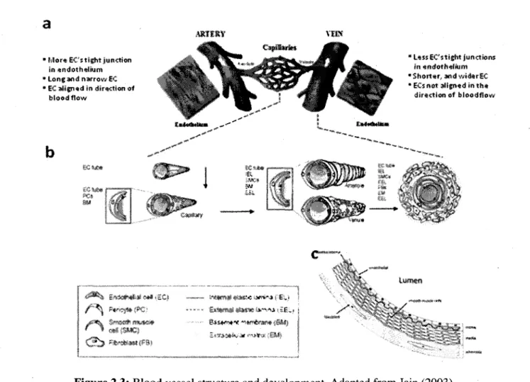
The existence of capillaries in blood vessels and lung of frogs was first reported by Marcello Malpighi in 1661 [Hillen et al. 2006]. After the invention of biological microscopes by Antonie van Leeuwenhoek (1632-1727) and his colleagues and further development of fluorescence techniques, the anatomy of blood vessels was revealed [Hillen et al. 2006]. Blood vessels are composed of several cell types and the main ones are smooth muscle cells (SMC), pericytes, fibroblasts and endothelial cells [Jain 2003; Ko et al. 2007].
Smooth muscle cells are found in blood vessels, such as in middle layer (i.e., tunica media) of large and small blood vessels, in lymphatic vessels, uterus, and in the gastrointestinal and respiratory systems [Hillen et al. 2006; Jain 2003]. Behind the basic function of vascular SMC
in blood vessels i.e., to maintain vessels integrity and support the endothelium [Auerbach et al. 2003; Hillen et al. 2006], they are highly specialized cells for the regulation of blood vessel diameter, vessel contraction, blood pressure and flow distribution [Auerbach et al. 2003; Owens et al. 2004]. Furthermore, SMC synthesize the connective tissue matrix of the vessel wall, which is composed of elastin, collagen, and proteoglycans [Auerbach et al. 2003; Hillen et al. 2006]. SMC show a very low level of proliferation during normal development but become more proliferative in response to vessel injury [Jain 2003].
There are some markers to identify smooth muscle cells, including smooth muscle cc-actin (SMaA) [Owens et al. 2004], smooth muscle myosin heavy chain (SM-MHC) [Bramfeld et al. submitted-2010; Hungerford and Little 1999], SM22cc, and calponin [Duband et al. 1993]. To date, SMaA is the most commonly used marker of SMC, which represents up to 70% of the actin population in vascular SMC [Bramfeld et al. submitted-2010; Hungerford and Little
1999; Jung etal. 2002].
Pericytes are perivascular-specific cells that are associated with capillaries and blood microvessel development [Hillen 2006; Jain 2003]. Pericytes have the capacity to differentiate into other cell types, including SMC, fibroblasts, and osteoblasts [Jung et al. 2002; Owens et al. 2004]. During blood microvessel development, the coverage of capillary by pericytes is important for the maturation, remodeling and maintenance of the vascular system via the secretion of growth factors and/or modulation of the ECM [Allt and Lawrenson 2001; Jung et al. 2002]. The important role of pericytes in capillary development in vitro has been studied by some researchers. For example, endothelial cells co-cultured with pericytes, separated by a cellulose membrane, resulted in inhibition of capillary growth, while when pericytes were in contact or close to endothelial cells; endothelial cell growth and capillary development were observed [Orlidge et al. 1987].
Fibroblasts are a cell type mostly found in connective tissues, including blood vessels, cartilage, soft tissues (e.g., dermis) and bone. A connective tissue can be defined as a tissue that wraps, connects, nourishes and supports all other tissues and organs [Becker et al. 2009]. Fibroblasts originate from mesenchymal cells - mesenchymal cells are progenitor cells