RESEARCH ARTICLE
Intermicrobial interaction: Aspergillus
fumigatus siderophores protect against
competition by Pseudomonas aeruginosa
Gabriele SassID1*, Shajia R. Ansari1, Anna-Maria Dietl2, Eric De´zielID3, Hubertus Haas2,
David A. Stevens1,4
1 California Institute for Medical Research, San Jose, California, United States of America, 2 Division of Molecular Biology, Biocenter, Medical University of Innsbruck, Innsbruck, Austria, 3 INRS-Institut Armand-Frappier, Laval, Quebec, Canada, 4 Division of Infectious Diseases and Geographic Medicine, Department of Medicine, Stanford University School of Medicine, Stanford, California, United States of America
Abstract
Pseudomonas aeruginosa and Aspergillus fumigatus are pathogens frequently
co-inhabit-ing immunocompromised patient airways, particularly in people with cystic fibrosis. Both microbes depend on the availability of iron, and compete for iron in their microenvironment. We showed previously that the P. aeruginosa siderophore pyoverdine is the main instru-ment in battling A. fumigatus biofilms, by iron chelation and denial of iron to the fungus. Here we show that A. fumigatus siderophores defend against anti-fungal P. aeruginosa effects.
P. aeruginosa supernatants produced in the presence of wildtype A. fumigatus planktonic
supernatants (Afsup) showed less activity against A. fumigatus biofilms than P. aeruginosa supernatants without Afsup, despite higher production of pyoverdine by P. aeruginosa. Supernatants of A. fumigatus cultures lacking the sidA gene (AfΔsidA), unable to produce
hydroxamate siderophores, were less capable of protecting A. fumigatus biofilms from P.
aeruginosa supernatants and pyoverdine. AfΔsidA biofilm was more sensitive towards
inhib-itory effects of pyoverdine, the iron chelator deferiprone (DFP), or amphothericin B than wildtype A. fumigatus biofilm. Supplementation of sidA-deficient A. fumigatus biofilm with A.
fumigatus siderophores restored resistance to pyoverdine. The A. fumigatus siderophore
production inhibitor celastrol sensitized wildtype A. fumigatus biofilms towards the anti-fun-gal activity of DFP. In conclusion, A. fumigatus hydroxamate siderophores play a pivotal role in A. fumigatus competition for iron against P. aeruginosa.
Introduction
Ecosystems of pathogens have been described with regard to a multitude of diseases [1–3]. The bacteriumPseudomonas aeruginosa and the fungus Aspergillus fumigatus form such an
ecosys-tem, e.g. when chronically colonizing the lungs of cystic fibrosis (CF) individuals [4–7]. Both pathogens have been associated with deterioration of lung function [4–17], and their com-bined presence in airways of CF patients seems to aggravate disease progression [18,19].P. a1111111111 a1111111111 a1111111111 a1111111111 a1111111111 OPEN ACCESS
Citation: Sass G, Ansari SR, Dietl A-M, De´ziel E, Haas H, Stevens DA (2019) Intermicrobial interaction: Aspergillus fumigatus siderophores protect against competition by Pseudomonas
aeruginosa. PLoS ONE 14(5): e0216085.https:// doi.org/10.1371/journal.pone.0216085 Editor: Olaf Kniemeyer, Leibniz-Institut fur Naturstoff-Forschung und Infektionsbiologie eV Hans-Knoll-Institut, GERMANY
Received: January 4, 2019 Accepted: April 12, 2019 Published: May 8, 2019
Copyright:© 2019 Sass et al. This is an open access article distributed under the terms of the
Creative Commons Attribution License, which permits unrestricted use, distribution, and reproduction in any medium, provided the original author and source are credited.
Data Availability Statement: All relevant data are within the manuscript and its Supporting Information files.
Funding: This research was funded by John Flatley (CIMR no. 3770) to D.A.S., Child Health Research Institute, Stanford Transdisciplinary Initiatives Program (CIMR no. 3777) to D.A.S., Austrian Science Fund/Infect-ERA program (FWF grant I1616/Infect-ERA project AspMetNet) to H.H., and HOROS program (W1253) to A.-M.D. The funders
aeruginosa and A. fumigatus also are prominent opportunistic pathogens in
immune-compro-mised patients, particularly in those with neutropenia [20,21].
Previous studies have focused onA. fumigatus inhibition caused by P. aeruginosa products
such as pyocyanin (5-N-methyl-1-hydroxyphenazine) [22–25], 1-hydroxyphenazine [22,24,25], phenazine-1-carboxamide and phenazine-1-carboxylic acid [25]. We recently reported that theP. aeruginosa product pyoverdine is the major mediator of P. aeruginosa
inhibitory function towardsA. fumigatus biofilms [26]. Pyoverdine, the major siderophore of
P. aeruginosa [27,28], strongly binds to iron, which is an essential co-factor for bothP. aerugi-nosa and A. fumigatus [29–31]. Pyoverdine-bound iron is no longer available forA. fumigatus,
starvingA. fumigatus of iron, and resulting in fungistasis [26]. The question arose whetherA. fumigatus could counteract P. aeruginosa inhibition. Here we provide evidence that A. fumiga-tus hydroxamate siderophores in times of iron shortage, created by a competing microbe,
ensure availability of the essential co-factor iron exclusively to the fungus. Concomitantly, interference withA. fumigatus siderophore production renders the fungus more sensitive to
anti-fungal effects of iron chelators, and possibly more sensitive even to effects of anti-fungal drugs not involved in iron chelation, like amphotericin B.
Materials and methods
Materials
Pyoverdine (PYOV), 3-hydroxy-1,2-dimethyl-4(1H)pyridine (deferiprone, DFP), celastrol, 2,3-bis[2-methoxy-4-nitro-5-sulfophenyl]-2H-tetrazolium-5-carboxanilide inner salt (XTT), and menadione were purchased from Sigma-Aldrich (St. Louis, MO). Amphotericin B (AmB) was derived from X-Gen Pharmaceuticals Inc. (Horseheads, NY). Chrome Azurol S (CAS) was purchased from MP Biomedicals (Solon, OH). Ferri- and desferri-triacetylfusarinine C (TAFC, DF-TAFC) were purified as described previously [32].
Isolates
All isolates used in this study are summarized inTable 1.
The work flow for the following procedures is summarized in
S1 Fig
.
A.
fumigatus supernatant production
A. fumigatus conidia were inoculated into RPMI 1640 medium (RPMI, Lonza, Walkersville,
MD) at 2.5x10⁴conidia/ml.A. fumigatus suspensions were incubated at 37˚C for 48h Table 1. Isolates used in this study.
Organism Isolate Description ATCC Reference
A. fumigatus 10AF Virulent patient isolate 90240 [33,34]
A. fumigatus AF13073 Parental strain for AfΔsidA 13073
A. fumigatus AfΔsidA L-ornithine-N5-mono-oxygenase deficientA. fumigatus mutant strain [35]
A. fumigatus AF46645 Parental strain for AfΔsidC and AfΔsidF 46645
A. fumigatus AfΔsidC Deficient for the hydroxamate siderophores ferricrocin (FC) and hydroxy-FC (HFC) [36]
A. fumigatus AfΔsidF Deficient for the hydroxamate siderophores fusarinine C (FsC) and triacetylfusarinine C (TAFC). [36]
A. fumigatus AfS77 Derivate of ATCC 46645 [37]
P. aeruginosa PA14 Parental strain forpvdD- and pvdD-pchE- [38]
P. aeruginosa pvdD- Pyoverdine deficient mutant [39]
P. aeruginosa pvdD-pchE- Pyoverdine/pyochelin deficient mutant [26]
https://doi.org/10.1371/journal.pone.0216085.t001
had no role in study design, data collection and analysis, decision to publish, or preparation of the manuscript.
Competing interests: The authors have declared that no competing interests exist.
Abbreviations: Af, Aspergillus fumigatus; Pa, P.
Aeruginosa; PS, planktonic P. aeruginosa culture
filtrate; PYOV, pyoverdine; BCAM, metabolic assay of A. fumigatus biofilm formation on agar; DFP, deferiprone; AmB, amphotericin B; FC, ferricrocin; HFC, hydroxy-ferricrocin; FsC, fusarinin C; TAFC, triacetylfusarinine C; DF-TAFC, desferri-triacetylfusarinine C; CAS, chrome azurol S.
(S1 Fig).A. fumigatus supernatants (Afsup) were filtered (0.22 μm) for sterility after the
growth period.
Pseudomonas supernatant production and pyoverdine measurement
PA14 supernatants were prepared as detailed previously [40]. Briefly,P. aeruginosa [5 x 10⁷
cells/ml] was inoculated into RPMI 1640 medium, or mixtures of RPMI and Afsup, and incu-bated at 37˚C for 24h. Bacterial growth was measured at 600 nm with a spectrophotometer (Genesys 20, Thermo Fisher Scientific Inc., Waltham, MA). Bacterial cultures were centrifuged at 200 xg for 30 min at room temperature, and filtered (0.22 μm). Pyoverdine production in
the supernatant was measured as described previously [41] at 405 nm. Pyoverdine measure-ments were normalized to bacterial growth using the formula: Relative PYOV expression = OD405 / OD600. At the concentrations used in this study, pyoverdine, a colored substance, did not interfere with the colorimetric XTT assay used for determination of fungal metabo-lism. PYOV concentrations in undilutedP. aeruginosa supernatants are about 30 μM.
Pyover-dine concentrations in sputum have been shown to be between 0.3 and 51μM [42].
Assay for the measurement of metabolism of
A. fumigatus forming (BCAM
assay, Bioassay-Conidia-Agar-Metabolic) or preformed (BHAM assay,
Bioassay-Hyphae-Agar-Metabolic) biofilms
BCAM and BHAM assays were performed as described previously [26]. In these assays,A. fumigatus grows out into biofilms covering the agar surface. Briefly, RPMI agar containing
2.5x10⁴to 105A. fumigatus conidia/ml agar (as specified for different experiments in the
Results section) was distributed into sterile flat-bottom 96 well cell culture plates (COSTAR, Corning, NY) at 100μl/well. Upon agar solidification, wells were either incubated at 37˚C for 24 hours before loading (= BHAM assays), or immediately loaded with 100μl of test sub-stances (= BCAM assays). Control wells on each test plate contained 100μl of RPMI 1640 medium, allowing the conversion of test results to % of the RPMI control (= 100%). Loaded plates were incubated at 37˚C for 24 hours. Fungal metabolism was determined by XTT meta-bolic assay at 490 nm [40,43]. Menadione (vitamin K3) was used as an ingredient in the XTT metabolic assay, boosting the reduction of tetrazolium salts to formazans. XTT assays were evaluated using a plate reader (Opsys MR, DYNEX Technologies, Chantilly, VA). Although XTT is a measure of metabolic activity of cells, previous studies ofA. fumigatus have indicated
that XTT results are linear with mass, and equated XTT result with dry weight [44–46].
Aspergillus growth assays
AfΔsidA (104
conidia) was point-inoculated on 2 ml solid minimal medium [47] in the pres-ence of 50–600μl PA14 wildtype or PA14 PaΔpvdD bacterial supernatant with or without sup-plementation of FeSO4[1μM]. Radial fungal growth was scored after incubation of the plates
for 48 hours at 37˚C.
Chrome azurol S (CAS) assay
For measurement of siderophore production 10x CAS assay reagent was prepared as described previously [48]. One part 10x CAS reagent was combined with 9 parts Afsups in RPMI, and incubated at 37˚C for 24 hours. Mixtures were measured using the plate reader, and compared to RPMI not containing CAS reagent or RPMI/1x CAS reagent as reference points.
Statistical analysis
Results were analyzed using Student’st test, if two groups were compared, and by 1-way
ANOVA, combined with a Tukey’s post-test for multiple comparisons. Data reported as per-centages of the control value were compared after arcsin transformation of the proportions [26]. All data in this study are expressed as a mean± SD.The number of replicates in each assay is four or higher. Assays were repeated at least twice, and a representative experiment is shown. Supporting information on data sets used in this study is provided inS1 Table.
Results
A. fumigatus supernatants induce pyoverdine production by P. aeruginosa
Fungal supernatants (Afsup), produced by planktonic growth ofA. fumigatus strain 10AF in
RPMI (experimental setup described inS1 Fig), induced pyoverdine production byP. aerugi-nosa in a dose-dependent manner, with 10% Afsup still significantly inducing pyoverdine
production. As pyoverdine is induced in response to iron shortage, increased pyoverdine pro-duction here suggests sequestration of iron from the growth medium by Afsup (Fig 1A).
Concentrations of Afsup higher than 10% interfered with bacterial growth in a concentra-tion-dependent manner (Fig 1B). As iron is a major co-factor for microbial growth, the reason for inhibitory effects of Afsup onP. aeruginosa growth might be a reaction to iron denial.
Although gliotoxin has been suggested as an anti-microbial factor [49], in our hands superna-tants produced by anA. fumigatus mutant unable to produce gliotoxin [50] affectedP. aerugi-nosa growth to a similar degree as supernatants produced by its parent (S2 Fig). Distilled water (25%), instead of Afsup (25%) duringP. aeruginosa supernatant preparation did not result in
interference withP. aeruginosa effects on A. fumigatus biofilm metabolism, indicating that P. aeruginosa supernatant dilution by Afsups was not the reason for the protective effects of
Afsups (S3 Fig).
In order to verify that Afsup indeed induced production of pyoverdine, we used a PA14 mutant not able to produce pyoverdine (PaΔpvdD) [39]. With or without the presence of Afsup, PaΔpvdD supernatant did not absorb at 405 nm, confirming that Afsup did not induce production of an unknownP. aeruginosa product detectable at 405 nm (S4A Fig).
Afsup protects
A. fumigatus forming biofilm from P. aeruginosa
anti-fungal activity and pure pyoverdine
Pyoverdine has detrimental effects on Af biofilm metabolism [26]. Surprisingly, although con-taining high concentrations of pyoverdine (Fig 1A),P. aeruginosa supernatants produced in
the presence of Afsup were less inhibitory for forming (Fig 1C) or preformed (Fig 1D)A. fumi-gatus biofilms than P. aeruginosa supernatants produced without Afsups,whereas Afsups up to
50% did not affectA. fumigatus biofilms when administered alone. Similarly, when P. aerugi-nosa supernatants and Afsups were prepared separately, their combination was less inhibitory
toA. fumigatus biofilms than P. aeruginosa supernatant alone (Fig 1E).
Presumably owing to their lack of pyoverdine production, supernatants of PaΔpvdD were less inhibitory to 10AF biofilms than PA14 supernatants. The presence of Afsup further decreased the inhibitory activity of PaΔpvdD supernatants to RPMI control levels (S4B Fig). Protective Afsup effects were also observed when Afsups were combined with pure pyoverdine (Fig 1F).
Taken together, these data indicate that despite its ability to induce pyoverdine production byP. aeruginosa, Afsup protects A. fumigatus biofilms.
Stability of protective Af supernatant effects
In order to determine the reason for protection of A. fumigatus biofilms by Afsup, we first tested stability of 10AFsup to heat, and long-term storage. 10AFsup was heated to 56˚C or 90˚C for 30 minutes, or subjected to three freeze-thaw cycles. Treated or untreated 10AFsups were diluted to 25%, combined withP. aeruginosa supernatants, and tested for effects on A. fumigatus biofilm metabolism. Our results show that heat does not destroy the protective
com-pound in Afsup (Fig 2A). Repeated freeze-thaw cycles diminished, but did not abolish protec-tion (Fig 2A). We also kept 10AFsup at 4˚C for 12 months, and measured protecprotec-tion from Fig 1.A. fumigatus supernatant effects on P. aeruginosa growth and pyoverdine production. Planktonic 10AF
supernatant (10AFsup) was diluted in RPMI from 50% to 0% 10AFsup, and incubated withP. aeruginosa cells (5x10⁷/ml) at 37˚C for 24h. Relative pyoverdine (PYOV) concentrations (A) were calculated using the quotient A405 (PYOV)/A600 (bacterial growth: B). Supernatants shown in A and B, as well as PA14 supernatant not containing 10Afsup, were compared with respect to their activities on 10AF forming biofilm (C: BCAM) or preformed biofilm (D: BHAM) metabolism. E: 10AF forming biofilm was incubated with 10 or 25% PA14 wildtype supernatant with or without the addition of 25% 10AFsup for 24 hours. Effects on 10AF forming biofilm metabolism were evaluated by XTT assay. F: 10AF forming biofilm was incubated with 5 or 10μM pyoverdine (PYOV) with or without the addition of 25% 10AFsup for 24 hours. Effects on 10AF forming biofilm metabolism were evaluated by XTT assay. Statistics for A and B: 1way ANOVA, RPMI (white bar) vs. all groups containing Afsup (black bars). Statistics for C and D: 1way ANOVA, PA14 supernatant (white striped bar) vs. PA14 supernatant containing 10AFsup (grey striped bars). Other comparisons by t-Test as indicated by the ends of the brackets. Statistics for E and F: t-Test, RPMI (white bar) vs. all other bars. Other comparisons as indicated by the ends of the brackets. One, two or three asterisks = p � 0.05, p � 0.01 or p � 0.001, respectively.
https://doi.org/10.1371/journal.pone.0216085.g001
pyoverdine every 3 months. The protective potential of Afsup was almost constant over the 12 months period (Fig 2B). When 10AFsup was kept at room temperature for 12 months, protec-tion was marginally lower, but still significant (Fig 2B). Taken together, our data suggest that the protective compound in 10AFsup is stable.
A. fumigatus siderophores protect A. fumigatus biofilm from P. aeruginosa
anti-fungal activity
Knowing that iron is a crucial factor forA. fumigatus biofilm, that pyoverdine inhibitory
activ-ity is owing to withholding iron from the fungus, and that the protective compound in Afsup Fig 2. Stability of Afsup. A: Mixtures (25%) of freshly prepared 10AFsup in RPMI were kept at 4˚C, or heated to 56˚C or 90˚C for 30 minutes, or subjected to 3 freeze-thaw cycles. Treated 10AFsups (25% in RPMI), were combined with 20%P. aeruginosa supernatants, and tested for effects on A. fumigatus forming biofilm metabolism. B: 10AFsup was
stored at 4˚C for up to 12 months, and tested for protective activity against 5μM pyoverdine (PYOV) every 3 months. A portion of the 10AFsup was stored at room temperature (RT), and tested after 12 months of storage. Protective activity was tested using a BCAM assay. Statistics for A: t-Test, PA14 supernatant (grey bar) vs. all other bars. Other comparisons as indicated by the ends of the brackets.�indicate significant increases,■ indicate significant decreases.
Statistics for B: t-Test, pyoverdine (white striped bar) vs. all other bars. One, two or three asterisks or squares = p � 0.05, p � 0.01 or p � 0.001, respectively.
is stable (Fig 2), we investigated the hypothesis that the protective compound might be anA. fumigatus siderophore. We produced supernatant of an A. fumigatus mutant lacking sidA, a
gene crucial for the production of all four hydroxamate siderophores (AfΔsidAsup), and com-pared to wildtype Afsup, produced by the AfΔsidA parent AF13073 (AF13073sup). A CAS assay confirmed the lack of siderophores in AfΔsidAsup (Fig 3A). Dilutions (25%) of AF13073sup and AfΔsidAsup were incubated with PA14 or PaΔpvdD. AF13073sup stimulated pyoverdine production by PA14 significantly more than AfΔsidA sup (Fig 3B). AfΔsidAsup also showed less protection forA. fumigatus biofilm against P. aeruginosa anti-fungal activity than AF13073sup
(Fig 3C). When siderophore-deficient fungus was treated with pyoverdine, significant damage was induced (Fig 3D), whereas Afsup derived from either 10AF or AF13073 wildtype strains protected AfΔsidA from pyoverdine-induced damage (Fig 3D). It has to be noted that neither the absence of pyoverdine nor the presence of Afsup from siderophore-deficient fungus pre-ventedP. aeruginosa anti-fungal activity completely.
In comparison to wildtypeA. fumigatus, AfΔsidA has a growth disadvantage due to missing
Fe3+uptake, which requires siderophores. 10AF or AF13073 wildtype supernatants, containing siderophores and iron, partially compensated AfΔsidA disadvantages, as indicated by higher XTT values for AfΔsidA in the presence of Afsups (Fig 3D). In conclusion,A. fumigatus
sidero-phores are able to protectA. fumigatus biofilms against P. aeruginosa anti-fungal activity.Fig 3Calso shows that AfΔsidA sup was able to provide protection for A. fumigatus biofilm from PaΔpvdD supernatant, whereas there was no protection against PA14 wildtype sup by either Fig 3.A. fumigatus siderophores protect A. fumigatus biofilm from P. aeruginosa anti-fungal activity. A:
Planktonic supernatants produced by anA. fumigatus mutant lacking hydroxamate siderophore production (AfΔsidA)
or its parental strain (AF13073) were subjected to siderophore production measurement by CAS assay. B: RPMI, or 25% AfΔsidA or AF13073 supernatant in RPMI, were inoculated with PA14 wildtype or the PA14 mutant PaΔpvdD [5x10⁷cells/ml], and incubated at 37˚C for 24h. Pyoverdine production was measured. C: Supernatants obtained in B (middle and right sets of 3 bars), as well as 25% AfΔsidA or AF13073 supernatants in RPMI (left 3 bars) were tested for activity againstA. fumigatus biofilm formation. D: AfΔsidA forming biofilm was incubated with 5 μM pyoverdine
(PYOV) with or without the addition of 25% 10AFsup or AF13073sup for 24 hours. Effects on forming biofilm metabolism were evaluated by XTT assay. Statistics: t-Test. Comparisons without brackets: B: RPMI vs.A. fumigatus
supernatants for each bacterial strain. C: RPMI vs. all other bars. D: RPMI (leftmost white bar) vs. all other bars. Other comparisons as indicated by the ends of the brackets.�indicate significant decreases,■ indicate significant increases.
One, two, or three asterisks or squares = p � 0.05, or p � 0.01, or p � 0.001, respectively.
https://doi.org/10.1371/journal.pone.0216085.g003
Afsup. This finding indicates thatA. fumigatus hydroxamate siderophores are crucial for
pro-tection from detrimental pyoverdine effects, but that Afsup seems to contain other compounds which are able to protectA. fumigatus biofilm when the Pasup challenge lacks the powerful
inhibitor pyoverdine.
Af
ΔsidA is more sensitive to P. aeruginosa anti-fungal activity and
pyoverdine than its wildtype
AfΔsidA-derived supernatants were significantly less protective against pyoverdine than wild-type supernatants (Fig 4A). AfΔsidA is lacking the intracellular hydroxamate siderophores ferricrocin (FC) and hydroxy-ferricrocin (HFC), as well as the extracellular hydroxamate side-rophores fusarinin C (FsC) and triacetylfusarinine C (TAFC). UsingA. fumigatus mutants
with specific mutations in intracellular (AfΔsidC), or extracellular hydroxamate siderophores (AfΔsidF), we found that a lack of extracellular siderophores significantly interfered with pro-tection from pyoverdine byA. fumigatus supernatants (Fig 4A). Protective effects of AfΔsidF
sup were significantly higher than protective effects of AfΔsidA sup (Fig 4A), indicating that there might be some contribution to protection by other molecules missing in AfΔsidA sup. Fig 4Aalso shows that supernatants, derived from three differentA. fumigatus wildtypes
(AF13073, AF46645, AfS77) protected forming biofilm of a fourthA. fumigatus wildtype
(10AF), indicating that protection is not strain specific.
PA14 supernatants, prepared in RPMI, as well as pure pyoverdine, were significantly more inhibitory during the formation ofA. fumigatus biofilms derived from AfΔsidA conidia than
they were for biofilms derived from AF13073 conidia (Fig 4B).A. fumigatus mutants lacking
either intracellular (AfΔsidC), or extracellular hydroxamate siderophores (AfΔsidF) showed increased sensitivity to PA14 supernatants or pure pyoverdine, compared to their wildtype AF46645 (Fig 4B). The loss of extracellular hydroxamate siderophores was more important for sensitivity than the loss of intracellular hydroxamate siderophores (Fig 4B). Using pure TAFC, or desferri-TAFC (DF-TAFC) we found complete protection from pyoverdine anti-fungal activity (Fig 4C), confirming the importance forA. fumigatus siderophores for protection
fromP. aeruginosa anti-fungal activity.
As observed inFig 2, the protective compound in Afsup was stable to prolonged heat treat-ment (90˚C, 30 min.). After being subject to the same treattreat-ment pure TAFC and DF-TAFC still significantly protected from pyoverdine toxicity (S5 Fig), further supporting the assump-tion thatA. fumigatus siderophores are the protective compound in Afsup. It was also noted
that pyoverdine was heat stable (S5 Fig).
The absence of A. fumigatus hydroxamate siderophores might have
therapeutic relevance
Compared to wildtypeA. fumigatus (AF13073), AfΔsidA growth on plate was more affected
with the highest concentration (600μl) of PA14 supernatant blocking growth (Fig 5A). Like-wise, the IC50 of pyoverdine for AF13073-derived forming biofilm was about 4 times higher than the IC50 for AfΔsidA-derived forming biofilm (Fig 5B). Recently, the iron chelator deferi-prone (DFP), which similar to pyoverdine, exerts anti-fungal activity by denying iron fromA. fumigatus biofilms, has been proposed to be useful in anti-fungal therapy [51,52]. We tested effects of DFP on AfΔsidA, or its wildtype, and found significantly higher sensitivity of A.
fumi-gatus biofilms to DFP when siderophore production was missing (Fig 5C). Genetic inhibition of siderophore production also increased anti-fungal effects of amphotericin B (AmB), an anti-fungal agent used against serious fungal infections, not only byAspergillus, but also by
Fig 4. AfΔsidA is more sensitive towards PA14 or pure pyoverdine than its wildtype. A: Mixtures (25%) of freshly prepared AF13073, AfΔsidA, AF46645, AfΔsidC, AfΔsidF, or AfS77 supernatants were combined with pyoverdine [10μM], and tested for effects on 10AF forming biofilm metabolism. Fungal metabolism was measured by XTT assay. Measurements for controls (no pyoverdine) in each group were regarded as 100%. Statistics: t-Test, for each group: no pyoverdine (grey bar) vs. pyoverdine (black bar). Other comparison as indicated by the ends of the bracket. B: AF13073, AfΔsidA, AF46645, AfΔsidC, AfΔsidF or AfS77 BCAM assays were incubated with either RPMI, PA14 supernatant, or 5μM pyoverdine. Fungal metabolism was measured by XTT assay. For each fungus RPMI control measurements were regarded as 100%. Statistics: t-Test, comparison: RPMI (white bars) vs. PA14 supernatant (grey bars), or pyoverdine (black bars) for each fungus. Other comparisons as indicated by the ends of the brackets. C: A 10AF BCAM assay was incubated with either RPMI, pyoverdine [10μM], TAFC [5 or 10 μM], DF-TAFC [5 or 10 μM], or combinations of pyoverdine and TAFC or DF-TAFC. Fungal metabolism was measured by XTT assay. RPMI control measurements were regarded as 100%. Statistics: t-Test, comparison: RPMI (white bar) vs. all other bars. Other comparisons as indicated by the ends of the brackets. One, two or three asterisks = p � 0.05, p � 0.01 or p � 0.001, respectively.
https://doi.org/10.1371/journal.pone.0216085.g004
Fig 5. Absence of hydroxamate siderophores sensitizesA. fumigatus. A: RPMI was inoculated with PA14 [5x10⁷
cells/ml], incubated for 24 hours, and the culture supernatant was sterile filtered. Growth of point inoculated AF13073 or AfΔsidA (104conidia) on 3 ml solid minimal medium in the presence of 1
μM FeSO4plus 50–600μl of the sterile
filtered supernatants was compared after incubation for 48 h at 37˚C. B: AfΔsidA (white bars) or AF13073 (black bars) BCAM assays were incubated with either RPMI or different concentrations of pyoverdine. Fungal metabolism was measured by XTT assay. Statistics: t-Test. For each fungus RPMI controls were regarded as 100%. RPMI controls for each fungus vs. all pyoverdine concentration. Other comparisons as indicated by the ends of the brackets. C: Wildtype
As a pharmacological complementation of our data obtained using AfΔsidA, we investi-gated effects of the SidA-biosynthesis inhibitor celastrol [54]. Celastrol showed anti-fungal activity when used alone at concentrations above 5μM (Fig 6). When combined with DFP, celastrol significant enhanced anti-fungal effects by DFP (Fig 6).
Discussion
Fungal and bacterial biofilms e.g. frequently found co-inhabiting lungs of persons suffering from cystic fibrosis, represent a potentially severe pathogenicity factor. The present study mainly focuses on events during formation ofA. fumigatus biofilm. In previous studies [40] and in studies by many others, it has been shown that biofilm formation byA. fumigatus is
substantial within the first 16 hours of incubation. We have also performed many of the studies described in the present communication against fully formedA. fumigatus biofilms that
develop over the subsequent 24 hours of incubation, and found the same phenomena, although to a lesser degree than in the earlier phase ofA. fumigatus biofilm formation, as
illus-trated inFig 1Cvs.1D. This may suggest that iron is more important for the initial develop-ment ofA. fumigatus biofilms.
The human body contains free iron levels of 10−24M [55]. Free iron levels are decreased during infections due to increased levels of ferritin and the release of lactoferrin from neutro-phils [56]. In the lungs of cystic fibrosis patients,P. aeruginosa and A. fumigatus, which both
are crucially dependent on the availability of free iron for metabolism and growth, aggravate disease pathology [4–7]. Under low iron conditions, these organisms are forced to compete for resources in the same environment [29,30].
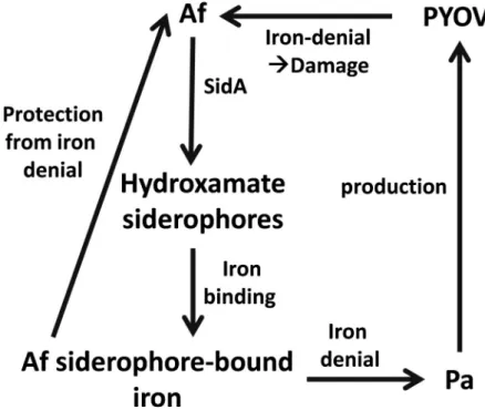
As summarized inFig 7, forP. aeruginosa as well as A. fumigatus a lack of iron is the signal
to increase production of siderophores [27,28]. Siderophores specifically chelate ferric iron with a high affinity [57]. Siderophores are of different types, based on the way the iron is (AF13073) or AfΔsidA forming biofilms were incubated with DFP [0.125–2 mM] at 37˚C for 24 hour. Fungal metabolism was measured by XTT assay. Statistics: t-Test, RPMI vs. all other bars of the same group. Other comparisons as indicated by the ends of the brackets. D: Wildtype (AF13073) or AfΔsidA forming biofilms were incubated with AmB [19–312 ng/ml] at 37˚C for 24 hour. Fungal metabolism was measured by XTT assay. Statistics: t-Test, RPMI vs. all other bars of the same group. Other comparisons as indicated by the ends of the brackets. One, two or three asterisks = p � 0.05, p � 0.01 or p � 0.001, respectively.
https://doi.org/10.1371/journal.pone.0216085.g005
Fig 6. Celastrol sensitizesA. fumigatus for anti-fungal activity of DFP. Forming wildtype A. fumigatus biofilm
(10AF) was incubated with 1, 2.5, or 5μM celastrol, 0.25–1 mM of DFP, or combinations of these two substances at 37˚C for 24 hour. Fungal metabolism was measured by XTT assay. Statistics: t-Test. Bars without DFP (leftmost group) vs. all other bars with the same celastrol concentration. Other comparisons as indicated by the ends of the brackets. One, two or three asterisks = p � 0.05, p � 0.01 or p � 0.001, respectively.
https://doi.org/10.1371/journal.pone.0216085.g006
complexed: phenolate-, catecholate-, hydroxamate-, carboxylate-, or mixed type of sidero-phores have been described [58].
TheP. aeruginosa siderophore pyoverdine is a composite (mixed) siderophore comprising
a peptide chain and a chromophore [59]. Pyoverdines bind iron with very high affinity, are able to acquire iron from transferrin, and their production is absolutely needed in mouse pul-monary infections [60–62]. We have described pyoverdine to be thePseudomonas-derived key
inhibitor ofA. fumigatus in their intermicrobial competition via iron sequestration under low
iron conditions [26]. We note that the loss of pyoverdine did not preventP. aeruginosa
anti-fungal activity completely, however. Pyochelin, the second siderophore ofP. aeruginosa, is
pro-duced by allP. aeruginosa isolates, but its affinity for iron is much lower compared to
pyover-dine [63,64]. Pyoverpyover-dine does not act as a xenosiderophore forA. fumigatus [26], thus withholding iron from the fungus, and inducing anti-fungal effects [26].P. aeruginosa does
not seem to be able to useA. fumigatus siderophores either. Our results show that Afsups
pro-voke increased pyoverdine production byP. aeruginosa, indicating that there is a paucity of
iron in the medium. IfP. aeruginosa could use iron bound to A. fumigatus siderophores, there
would be an abundancy of iron available to the bacterium, and hence no increase in pyover-dine production. We here for the first time provide evidence thatA. fumigatus is able to use
iron bound to its hydroxamate siderophores as the main defense againstP. aeruginosa
compe-tition for iron. These findings are summarized inFig 7. Our results usingA. fumigatus mutants
defective in hydroxamate siderophore production also indicate that additional defense mecha-nisms might be in place, since supernatants derived from these mutants still partially protected fromP. aeruginosa toxicity. Other microorganisms have developed defense mechanisms
againstP. aeruginosa not based on protective siderophore production. Candida albicans
appears to defend itself againstP. aeruginosa in part by down-regulating P. aeruginosa
sidero-phore production [65].
Fig 7. Summary. In need for ironP. aeruginosa (Pa) produces its siderophore pyoverdine (PYOV). PYOV-chelated
iron is not available toA. fumigatus (Af), resulting in iron deficiency and damage to the fungus. Anti-fungal activity in
part is counter-balanced by SidA-dependentA. fumigatus hydroxamate siderophores, providing iron to the fungus,
further denying iron fromP. aeruginosa.
Anti-bacterialA. fumigatus supernatant effects as a reason for protective effects against P. aeruginosa anti-fungal activity are highly unlikely. In the presence of Afsup, P. aeruginosa is
able to even produce more pyoverdine, which requires functional bacterial metabolism. Also, Afsup protects fromP. aeruginosa supernatants produced without Afsup being present, and
Afsup, as well as pureA. fumigatus siderophores, protect from pure pyoverdine. Additionally,
Afsups derived from a giotoxin mutant affected bacterial growth To the same degree as wild-type Afsups. The most plausible explanation for anti-bacterial effects of Afsups is depletion of essential factors in the medium, especially that of iron.
To overcome iron starvation,A. fumigatus produces its own siderophores [35].A. fumiga-tus is able to produce four hydroxamate-containing siderophores: ferricrocin (FC) as well as
hydroxyferricrocin (HFC) for intracellular iron trafficking, and fusarinine C (FsC) as well as its derivative triacetylfusarinine C (TAFC) for extracellular iron scavenging [36,66,67]. The first step in the biosynthesis of all four hydroxamate-containing siderophores is catalyzed by the enzyme L-ornithine N5-monooxygenase, termed SidA [30,67]. SidA catalyzes oxygen and NADPH-dependent hydroxylation of L-ornithine to N5-L-hydroxyornithine, a crucial step for the biosynthesis of hydroxamate-containing siderophores [35]. In a similar fashion toP. aeru-ginosa siderophores, A. fumigatus siderophores are essential for pathogenesis, as the AfΔsidA
strain is unable to establish invasive aspergillosis in a mouse model [35,56]. We show that in contrast to wildtypeA. fumigatus supernatants, supernatants derived from AfΔsidA were
unable to protectA. fumigatus biofilms from detrimental effects of P. aeruginosa supernatants,
or pyoverdine. This finding indicates the relevance ofA. fumigatus siderophores for protection
ofA. fumigatus from P. aeruginosa-induced iron denial, and was supported by our finding that A. fumigatus siderophores (FC as well as TAFC) each could partially protect A. fumigatus from
detrimentalP. aeruginosa effects (Fig 4A and 4B). Pure preparations of the sideropore TAFC protectedA. fumigatus from pyoverdine, even after heat treatment (Fig 4C, andS5 Fig). Pro-tection by TAFC and its desferri form DF-TAFC was about equal, indicating that TAFC very efficiently binds free iron in medium, before pyoverdine can do the same. TAFC-bound iron does not seem to be transferable to pyoverdine, and exclusively is available toA. fumigatus.
A. fumigatus lacking hydroxamate siderophores, especially of the extracellular type, was
more susceptible to pyoverdine (Fig 4B). The most pronounced detrimental effects of pyover-dine were observed when all four siderophores were missing.
A lack of siderophores, especially owing to a loss in SidA, renders the fungus more sensitive to iron denial by either pyoverdine (Fig 5B), or the clinically used iron chelator deferiprone (DFP,Fig 5B). Siderophore deficiency even sensitized the fungus to effects of amphotericin B (AmB,Fig 5C). While sensitization to DFP might be expected knowing that iron chelation by pyoverdine powerfully inhibits the fungus, sensitization to AmB is more surprising. It might be that a struggle for iron takes away energy from the fungus, and dampens intrinsic defense mechanisms, or that the membrane action of AmB [53] may adversely affect iron flux in the fungus.
As a pharmacological analog tosidA knockout we used celastrol treatment [54]. Celastrol, a pentacyclic triterpenoid that belongs to the family of quinone methides, exerts potent anti-can-cer and anti-metastatic [68,69], anti-inflammatory [70,71], and antioxidant [72] activities. Recently celastrol was identified as a noncompetitive inhibitor of SidA production [54]. Inhibi-tion of SidA producInhibi-tion by celastrol is detrimental toA. fumigatus growth [54]. We observed inhibitory effects of celastrol onA. fumigatus metabolism as well (Fig 6). Since celastrol has numerous effects [68–71] it can’t be excluded that effects onA. fumigatus are not solely owed
to inhibition of siderophores production.
SidA-deficiency or addition of celastrol toA. fumigatus wildtype cultures resulted not only
in reduced fungal growth [54], and reducedA. fumigatus biofilm metabolism (Fig 6), but also Aspergillus counter-balances Pseudomonas’ anti-fungal activity
in increased sensitivity towards the iron chelator DFP. DFP is clinically used to treat iron over-load, as in thalassemia major [73], but also interferes with iron needs of bacteria [74], andA. fumigatus biofilms [51]. Given that celastrol does not have unwanted effects on the host it might be quite useful in supporting anti-fungal therapy.
Previous studies have focused onP. aeruginosa products and their inhibition of A. fumiga-tus, at high P. aeruginosa product concentrations. Such studies have not considered the
possi-ble response ofA. fumigatus at the onset of P. aeruginosa competition. We here show that A. fumigatus uses its siderophores to counter-balance iron denial by P. aeruginosa. In vivo, the
winning microbe in this competition might be the one which unleashes its products first, and in the greatest quantity.A. fumigatus siderophores seem to also strengthen the fungus against
certain types of therapy. Therefore, interference with siderophore production might boost existing therapy againstA. fumigatus.
Supporting information
S1 Fig. Overview of the experimental setup. Af:Aspergillus fumigatus; Afsup: planktonic A. fumigatus supernatant, Pa: Pseudomonas; Pasup: planktonic P. aeruginosa supernatant, PYOV:
pyoverdine; (TIF)
S2 Fig. Gliotoxin content in Afsup is not likely to affectP. aeruginosa. P. aeruginosa cells (5 x 10⁷/ml) were incubated with planktonic supernatants (25%) derived from AF5322 wildtype, AFgliΔP (gliotoxin mutant), or AFgliPR (reversion of the gliotoxin mutant) at 37˚C for 24h. Bacterial growth (A600: A), and pyoverdine (PYOV; A405) were measured, and relative pyo-verdine concentration (B) was calculated using the quotient A405/A600. Statistics by t-Test: PA14 supernatant, not containing Afsup (white bar) vs. PA14 supernatants containing Afsup. Two or three asterisks = p � 0.01 or p � 0.001, respectively.
(TIF)
S3 Fig. Reduction of nutrients affects bacterial growth, but does not result in protection of A. fumigatus from P. aeruginosa toxicity. P. aeruginosa cells (5 x 10⁷/ml) were incubated in RPMI 1640 medium containing 25% 10AFsup, or 25% sterile water, at 37˚C for 24h. A: Bacte-rial growth (A600) was measured. Supernatants derived from A were tested for toxicity against
A. fumigatus biofilm formation (XTT assay: B). Statistics by t-Test: A: PA14 supernatant
pre-pared without Afsup or water addition (white bar) vs all other bars. B: RPMI (while bar) vs. all other bars. Other comparisons as indicated by the ends of the brackets. Two or three
asterisks = p � 0.01 or p � 0.001, respectively. (TIF)
S4 Fig. Afsup induces pyoverdine and protects fromP. aeruginosa anti-fungal activity. A: RPMI was inoculated with PA14 wildtype or the PA14 mutant PaΔpvdD (5x10⁷
cells/ml), with (black bars) or without (white bars) the presence of 25% 10AFsup, and incubated at 37˚C for 24h. Pyoverdine production was measured. B: Samples produced in A were used in a BCAM assay, and compared to metabolism of 10AF forming biofilm in the presence of RPMI or 25% 10AFsup, incubated without bacteria. Statistics: t-Test, as indicated by the ends of the brackets. Two or three asterisks = p � 0.01 or p � 0.001, respectively.
(TIF)
S5 Fig. TAFC and DF-TAFC are stable to prolonged heat treatment. A 10AF BCAM assay
was incubated with RPMI, TAFC [10μM], DF-TAFC [10 μM], either fresh or heat treated (90˚C for 30 min), and combined with pyoverdine (not heated) [PYOV, 10μM]. Fungal
metabolism was measured by XTT assay. Control (RPMI incubation without heat treatment) was regarded as 100%. Statistics: t-Test, comparison: PYOV without heat treatment vs. all other PYOV-containing bars. Other comparisons as indicated by the ends of the brackets. One, two or three asterisks = p � 0.05, p � 0.01 or p � 0.001, respectively. Comparison of heat treatment of PYOV to unheated PYOV is also shown.
(TIF)
S1 Table. Data sets used in this study.
(PDF)
Acknowledgments
The authors thank Marife Martinez for excellent technical support. TheA. fumigatus mutant
strain AfΔsidA was kindly provided by M. M. Moore, Department of Biological Sciences, Simon Fraser University, Burnaby, Canada.
Author Contributions
Conceptualization: Gabriele Sass, Eric De´ziel, Hubertus Haas, David A. Stevens. Data curation: Gabriele Sass, Shajia R. Ansari.
Formal analysis: Gabriele Sass.
Funding acquisition: Anna-Maria Dietl, Hubertus Haas, David A. Stevens. Investigation: Gabriele Sass, Shajia R. Ansari.
Methodology: Gabriele Sass, Anna-Maria Dietl, David A. Stevens. Project administration: David A. Stevens.
Resources: Anna-Maria Dietl, Eric De´ziel, Hubertus Haas.
Supervision: Gabriele Sass, Eric De´ziel, Hubertus Haas, David A. Stevens. Validation: Gabriele Sass, David A. Stevens.
Visualization: Gabriele Sass, Anna-Maria Dietl.
Writing – original draft: Gabriele Sass, David A. Stevens.
Writing – review & editing: Gabriele Sass, Anna-Maria Dietl, Eric De´ziel, Hubertus Haas.
References
1. Lerner A, Arleevskaya M, Schmiedl A, Matthias T. Microbes and viruses are bugging the gut in celiac disease. Are they friends or foes? Front Microbiol. 2017; 8: 1392. eCollection 2017. Review.https://doi. org/10.3389/fmicb.2017.01392PMID:28824555
2. de Souza HSP, Fiocchi C, Iliopoulos D. The IBD interactome: an integrated view of aetiology, pathogen-esis and therapy. Nat Rev Gastroenterol Hepatol. 2017; 14: 739–749.https://doi.org/10.1038/nrgastro. 2017.110PMID:28831186
3. Wang J, Li F, Tian Z. Role of microbiota on lung homeostasis and diseases. Sci China Life Sci. 2017; 60: 1407–1415. Review.https://doi.org/10.1007/s11427-017-9151-1PMID:29019144
4. Williams HD, Davies JC. Basic science for the chest physician: Pseudomonas aeruginosa and the cys-tic fibrosis airway. Thorax. 2012; 67: 465–467.https://doi.org/10.1136/thoraxjnl-2011-201498PMID:
22382597
5. Smyth AR, Hurley MN. Targeting the Pseudomonas aeruginosa biofilm to combat infections in patients with cystic fibrosis. Drugs Fut. 2010; 35: 1007–1014.
6. Folkesson A, Jelsbak L, Yang L, Johansen HK, Ciofu O, Høiby N, et al. Adaptation of Pseudomonas
aeruginosa to the cystic fibrosis airway: an evolutionary perspective. Nat Rev Microbiol. 2012; 10: 841–
851. Review.https://doi.org/10.1038/nrmicro2907PMID:23147702
7. Sabino R, Ferreira JA, Moss RB, Valente J, Verı´ssimo C, Carolino E, et al. Molecular epidemiology of
Aspergillus collected from cystic fibrosis patients. J Cyst Fibros. 2015; 14: 474–481.https://doi.org/10. 1016/j.jcf.2014.10.005PMID:25459562
8. Fillaux J, Bre´mont F, Murris M, Cassaing S, Rittie´ JL, Te´tu L, et al. Assessment of Aspergillus sensitiza-tion or persistent carriage as a factor in lung funcsensitiza-tion impairment in cystic fibrosis patients. Scand J Infect Dis. 2012; 44: 842–847.https://doi.org/10.3109/00365548.2012.695454PMID:22831545
9. Speirs JJ, van der Ent CK, Beekman JM. Effects of Aspergillus fumigatus colonization on lung function in cystic fibrosis. Curr Opin Pulm Med. 2012; 18: 632–638.https://doi.org/10.1097/MCP.
0b013e328358d50bPMID:22965276
10. Ramsey KA, Ranganathan S, Park J, Skoric B, Adams AM, Simpson SJ, et al. Early respiratory infec-tion is associated with reduced spirometry in children with cystic fibrosis. Am J Respir Crit Care Med. 2014; 190: 1111–1116.https://doi.org/10.1164/rccm.201407-1277OCPMID:25321321
11. de Boer K, Vandemheen KL, Tullis E, Doucette S, Fergusson D, Freitag A, et al. Exacerbation fre-quency and clinical outcomes in adult patients with cystic fibrosis. Thorax. 2011; 66: 680–685.https:// doi.org/10.1136/thx.2011.161117PMID:21680566
12. Nicolai T, Arleth S, Spaeth A, Bertele-Harms RM, Harms HK. Correlation of IgE antibody titer to Af with decreased lung function in cystic fibrosis. Pediatr Pulmonol. 1990; 8: 12–15. PMID:2405341
13. Forsyth KD, Hohmann AW, Martin AJ, Bradley J. IgG antibodies to Aspergillus fumigatus in cystic fibro-sis: a laboratory correlate of disease activity. Arch Dis Child. 1988; 63: 953–957. PMID:3046514
14. Schønheyder H, Jensen T, Høiby N, Andersen P, Koch C. Frequency of Aspergillus fumigatus isolates and antibodies to Aspergillus antigens in cystic fibrosis. Acta Pathol Microbiol Immunol Scand B. 1985; 93: 105–112. PMID:3893030
15. Coughlan CA, Chotirmall SH, Renwick J, Hassan T, Low TB, Bergsson G, et al. The effect of Aspergillus
fumigatus infection on vitamin D receptor expression in cystic fibrosis. Am J Respir Crit Care Med.
2012; 186: 999–1007.https://doi.org/10.1164/rccm.201203-0478OCPMID:22904183
16. MirkovićB, Lavelle GM, Azim AA, Helma K, Gargoum FS, Molloy K, et al. The basophil surface marker CD203c identifies Aspergillus species sensitization in patients with cystic fibrosis. J Allergy Clin Immu-nol. 2016; 137: 436–443.https://doi.org/10.1016/j.jaci.2015.07.045PMID:26388311
17. Baxter CG, Moore CB, Jones AM, Webb AK, Denning DW. IgE-mediated immune responses and air-way detection of Aspergillus and Candida in adult cystic fibrosis. Chest. 2013; 143: 1351–1357.https:// doi.org/10.1378/chest.12-1363PMID:23139075
18. Shoseyov D, Brownlee KG, Conway SP, Kerem E. Aspergillus bronchitis in cystic fibrosis. Chest. 2006; 130: 222–226.https://doi.org/10.1378/chest.130.1.222PMID:16840406
19. Amin R, Dupuis A, Aaron SD, Ratjen F. The effect of chronic infection with Aspergillus fumigatus on lung function and hospitalization in patients with cystic fibrosis. Chest. 2010; 137: 171–176.https://doi. org/10.1378/chest.09-1103PMID:19567494
20. de Bentzmann S, Ple´siat P. The Pseudomonas aeruginosa opportunistic pathogen and human infec-tions. Environ Microbiol. 2011; 13: 1655–1665.https://doi.org/10.1111/j.1462-2920.2011.02469.x
PMID:21450006
21. Walsh TJ, Stevens DA. Aspergillosis. Chapter 347, Cecil Textbook of Medicine, 24th ed. Goldman L, Schafer A, eds.; Elsevier; 2011.
22. Mangan A. Interactions between some aural Aspergillus species and bacteria. J Gen Microbiol. 1969; 58: 261–266.https://doi.org/10.1099/00221287-58-2-261PMID:4982824
23. Blyth W, Forey A. The influence of respiratory bacteria and their biochemical fractions on Aspergillus
fumigatus. Sabouraudia. 1971; 9: 273–282. PMID:5002653
24. Kerr JR, Taylor GW, Rutman A, Høiby N, Cole PJ, Wilson R. Pseudomonas aeruginosa pyocyanin and 1-hydroxyphenazine inhibit fungal growth. J Clin Pathol. 1999; 52: 385–387. PMID:10560362
25. Briard B, Bomme P, Lechner BE, Mislin GL, Lair V, Pre´vost MC, et al. Pseudomonas aeruginosa manip-ulates redox and iron homeostasis of its microbiota partner Aspergillus fumigatus via phenazines. Sci Rep. 2015; 5: 8220.https://doi.org/10.1038/srep08220PMID:25665925
26. Sass G, Nazik H, Penner J, Shah H, Ansari SR, Clemons KV, et al. Studies of Pseudomonas
aerugi-nosa mutants indicate pyoverdine as the central factor in inhibition of Aspergillus fumigatus biofilm. J
Bacteriol. 2017. pii: JB.00345-17.https://doi.org/10.1128/JB.00345-17
27. Cornelis P, Dingemans J. Pseudomonas aeruginosa adapts its iron uptake strategies in function of the type of infections. Front Cell Infect Microbiol. 2013; 3: 75. Review.https://doi.org/10.3389/fcimb.2013. 00075PMID:24294593
28. Schalk IJ, Guillon L. Pyoverdine biosynthesis and secretion in Pseudomonas aeruginosa: implications for metal homeostasis. Environ Microbiol. 2013; 15: 1661–73. Review. https://doi.org/10.1111/1462-2920.12013PMID:23126435
29. Minandri F, Imperi F, Frangipani E, Bonchi C, Visaggio D, Facchini M, et al. Role of iron uptake systems in Pseudomonas aeruginosa virulence and airway infection. Infect Immun. 2016; 84: 2324–2335.
https://doi.org/10.1128/IAI.00098-16PMID:27271740
30. Haas H. Iron—a key nexus in the virulence of Aspergillus fumigatus. Front Microbiol. 2012; 3: 28. eCol-lection 2012.https://doi.org/10.3389/fmicb.2012.00028PMID:22347220
31. Matthaiou EI, Sass G, Stevens DA, Hsu JL. Iron: an essential nutrient for Aspergillus fumigatus and a fulcrum for pathogenesis. Current Opinion Infect Dis. 2018; 31: 506–511.
32. Oberegger H, Schoeser M, Zadra I, Abt B, Haas H. SREA is involved in regulation of siderophore bio-synthesis, utilization and uptake in Aspergillus nidulans. Mol Microbiol. 2001; 41: 1077–89. PMID:
11555288
33. Denning DW, Clemons KV, Hanson LH, Stevens DA. Restriction endonuclease analysis of total cellular DNA of Aspergillus fumigatus isolates of geographically and epidemiologically diverse origin. J Infect Dis. 1990; 162: 1151–1158. PMID:1977804
34. Denning DW, Stevens DA. Efficacy of cilofungin alone and in combination with amphotericin B in a murine model of disseminated aspergillosis. Antimicrob Agents Chemother. 1991; 35: 1329–1333. PMID:1929289
35. Schrettl M, Bignell E, Kragl C, Joechl C, Rogers T, Arst HN Jr, et al. Siderophore biosynthesis but not reductive iron assimilation is essential for Aspergillus fumigatus virulence. J Exp Med. 2004; 200: 1213– 1219.https://doi.org/10.1084/jem.20041242PMID:15504822
36. Schrettl M, Bignell E, Kragl C, Sabiha Y, Loss O, Eisendle M, et al. Distinct roles for intra- and extracellu-lar siderophores during Aspergillus fumigatus infection. PLoS Pathog. 2007; 3:e128.
37. Hartmann T, Du¨mig M, Jaber BM, Szewczyk E, Olbermann P, Morschha¨user J, et al. Validation of a self-excising marker in the human pathogen Aspergillus fumigatus by employing the beta-rec/six site-specific recombination system. Appl Environ Microbiol. 2010; 76: 6313–6317.https://doi.org/10.1128/ AEM.00882-10PMID:20656854
38. Rahme LG, Stevens EJ, Wolfort SF, Shao J, Tompkins RG, Ausubel FM. Common virulence factors for bacterial pathogenicity in plants and animals. Science 1995; 268: 1899–1902. PMID:7604262
39. Liberati NT, Urbach JM, Miyata S, Lee DG, Drenkard E, Wu G, et al. An ordered, nonredundant library of Pseudomonas aeruginosa strain PA14 transposon insertion mutants. Proc Natl Acad Sci U S A. 2006; 103: 2833–2838.https://doi.org/10.1073/pnas.0511100103PMID:16477005
40. Ferreira JA, Penner JC, Moss RB, Haagensen JA, Clemons KV, Spormann AM, et al. Inhibition of
Aspergillus fumigatus and its biofilm by Pseudomonas aeruginosa is dependent on the source,
pheno-type and growth conditions of the bacterium. PLoS One. 2015; 10: e0134692.https://doi.org/10.1371/ journal.pone.0134692PMID:26252384
41. Wilderman PJ, Vasil AI, Johnson Z, Wilson MJ, Cunliffe HE, Lamont IL, et al. Characterization of an endoprotease (PrpL) encoded by a PvdS-regulated gene in Pseudomonas aeruginosa. Infect Immun. 2001; 69: 5385–5394.https://doi.org/10.1128/IAI.69.9.5385-5394.2001PMID:11500408
42. Martin LW, Reid DW, Sharples KJ, Lamont IL. Pseudomonas siderophores in the sputum of patients with cystic fibrosis. Biometals. 2011; 24: 1059–67.https://doi.org/10.1007/s10534-011-9464-zPMID:
21643731
43. Scudiero DA, Shoemaker RH, Paull KD, Monks A, Tierney S, Nofziger TH, et al. Evaluation of a soluble tetrazolium/formazan assay for cell growth and drug sensitivity in culture using human and other tumor cell lines. Cancer Res. 1988; 48: 4827–4833. PMID:3409223
44. Meletiadis J, Mouton JW, Meis JFGM, Bouman BA, Donnelly JP, Verweij PE, et al. Colorimetric assay for antifungal susceptibility testing of Aspergillus species. J Clin Microbiol. 2001; 39: 3402–3408.https:// doi.org/10.1128/JCM.39.9.3402-3408.2001PMID:11526191
45. Moss BJ, Kim Y, Nandakumar MP, Marten MR. Quantifying metabolic activity of filamentous fungi using a colorimetric XTT assay. Biotechnol Prog. 2008; 24: 780–783.https://doi.org/10.1021/bp070334t
PMID:18386938
46. Pierce CG, Uppuluri P, Tristan AR, Wormley FL, Mowat E, Ramage G, et al. A simple and reproducible 96-well plate-based method for the formation of fungal biofilms and its application to antifungal suscepti-bility testing. Nature Protocols. 2008; 3: 1494–1500.https://doi.org/10.1038/nport.2008.141PMID:
18772877
47. Pontecorvo G, Roper JA, Hemmons LM, Macdonald KD, Bufton AWJ. The genetics of Aspergillus
nidu-lans. Adv Genet. 1953; 5: 141–238. PMID:13040135
48. Andrews MY, Santelli CM, Duckworth OW. Layer plate CAS assay for the quantitation of siderophore production and determination of exudation patterns for fungi. J Microbiol Methods. 2016; 121: 41–43.
https://doi.org/10.1016/j.mimet.2015.12.012PMID:26712125
49. Reece E, Doyle S, Greally P, Renwick J, McClean S. Aspergillus fumigatus inhibits Pseudomonas
aeru-ginosa in co-culture: Implications of a mutually antagonistic relationship on virulence and inflammation
in the CF airway. Front Microbiol. 2018; 9: 1205. eCollection 2018.https://doi.org/10.3389/fmicb.2018. 01205PMID:29922270
50. Sugui JA, Pardo J, Chang YC, Zarember KA, Nardone G, Galvez EM, Mu¨llbacher A, Gallin JI, Simon MM, Kwon-Chung KJ. Gliotoxin is a virulence factor of Aspergillus fumigatus: gliP deletion attenuates virulence in mice immunosuppressed with hydrocortisone. Eukaryot Cell. 2007; 6: 1562–1569.https:// doi.org/10.1128/EC.00141-07PMID:17601876
51. Nazik H, Penner JC, Ferreira JA, Haagensen JA, Cohen K, Spormann AM, et al. Effects of iron chela-tors on the formation and development of Aspergillus fumigatus biofilm. Antimicrob Agents Chemother. 2015; 59: 6514–6520.https://doi.org/10.1128/AAC.01684-15PMID:26239975
52. Hsu JL, Clemons KV, Manouvakhova O, Inayathulla M, Tu AB, Sobel RA, et al. Microhemorrhage-asso-ciated tissue iron enhances the risk for Aspergillus fumigatus invasion in murine airway transplantation. Sci Transl Med. 2018; 10(429). pii: eaag2616.https://doi.org/10.1126/scitranslmed.aag2616
53. Stevens DA. Systemic antifungal agents. Cecil Textbook of Medicine, 25th ed. Chapter 331; Goldman L Schafer A, eds; Elsevier; 2015.
54. Martı´n Del Campo JS, Vogelaar N, Tolani K, Kizjakina K, Harich K, Sobrado P. Inhibition of the flavin-dependent monooxygenase siderophore A (SidA) blocks siderophore biosynthesis and Aspergillus
fumigatus growth. ACS Chem Biol. 2016; 11: 3035–3042.https://doi.org/10.1021/acschembio.6b00666
PMID:27588426
55. Potrykus J, Ballou ER, Childers DS, Brown AJP. Conflicting interests in the pathogen−host tug of war: Fungal micronutrient scavenging versus mammalian nutritional immunity. PLoS Pathog. 2014; 10: e1003910.https://doi.org/10.1371/journal.ppat.1003910PMID:24626223
56. Hissen AHT, Chow JMT, Pinto LJ, Moore MM. Survival of Aspergillus fumigatus in serum involves removal of iron from transferrin: the role of siderophores. Infect Immun. 2004; 72: 1402−1408.https:// doi.org/10.1128/IAI.72.3.1402-1408.2004PMID:14977945
57. Braun V, Killmann H. Bacterial solutions to the iron-supply problem. Trends Biochem Sci. 1999; 24: 104–109.doi:10.1016/S0968-0004(99) 01359–6 PMID:10203757
58. Khan A, Singh P, Srivastava A. Synthesis, nature and utility of universal iron chelator—Siderophore: A review. Microbiol Res. 2017. pii: S0944-5013(17)30673-0. Review.
59. Visca P, Imperi F, Lamont IL. Pyoverdine siderophores: from biogenesis to biosignificance. Trends Microbiol. 2007; 15: 22–30. Review.https://doi.org/10.1016/j.tim.2006.11.004PMID:17118662
60. Albrecht-Gary AM, Blanc S, Rochel N, Ocacktan AZ, Abdallah MA. Bacterial iron transport: coordination properties of pyoverdin PaA, a peptidic siderophore of Pseudomonas aeruginosa. Inorg Chem. 1994; 33: 6391–6402.https://doi.org/10.1021/ic00104a059
61. Meyer JM. Pyoverdines: pigments, siderophores and potential taxonomic markers of fluorescent
Pseu-domonas species. Arch Microbiol. 2000; 174: 135–142.https://doi.org/10.1007/s0020300001PMID:
11041343
62. Imperi F, Massai F, Facchini M, Frangipani E, Visaggio D, Leoni L, et al. Repurposing the antimycotic drug flucytosine for suppression of Pseudomonas aeruginosa pathogenicity. Proc Natl Acad Sci USA. 2013; 110: 7458–7463.https://doi.org/10.1073/pnas.1222706110PMID:23569238
63. Ankenbauer RG, Toyokuni T, Staley A, Rinehart KL Jr, Cox CD. Synthesis and biological activity of pyo-chelin, a siderophore of Pseudomonas aeruginosa. J Bacteriol. 1988; 170: 5344–5351. PMID:3141386
64. Brandel J, Humbert N, Elhabiri M, Schalk IJ, Mislin GL, Albrecht-Gary AM. Pyochelin, a siderophore of
Pseudomonas aeruginosa: physicochemical characterization of the iron(III), copper(II) and zinc(II)
com-plexes. Dalton Trans. 2012; 41: 2820–2834.https://doi.org/10.1039/c1dt11804hPMID:22261733
65. Lopez-Medina E, Fan D, Coughlin LA, Ho EX, Lamont IL, Reimmann C, et al. Candida albicans inhibits
Pseudomonas aeruginosa virulence through suppression of pyochelin and pyoverdine biosynthesis.
PLoS Pathog. 2015; 11: e1005129. eCollection 2015 Aug.https://doi.org/10.1371/journal.ppat. 1005129PMID:26313907
66. Haas H. Fungal siderophore metabolism with a focus on Aspergillus fumigatus. Nat Prod Rep. 2014; 31: 1266–1276.https://doi.org/10.1039/c4np00071dPMID:25140791
67. Blatzer M, Schrettl M, Sarg B, Lindner HH, Pfaller K, Haas H. SidL, an Aspergillus fumigatus transacety-lase involved in biosynthesis of the siderophores ferricrocin and hydroxyferricrocin. Appl Environ Micro-biol. 2011; 77: 4959−4966.https://doi.org/10.1128/AEM.00182-11PMID:21622789
68. Tiedemann RE, Schmidt J, Keats JJ, Shi C-X, Zhu YX, Palmer SE, et al. Identification of a potent natural triterpenoid inhibitor of proteosome chymotrypsin-like activity and NF- B with antimyeloma activity in vitro and in vivo. Blood. 2008; 113: 4027–4037.https://doi.org/10.1182/blood-2008-09-179796PMID:
19096011
69. Zhu H, Liu XW, Cai TY, Cao J, Tu CX, Lu W, et al. Celastrol acts as a potent antimetastatic agent target-ing beta1 integrin and inhibittarget-ing cell-extracellular matrix adhesion, in part via the p38 mitogen-activated protein kinase pathway. J Pharmacol Exp Ther. 2010; 334: 489–499.https://doi.org/10.1124/jpet.110. 165654PMID:20472666
70. Kim DH, Shin EK, Kim YH, Lee BW, Jun JG, Park JH, et al. Suppression of inflammatory responses by celastrol, a quinone methide triterpenoid isolated from Celastrus regelii. European Journal of Clinical Investigation. 2010; 39: 819–827.
71. Venkatesha SH, Yu H, Rajaiah R, Tong L, Moudgil KD. Celastrus-derived celastrol suppresses autoim-mune arthritis by modulating antigen-induced cellular and humoral effector responses. Journal of Bio-logical Chemistry. 2011; 286: 15138–15146.https://doi.org/10.1074/jbc.M111.226365PMID:
21402700
72. Allison AC, Cacabelos R, Lombardi VRM, Alvarez XA, Vigo C. Celastrol, a potent antioxidant and anti-inflammatory drug, as a possible treatment for Alzheimer’s disease. Progress in Neuro-Psychopharma-cology and Biological Psychiatry. 2001; 25: 1341–1357. PMID:11513350
73. Savulescu J. Thalassaemia major: The murky story of deferiprone. BMJ. 2004; 328: 358–359.https:// doi.org/10.1136/bmj.328.7436.358PMID:14962851
74. Paradkar PN, De Domenico I, Durchfort N, Zohn I, Kaplan J, Ward DM. Iron depletion limits intracellular bacterial growth in macrophages. Blood. 2008; 112: 866–874. https://doi.org/10.1182/blood-2007-12-126854PMID:18369153
![Fig 4. AfΔsidA is more sensitive towards PA14 or pure pyoverdine than its wildtype. A: Mixtures (25%) of freshly prepared AF13073, AfΔsidA, AF46645, AfΔsidC, AfΔsidF, or AfS77 supernatants were combined with pyoverdine [10 μM], and tested for effects on 10](https://thumb-eu.123doks.com/thumbv2/123doknet/2932807.78063/9.918.299.743.119.794/afδsida-sensitive-pyoverdine-mixtures-afδsida-afδsidc-supernatants-pyoverdine.webp)