Wobble tRNA modi
fication and hydrophilic amino
acid patterns dictate protein fate
Francesca Rapino
1,2,3,4,14
✉
, Zhaoli Zhou
1,2,3,5,14
, Ana Maria Roncero Sanchez
1,2,3,14
, Marc Joiret
1,2,3,6
,
Christian Seca
1,2,3
, Najla El Hachem
1,2,3
, Gianluca Valenti
3,7
, Sara Latini
1,2,3
, Kateryna Shostak
2,3,8
,
Liesbet Geris
2,3,6
, Ping Li
5
, Gang Huang
5
, Gabriel Mazzucchelli
2,3,9
, Dominique Baiwir
2,3,9
,
Christophe J. Desmet
2,3,10,11
, Alain Chariot
2,3,8,12
, Michel Georges
2,3,11,13
& Pierre Close
1,2,3,12
✉
Regulation of mRNA translation elongation impacts nascent protein synthesis and integrity
and plays a critical role in disease establishment. Here, we investigate features linking
reg-ulation of codon-dependent translation elongation to protein expression and homeostasis.
Using knockdown models of enzymes that catalyze the mcm
5s
2wobble uridine tRNA
modi
fication (U
34-enzymes), we show that gene codon content is necessary but not suf
ficient
to predict protein fate. While translation defects upon perturbation of U
34-enzymes are
strictly dependent on codon content, the consequences on protein output are determined by
other features. Speci
fic hydrophilic motifs cause protein aggregation and degradation upon
codon-dependent translation elongation defects. Accordingly, the combination of codon
content and the presence of hydrophilic motifs de
fine the proteome whose maintenance
relies on U
34-tRNA modi
fication. Together, these results uncover the mechanism linking
wobble tRNA modification to mRNA translation and aggregation to maintain proteome
homeostasis.
https://doi.org/10.1038/s41467-021-22254-5
OPEN
1Laboratory of Cancer Signaling, University of Liège, Liège, Belgium.2GIGA-Institute, University of Liège, Liège, Belgium.3University of Liège, Liège, Belgium. 4Department of Radiation Oncology (Maastro), GROW School for Oncology and Developmental Biology, Maastricht University Medical Centre+,
Maastricht, The Netherlands.5Shanghai Key Laboratory of Molecular Imaging, Shanghai University of Medicine and Health Sciences, Shanghai, China.
6Biomechanics Research Unit, University of Liège, Liège, Belgium.7Unité de Recherche Transitions, University of Liège, Liège, Belgium.8Laboratory of
Medical Chemistry, University of Liège, Liège, Belgium.9Mass Spectrometry Laboratory, System Biology and Chemical Biology, University of Liège,
Liège, Belgium.10Laboratory of Cellular and Molecular Immunology, University of Liège, Liège, Belgium.11Faculty of Veterinary Medicine, University of Liege,
Liege, Belgium.12WELBIO, University of Liege, Liege, Belgium.13Unit of Animal Genomics, University of Liège, Liège, Belgium.14These authors contributed equally: Francesca Rapino, Zhaoli Zhou, Ana Maria Roncero Sanchez. ✉email:[email protected];[email protected]
123456789
A
dequate regulation of mRNA translation rate and speed is
essential for cellular homeostasis. mRNA translation
impacts protein output, but it also regulates co-translational
processes that assist nascent polypeptides. The speed of ribosomes
on mRNA during translation is not uniform and an increasing
amount of evidence has demonstrated that translation kinetics play
a prominent role in proteostasis by impacting co-translational
protein folding, translation
fidelity, or protein quality control
pathways
1–9. In addition, cells must not only promote accurate
folding but also have to prevent accumulation of misfolded proteins
that may arise from errors in translation, aberrant mRNAs, or
defects in the chaperone machinery
10,11.
Local translation elongation rate is tightly regulated at the level of
individual codons through the integration of different layers,
including tRNA abundance and modification, tRNA selection, and
codon usage. In this context, wobble uridine (U
34) tRNA
mod-ifications, including the addition of both methoxycarbonylmethyl
(mcm
5) and thiol (s
2), are crucial for the decoding of specific
codons, mainly AA-ending codons (i.e., AAA, GAA, CAA)
12–19.
Consequently, loss of these modifications was shown to lower
expression of functionally important proteins whose mRNA is
enriched in such codons, a process that can be rescued by the
replacement of AA-ending codons by synonymous, U
34-tRNA
modification independent, AG-ending codons
19,20. The enzymatic
pathway leading to the addition of the mcm
5s
2modification at U
34
-tRNAs (U
34-enzymes) encloses the acetyltransferase complex
Elon-gator, a six subunit complex (ELP1-6) where ELP3 harbors the
catalytic activity, the methyltransferase ALKBH8 (Alkylation repair
homolog 8) and the thiouridylase complex composed of CTU1/2
proteins (Cytosolic thiouridylase proteins 1/2)
12,14,15,21,22. Ribosome
profiling experiments demonstrate that loss of U
34-enzymes leads to
ribosome accumulation (i.e., translation elongation defects) mainly
at AA-ending codons (i.e., AAA, GAA, CAA)
16–19. However, the
exact consequences of this codon-specific translation pausing on
subsequent protein expression and homeostasis still remain elusive.
Recent studies showed that loss of U
34-enzymes leads to increase
protein aggregation
17,19. However, the mechanisms linking
codon-dependent translation dysregulation to protein aggregation remain
poorly understood.
tRNA modifications emerge as key players in development
and cancer. In particular, loss of the U
34-enzyme Elp3 in murin
cortical progenitors results in neurogenesis defects and
micro-cephaly through induction of an unfolded protein response
(UPR)
18. Also, altered proteostasis consequent to Elp3 loss
affects the development of the cochlea and results in deafness
23.
Mutations in Elongator genes are associated with
neurodegen-erative disorders
24. In cancer, we and others have recently
uncovered a key role of U
34-enzymes in tumor development,
metastasis and resistance to therapy by promoting efficient
codon-specific translation. ELP3 was shown to drive
Wnt-dependent intestinal tumor initiation by maintaining a
sub-population of Lgr5
+/Dclk1
+/Sox9
+cancer stem cells
25. Also,
U
34-enzymes regulate breast cancer metastasis through the
translation of the pro-invasive DEK oncoprotein
20.
Further-more, U
34-enzymes promote resistance to targeted therapy in
melanoma, through regulation of HIF1A mRNA translation in
a codon-dependent manner
19. Finally, Elongator and other
U
34-enzymes were identified as a key determinant of
gemcita-bine sensitivity in gallbladder cancer by regulating the mRNA
translation of hnRNPQ
26. Together, these evidences highlight
the role of U
34-enzymes in cancer by promoting the mRNA
translation of key oncoproteins
22. However, the extent of the
proteome subset whose expression relies on the U
34-tRNA
modification pathway remains to be determined.
Here, we took advantage of loss-of-function models of
U
34-enzymes to investigate the impact of codon-dependent
regulation of translation elongation on proteome expression
and maintenance. Surprisingly, we
find that gene codon
con-tent is necessary but not sufficient to predict protein fate upon
defective U
34-tRNA modification. Patterns of hydrophilic
pentasequences link defects in codon-dependent translation
elongation to protein aggregation and subsequent degradation.
Results
Analysis of U
34-codons frequency across the human orfeome
uncovers translational targets of the U
34-tRNA modi
fication
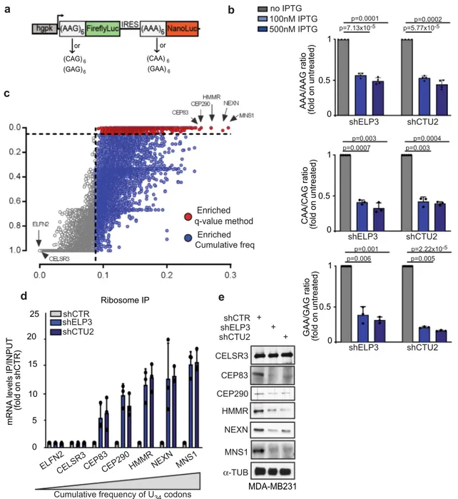
pathway. Biological phenotypes associated with modulation of
the mcm
5s
2wobble uridine tRNA modification have been
associated with decoding of specific codon during translation
elongation, mainly of three codons AAA, GAA, and CAA (U
34-codons)
16–19. We designed dual-luciferase constructs encoding
six in frame repetitions of U
34-codons (xAA) upstream of
NanoLuc and six in frame repetitions of the corresponding
synonymous codon (xAG), whose decoding does not require
U
34-tRNA modification, upstream of FireflyLuc (Fig.
1
a and
Supplementary Fig. 1a). Depletion of the U
34-enzymes, ELP3,
or CTU2, strongly decreased NanoLuc activity but did not
impact FireflyLuc activity (Fig.
1
b and Supplementary Fig.
1b-d), and no effect was seen using a construct lacking the codon
repetitions (Supplementary Fig. 1e-f). Together with previous
evidences
19,20,27–29, these data indicate that decoding the -AA
ending codons requires U
34-enzymes.
In order to identify the mechanisms linking codon-dependent
translational optimization and protein fate, we computed the
frequency bias across the human genome of the three U
34-codons
(i.e., AAA, GAA, and CAA). To this end, we calculated a q value
for each U
34-codon in every gene of the human orfeome in order
to define codon enrichment in a statistical manner (q < 0.05, see
the
“Methods” section). Interestingly, 4331 genes display a high
frequency of at least one of the three codons (q value <0.05;
Supplementary Fig. 1g), among which 608 genes are strongly
enriched in all three codons (Fig.
1
c and Supplementary Fig. 1h).
In line with previously published evidence in S. cerevisae, genes
enriched in these codons significantly cluster in biological
pathways related to the cell cycle
30,31(Supplementary Fig. 1i,
Supplementary Data1) as well as in specific protein families,
including kinesins (Supplementary Fig. 1j). As expected,
deple-tion of ELP3 and CTU2, two of the U
34-enzymes
12,13,22, leads to a
strong increase in ribosome occupancy (i.e., defect in translation
elongation) at the top
five transcripts enriched in U
34-codons
(i.e., CEP83, CEP290, HMMR, NEXN, and MNS1; Fig.
1
d and
Supplementary Fig. 1k). This correlates with a strong decrease in
their protein expression levels (Fig.
1
e). No difference in the
expression levels of the corresponding mRNAs was detected
(Supplementary Fig. 1l). These data indicate that high abundance
of U
34-codons in transcripts predicts dependency toward U
34-enzymes for correct mRNA translation and corresponding
protein expression.
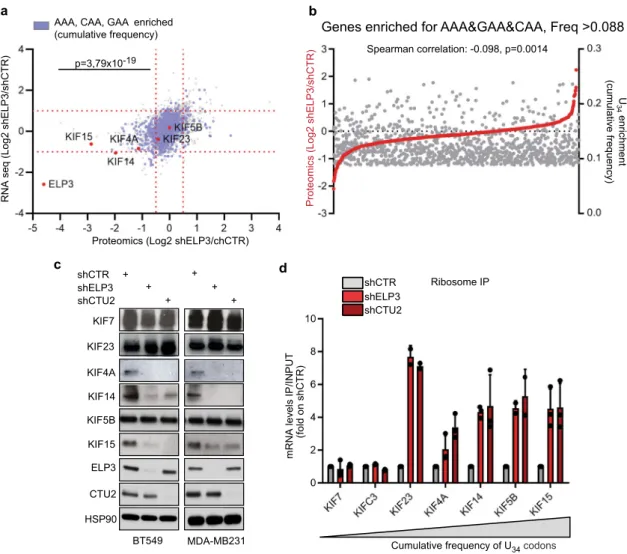
mRNA codon content is not suf
ficient to predict protein
abundance upon loss of the U
34-enzymes. In order to assess if
the enrichment of U
34-codons in mRNAs is sufficient to predict
their protein expression levels upon loss of U
34-enzymes, we
performed RNA-seq and label-free quantitative proteomics upon
ELP3 depletion (Fig.
2
a and Supplementary Fig. 2a-b).
Surpris-ingly, even though downregulated proteins were significantly
enriched in U
34-codons in their corresponding mRNA as
com-pared to the genome ORFs content (χ
2test, p
= 3.79 × 10
−19;
Fig.
2
a and Supplementary Data 2), a large number of genes
enriched in U
34-codons remained unchanged in their protein
changes at the mRNA steady-state levels were detected for U
34-enriched genes upon ELP3 depletion (Supplementary Fig. 2b and
Supplementary Data 2). In agreement with our codon-content
analysis, many members of the kinesin family were found
downregulated at the protein, but not the mRNA, level in the
absence of ELP3 (i.e., KIF4A, KIF14, KIF15, KIF13B, KIF5A,
KIF5C, and KIF1B; gene family enrichment analysis, p
= 1.10 ×
10
−13) (Fig.
2
c and Supplementary Fig. 2d-e). Surprisingly, other
kinesins, such as KIF5B or KIF23, even though strongly enriched
in U
34-codons (i.e., KIF5B: 17.11% and KIF23: 13.4%), were not
downregulated in ELP3-depleted cells (Fig.
2
a, c and
Supple-mentary Data 2). Strikingly, ribosomes invariably accumulate at
kinesin transcripts enriched in U
34-codons (i.e., KIF4A, KIF14,
KIF15, KIF5B, and KIF23), independently of their protein
expression levels, and with no change in the corresponding
mRNA levels (Fig.
2
d and Supplementary Fig. 2f). Therefore, the
U
34-codon content of mRNAs is predictive of changes in
ribo-some translational elongation, but is not sufficient to predict
protein expression output in the absence of U
34-enzymes.
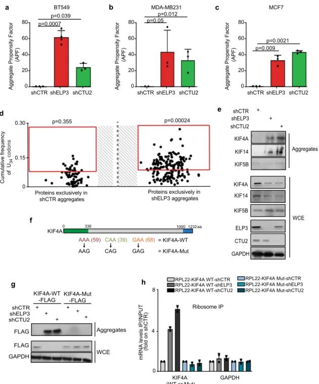
Loss of U
34-enzymes leads to protein aggregation in a
codon-dependent manner. Defective translation elongation has been
associated with increased protein misfolding and aggregation
32.
Accordingly, loss of U
34-enzymes leads to increased protein
b
(CAG)6 (GAG)6 (CAA)6 (GAA)6 or ora
AAA/AAG ratio (fold on untreated) 0 0.5 1 shELP3 shCTU2 p=0.0001 p=7.13x10⁻⁵ p=0.0002 p=5.77x10⁻⁵c
CAA/CAG ratio (fold on untreated) 0 0.5 1 shELP3 shCTU2 p=0.003 p=0.0007 p=0.0004 p=0.003 GAA/GAG ratio (fold on untreated) 0 0.5 1 shELP3 shCTU2 p=0.001 p=0.006 p=2.22x10⁻⁵ p=0.005 100nM IPTG 500nM IPTG no IPTG MDA-MB231 shCTR shELP3 shCTU2 + + CELSR3 CEP290 α-TUB HMMR NEXN + CEP83 MNS1e
mRNA levels IP/INPUT
(fold on shCTR) 5 10 20 ELFN2 15
CELSR3CEP83CEP290 HMMRNEXN MNS1 0
25 shCTR
shELP3 shCTU2
Cumulative frequency of U34 codons
d
Ribosome IPEnriched q-value method Enriched Cumulative freq
Fig. 1 mRNA codon content predicts translation dependency on U34-enzymes. a Schematic representation of the dual-luciferase construct. b NanoLuc/ Firefly luminescence ratio of MDA-MB231 cells stably expressing the indicated constructs and depleted or not of ELP3 or CTU2 upon IPTG treatment. Data are normalized on non-treated conditions (IPTG 0μM, n = 3–4 independent experiments, two-sided t-test, data are mean + s.d). c AAA, CAA, and GAA codon frequency andq value were computed for each human gene, red and blue dots represent genes enriched in the three U34-codons (AAA&GAA&CAA q value <0.05 = red dots; AAA&GAA&CAA cumulative frequency >0.088 = blue dots). d qRT-PCR after ribosome immunoprecipitation in MDA-MDMB231 cells.n = 2 independent experiments, two-sided t-test, data are mean + s.d. e Levels of indicated proteins were determined in cells by western blot (n = 1 replicate).
aggregation in a variety of cell lines (Fig.
3
a–c)
19,33. We surmised
that defects in ribosome kinetics upon loss of U
34-enzymes might
result in defective protein expression only for proteins that are
integrated into aggregates for clearance. To test this hypothesis,
we purified the aggregates formed in ELP3-depleted cells and we
assessed their protein content by quantitative proteomics
(Fig.
3
d). Strikingly, proteins exclusively detected in aggregates
upon ELP3 depletion were strongly biased toward U
34-codons in
their corresponding mRNAs (p
= 0.00024 vs p = 0.355 in control
aggregates compared to human orfeome). Also, previously
iden-tified targets of U
34-enzymes, KIF4A and KIF14, but not KIF5B,
were detected by western blot in protein aggregates upon loss of
U
34-enzymes (Fig.
3
e). Of note, KIF4A and KIF14 protein levels
were partially rescued after blocking the autophagic
flux with
chloroquine, but not after proteasome inhibition (Supplementary
Fig. 3a). Importantly, the systematic recoding of KIFA4A cDNA
(i.e., all AAA, CAA, GAA codons were mutated into their
synonymous, U
34-modification independent, codons AAG, CAG,
GAG—KIF4A-Mut, as in refs.
19,20; Fig.
3
f) abolished the
trans-lation defects observed upon U
34-enzymes depletion (Fig.
3
g),
prevented KIF4A aggregation and restored KIF4A expression
levels (Fig.
3
h). Of note, no changes in mRNA levels nor in global
aggregation propensity were observed upon ELP3 depletion
among cells overexpressing the KIF4A-WT or -Mut constructs
(Supplementary Fig. 3b-c). This shows that aggregation of protein
targets of the U
34-tRNA modification pathway (U
34-targets)
is caused by codon-dependent translational defects upon loss
of U
34-enzymes. Taken together, these data indicate that U
34-enzymes prevent the aggregation and degradation of U
34-targets
through codon-dependent regulation of mRNA translation.
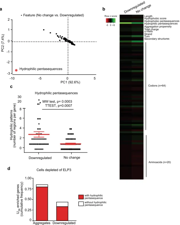
A penta-hydrophilic amino acid pattern links translation
defects to protein aggregation in targets of the U
34-tRNA
modification pathway. Surprisingly, the above analyses show
that defective translation (i.e., ribosome accumulation) in U
34-enzymes-depleted cells does not systematically correlate with
protein aggregation and with subsequent differences in protein
expression levels. Indeed, a number of genes are significantly
enriched in U
34-codons but they remain normally expressed at
the protein level in cells defective in U
34-enzymes (Figs.
1
e,
2a–c
). We surmised that additional features may dictate protein
aggregation in the context of defective codon-dependent
translation. To identify these features, we performed a series
of analyses to identify specific parameters that differentiate
proteins that are downregulated versus unchanged upon
ELP3 depletion, despite the sharing of high U
34-codon content
Fig. 2 mRNA codon content poorly predicts protein expression fate upon loss of U34-enzymes. a Dot plot of RNA-seq and proteomics of BT549 cells depleted or not of ELP3 (n = 3 independent experiments); genes enriched in U34-codons are shown in blue. U34-codons enrichment in downregulated proteins was calculated byχ2test (two-sided).b, Frequency of U
34-codons and protein expression in BT549 cells depleted of ELP3 was plotted, correlation was calculated by Spearman test (two-sided).c Levels of indicated proteins were determined in cells depleted of ELP3 or CTU2 by western blot (n = 4 replicates).d qRT-PCR after ribosome immunoprecipitation in MDA-MDMB231 cells.n = 2 independent experiments, data are mean + s.d.
(i.e., AAA, CAA, GAA; q value <0.05; Supplementary Fig. 4a).
First, we quantified features linked to mRNAs, namely,
expression levels, length, codon content, and U
34-codons
dis-tribution. No significant difference is observed between the two
groups (Supplementary Fig. 4b-e). Hence, for each protein of
the two groups, we assessed features linked to protein structure
(i.e., number of
α-helix and β-sheets), amino acid content, and
protein aggregation propensity (calculated by the Zyggregator
method
34). Again here, no statistical difference between the two
groups is observed (Supplementary Fig. 5a-c). Finally, we
Fig. 3 Loss of U34-enzymes leads to protein aggregation in a codon-dependent manner. a–c Protein aggregation was measured by FACS in BT549 (a), MDA-MD231 (b), and MCF7 (c) upon loss of ELP3 or CTU2 (n = 3 independent experiments, two-tail t-test, data are mean + s.d). d Protein content of aggregates of MCF7 cells depleted or not of ELP3 were assessed by proteomics (n = 1 independent experiment); proteins exclusively found in shELP3 or shCTR conditions are shown. Enrichment of U34-codons was assessed byχ2test (two-sided).e Levels of indicated proteins were determined by western blot in whole-cell extracts (WCE; loaded 30μg) and aggregates (isolated from >1 mg of protein extract) of MCF7 cells depleted of ELP3 or CTU2 (n = 2 replicates). f Schematic representation of KIF4A mutant. The kinesin domain (green) and the globular region (blue) of the protein are indicated.g Protein levels of whole-cell extracts (WCE) and aggregates of MCF7 cells stably overexpressing FLAG-RPL22 and the identified KIF4A mutants in the presence or absence of ELP3 or CTU2 (n = 3 replicates). h qRT-PCR after ribosome immunoprecipitation of indicated cells. n = 2 independent experiments, data are mean+ s.d.further evaluated each of the parameters linked to aggregation
propensity separately. Strikingly, we found that the presence of
patterns of
five hydrophilic amino acids significantly segregated
proteins that are downregulated upon depletion of ELP3 (p
=
0.0003; Fig.
4
a–c), while total charge, hydrophobic score,
and patterns of
five hydrophobic amino acids was similar
between both groups (Supplementary Fig. 5d-f). In line with
these data, proteins harboring hydrophilic pentasequences very
significantly cluster in both ELP3-downregulated proteins as
well as in proteins specifically incorporated into aggregates
upon ELP3 depletion, indicating a central role of the identified
motif in promoting inclusion into aggregates of U
34-target
proteins for degradation (Fig.
4
d, Supplementary Data 3).
Together, these analyses highlighted that the presence of
hydrophilic pentasequences is a key feature to predict protein
integration into aggregates upon depletion of U
34-enzymes.
Fig. 4 A penta-hydrophilic amino acid pattern links translation defects to protein aggregation in U34-enzymes depleted cells. a, b Principal component analysis (a) and heat map representation (b) of the assessed features in U34-enriched (q value <0.05) downregulated or unaffected proteins upon depletion of ELP3.c Number of hydrophilic pentasequences was assessed in U34-enriched (q value <0.05) downregulated (n = 58) or unaffected proteins (n = 56) upon depletion of ELP3. Two-tails t-test and Mann–Whitney U (MW) test were performed to assess significance. Data are mean +/− SEM. d Presence of hydrophilic pentasequences was assessed in U34-enriched (frequency of U34-codon >0.088) downregulated proteins (BT549) or in aggregates (MCF7) cells depleted of ELP3.
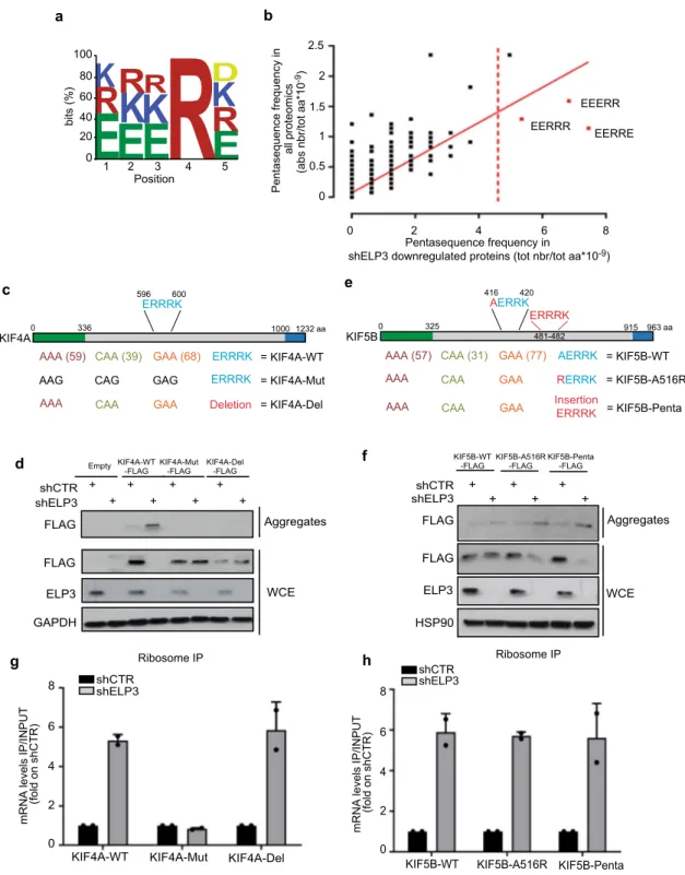
Specific hydrophilic motifs dictate protein aggregation upon
loss of U
34-enzymes. Having uncovered the importance of
hydrophilic pentasequence patterns in the definition of protein
targets of the U
34-tRNA modification pathway, we combined
all identified pentasequences and highlighted a consensus motif
[EKR]-[EKR]-[EKR]-R-[DEKR] (Fig.
5
a). This motif is present in
10.16% of the protein-coding genes (Supplementary Fig. 6a) and
is strongly associated with the frequency of U
34-codons in genes
(χ
2test: p
= 9.84 × 10
−48; Supplementary Data 4). Furthermore, the
motif discriminates proteins that are downregulated (χ
2test: p
=
3.88 × 10
−18; Fig.
5
b) and directed into aggregates upon ELP3
depletion (χ
2test: p
= 8.4 × 10
−8; Supplementary Data 4). Finally,
the combination of the two features, the U
34-codon content and
the presence of hydrophilic motifs, better predicts protein
expres-sion levels downstream of the U
34-tRNA modification pathway
(χ
2test downregulated proteins vs. no change: p
= 6.58 × 10
−10;
Fig. 5 Specific hydrophilic motifs dictate protein aggregation upon loss of U34-enzymes. a Identified hydrophilic motif. b Assessment of the specific hydrophilic pentasequences (in red) overrepresented in proteins downregulated upon ELP3 depletion. Linear regression is plotted.c, e Schematic representation of KIF4A (c) and KIF5B (e) proteins and mutants. Hydrophilic motifs are indicated. The kinesin domain (green) and the globular region (blue) of the proteins are indicated.d, f Levels of indicated proteins were determined by western blot in whole-cell extract (WCE) and aggregates of MCF7 cells overexpressing the indicated mutants of KIF4A (d) and KIF5B (f) and depleted of ELP3 (n = 2 replicates). g, h qRT-PCR after ribosomeSupplementary Data 4). Of note, we did not
find any correlation
between the presence and the distribution of the motifs and protein
length (Supplementary Fig. 6b-c). Finally, we computed the
con-servation scores of the identified motif (Supplementary Fig. 6d)
against random sequences of
five contiguous amino acids across 10
mammalian species (Supplementary Fig. 6e). Interestingly, we show
that the motif is conserved (p
= 7.05 × 10
−20) suggesting a selection
pressure during evolution.
To further assess the biological relevance of the identified motif
in determining protein fate upon U
34-tRNA
modification-dependent translation elongation defects, we took advantage of
two kinesin proteins whose mRNAs are both highly enriched in
U
34-codons: (1) KIF4A, which harbors a consensus motif (i.e.,
ERRRK; Fig.
5
c), and is detected in protein aggregates upon
depletion of U
34-enzymes (Fig.
5
d); and (2) KIF5B, which does not
contain any consensus motif (Fig.
5
e), is not downregulated and
therefore not detected in protein aggregates upon depletion of U
34-enzymes (Fig.
5
f). We deleted the hydrophilic motif in the KIF4A
sequence and inserted one in the KIF5B sequence and we assessed
protein expression of the constructs upon depletion of U
34-enzymes. Strikingly, the deletion of the ERRRK motif in KIF4A
(KIF4A-Del; Fig.
5
c) is sufficient to rescue its expression level and to
prevent KIF4A aggregation in the absence of ELP3 (Fig.
5
d). This
effect does not rely on changes in mRNA translation elongation
(i.e., ribosome occupancy), which remains unaffected by the
absence of the motif (Fig.
5
g and Supplementary Fig. 7a).
Conversely, the addition of the ERRRK motif in the KIF5B cDNA
sequence (either by a single amino acid
permutation—KIF5B-A516R—or by insertion of the whole ERRRK
sequence—KIF5B-Penta; Fig.
5
e) was sufficient to promote KIF5B protein aggregation
and downregulation upon ELP3 depletion (Fig.
5
f). Again, the
addition of the motif does not affect KIF5B mRNA translation
elongation (Fig.
5
h and Supplementary Fig. 7b). These data indicate
that the presence of the hydrophilic motif is necessary to promote
protein aggregation and downregulation of genes that undergo
translation elongation defects upon depletion of U
34-enzymes.
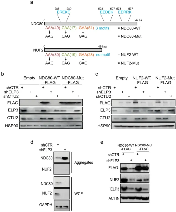
We then wanted to assess if the presence of a hydrophilic
motif could discriminate the proteins whose downregulation is
directly dependent on the loss of the U
34-tRNA modification (i.e.,
proteins incorporated in aggregates in a codon-dependent
manner) from those that are indirectly downregulated in these
conditions. To this aim, we selected two proteins that share
similar U
34-codons frequency, are downregulated upon ELP3
depletion and are part of the same kinetochore complex
35. We
generated U
34-codon mutants cDNA of NCD80 and NUF2 (as
before; Fig.
6
a) and we assessed their expression levels upon U
34-enzymes depletion. Surprisingly, even though NCD80 and NUF2
are in the same protein complex, share a similar frequency of U
34-codons and display comparable ribosome accumulation at their
mRNA upon U
34-enzymes loss (Supplementary Fig. 7c), the
systematic recoding of U
34-codons by their synonymous only
rescued NCD80 expression (Fig.
6
b) but did not impact NUF2
levels (Fig.
6
c). Importantly, only NCD80 harbors hydrophilic
motifs in its sequence (Fig.
6
a) and is therefore detected in
protein aggregates upon ELP3 depletion (Fig.
6
d). Conversely,
NUF2 sequence does not contain any hydrophilic motif and it is
never detected in protein aggregates upon ELP3 depletion
(Fig.
6
d). Hence, NUF2 downregulation is partially reverted
upon expression of the NDC80-Mut construct (Fig.
6
e). This
indicates that NUF2 downregulation likely results from the loss of
its partner NDC80 and not from an intrinsic dependence on the
U
34-tRNA modification pathway. These data reveal that the
presence of the hydrophilic motif discriminates between direct
(i.e., NDC80) and indirect (i.e., NUF2) targets of the U
34-tRNA
modification pathway. All together this set of data demonstrates
that the presence of hydrophilic motifs is necessary to mediate
protein aggregation and clearance of U
34-targets upon loss of
U
34-enzymes.
The charged/ampholytic nature of the hydrophilic motif
mediates protein aggregation upon depletion of U
34-enzymes.
The identified hydrophilic pentamer is defined by its unique
amino acid composition, exclusively enclosing charged
resi-dues, but also presenting an ampholytic nature (i.e., has the
characteristics of both an acid and a base). We surmised that
this unique feature might be the reason why proteins are
sub-jected to aggregation upon depletion of U
34-enzymes. In line
with this hypothesis, we tested two non-exclusive models that
may explain the role of the hydrophilic motif in protein
aggregation. First, the ampholytic nature of the motif may drive
interaction with the ribosome exit tunnel during translation
elongation. The ribosome exit tunnel is a negatively charged
cavity from which the nascent proteins arise
36. Different factors
are involved in facilitating or opposing the exit of the nascent
peptide from the ribosome tunnel, among these, tRNA
avail-ability, protein secondary structure formation and the presence
of charged amino acids in the nascent peptide
37–39. We recently
computed the ribosome exit tunnel electrostatics in a model to
quantify the energetic needs required to effectively exit nascent
peptides from the ribosome tunnel
40. Using this model, we
found that the presence of the hydrophilic motif in the KIF4A
protein sequence represents a region of high energetic needs in
order to exit the ribosome tunnel and to progress toward
translation elongation (Fig.
7
a). Conversely, the deletion of the
hydrophilic motif in the KIF4A protein sequence (i.e., in
KIF4-Del), which prevents KIF4A aggregation upon depletion of U
34-enzymes, alters the local energetic barrier, which is predicted
to favor ribosome movement across this region (Fig.
7
b). This
observation indicates that the presence of the hydrophilic motif
brings an additional energetic barrier during translation
elon-gation, which may be the cause of protein misfolding and
aggregation.
Second, recent evidences describe the propensity of low
complexity regions (LCR) rich in charged amino acids to regulate
protein phase behavior, to promote liquid–liquid phase
separa-tion (LLPS) and to trigger protein aggregasepara-tion
41–45. Therefore, we
speculated that the ampholytic nature of the hydrophilic motif
might trigger LLPS and auto-aggregation among the U
34-target
proteins, upon U
34-enzymes depletion. We visualized KIF4A-WT
or KIF4A-Del (i.e., that does not harbor the hydrophilic motif)
proteins in MCF7 cells by immunofluorescence. Interestingly,
upon depletion of U
34-enzymes, KIF4A-WT aggregates in small
punctuated structures, similar to SHP2 mutants-LLPS associated
structures
46(Fig.
7
c). Strikingly, KIF4A-Del is never found in
these structures in U
34-enzymes depleted cells (Fig.
7
c). We also
assessed the amyloid aggregation propensity in KIF4A-WT and
KIF4A-Del using the TANGO algorithm
47. Interestingly, the
hydrophilic motif we identified in KIF4A is predicted to have
high propensity to undergo amyloid aggregation (Fig.
7
d).
Moreover, its deletion (i.e., in KIF4A-Del) causes a net reduction
in amyloid aggregation propensity in KIF4A, as measured by the
total area under the curve (KIF4A-WT
= 163231 and
KIF4A-Del= 146070), and a change in the maximal amyloid aggregation
peak (KIF4A-WT max peak—intensity 34359—is found in bin 9
that harbors the motif, while KIF4A-Del max peak—intensity
24352—is found in bin 14) (Fig.
7
d). Conversely, deletion of the
motif did not affect KIF4A general aggregation propensity
(Supplementary Fig. 8a) nor its presence was found associated
with aggregation propensity calculated by the Zyggregator
method (Supplementary Fig. 8b). This indicates that the presence
of the charged/ampholytic hydrophilic motif may trigger
aggregation through changes in protein phase behavior and LLPS
upon depletion of U
34-enzymes.
Discussion
Recently, the role of tRNA biology in health and disease has
gained an increasing amount of attention
11,48,49. The
develop-ment of ribosome sequencing techniques allowed to assess the
exact role of tRNA modifications, such as mcm
5s
2U
34
-tRNA
modification, in translation regulation and specific codon
decoding
16,17,19. Currently, it is assumed that impairments in
ribosome kinetics at mRNAs are directly reflected in changes in
expression of the corresponding proteins. Nevertheless, very few
studies focus on the mechanisms linking codon-specific ribosome
pausing and protein homeostasis. Specifically, how perturbation
of codon-dependent-translation impacts subsequent protein
expression output remains poorly understood. Surprisingly, here
we show that codon-dependent translation elongation defects
caused by the depletion of U
34-enzymes are not systematically
associated with changes in protein expression. While translation
elongation defects upon perturbation of U
34-tRNA modification
Fig. 6 The presence of hydrophilic motifs discriminates between direct and indirect targets of the U34-tRNA modification pathway. a Schematic representation of the NDC80 and NUF2 protein sequences. Hydrophilic motifs are indicated in blue.b Protein levels of indicated proteins in cells overexpressing control or NDC80-WT-FLAG or NDC80-Mut-FLAG constructs in the presence or absence of ELP3 or CTU2 (n = 2 replicates). c Protein levels of indicated proteins in cells overexpressing control or NUF2-WT-FLAG or NUF2-Mut-FLAG constructs in the presence or absence of ELP3 or CTU2 (n = 2 replicates). d Levels of indicated proteins were determined by western blot in whole-cell extracts (WCE) and aggregates of MCF7 cells depleted of ELP3 (n = 2 replicates). e Levels of indicated proteins were determined by western blot in cells overexpressing NDC80-WT-FLAG or NDC80-Mut-FLAG constructs in the presence or absence of ELP3 (n = 1 replicate).
are strictly dependent on codon content, the consequences of
these defects on protein output are determined by other features.
We identify hydrophilic amino acid motifs as essential to dictate
protein aggregation and subsequent degradation in contexts
of defective translation elongation at U
34-codons. Therefore, we
propose a model in which codon-dependent regulation of
translation elongation by wobble tRNA modification and
maintenance of protein integrity are two independent processes
(see the model in Fig.
8
). Importantly, the previously identified
protein targets of the U
34-tRNA modification pathway (i.e.,
SOX9
25, DEK
20, HIF1α
19, and hnRNPQ
26; Fig.
1
c–e) all harbor
at least one hydrophilic motif in their sequences, supporting
the model here proposed (Supplementary Fig. 8c). Of note, a
portion of genes enriched in U
34-codons and downregulated
upon depletion of U
34-enzymes do not harbor hydrophilic
motifs and are not directed toward aggregation (for instance
NUF2; see Supplementary Table 3). The mechanism leading to
their downregulation upon depletion of U
34-enzymes remains
to be understood.
How the presence of hydrophilic motifs mediate protein
aggregation remains to be precisely established. The motif
strongly differs from known chaperone- and
cochaperone-binding sites, which are generally hydrophobic
50. In contrast,
the here identified motif shares some similarities with the motifs
responsible for chaperone-mediated autophagy (CMA) (i.e.,
KFERQ and derivatives)
51. Nevertheless, the unbiased search for
CMA motifs
51showed no overlap with the hydrophilic motifs
identified herein, making this mechanism of degradation unlikely
Fig. 7 The ampholytic nature of the motif drives protein aggregation upon depletion of U34-enzymes. a, b Axial forces profile on nascent chain caused by the electrostatic interaction in the ribosome exit tunnel (a) wild-type KIF4A; (b) ERRRK deleted KIF4A. The axial forces profiles and local mechanical work acting on the two chains are different upon incorporation of residues 596–646 at the peptidyl-transferase center (PTC). Schematic representation of ribosome–protein interaction, on the left. c Anti-Flag immunofluorescence of MCF7 cells depleted or not of ELP3 and overexpressing WT or KIF4A-Del in control or after 6 h of treatment with 100μM chloroquine. Arrows depict KIF4A aggregates (n = 2 replicates). d Amyloid aggregation profiles of KIF4A-WT and KIF4A-Del calculated by using the TANGO algorithm.(Supplementary Fig. 8d-e). Previous studies highlighted the
potential role of hydrophilic stretches as gatekeepers for the
correct folding of nascent proteins due to their proximity to
aggregation-prone regions
52–55. Although, we do not exclude this
possibility, in the KIF4A protein sequence, the hydrophilic motif
sits in the proximity of low aggregation-prone regions. Rather,
our data show that the unique amino acid composition of the
motif may underlie protein aggregation upon U
34-enzymes
depletion. Indeed, we show that the charged and ampholytic
nature of the motif may affect phase behavior of the U
34-protein
targets, possibly triggering LLPS and consequently promoting
protein aggregation upon depletion of U
34-enzymes. For example,
the here identified motif shares physical characteristics with
fragments in the low complexity (LC) domains in the U1-70K
protein, that have been shown to drive LLPS and aggregation in
cells
41. Also, arginine-rich domains mediate LLPS via electrostatic
interactions, which therefore requires multiple proteins or motifs.
Why would the perturbation of U
34-tRNA modification and the
subsequent codon-dependent deregulation of translation favor
this mechanism is not known. It is possible that the translational
defects seen upon depletion of U
34-enzymes might result in
protein downregulation, only if the exposure of charged/
ampholytic motifs is sufficient to trigger LLPS-driven aggregation
that will lead to clearance by the lysosome.
The charged/ampholytic nature of the pentamer could trigger
aggregation also by its interaction with the ribosome exit
channel tunnel during translation elongation. Previous studies
demonstrated that factors involved in facilitating or opposing
the exit of the nascent peptide from the ribosome tunnel can
impact on protein folding
56. Interestingly, in KIF4A, the
identified hydrophilic motif represents a region of high
ener-getic needs in order to exit the ribosome tunnel and to progress
toward translation elongation (Fig.
7
c). Conversely, its deletion,
which prevents incorporation of KIF4A in aggregates upon
depletion of the U
34-enzymes (Fig.
5
d), alters the local energetic
barrier, likely favoring ribosome movement across this region
(Fig.
7
d). It is therefore tempting to speculate that the
addi-tional energetic barrier brought by the presence of the
hydro-philic motif could lead to an excessive ribosome pausing
and may be the cause of protein misfolding and aggregation.
Further work is required to further clarify these mechanisms.
Our data open the possibility that the uncoupling of translation
elongation defects and maintenance of protein homeostasis may
generally apply to other tRNA modification pathways.
Under-standing the mechanisms linking codon-dependent translation
defects to maintenance of protein expression for all the still
poorly studied tRNA modification pathways will help define the
importance of this layer of regulation in proteome expression.
Methods
Cell lines and reagents. Breast cancer cell lines (BT549, MDA-MB231, and MCF7) were from ATCC and were grown in Dulbecco’s modified Eagle’s medium (Gibco) containing 10% of fetal bovine serum, 1% glutamine, and 1%
penicillin–streptomycin. Lines were tested for mycoplasma contamination on a regular basis.
The antibodies, primers, and shRNA sequences used in this study are listed in Supplementary Data 5.
Western blot and immunoprecipitation. Cells were lysed in lysis buffer (50 mM Tris-HCl pH 7.5, containing 2 mM EDTA, 150 mM sodium chloride, 0.5% sodium deoxycholate, 50 mM sodiumfluoride, 1% NP-40, and 0.1% SDS, Complete™ proteinase inhibitor cocktail (Roche)) and protein determination was assessed using BCA Protein Assay Kit from ThermoFisher and according to manufacturer’s instructions.
qPCR. Total RNA was extracted using the E.Z.N.A. Total RNA Kit I (OMEGA Bio-Tek) according to the manufacturer’s instructions. Subsequently, 1 μg of total RNA was used to synthetize the corresponding cDNA using the RevertAid H Minus First Strand cDNA Synthesis Kit (ThermoFisher). Quantitative evaluation of selected genes was determined by SYBR-Green-based qPCR performed using the Light Cycler 480 (Roche). All determinations were performed in triplicates. The relative expression of the target gene transcript and reference gene transcript was calculated asΔΔCt.
Viral infections. For stable lentiviral shRNA infections, all constructs were purchased from Sigma. The kinesins FLAG-tag overexpression plasmids (KIF4A, KIF5B, and mutants) were generated from the pLV-Puro-hPGK plasmid by Vectorbuilder. NUF2 and NDC80 FLAG-tag overexpression plasmids and codon mutants were generated from the Lego-Ig2 plasmid by Genscript. The Lucifer-ase/Nanoluciferase constructs were derived from pLV-Puro-hPGK and gener-ated by Vectorbuilder (maps are available upon request). Lentiviral infections were carried out as previously described57. Briefly, in all cases, HEK293-LentiX producer cells were transfected with 12μg of lentiviral vector, 5 μg VSVG, and 12μg R.891 and Mirus (3:1) for 48 h. Supernatant containing virus was filtrated using a 45 µmfilter and polybrene was added to a final concentration of 8 µg/ml, and virus were added to target cells. Cells were selected for 3 days with 1μg/ml puromycin. Knockdown or overexpression efficiency was evaluated by RT-qPCR and/or by western blot analysis.
Ribosome immunoprecipitation. Ribosome pulldown was performed as pre-viously described19. Briefly, cells were stably transduced with RPL22-FLAG expressing lentiviral construct and treated for 15 min with 100μg/ml cyclohex-imide. Then, cells were lysed in 20 mM HEPES-KOH (pH 7.3), 150 mM KCl, 10 mM MgCl2, 1% NP-40, EDTA-free protease inhibitors, 0.5 mM DTT, 100μg/ml cycloheximide, and 10μl/ml rRNasin and Superasin in RNase-free water, and incubated overnight with anti-FLAG beads. After incubation, beads were washed five times with a high salt buffer (350 mM KCl) and three times with a low salt buffer (150 mM KCl). Beads were then washed in RNA extracting buffer with β-mercaptoethanol as previously described and underwent RT-qPCR. Immunofluorescence. 200000 MCF7 cells depleted or not of ELP3 were seeded and treated as indicated. Cells werefixed in 4% PAF and permeabilized in PBS supplemented with Triton 0.2%. Primary antibody was kept overnight at 4 °C in PBS supplemented with 0.1% TRITON and 5% BSA (dilution: 1/200). Appropriate secondary antibody (dilution: 1/1000) and DAPI were added for 1 h at room temperature in PBS supplemented with TRITON 0.1% and BSA 3%. Images were acquired by confocal microscopy (confocal Leica SP5).
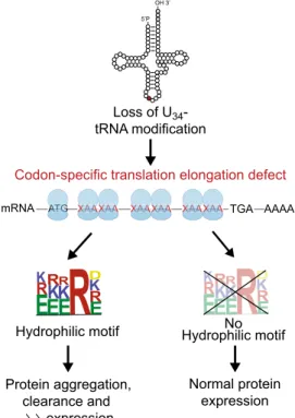
5’P OH 3’ Loss of U34 -tRNA modification ATG ATG A A A G A A GTGTG A ATG A ATG A A A GGG ATG A A G
A XAAXAAXAAXAAXAAXAAXAAXAAXAAXAAAAXAAXAAXAAXAAXAAXAAXAAXAAXAAXAAAA XAAXAAXAAXAAXAAXAAXAAXAAXAAXAAAAXAAXAAXAAXAAXAAXAAXAAXAAXAAXAAAA XAAXAAXAAXAAXAAXAAXAAXAAXAAXAAAAXAAXAAXAAXAAXAAXAAXAAXAAXAAXAAAA TGA AAAA mRNA
Codon-specific translation elongation defect
K
R
E
E
E
R
E
R
K
K
RK
R
DK
R
K
K
E
E
R
R
E
R
E
E
E
R
K
R
R
K
K
K
RR
E
E
RR
E
E
K
R
R
DR
R
Hydrophilic motif Hydrophilic motifNo
Protein aggregation, clearance and expression
Normal protein expression
Fig. 8 Proposed model. Loss of U34-enzymes invariably determines pausing and accumulation of ribosomes on transcripts rich in XAA codons. The result of this pausing on protein expression is determined by the presence of hydrophilic motifs that mediate protein aggregation and subsequent clearance. See text for details.
RNA sequencing. BT549 cells were stably depleted of ELP3 by shRNA (see the “Viral infections” section); a scramble shRNA was used as control. Total RNAs from three independent infections were extracted using TRIzol (Life Technologies), according to the manufacturer’s protocol. RNA integrity was verified on the Bioanalyser 2100 with RNA 6000 Nano chips, and RIN scores were >9 for all samples. The Illumina Truseq RNA Sample Preparation kit V2 was used to prepare libraries from 500 ng total RNAs. PolyA RNAs were purified with polyT-coated magnetic beads, chemically fragmented, and used as template for cDNA synthesis using random hexamers. cDNA ends were subsequently end-blunted, adenylated at 3′OH extremities, and ligated to indexed adaptors. Finally, the adapters ligated library fragments were enriched by PCR following Illumina’s protocol and purified. Libraries were validated on the Bioanalyser DNA 1000 chip and quantified by qPCR using the KAPA library quantification kit. Sequencing was performed on HiSeq2000 in paired-end 2 × 100 base protocol. For data analysis, the nf-core-rnaseq pipeline was used.
Proteomic study. BT549 cells depleted or not of ELP3 by shRNA were lysed in SDS 2% buffer containing 50 mM Tris-HCl pH 8, 150 mM sodium chloride, 10 mM sodiumfluoride, and 1 mM trisodium phosphate, Complete™ proteinase inhibitor cocktail (Roche). At least 1 mg of each sample was sent for mass spec-trometry analysis using LC/MS/MS (MsBioworks, LLC). Samples were TCA pre-cipitated overnight at−20 °C in 90/10 acetonitrile/water incorporating 20% TCA. Precipitates were washed, pelleted, and resuspended in 5 M urea, 50 mM Tris-HCl, pH 8.0, Complete™ proteinase inhibitor cocktail (Roche) and quantitated by Qubit fluorometry (Life Technologies). A 50 µg aliquot per sample was reduced using dithiothreitol, alkylated with iodoacetamide, and then subjected to digestion with trypsin (Promega) for 18 h at 37 °C at an enzyme-to-substrate ratio of 1:50. The digestion was quenched with formic acid and peptides cleaned using solid-phase extraction (SPE) using the Empore C18 plate (3 M). A 2 µg aliquot of each sample was analyzed by nano LC/MS/MS with a Waters M-Class HPLC system interfaced to a Fusion Lumos tandem mass spectrometer (ThermoFisher). Peptides were loaded on a trapping column and eluted over a 75 µm analytical column at 350 nL/ min; both columns were packed with Luna C18 resin (Phenomenex). A 4 h gra-dient was employed. The mass spectrometer was operated in data-dependent mode, with MS and MS/MS performed in the Orbitrap at 60,000 FWHM resolu-tion and 15,000 FWHM resoluresolu-tion, respectively. APD was turned on. The instrument was run with a 3-s cycle for MS and MS/MS. The experiment was done in triplicate.
Thermo RAWfiles were processed using MaxQuant v1.6.2.3 (Max Planck Institute). Production data were searched against the combined forward and reverse Swissprot H. sapiens protein database using Andromeda. Data werefiltered 1% protein and peptide-spectrum match (PSM) level false discovery rate (FDR) and requiring at least one unique peptide per protein. Label-free protein quantitation was based on the extracted ion chromatograms of detected peptides; data were normalized using the embedded LFQ algorithm.
Protein aggregates determination. Protein aggregates formation was monitored by FACS using PROTEOSTAT® Aggresome Detection Kit (ENZO) according to manufacturer instructions and as previously described19. Fluorescence was mea-sured by FACS by FL3 channel. Aggresome propensity factor (APF) was calculated using the formula: 100*(mean fluorescence intensity depleted − mean fluorescence intensity control)/meanfluorescence intensity depleted.
Isolation of protein aggregates was performed as previously described19. Briefly, cells were pelleted, snap-frozen in liquid nitrogen, and resuspended in lysis buffer (20 mM Na-phosphate, pH 6.8, 10 mM DTT, 1 mM EDTA, 0.1% Tween, 1 mM PMSF, protease inhibitor cocktail [Roche] and DNAse I). Lysates were kept 20 min at room temperature and sonicated. After centrifugation at 200 × g for 20 min at 4 ° C, supernatants were collected and protein dosage performed. Protein extracts were then centrifuged for 20 min 16,000 × g at 4 °C. Pellets were washed (20 mM Na-phosphate, pH 6.8, 1 mM PMSF, protease inhibitor cocktail, 2% NP-40) and sonicated. Final pellets were washed with buffer with no NP-40 and sonicated again. For protein detection by western blot, pellets were resuspended in 1% SDS.
For proteomics analysis, aggregates were purified from the cytoplasmic fraction after cyto-nuclear extraction. Briefly, cells were lysate with cytoplasmic lysis buffer (1 M Tris-HCl pH 7.9, 0.2 CaCl2, 0.25 M EDTA, 2 M MgCl2, protease inhibitors) and supernatant was collected after centrifugation (4 °C for 10 min at 17,000 × g). After aggregates isolation, pellets were washed twice in 1× PBS, and analyzed by LC/MS/MS. Luminescence. Luminescence of indicated stably transduced MDA-MB231 cell lines was measured using Promega Dual-Luciferase® Reporter Assay according to manufacturer instructions. Data were expressed as fold of nanoluc/firefly ratio. Computational analysis of gene-specific codon usage (GSCU) patterns. We downloaded 23,099 homo sapiens gene sequences from the human genome data-base (HGD), and then analyzed each gene using our GSCU algorithm. The GSCU algorithm interrogated a gene from the start to the stop codon. The frequency of a specific codon (i.e., AAA, GAA, and CAA) used in a gene was counted and further compared with the codon frequency in another gene, which was randomly picked up. 10,000 times comparison was done. By such comparison we got the p-value,
which means the possibility that the randomly picked up gene has higher frequency of that specific codon than the interest one. We then used q package with the R program, and we generated the q value for each gene. Using q= 0.05 as cut off, we picked up the genes enriched for the U34-codons AAA, GAA, and CAA. Codon enrichment analysis. Genes clustering was performed byfiltering for statistical increase in specific codons content (q < 0.05). Genes with AAA, CAA, and GAA q value <0.05 subsequently underwent Toppgene analysis in order to uncover enriched biological process and gene families.
Statistical analysis. Gene set enrichment analysis was performed using Toppfun software and GO_biological process database. A FDR q value lower than 0.05 was taken into account. Bubble graphs were created in excel: distance between the clusters was calculated as the frequency of overlapping genes between clusters. The first 30 significant pathways were considered.
Distribution of U34-codons across mRNAs was assessed by computing the U34 -codons present in genes. To this end, we divided into 100 bins the 116 genes of interest and calculated the frequency of the U34codons for each bin.
Kolmogorov–Smirnov test was performed to assess significance.
Principal component analyses were performed using the FactoMineR and Factoextra R packages. The script is available upon request. Chi-square test was performed to assess significance.
Aggregation propensity was evaluated by two methods: (1) the Zyggregator method34, (2) the TANGO algorithm47. Aggregation by the zyggregator method was calculated by using the log(k)= α0+ αhydr+ αss+ αch+ αpatformula; where α0indicates pH and temperature (here set to 0),αhydrindicates the Hydrophobic score calculated as the sum of the hydrophobic value from ref.58.αssindicated the absolute number of secondary structures such asα-helix and β-sheets according to Uniprot database,αchindicates the total charge of each protein calculated by adding the net charge of+1 or −1 for positively or negatively charged amino acids, respectively.αpatindicates a pattern offive consecutive hydrophobic or hydrophilic residues in the sequence; a value of+1 was given to each pentasequence. Aggregation calculated by the TANGO algorithm was performed assuming the following parameters: pH= 7; temperature = 298.15 K; ionic strength is 0.02 M; concentration= 1 M; no protection of N and C-term. Both beta-sheet aggregation and amyloid aggregation were assessed.
Analysis of the chaperone-mediated autophagy motifs was done by the KFERQ-finder software51.
The mapping of the electrostatic potential within the ribosomal exit tunnel and the energy needed by the nascent peptide to exit the tunnel was performed as in ref.40. Motif search. Hydrophilic pentasequences (n= 161) were aligned by ClustaIW in UNIGE tofind a consensus motif. Motif search ([EKR]-[EKR]-[EKR]-R-[DEKR]) in different datasets was performed by Motif search tool by Genome.net.
For the conservation analysis, 1466 multiple-alignments were conducted for each subset of 10 orthologs. The human protein was used as source for all the 9 target mammalian orthologs in order to be aligned using the CLUSTAL-W algorithm in Bio-Python. Conservation scores for all the hydrophilic
pentasequences and for random sequence of 5 contiguous residues were calculated (Shannon entropy conservation score).
Reporting summary. Further information on research design is available in the Nature Research Reporting Summary linked to this article.
Data availability
Proteomics data are available at ProteomeXchange Consortium (PXD019590; DOI: 10.6019/PXD019590 and PXD019620; DOI: 10.6019/PXD019620) and RNA-seq data are
available at ArrayExpress under accession codeE-MTAB-9206. Open access databases
used in this work include Swissprot (H. sapiens protein database,https://www.uniprot.
org/uniprot/?query=*&fil=organism%3A%22Homo+sapiens+%28Human%29+% 5B9606%5D%22+AND+reviewed%3Ayes); Human genome database (https://www.ncbi. nlm.nih.gov/grc/human), GO_biological process database (http://geneontology.org/). The
data supporting thefindings of this study are available from the corresponding authors
upon reasonable request. Source data are provided with this paper.
Received: 18 May 2020; Accepted: 5 March 2021;
References
1. Kim, S. J. et al. Translational tuning optimizes nascent protein folding in cells. Sciencehttps://doi.org/10.1126/science.aaa3974(2015).
2. Yu, C. H. et al. Codon usage influences the local rate of translation elongation to regulate co-translational protein folding. Mol. Cellhttps://doi.org/10.1016/j. molcel.2015.07.018(2015).
3. Collart, M. A. & Weiss, B. Ribosome pausing, a dangerous necessity for co-translational events. Nucleic Acids Res.https://doi.org/10.1093/nar/gkz763
(2019).
4. Thommen, M., Holtkamp, W. & Rodnina, M. V. Co-translational protein folding: progress and methods. Curr. Opin. Struct. Biol.https://doi.org/ 10.1016/j.sbi.2016.11.020(2017).
5. Pechmann, S. & Frydman, J. Evolutionary co nservation of codon optimality reveals hidden signatures of cotranslational folding. Nat. Struct. Mol. Biol.
https://doi.org/10.1038/nsmb.2466(2013).
6. Zhang, G. & Ignatova, Z. Folding at the birth of the nascent chain: Coordinating translation with co-translational folding. Curr. Opin. Struct. Biol.https://doi.org/10.1016/j.sbi.2010.10.008(2011).
7. Patil, A. et al. Translational infidelity-induced protein stress results from a deficiency in Trm9-catalyzed tRNA modifications. RNA Biol. 9, 37–41 (2012). 8. Drummond, D. A. & Wilke, C. O. Mistranslation-induced protein misfolding as a dominant constraint on coding-sequence evolution. Cellhttps://doi.org/ 10.1016/j.cell.2008.05.042(2008).
9. Stein, K. C. & Frydman, J. The stop-and-go traffic regulating protein biogenesis: How translation kinetics controls proteostasis. J. Biol. Chem.
https://doi.org/10.1074/jbc.REV118.002814(2019).
10. Balchin, D., Hayer-Hartl, M. & Hartl, F. U. In vivo aspects of protein folding and quality control. Sciencehttps://doi.org/10.1126/science.aac4354(2016). 11. Pereira, M. et al. Impact of tRNA modifications and tRNA-modifying
enzymes on proteostasis and human disease. Int. J. Mol. Sci.https://doi.org/ 10.3390/ijms19123738(2018).
12. Huang, B., Johansson, M. J. O. & Byström, A. S. An early step in wobble uridine tRNA modification requires the Elongator complex. RNA 11, 424–436 (2005).
13. Leidel, S. et al. Ubiquitin-related modifier Urm1 acts as a sulphur carrier in thiolation of eukaryotic transfer RNA. Nature 458, 228–232 (2009). 14. Songe-Møller, L. et al. Mammalian ALKBH8 possesses tRNA
methyltransferase activity required for the biogenesis of multiple wobble uridine modifications implicated in translational decoding. Mol. Cell. Biol. 30, 1814–1827 (2010).
15. Dewez, M. et al. The conserved Wobble uridine tRNA thiolase Ctu1-Ctu2 is required to maintain genome integrity. Proc. Natl Acad. Sci. USA 105, 5459–5464 (2008).
16. Zinshteyn, B. & Gilbert, W. V. Loss of a conserved tRNA anticodon modification perturbs cellular signaling. PLoS Genet. 9, e1003675 (2013). 17. Nedialkova, D. D. & Leidel, S. A. Optimization of codon translation rates via
tRNA modifications maintains proteome integrity. Cell 161, 1–13 (2015). 18. Laguesse, S. et al. A dynamic unfolded protein response contributes to the
control of cortical neurogenesis. Dev. Cell 35, 553–567 (2015). 19. Rapino, F. et al. Codon-specific translation reprogramming promotes
resistance to targeted therapy. Nature 558, 605–609 (2018). 20. Delaunay, S. et al. Elp3 links tRNA modification to IRES-dependent
translation of LEF1 to sustain metastasis in breast cancer. J. Exp. Med. 213, 2503–2523 (2016).
21. Leidel, S. et al. Ubiquitin-related modifier Urm1 acts as a sulphur carrier in thiolation of eukaryotic transfer RNA. Nature 458, 228–232 (2009). 22. Rapino, F., Delaunay, S., Zhou, Z., Chariot, A. & Close, P. tRNA modification:
is cancer having a wobble? Trends Cancer 3, 249–252 (2017).
23. Freeman, S. et al. Proteostasis is essential during cochlear development for neuron survival and hair cell polarity. EMBO Rep.https://doi.org/10.15252/ embr.201847097(2019).
24. Hawer, H. et al. Roles of elongator dependent tRNA modification pathways in neurodegeneration and cancer. Geneshttps://doi.org/10.3390/genes10010019
(2019).
25. Ladang, A. et al. Elp3 drives Wnt-dependent tumor initiation and regeneration in the intestine. J. Exp. Med. 212, 2057–2075 (2015). 26. Xu, S. et al. Genome-wide CRISPR screen identifies ELP5 as a determinant of
gemcitabine sensitivity in gallbladder cancer. Nat. Commun.https://doi.org/ 10.1038/s41467-019-13420-x(2019).
27. Chan, C. T. Y. et al. Reprogramming of tRNA modifications controls the oxidative stress response by codon-biased translation of proteins. Nat. Commun. 3, 937 (2012).
28. Endres, L., Dedon, P. C. & Begley, T. J. Codon-biased translation can be regulated by wobble-base tRNA modification systems during cellular stress responses. RNA Biol. 12, 603–614 (2015).
29. Goffena, J. et al. Elongator and codon bias regulate protein levels in mammalian peripheral neurons. Nat. Commun.https://doi.org/10.1038/ s41467-018-03221-z(2018).
30. Patil, A. et al. Increased tRNA modification and gene-specific codon usage regulate cell cycle progression during the DNA damage response. Cell Cycle 11, 3656–3665 (2012).
31. Frenkel-Morgenstern, M. et al. Genes adopt non-optimal codon usage to generate cell cycle-dependent oscillations in protein levels. Mol. Syst. Biol.
https://doi.org/10.1038/msb.2012.3(2012).
32. Jacobson, G. N. & Clark, P. L. Quality over quantity: optimizing co-translational protein folding with non-’optimal’ synonymous codons. Curr. Opin. Struct. Biol.https://doi.org/10.1016/j.sbi.2016.06.002(2016). 33. Nedialkova, D. D. & Leidel, S. A. Optimization of codon translation rates via
tRNA modifications maintains proteome integrity. Cellhttps://doi.org/ 10.1016/j.cell.2015.05.022(2015).
34. Tartaglia, G. G. & Vendruscolo, M. The Zyggregator method for predicting protein aggregation propensities. Chem. Soc. Rev.https://doi.org/10.1039/ b706784b(2008).
35. DeLuca, J. G. et al. Hec1 and Nuf2 are core components of the kinetochore outer plate essential for organizing microtubule attachment sites. Mol. Biol. Cellhttps://doi.org/10.1091/mbc.E04-09-0852(2005).
36. Lu, J., Kobertz, W. R. & Deutsch, C. Mapping the electrostatic potential within the ribosomal exit tunnel. J. Mol. Biol.https://doi.org/10.1016/j.
jmb.2007.06.038(2007).
37. Sharma, A. K., Ahmed, N. & O’Brien, E. P. Determinants of translation speed are randomly distributed across transcripts resulting in a universal scaling of protein synthesis times. Phys. Rev. Ehttps://doi.org/10.1103/
PhysRevE.97.022409(2018).
38. Tuller, T. et al. Composite effects of gene determinants on the translation speed and density of ribosomes. Genome Biol. https://doi.org/10.1186/gb-2011-12-11-r110(2011).
39. Gorochowski, T. E., Ignatova, Z., Bovenberg, R. A. L. & Roubos, J. A. Trade-offs between tRNA abundance and mRNA secondary structure support smoothing of translation elongation rate. Nucleic Acids Res.https://doi.org/ 10.1093/nar/gkv199(2015).
40. Joiret, M., Rapino, F., Close, P. & Geris, L. Ribosome exit tunnel electrostatics. Preprint at bioRxivhttps://doi.org/10.1101/2020.10.20.346684(2020). 41. Xue, S. et al. Low-complexity domain of U1-70K modulates phase separation
and aggregation through distinctive basic-acidic motifs. Sci. Adv.https://doi. org/10.1126/sciadv.aax5349(2019).
42. Lin, Y. H., Forman-Kay, J. D. & Chan, H. S. Theories for sequence-dependent phase behaviors of biomolecular condensates. Biochemistryhttps://doi.org/ 10.1021/acs.biochem.8b00058(2018).
43. Franzmann, T. M. & Alberti, S. Prion-like low-complexity sequences: Key regulators of protein solubility and phase behavior. J. Biol. Chem.https://doi. org/10.1074/jbc.TM118.001190(2019).
44. Babinchak, W. M. & Surewicz, W. K. Liquid–liquid phase separation and its mechanistic role in pathological protein aggregation. J. Mol. Biol.https://doi. org/10.1016/j.jmb.2020.03.004(2020).
45. Molliex, A. et al. Phase separation by low complexity domains promotes stress granule assembly and drives pathologicalfibrillization. Cellhttps://doi.org/ 10.1016/j.cell.2015.09.015(2015).
46. Zhu, G. et al. Phase separation of disease-associated SHP2 mutants underlies MAPK hyperactivation. Cellhttps://doi.org/10.1016/j.cell.2020.09.002(2020). 47. Fernandez-Escamilla, A. M., Rousseau, F., Schymkowitz, J. & Serrano, L.
Prediction of sequence-dependent and mutational effects on the aggregation of peptides and proteins. Nat. Biotechnol.https://doi.org/10.1038/nbt1012
(2004).
48. Santos, M., Fidalgo, A., Varanda, A. S., Oliveira, C. & Santos, M. A. S. tRNA deregulation and its consequences in cancer. Trends Mol. Med.https://doi.org/ 10.1016/j.molmed.2019.05.011(2019).
49. Torres, A. G., Batlle, E. & Ribas de Pouplana, L. Role of tRNA modifications in human diseases. Trends Mol. Med. 20, 306–314 (2014).
50. Koldewey, P., Stull, F., Horowitz, S., Martin, R. & Bardwell, J. C. A. Forces driving chaperone action. Cellhttps://doi.org/10.1016/j.cell.2016.05.054
(2016).
51. Kirchner, P. et al. Proteome-wide analysis of chaperone-mediated autophagy targeting motifs. PLoS Biol.https://doi.org/10.1371/journal.pbio.3000301
(2019).
52. Pawar, A. P. et al. Prediction ofprone’ and ‘aggregation-susceptible’ regions in proteins associated with neurodegenerative diseases. J. Mol. Biol.https://doi.org/10.1016/j.jmb.2005.04.016(2005).
53. Rousseau, F., Serrano, L. & Schymkowitz, J. W. H. How evolutionary pressure against protein aggregation shaped chaperone specificity. J. Mol. Biol.https:// doi.org/10.1016/j.jmb.2005.11.035(2006).
54. Varadi, M., Zsolyomi, F., Guharoy, M., Tompa, P. & Levy, Y. K. Functional advantages of conserved intrinsic disorder in RNA-binding proteins. PLoS ONEhttps://doi.org/10.1371/journal.pone.0139731(2015).
55. Boeynaems, S. et al. Phase separation of C9orf72 dipeptide repeats perturbs stress granule dynamics. Mol. Cellhttps://doi.org/10.1016/j.molcel.2017.02.013
(2017).
56. Lu, J. & Deutsch, C. Electrostatics in the ribosomal tunnel modulate chain elongation rates. J. Mol. Biol.https://doi.org/10.1016/j.jmb.2008.08.089
(2008).
57. Close, P. et al. Transcription impairment and cell migration defects in elongator-depleted cells: implication for familial dysautonomia. Mol. Cell 22, 521–531 (2006).
58. Monera, O. D., Sereda, T. J., Zhou, N. E., Kay, C. M. & Hodges, R. S. Relationship of sidechain hydrophobicity andα‐helical propensity on the stability of the single‐stranded amphipathic α‐helix. J. Pept. Sci.https://doi. org/10.1002/psc.310010507(1995).
Acknowledgements
We are grateful to the GIGA-proteomics, imaging, genomics, and viral vector facilities for their assistance. This study was supported by an Incentive Grant for Scientific Research (MIS F:4532.13) from the FNRS, the Concerted Research Action Program (tRAME), and Special Research Funds (FSR: C-15/44) at the University of Liege, the Belgian Foundation against Cancer (F/2016/840 and F/2019/097), as well as by the Walloon Excellence in Life Sciences and Biotechnology (WELBIO). We also thank the National Natural Science Foundation of China (NSFC, No. 81803581) and Shanghai Institutions of Higher
Learning-2018 (TPLearning-2018080, JZLearning-2018005). We are also grateful to the“Foundation Leon Fredericq” for
itsfinancial support. N.E.H. is Research Fellow, F.R. and C.D. are Research Associate, A.C.
is Research Director, and P.C. is Senior Research Associate at the FNRS, respectively.
Author contributions
F.R. and P.C. designed the study and analyzed experimental data; F.R., Z.Z., G.V., P.L., M.J., M.G., and P.C. performed software and bioinformatic analyses; F.R., Z.Z., A.M.R.S., N.E.H., S.L., and K.S. performed experiments; G.M. and D.B. performed proteomics, P.C. provided funding. A.C., G.H., C.D., L.G., and P.C. provided resources; F.R. and P.C. wrote the paper. All authors discussed the results and commented on the manuscript.
Competing interests
A.C., P.C., and F.R. are named on a Belgian patent application (BE2021/5237) related to the cell-based method described in this manuscript. The other authors declare no competing interests.
Additional information
Supplementary informationThe online version contains supplementary material
available athttps://doi.org/10.1038/s41467-021-22254-5.
Correspondenceand requests for materials should be addressed to F.R. or P.C.
Peer review informationNature Communications thanks Juana Diez Anton and other,
anonymous, reviewer(s) for their contributions to the peer review of this work. Peer review reports are available.
Reprints and permission informationis available athttp://www.nature.com/reprints
Publisher’s note Springer Nature remains neutral with regard to jurisdictional claims in
published maps and institutional affiliations.
Open Access This article is licensed under a Creative Commons
Attribution 4.0 International License, which permits use, sharing, adaptation, distribution and reproduction in any medium or format, as long as you give appropriate credit to the original author(s) and the source, provide a link to the Creative Commons license, and indicate if changes were made. The images or other third party
material in this article are included in the article’s Creative Commons license, unless
indicated otherwise in a credit line to the material. If material is not included in the article’s Creative Commons license and your intended use is not permitted by statutory regulation or exceeds the permitted use, you will need to obtain permission directly from
the copyright holder. To view a copy of this license, visithttp://creativecommons.org/
licenses/by/4.0/. © The Author(s) 2021