The
active sites of the f-lactamases
of
Streptomyces
cacaoi
and
Streptomyces albus
G
Fabien DE MEESTER,* Bernard JORIS,* Mauro V.LENZINI,* Philippe DEHOTTAY,* Thomas ERPICIUM,* Jean DUSART,* Daniel KLEIN,* Jean-Marie GHUYSEN,* Jean-Marie
FRERE*t
andJozef VAN BEEUMEN *Laboratoires de Microbiologieetd'Enzymologie,UniversitedeLiege,Institut de Chimie,B6, B-4000 SartTilman(Liege 1),andtLaboratoriumvoorMicrobiologie, Rijksuniversiteit Gent,K. L.Ledeganckstraat35, B-9000Gent, Belgium
The active-site serine of the extracellular
,3-lactamases
ofStreptomyces cacaoi and Streptomyces albus G has beenlabelled with,8-iodopenicillanate.
Thedetermination of the sequence of the labelledpeptidesobtained after trypsin digestion of the denatured proteins indicate both enzymes to be class A ,J-lactamases. Surprisingly the two Streptomyces enzymes donot appear to be especially homologous, andnoneof themexhibited a high degree ofhomology with the Streptomyces R61 DD-peptidase. Our data confirm that, as afamilyofhomologous enzymes, class A is ratherheterogeneous, withonlya small number of conserved residues in allmembers of the class.
INTRODUCTION
The properties of ,6-lactamases synthesized and excreted by two strains of Streptomyces have been studied in the lastfew years (Duez et al., 1981; Ogawara et al., 1981; Frere et al., 1982; Lenzini & Frere, 1985). Those enzymes are most activeagainstclassical penicillins, but also hydrolyse cephalosporins at a non-negligible rate. Such a substrate profile and their insensitivity to metal-chelating agents distinguish them from classes C and B
fl-lactamases
respectively. Detailed studies of their interactions withf,-iodopenicillanate,
an inactivator of serine/3-lactamases,
indicated clear similarities with thegeneral properties of class A enzymes (De Meester etal.,
1986), although the inactivation of the S. cacaoi
,B-lactamasewasdistinctlyslower than thatof most class A enzymes (Lenzini & Frere, 1985).
Recently, striking similarities were found in the relative positions of most secondary-structure elements of the class A
,1-lactamases
of Bacillus cereus and Bacilluslicheniformis and those of another Streptomyces extracellular enzyme: the penicillin-sensitiveDD-pepti-dase of Streptomyces R61 (Kelly etal., 1986; Samraoui etal., 1986). Surprisingly,attheprimary-structure level,
more homologies were found between theDD-peptidase
and class C
f-lactamases
than with the class A enzymes(Joriset al., 1987a; Duezet al., 1987). In consequence, it became extremely important to obtain sequence and
structuredata on theStreptomyces
,J-lactamases,
in orderto compare those enzymes with a
penicillin-sensitive
DD-peptidase produced by bacteria of a closely related
species. Parallel studies were thus undertaken to label,
isolate and sequence active-site peptides of the two
Streptomyces
,-lactamases
and todetermine the nucleo-tide sequences of the corresponding genes. We here present the results obtained bythefirst approach.MATERIALS AND METHODS
Enzymes
Tos-Phe-CH2Cl-treated trypsin was purchased from
Millipore Corp. (Freehold, NJ, U.S.A.). The
Strepto-myces cacaoi and the StreptoStrepto-myces albus G
fl-lactamases
wereproducedbyStreptomyces lividans after transforma-tion by pIJ702 plasmid derivatives pDML51 and pDML6 respectively (Dehottay et al., 1986; Lenzini et al., 1985). For production of the S. cacaoi enzyme, S. lividans was grown in E9 medium (Dehottay et al.,
1986). After 48 h at 28 'C,theculture filtrate was separ-ated from themyceliumbycentrifugationand the enzyme was purified by using a slight modification of the pro-cedure described by Ogawara et al.(1981). Similarly, for
production of the S. albus G enzyme, S. lividans was grown in Lennox (1955)medium.After 48 h at 28 'C, the culture filtratewascollected andthe enzymewaspurified bytheprocedure describedby Duezet al. (1981).
Chemicals
Dithiothreitol andNbs2 were purchased from Janssen, Beerse, Belgium. The sample of
,-iodo[,J-methyl-3H]-penicillanic acid
(flIP)
was that prepared previously(DeMeester etal., 1985).
Spectra
U.v. spectra were recorded on a Beckman DU8 or a
LKBUltrospec 4015 spectrophotometer.
Peptide analysis andsequencing
Aminoacidcompositionsaftera24h
hydrolysis
of the peptideinazeotropic
HCIwereobtainedby using
aD-300Dionex analyser
equipped
with a Waters column andSpectra Physics detector.
Sequencing
wasperformed
onAbbreviations used: Tos-Phe-CH,Cl, tosylphenylalanylchloromethane ('TPCK'); Nbs2, 5,5'-dithiobis-(2-nitrobenzoic acid) ('DTNB')' flIP,
fl-iodopenicillanic acid;f.p.l.c.,fastproteinliquid chromatography. t Towhomcorrespondenceandreprintrequestsshould besent.
F.DeMeesterandothers
T
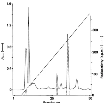
25 Fractionno. .-0 ._ 0 co._-Fig. 1. Purification of the labelled peptide obtained by trypsin
digestion of the S. cacaoiI)-lactamase on the Pro-RPC
column usinggradient A
A 0.5 ml portion, representing one-third of the total
material, wasinjected. The Figure showsthe absorbance profileat 215nm( ),the radioactivity of the fractions
(----) and the gradient(---) reaching 76% ofbuffer B (see
the text) after fraction 50. The radioactivity was determinedonportionsrepresenting 10% of the volume of
eachfraction.Forotherdetails,see the text.
a 470-A Applied Biosystems gas-phase sequenator as described previously (Joris et al., 1985). N-Terminal
residuesweredeterminedby dansylation
(Hartley,
1970).Peptidepurification
This was done with the help of a f.p.l.c. apparatus
(Pharmacia, Uppsala, Sweden)equipped withaPro-RPC column (0.5cmx5cm). The buffers and gradients were asfollows.Buffers A and Bcontained 10mM-NH4HCO3 respectively in 100% water and in water/acetonitrile
(2:2,v/v). Buffers C and D contained 0.1
%
trifluoroace-tic acidrespectively in 100% water and inwater/aceto-nitrile(3:7,v/v). Gradient A started with 1 ml of buffer A and went from 0 to 100% of buffer B over 18 ml. Gradient B started with 1 ml of buffer A andwentfrom 0to65
%
of bufferBover20 ml.Gradient C started with 1 ml of buffer C and went from 0 to 80% ofbuffer D over 20 ml. In all cases the flow rate was 0.3 ml min-' and 0.3 mlfractions werecollected.RESULTS
The active-site
peptide
of thefl-lactamase
ofS. cacaoi The enzyme (6 mg, i.e. 176nmol) in 3ml of 10 mM-Tris/HCIbuffer,pH 7.2,containing 10% ethylene glycolwas added with 25,1 of
1O
mM-[3H]/JIP
(inactivator/enzyme ratio 1.4:1). Inactivation was
complete
after 10 minat30'C. A u.v. spectrum wasrecorded.,
indicatingtheexpected
A325/A280
ratio of0.44 (De Meester et al.,A
0.125
250 300 350
Wavelength (nm)
Fig. 2. U.v. spectrum of thepurifiedpeptideobtainedbytrypsin
digestion
ofthe S. cacaoiII-lactamase
The solution contained about 8 nmol ofpeptide(on the basisof theradioactivity)in 300,Aofwater.
Table 1. Aminoacidcompositionoftheactive-sitepeptides
Peptide... S. cacaoi S. albusG
Amino Amount No. of Amount No. of
acid (nmol) residues* (nmol) residues*
Lys 1.8 1 0.69 1 His - - - -Arg - - - -Asx - - 0.57 1 Thr 2.0 1 - -Ser 1.7 1 0.68 1 Glx - - 0.56 1 Pro - - 0.62 1 Gly 2.3 1 0.31 0 Ala 1.5 1 0.64 1
2-Cys
- - Pt (1) ? Val - - 0.65 1 Met - - 0.47 1 Ile - - - -Leu - - 0.58 1 Tyr 0.9 1 - -Phe 3.0 2 1.01 2 Total... 8 11-12* Nearestwhole number.
t P, presentbutnotaccuratelymeasured.
1986). The sample was dialysed for 12 h against water and the determination of the radioactivity of a portion
indicated an inactivator/enzyme ratio of 0.96:1. After freeze-drying, the powder was dissolved in 250 1l of
100mM-NH4HCO3 containing 100
,#M-CaCl2
and8M-urea.Thesamplewasincubated for 60 minat37° C,
diluted with 400
#1
of thesamebuffer devoid ofureaand,after addition of 600 ,ug oftrypsin,further incubated at
Streptomyces cacaoi Streptomyces albusG Klebsiella pneumoniae Klebsiella aerogenes Bacillus licheniformis Bacillus cereus Staphylococus aureus Plasmid RTEM2 Rhodopseudomonas capsulata Pseudomonas aeruginosa (plasmid) Class C (consensus) OXA 2 Streptomyces R61 DD-carboxypeptidas 62 [Ala 65
Asp Glu Arg] Ala Asp Glu Leu
Pro Asp Glu Arg Pro Asn Glu Arg Ser Asp Lys Pro Glu Glu Glu Asp Glu
Arg Arg
Leu
Gly Asp Glu Arg
Gln Pro Glu Ser Gln Glu Thr Leu
Lys Lys Arg
Thr Thr Asp Arg Phe Phe Phe Phe Phe Phe Phe Phe Phe Phe Phe ,Tyr lPhe
Ala Tyr Gly Pro Met Cys
Ala Met Cys
Ala Met Asn
Ala Phe Ala
Ala Phe Ala
Ala Tyr Pro Met
Ala
Met Leu Met Asn Pro Leu Asn
Glu Leu Ile Val Ser Pro Gly Ala
Arg Val Gly 70 Ser Ser Ser Ser Ser Ser Ser Ser Ser Ser Ser Ser Thr Phe Val Phe Thr Ser Thr Ser Thr Ile Thr Tyr Thr Ser Thr Phe Thr Val Thr Val Ile Leu Thr SerVal His Ser Phe Thr 73 Lys Lys Lys Lys Lys Lys Lys Lys Lys Lys Lys Lys Lys References
}
The present study Jorisetal. (1987b) Emanuel et al. (1986)I
Ambler(1980)J. 1. Campbell & R. P. Ambler, personal communication
Joris et al.(1986)
Dale eta/. (1985) Duezeta/. (1987)
Fig. 3. Comparison between the sequences of the active-site peptides of the
corresponding areas of other class A,8-lactamases S. cacaoi and S. albus G 8-lactamases and those of
Thehighly conserved sequences of class C ,-lactamases and those of the OXA-2 enzyme (class A?) and of the Streptomyces R61 DD-peptidasearealso shown. Residues 62-65 in theS. cacaoi enzyme were deduced from the partial nucleotide sequence
ofthe gene(M.V.Lenzini, unpublished work). The numbering is that of Ambler (1980).
37° C for 3 h. ThedigestwasfilteredthroughaSephadex
G-25 column(100cm x1 cm)in 50
mM-NH4HCO3.
Two groups of radioactive fractions were detected: the first group(25% oftotalradioactivity,Kay. =0.16)probably correspondedtouncompletely digestedmaterial andwasnotfurtherexamined.Thefractions
corresponding
tothe second group(75%,
Ka,.
=0.33) were pooled andfreeze-dried. The drypowderwas dissolvedin 1.5 ml of buffer A and 0.5 ml aliquots were injected into the Pro-RPCcolumn.Fig. 1 shows the elution
profile
forone of the threerunsperformed
withgradient
A. Fractions 35-37were pooledand freeze-dried. Fig. 2 showsa u.v. spectrum of thepeptide
after redissolution of thedry
powderin water. The N-terminal residue was identified
by using 3nmol of material.
Only
phenylalanine
was found. The amino acidcompositionafterhydrolysiswith 6 M-HCI of 1.8 nmol is shown in Table 1.The sequence determined with 8 nmol of material and thegas-phase
sequenator is shown inFig. 3.
Active-sitepeptideof thef-lactamase of S. albusG In afirstexperiment,140 nmol of enzyme(70o purity)
in 10mM-Tris/HCl buffer, pH 7.2, containing 8% glycerol and 8% ethylene glycol was dialysed
against
50 mM-sodium
phosphate
buffer,
pH 7.0,containing
1 M-NaCl,8
%
glyceroland 8%
ethyleneglycol.Thehighsalt concentration was used to decrease the ratio of
turnover of
flIP
to inactivation (see De Meester et al.,1986). To thedialysed solution, 250,ul of 10
mM-[3H],8IP
were added, yielding an inactivator/enzyme ratio of 9.5:1. After 10 min at 30 ° C, inactivation was complete and themixturewasexhaustively dialysed againstwater.
The ratio A325/A280 was 0.31, in good agreement with
previous results whenadjusted for the presence of
3000
impurity in the initial preparation. Similarly, after the
samecorrection, determination ofthe radioactivity ofa portion indicated a bound inactivator/enzyme ratio of 0.98:1. The sample was freeze-dried and the powder
dissolved in 300,tl of 100mM-NH4HCO3, containing 0.1 mM-CaCl2and 8M-urea. Thesolutionwasincubated for 2 hat37° Cand 300,1 of thesamebuffer,devoid of urea, were added, together with 5001ul of trypsin. The mixture wasincubated for 5h at 37'C. The digest was filtered through the Sephadex G-25 column, butmostof the radioactivity was excluded. A second filtration was
then performed through a Sephadex G-50 column
(150cmx1 cm). The radioactive fractions, centred at a
Kav.
value of 0.2, were pooled and freeze-dried. Thepowderwasdissolved in 1.5 ml of 10
mM-NH4HCO3;
thesamplewasinjectedinthe Pro-RPC column
(three runs)
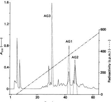
and waseluted withgradient B. Twomajor radioactive
peakswereobserved
(Fig
.4).Thecorresponding
fractions Vol.F. De Meester and others
40 60
Fractionno.
Fig. 4. Chromatographic profile of the first trypsin digest of the
S. albus G fl-lactamase on the Pro-RPC column using
gradient B
A 0.5 ml aliquot, representing one-third of the total material,wasinjected. The gradient (---) reached 56%
of Bafterfraction 63. For conditions and symbols,seethe
legendtoFig. 1.Thecontentsof fractions 30-32(peptide AG3) were also analysed. Only one N-terminal residue
(leucine)wasfound.
I
/ /
werepooled and the N-terminal residue determined for each group by using 1 nmol ofpeptide (on the basis of
the radioactivity). For peptide AG2, alanine was found as the unique N-terminal residue. For peptide AGI,
several residues were found(including alanine), and the
peptide was not further analysed. The sequence deter-mined on 2 nmol of peptide AG2 is given in Fig. 3. However, the amino acid composition (not shown) did
not exactly correspond to this sequence and, on sequencing,asecondpeptide, representingabout20% of the major one, was consistently found. This
peptide
appeared to be larger than the peptide presentedin Fig.
3, since a sharp decline in the total yield was observed after the 12th residue of the major peptide and several
residues were still detected with a similarly low yield.
Difficulties had beenencounteredbefore with a cysteine-containing peptide from Klebsiella pneumoniae (Joris et al., 1978b), and we decided to perform a second
digestion after blocking the free thiol(s) rather than
trying to furtherpurify peptideAG2.
Asecond experiment was performed where 63 nmol of enzyme in 1 ml of 50mM-sodium phosphate, pH 7.0, containing 1 M-NaCl,
8%
glycerol and8%
ethylene glycol were inactivated by 500 nmol of[3H],fIP.
After dialysis and freeze-drying, the powder was dissolved in 2ml of 8M-urea (in water), containing 0.7 mM-dithiothreitol, and the solution was incubated at 37 ° C for 4h. A 10 mm solution ofNbs2 was then added toyield a final concentration of 1.5 mm, and the mixture was incubated at 37 ° C for 30 min. The solution was
exhaustivelydialysed againstwaterandfreeze-dried. The
powder was dissolved in 500
,1l
of 100 mM-NH4HCO3,'100 -7 E C) 4-d ~0 .0 cr Fractionno.
Fig. 5. Purificationof thepeptidelabelled with bothNbs2andplPonthePro-RPC columnusinggradientC
Onlyone run wasperformed. The gradient (---) reached 86% of buffer D (see the text) after 60 fractions. Forconditions
andsymbols, seethelegendtoFig. 1.
1 20
430
-300
(a) 272 329 386 4 A 0.3- (b) 0.2- 0.1-0 I. , 215 272 329 386 Wavelength(nm) Fig. 6. U.v. spectra of the peptide labelled wi
fliP before (a) and after (b) addition
concn.)-mercaptoethanol
containing 100
pM-CaCl2
and 8 M-urea,Eincubated at 37 ° C for 2 h. Trypsin (150 water) was then added and the samj
incubated for 5 h at 37 ° C, and filter( Sephadex G-50 column. The radioactivc pooled, freeze-dried, and the powder M 600,ul of water. The sample was injected4
column and eluted with gradient C (Fi shows theu.v. spectrum of fraction 45 aft andredissolution in 300 ,ulof water. TheI
added with0.33mM-mercaptoethanol (fi:
a new spectrum was recorded after 10 mi 6(b) clearly shows a decreaseof A318 and of a newchromophore centred at 416 nm ding to free Nbs2- (5-thio-2-nitrobenzoa
The sample (fraction 45) was dry-e hydrolysed with 6M-HCl. The results of analysis given in Table 1 confirmed the the peptide whose sequence isgiven in F sequence was obtained by determinatio
nucleotide sequence of the gene coding
(P. Dehottay, unpublished work). DISCUSSION
Themost striking characteristic of bo peptides is the presence of the sequence
Phe-Xaa-Xaa-Xaa-Ser* -Xaa-X around the active-site serine residue (Se sequence hasbeen observed in all
/J-lactar
far, with the sole exception of the OXA-]
phenylalanine is replaced bytyrosine (but that could be considered as a minor change), and corresponds to
residues 66-73 in Ambler's (1980) numbering. The general comparison of all known ,1-lactamases sequences around the active-site serine residue depicted in Fig. 3 establishes that the Streptomyces cacaoi enzyme is a member ofclass A and strongly suggests that the S. albus G enzyme may also be so, in agreement with the predictionsmade onthe basis of the interactions between those enzymes and flIP. The S. cacaoi ,?-lactamase appears as a rather conventional class A enzyme. The Asp-Glu-Arg (63-65) sequence, residues Ala-67 and W43 500 Thr-71, and aromatic residues in positions 68 and 72 are observed in several other class A enzymes. Only one residue(Gly-69) istypical of class C enzymesand is also found in ahomologouspositionin the R61DD-peptidase.
Incontrast,the S.albus G
,-lactamase
has twotypicalclass C residues: the most striking is Leu-65, which occupies aposition where arginine is found in all class A
enzymes (with the exception of the Rhodopseudomonas capsulata enzyme), but also in OXA 2 and the
DD-peptidase. Val-71 is also aclass Ccharacteristic, which in this caseis also sharedby theDD-peptidase. However, Asp-63, Glu-64, Met-68 and an aromatic residue in position 72 indicate that the S. albus G enzyme belongs
to class A. While this work was in progress, nucleotide
sequences of the genes coding for both Streptomyces enzymes were obtained, which confirmed the homology
ith both Nbs2 and with class A enzymes (M. V. Lenzini & P.Dehottay,
of0.33mM (final unpublished work).
Our data underline another characteristic of class A: its heterogeneity. Indeed, if one excepts Phe-66, Ser-70 and Lys-73(whicharealso present in all other enzymes),
no
'.consensus,'
sequence of class Acanbewritten. At allandthe solution positions there is always at least one enzyme which jug in 0.5 ml of exhibits an individual behaviour, for instance Leu-65 in
ple
was further S. albus Gand R. capsulata (in all other other enzymes, ed through the arginine,) Leu-67 in R. capsulata (alanine or proline ine fractions were the others), Leu-68 in Pseudomonas (tyrosine, phenyl-vas dissolved in alanine ormethionine in the others) and Val-71 in S. albus onthe Pro-RPC G (threonine in the others). Some positions are highly ig. 5). Fig. 6(a) variable in class A enzymes, where the substitutions are terfreeze-drying certainly not conservative: positions 62 (Ala, Pro,
Ser,
sample was then Gly or Glu), 69 (Gly, Cys,Asn,
Ala, Met) and 72(Phe,
nal concn.),and Ser, Ile, Tyr, Val, His). Itshould
be noted that, in class in at 20'C. Fig. C, manysubstitutions are highly conservative (positions Lthe appearance 68 and 71 for instance).and correspon- It is also apparent, and rather surprising, that no
Lte).
marked homology is observed between the twoStrep-Iaporated
and tomycesf-lactamases,
in contrast with the two Bacillus the amino acid enzymes. Indeed, a stronger homology is found betweencomposition of the S. cacaoi and the B.
licheniformis
enzymes than 'ig. 3. The same between the two Streptomyces enzymes. An interesting in ofthe deoxy- characteristic of the S. albus G enzyme, shared only byfor the enzyme the
Klebsiella
pneumoniae/,-lactamase,
is the occurrence ofacysteineresidue next to the active-site serine residue. Various data (Joris et al., 1987b; P. Dehottay, unpub-lished work) indicate that the residue might be deeply buried in these enzymes.th Streptomyces When the comparison is extended to the OXA 2
f,-lactamase
and to the R61 DD-peptidase, it seems that aa-Lys the first enzyme is much closer to class A, which justifiesits classification as a 'honorary' class A member (the r*; Fig. 3). This term was coined by S. G. Waley). In contrast, and as masesstudiedso discussed elsewhere, the R61 DD-peptidase is distinctly 2enzyme, where closertoclass C enzymes(Duezetal., 1987),but it is also432 F. De Meester andothers
noteworthy that more similaritiesarefound between the peptidase and class A than between the two classes of
,/-lactamases.
When the sequences depicted in Fig. 3 (i.e. 17 proteins, since five class C enzymes are known) are compared pairwise, identities vary from 2to 12 residues outof 12 aligned (the bestscorehasbeen obtained with two class C enzymes). If the OXA-2fl-lactamase
is notincluded, Phe-66, Ser-70 and Lys-73 are always con-served, resulting in a minimum of 25% identity. However, as noted before, the homology between the various groups ofenzymes becomes much less distinct when the complete sequences are compared. One can only hope that X-ray-diffraction studies will, in the not-too-distant future, show the three-dimensional arrangement of those 12 residues and whether, in
addition to the active serine residue, the conserved phenylalanine (tyrosine in OXA 2) and lysine residues are
found in equivalent positions so that they can fulfil similar functions.
This work was supported in part by the Fonds de la
Recherche Scientifique Medicale, Brussels, Belgium (contract n° 3.4507.83), an Actionconcertee with the Belgian
Govern-ment (convention 79/84-II) and a Convention tripartite between the Region wallonne, Continental Pharma and the
University of Liege,J. D.andB. J. are,respectively, Chercheur qualifie and Charge de recherches du Fonds National de la Recherche Scientifique, Brussels, M.V. L. and P. D. are
indebtedto theMinistere del'Emploiet duTravail foraCadre
SpecialTemporaire,T. E.isaFellow from theIRSIA, Brussels.
REFERENCES
Ambler,R.P.(1980) Philos. Trans. R. Soc. London Ser. B289,
321-331
Dale,J.W.,Godwin,D., Massakowska, D., Stephenson, P. & Wall, S. (1985) FEBS Lett. 191,39-44
Dehottay, P., Dusart, J., Duez, C., Lenzini, M.V., Martial,
J. A., Frere, J. M.,Ghuysen,J. M.&Kieser,T.(1986)Gene
42, 31-36
De Meester, F.,Frere, J. M., Piette,J. L.&Vanderhaeghe,H. (1985) J. LabelledCompd. Radiopharm. 22, 415-425
De Meester, F., Frere, J. M.,Waley, S. G., Cartwright, S.J., Virden, R. &Lindberg, F. (1986)Biochem.J.239, 575-580 Duez, C., Frere, J. M., Klein, D., Noel, M., Ghuysen, J.M.,
Delcambe,L. &Dierickx, L. (1981)Biochem.J. 193, 75-82 Duez, C., Piron-Fraipont, C., Joris, B., Dusart, J., Urdea,
M.S., Martial,J. A.,Frere,J. M.&Ghuysen,J. M. (1987)
Eur.J.Biochem. 162, 509-518
Emanuel, E. L., Gagnon, J. & Waley, S. G. (1986)Biochem. J. 234,343-347
Frere, J. M., Dormans, C., Duyckaerts, C. & De Graeve, J.
(1982) Biochem.J.207, 437-444
Hartley, B. S. (1970)Biochem. J.119, 805-822
Joris, B., De Meester, F.,Galleni,M.,Reckinger, G., Coyette,
J., Frere, J. M.&Van Beeumen, J. (1985) Biochem.J. 228,
241-248
Joris, B., De Meester, F., Galleni, M., Masson, S., Dusart, J.,
Frere, J. M., Van Beeumen, J.,Bush,K.& Sykes, R.(1986)
Biochem. J. 239, 581-586
Joris, B., Jacques, P., Frere, J. M., Ghuysen, J. M. & Van Beeumen, J.(1987a)Eur. J. Biochem. 162,519-524
Joris, B., De Meester, F., Galleni, M., Frere, J. M. & Van Beeumen, J. (1987b)Biochem.J. 243, 561-567
Kelly, J. A.,Dideberg,O.,Charlier,P.,Wery, J. P.,Libert,M., Moews, P.C.,Knox, J. R., Duez, C.,Fraipont,C.,Joris,B., Dusart, J., Frere,J.M.&Ghuysen,J. M.(1986) Science 231,
1429-1431
Lennox, E. S. (1955)Virology1, 190-206
Lenzini,M. V. &Frere, J. M. (1985) J. EnzymeInhib. 1, 25-34 Lenzini, M.V., Erpicum, T. & Dusart, J. (1985) Arch. Int.
Physiol. Biochem. 93,B95
Ogawara,H.,Mantoku,A. &Shimada, S.(1981)J.Biol. Chem.
256, 2649-2655
Samraoui, B., Sutton, B. J., Todd, R.J., Artymiuk, P. J.,
Waley, S. G. & Phillips, D. C. (1986) Nature (London) 320, 378-380
Received 22 December 1986/9 February 1987; accepted 19 February1987