Université de Montréal
Characterization of p120-catenin, a novel RSK substrate in
the Ras/MAPK signalling pathway.
par Beichen Gao
Département de Biologie Moléculaire Faculté de Médecine
Mémoire présenté
en vue de l’obtention du grade de M.Sc en Biologie Moléculaire
option Générale
Janvier, 2017
Résumé
La voie de signalisation Ras/mitogen-activated protein kinase (Ras/MAPK) occupe un rôle central dans la régulation de différents processus biologiques tels que la croissance, la survie mais aussi la prolifération cellulaire. En réponse à des signaux extracellulaires, cette voie de signalisation mène à l’activation des protéines ERK1/2, impliquées dans l’activation de nombreux substrats cellulaires dont les protéines kinases RSK (p90 ribosomal S6 kinase). Ces protéines kinases sont, entre autres, impliquées dans l’invasion et la migration cellulaire mais les mécanismes responsables de ces phénomènes biologiques restent inconnus à ce jour.
Dans mon mémoire, je développe tout d’abord les travaux précédemment réalisés dans notre laboratoire, et identifie la protéine p120-Catenin (p120ctn), un composant majeur des jonctions adhérentes (AJ), comme un nouveau substrat de la voie Ras/MAPK. En utilisant notamment un anticorps phospho-spécificique, nous avons pu démontrer que p120ctn est phosphorylée sur la sérine 320, un nouveau site de phosphorylation, d’une manière dépendante des kinases RSK. D’autre part, nous avons trouvé que la signalisation Ras/MAPK réduit l’interaction entre les protéines p120ctn et N-cadhérine. Ainsi, nos observations suggèrent que l’activation de la voie Ras/MAPK est impliquée dans la diminution de l’adhérence entre cellules par la déstabilisation des AJ. Compte tenu du rôle primordial de la voie de signalisation Ras/MAPK dans le cancer, ce mécanisme nouvellement décrit pourrait contribuer à l’avancement des connaissances sur le développement des cancers dépendents de cette voie de signalisation.
Mots-clés : MAPK; RSK; p120ctn; jonctions adhérentes; cadhérine; phosphorylation; adhérence cellule-cellule;
Abstract
The Ras/MAPK (mitogen-activated protein kinase) signalling pathway is vital in regulating cell growth, survival and proliferation in response to extracellular signals. Positioned downstream in the pathway, the p90 ribosomal S6 kinase (RSK) family regulates cell invasion by weakening cell-cell adhesion, but the mechanisms involved remain elusive.
In this thesis, I expand upon previous work performed in our lab and identify p120ctn, a major component of adherens junctions (AJ), as a new substrate of the Ras/MAPK pathway. Using a phospho-specific antibody, we demonstrate that p120ctn is phosphorylated on a new phosphorylation site on S320 upon activation of MAPK signalling in a RSK-dependent manner. Furthermore, we show that Ras/MAPK signaling reduces p120ctn binding to N-cadherin, suggesting a new mechanism by which MAPK activity decreases cell-cell adhesion by destabilizing AJs. Finally, we designed and optimized two individual assays to be used in future experiments examining the effects of Ras/MAPK signalling on AJ function.
Taken together, our data identifies RSK as a regulator of p120ctn phosphorylation, and also implicates Ras/MAPK signalling in regulating cell-cell adhesion by destabilizing AJ through p120ctn. Given the role of Ras/MAPK signalling in cancer, this new mechanism may play a role in the development and progression of Ras-driven cancers.
Keywords : MAPK; RSK; p120ctn; adherens junction; cadherin; phosphorylation; cell-cell adhesion;
Table of Contents
Résumé ... i
Abstract ... ii
Table of Contents ... iii
List of Tables ... v
List of Figures ... vi
List of Abbreviations ... vii
Acknowledgements ... xi
INTRODUCTION ... 12
Ras/MAPK Signalling and RSK Kinases ... 13
The Ras/MAPK signalling pathway ... 13
The RSK kinase family ... 15
RSK substrates and functions ... 17
The role of RSK in cancer ... 19
Cell-Cell Adhesion and Adherens Junctions ... 21
Cell-Cell Adhesion Structures ... 21
Adherens junctions and cadherins ... 23
The p120-catenin Protein ... 26
Structure of p120ctn ... 26
p120ctn regulation of cell-cell adhesion ... 28
p120ctn and cadherins ... 30
p120ctn and RhoGTPases ... 31
p120ctn and transcriptional regulation ... 32
Regulation of p120ctn by phosphorylation ... 33
p120ctn in cancer ... 36
Rationale and Objectives ... 38
Rationale ... 38
Objectives ... 39
MATERIALS AND METHODS ... 40
Cell culture and transient transfection ... 41
DNA constructs and recombinant proteins ... 42
RNA interference ... 43
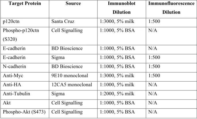
Antibodies ... 44
Cell lysis and lysate preparation ... 45
Bradford protein assay ... 46
Immunoprecipitation ... 46
SDS-PAGE gel separation and immunoblotting ... 46
Retroviral infection of cells ... 47
Immunofluorescence staining ... 48
Cellular impedance assay ... 50
RESULTS ... 52
SECTION 1: Ras/MAPK regulation of p120ctn phosphorylation. ... 53
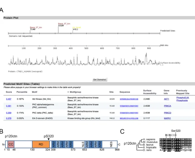
S320 on p120ctn is located within a highly conserved, AGC-kinase consensus sequence ... 53
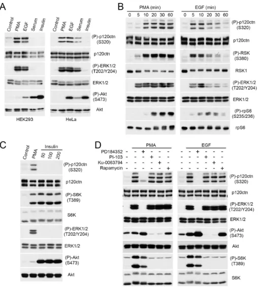
p120ctn is a substrate of the Ras/MAPK pathway ... 53
Phosphorylation of p120ctn on S320 is RSK-dependent ... 54
RSK regulates p120ctn phosphorylation on S320 in epithelial cell lines ... 55
SECTION 2: Ras/MAPK mediated effects on p120ctn ... 57
Ras/MAPK activity reduces p120ctn binding to N-cadherin ... 57
PMA Stimulation does not affect p120ctn localization ... 57
Site-specific mutation at S320 does not affect p120ctn localization ... 58
SECTION 3: Designing assays to examine AJ function ... 60
MCF-10A cells form stable, confluent monolayers in culture ... 61
p120ctn knockdown induces complete monolayer fragmentation ... 61
Calcium depletion increases monolayer fragmentation and triggers AJ disassembly ... 62
Site-specific mutation of S320 on p120ctn produce minor effects on monolayer adhesion ... 63 CHAPTER 3: FIGURES ... 65 DISCUSSION ... 78 General Findings ... 79 Regulation of S320 phosphorylation on p120ctn ... 80 S320 phosphorylation on p120ctn function ... 81
Ras/MAPK regulation of cell-cell adhesion through p120ctn ... 83
CONCLUSIONS AND PERSPECTIVES ... 85
List of Tables
Table 1: Effects of knockout and knockdown of p120ctn and related family members in different species. ... 29 Table 2: Identified phosphorylation sites on p120ctn and the effects of site-specific modifications on p120ctn function. ... 35 Table 3: Antibodies used in this study for immunoblots and immunofluorescence experiments. ... 44
List of Figures
Figure 1. Schematic representation of the Ras/MAPK pathway and RSK substrates ... 15 Figure 2. Schematic representation of the RSK isoforms and their structural features. ... 16 Figure 3. A basic schematic of cell-cell adhesion structures, and structural elements of adherens junctions. ... 23 Figure 4. The structural features of p120ctn including main domains, subcellular localization signals, phosphorylation sites, and alternative splicing sites. ... 27 Figure 5. Our proposed model of the effects of Ras/MAPK signalling on p120ctn function and AJ stability. ... 39 Figure 6. Analysis of S320 as a putative phosphorylation site on p120ctn. ... 65 Figure 7. Activation of the Ras/MAPK pathway induces phosphorylation of p120ctn on S320. ... 66 Figure 8. Ras/MAPK phosphorylation of p120ctn on S320 is RSK-dependent. ... 67 Figure 9. Activation of the Ras/MAPK pathway induces RSK-dependent p120ctn phosphorylation on S320 in epithelial cells. ... 68 Figure 10. Ras/MAPK activity reduces p120ctn binding to N-cadherin. ... 69 Figure 11. Validation of the p120ctn antibody for use in IF. ... 70 Figure 12. p120ctn remains localized at cell-cell contacts in HEK 293 cells in response to PMA stimulation ... 71 Figure 13. p120ctn remains localized at cell-cell contacts in A431 cells in response to PMA stimulation ... 72 Figure 14. Myc-tagged p120ctn S320 mutants localizes to cell-cell contacts in stably expressing MDCK cells. ... 73 Figure 15. MCF-10A cells generate confluent monolayers in comparison to Caco-2, A431, and IEC6 cells. ... 74 Figure 16. p120ctn knockdown promotes monolayer fragmentation. ... 75 Figure 17. Calcium depletion affects monolayer fragmentation and cell index. ... 76 Figure 18. Stable expression of p120ctn phosphomutants in A431 cells affects monolayer adhesion. ... 77
List of Abbreviations
a-cat a-catenin
b-cat b-catenin
AGC family protein kinases A, G and C
AJ adherens junction
Akt protein kinase B
AP-1 Activator protein 1
ARM Armadillo-domain
ARVCF Armadillo-repeat protein deleted in velocardio-facial syndrome BTB/POZ Bric à brac/Pox virus and zinc finger
CAMK Ca2+/calmodulin-dependent protein kinase CDKs cyclin-dependent kinases
cDNA complementary DNA
Chk1 checkpoint kinase 1
CREB cAMP response element-binding protein CTKD carboxyl-terminal kinase domain
E-cad E-cadherin
EC extracellular cadherin
EGF epidermal growth factor
eIF4G1 eukaryotic translation initiation factor 4 gamma 1 EMT epithelial-mesenchymal transition
EphA2 Ephrin type-A receptor 2
EPLIN epithelial protein lost in neoplasm ERK extracellular signal-regulated kinase EST expressed sequence tag
FERM four point one, Ezrin, Radixin and Moesin Gab2 Grb2-associated binder 2
GDI guanine nucleotide dissociation inhibitor GEF guanine exchange factor
Grb2 growth factor receptor-bound protein 2 IDC invasive ductal carcinoma
ILC invasive lobular carcinomas JAM junctional adhesive molecule
JMD juxtamembrane domain
JNK c-Jun N-terminal kinases
KIBRA expression enriched in kidney and brain KIBRA kidney and brain expressed protein MAPK mitogen-activated protein kinase
MDCK Madin-Darby canine kidney
MEK1/2 MAPK/ERK kinase 1 and 2
miRNA microRNA
Mrip Myosin phosphatase Rho-interacting protein mTOR mammalian target of rapamycin
N-cad N-cadherin
NES nuclear export signal
NLS nuclear localization sequence NSCLC non-small-cell lung cancer NTKD amino-terminal kinase domain
p120ctn p120-catenin
PBS phosphate buffered saline PDGF platelet-derived growth factor
PDK1 3’-phosphoinositide-dependent protein kinase 1 PI3K phosphatidylinositol-3-kinase
PKA protein kinase A
PKC protein kinase C
PSPL positional scanning peptide library PTB phosphotyrosine-binding
PTEN phosphatase and tensin homolog Raf regulator of a-fetoprotein
RD regulatory domain
Rheb Ras homolog enriched in brain
ROCK Rho kinase
RSK p90 ribosomal S6 kinase RTK receptor tyrosine kinase
S6K S6 kinase
SH2 Src homology 2
SH3P2 SH3 domain-containing protein 2
shRNA short hairpin RNA
siRNA small interfering RNA
SOS son of sevenless
TJ tight junction
TSC2 tuberous sclerosis complex 2
WT wild-type
Acknowledgements
There are so many people that I would like to thank for their help and support throughout my journey. First of all, I would like to thank my supervisor Dr. Philippe Roux for his support and guidance throughout this endeavour. Philippe, thank you teaching me to always think one step further, and for showing me that I should never be afraid to ask for feedback. I’m incredibly happy for the opportunities you have given me to do cutting edge research, and I’m proud of everything that I’ve accomplished in the lab under your mentorship!
I would also like to thank everyone in the Roux Lab, both past and present. Thank you Dr. Geneviève Lavoie for your patience while teaching me new lab techniques and for putting up with my endless “double-checking” question requests. Thank you Antoine Méant for being my fellow dreamer/idealist/teammate in the lab, and for all the interesting discussions we’ve had in the lab. Thank you Neethi Nandagopal for the endless snacks and crazy late night talks. If I continue on, this section will never end! So let me just give a big thank you to Léo Aubert, Thibault Houles, Sami Nourreddine and Justine Paradis for all your nuggets of wisdom and help in the lab throughout the years.
Outside of our own lab, I would like to thank Dr. Étienne Gagnon for enthusiastically supporting all my crazy ideas throughout the years. Moreover, I have to thank his student, Jordan Quenneville, for being an amazing friend who’s crazy enough to join me in all my crazy side-endeavours! I’m sure you’ll be doing some extraordinary things in the next few years man!
I’m incredibly grateful for the opportunity granted to me by the IRIC institute to study, train, and grow here. It has been such an amazing experience to work in a place with such cutting-edge technology. Not only that, but I also have to thank Pascale Le Thérizien, Julie Mantovani, Patrick Lacasse and Valérie De Rop for being the best administrative and student support team ever. All of you are always so friendly and approachable, and have always provided me with great advice when I needed it!
Finally, I would like to thank my mom and dad for their endless love, support, and FaceTime calls at the most inopportune times midway through an experiment. I couldn’t have done it without the two of you!
Ras/MAPK Signalling and RSK Kinases
The Ras/MAPK signalling pathway
Mitogen-activated protein kinase (MAPK) cascades are evolutionarily conserved signal transduction pathways in eukaryotic cells, allowing them to respond to various intracellular and extracellular stimuli. Many cell-surface receptors can stimulate Ras/MAPK signalling, such as G protein-coupled receptors (GPCRs), cytokine receptors, and receptor tyrosine kinases (RTKs). Traditionally organized, and characterized, by a three-tiered set of sequentially activated and evolutionarily conserved kinases, MAPK pathways act in cells to regulate a plethora of cellular processes, including gene expression, metabolism, motility, proliferation, survival and differentiation (Reviewed in [1]). In mammals, several essential MAPKs have been characterized, including the extracellular signal-regulated kinases 1/2 (ERK1/2), c-Jun amino (N)-terminal kinases 1/2/3 (JNK1/2/3), and p38 isoforms. Of these, the ERK1/2 module is the best studied, and plays a significant role in many human cancers, as evidenced by the many tumors showing hyperactivation of these kinases [2].
The core organization of the ERK1/2 module has been extensively studied, and, like many other traditional MAPK pathways, contains three sequentially acting kinases: A/B/C-Raf, MEK1/2, and ERK1/2. While other unconventional kinases have been shown to replace Raf function in a cell-specific manner, the canonical activation and regulation of the pathway remains consistent [3].
The ERK1/2 signalling cascade is most commonly activated in response to binding of extracellular growth factors, such as platelet-derived growth factor (PDGF), epithelial growth factor (EGF) and insulin, to cell surface receptors. While it has been demonstrated that GPCRs, cytokines and osmotic stress can also initiate ERK1/2 signalling, and that cAMP can regulate ERK1/2 activity in a cell-specific manner, the most well understood method of activation usually implicates RTKs (Fig. 1) [4].
Upon ligand binding, activation and dimerization of RTKs induces autophosphorylation of Tyr residues located on the intracellular domains of the receptors. These phosphorylated residues create docking sites for Src homology 2 (SH2) and phosphotyrosine-binding (PTB) domain-containing proteins, such as Grb2 (growth factor receptor-bound protein 2). Upon
the plasma membrane. SOS is best known for catalyzing the exchange of GDP for GTP bound to Ras, leading to its activation. Now active, Ras-GTP directly interacts with, and promotes the dimerization, of Raf protein kinases (A-, B- and C-Raf), which then phosphorylates its specific substrates MEK1/2 (MAPK/ERK kinase 1 and 2). The dual-specificity MEK1/2 kinases then phosphorylate the MAPKs known as ERK1/2 within a conserved Thr-Glu-Tyr (TEY) motif in their activation loop motif. Activation of ERK1/2 then leads to further propagation of the initial growth factor signal to a large variety of downstream substrates regulating, among other responses, gene transcription, cell growth, proliferation, motility and survival (Reviewed in [1]). In the context of cancer, many tumors harbour activating mutations within the genes encoding for initiating components of the ERK1/2 module, such as Ras and Raf – pointing to the oncogenic role of this pathway in driving cancer development and progression [2]. Thus, many cancers exhibit hyperactivation of ERK1/2 and deregulation of downstream biological functions, leading to tumor growth and survival.
Academic efforts to better characterize and understand this critical signalling pathway has revealed several important downstream effectors, including the 90 kDa ribosomal S6 kinases (RSKs), a family of Ser/Thr kinases that lie immediately downstream of the ERK1/2 kinases.
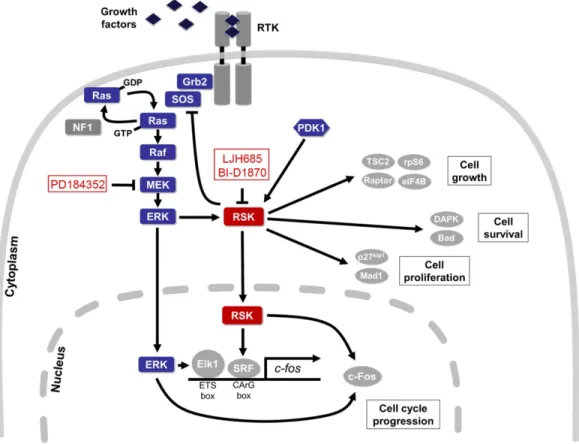
Figure 1. Schematic representation of the Ras/MAPK pathway and RSK substrates
Upon activation of cell surface RTKs, the Grb2/SOS complex is recruited to the plasma membrane and promotes GTP binding to Ras. Active Ras then initiates the signalling cascade leading to activation of the ERK1/2 MAPK signalling module, and RSK activation. Some of the known substrates of RSK are indicated here, including feedback inhibition of SOS to prevent hyperactivation of MAPK activity. Pharmacological compounds targeting MEK and RSK employed in this study are also highlighted.
(Adapted from [5])
The RSK kinase family
Initially identified in 1985 as two kinases, S6KI and S6KII, that phosphorylated ribosomal protein S6 (rpS6) in Xenopus laevis eggs (later renamed RSK), are one of the many substrates of ERK1/2 and are effector kinases capable of regulating many cellular processes through its own substrates [6]. In human cells, there are four different isoforms (RSK1-4) that share between 73-80% sequence homology, mostly diverging in their C- and N-terminus regions [5] (Fig. 2).
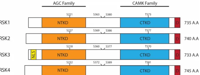
Figure 2. Schematic representation of the RSK isoforms and their structural features.
The 4 RSK isoforms all consist of the NTKD and CTKD connected by a linker region. All isoforms also contain a D-domain docking site for ERK1/2 association. Only isoform 3 contains a putative NLS domain. The phosphorylation sites required for RSK activation are indicated.
All four isoforms contain two highly conserved, yet functionally different, kinase domains connected by a linker region approximately 100-residues in length containing hydrophobic and turn motifs that are involved in RSK activation and regulation. These two distinct functional kinase domains appear to be a unique feature of RSKs, with the exception of the mitogen- and stress-activated protein kinases (MSKs), which are highly similar to the RSK [7]. The amino-terminal kinase domain (NTKD) shares high sequence homology with members of the AGC kinase family (including Akt, PKC and S6K1/2) and is the main functional domain for phosphorylation of RSK substrates and signal propagation. On the other end, the carboxyl-terminal kinase domain (CTKD) is similar to kinase domains found in the calcium/calmodulin-dependent protein kinases (CaMKs) and its only known function is to autophosphorylate RSK for the activation of NTKD in response to upstream signals from ERK1/2 [8]. Beyond the CTKD, an ERK1/2 docking site is located in the C-terminal region of RSK and is essential for the association of ERK1/2 with RSK for activation of the latter [9]. Finally, at the very end of the C-terminus is a PDZ domain-binding motif that varies slightly between the four RSK isoforms, and has been shown to play a role in regulating RSK interaction with PDZ domain-containing proteins during specific cellular processes, such as during synaptic transmission [10].
RSK activation requires a well-characterized and well-regulated sequence of phosphorylation events on four essential phosphorylation sites: Ser221, Ser363, Ser380 and Thr573 in human RSK1. Upon activation by mitogenic signals, ERK1/2 phosphorylates RSK on Thr573 in the activation loop region of the CTKD [11]. Now active, the CTKD and ERK1/2 coordinately phosphorylates Ser380 and Ser363, respectively, in the linker region of RSK [8, 12]. These phosphorylated residues generate a binding site for 3’-phosphoinositide-dependent protein kinase 1 (PDK1) – a constitutively active Ser/Thr kinase that has been shown to regulate members of the AGC kinase family [13]. Recruited PDK1 then phosphorylates Ser221 located within the activation loop of the NTKD, leading to its full activation and phosphorylation of RSK substrates [14].
Looking at their expression patterns, mRNA encoding RSK1-3 are present within all tissue types, while RSK4 appears to have low levels of expression in both embryonic and adult tissues. While RSK is ubiquitously expressed, tissue-specific differences in mRNA levels of RSK1-3 have been observed. RSK1 mRNA levels are commonly elevated in kidney, pancreas and lung tissue, while RSK2 and 3 are more predominantly expressed in the heart and skeletal muscle [15]. Northern Blot analysis identified RSK4 mRNA expression in the brain, heart, cerebellum, kidney and skeletal muscle, and is completely undetectable in the lung, liver and pancreas [16]. Specifically in the central nervous system, RSK isoforms have been shown to be expressed in different brain structures [17-19]. Studies into the expression of RSK isoforms during developmental stages also reveals distinct patterns of expression. RSK1 and 3 perhaps play a greater role in development as they are more highly expressed in fetal mouse tissue compared to RSK2 and 4 that show lower mRNA levels [20].
Taken together, these data suggest that while RSK isoforms may share high similarity, considering the differences in tissue distribution, and different expression patterns during developmental stages, the isoforms must regulate significantly different programs and serve different roles in a cell or tissue-specific manner.
RSK substrates and functions
To better understand the roles of RSKs in cells, various studies have been conducted with the aim to identify and characterize novel RSK substrates to build a greater understanding
consensus motif was initially believed to require the specific motif sequences: R/K-X-R-X-X-pS/T or R-R-X-R/K-X-R-X-X-pS/T [21]. More recently, using a positional scanning peptide library (PSPL), our lab confirmed that the RSK1 consensus motif resembles R-X-R-X-X-pS/T with a strong requirement for arginine residues at the -3 position, and a preference to phosphorylate serine over threonine [22]. Identification of the RSK consensus motif helps not only to provide confirmation of previously identified RSK substrates, but also creates the opportunity for high-throughput proteomic screening for novel RSK substrates.
Collectively, the current known substrates of RSKs indicate they play a role in regulating gene transcription, protein synthesis, cell-cycle progression, proliferation, survival, motility and migration. For the sake of brevity, I will only address a few of the identified substrates of interest here, but for further details, more in-depth reviews specifically addressing RSK substrates can be found [5, 23].
At the transcriptional level, RSK2 has been shown to phosphorylate and stabilize c-Fos in cells, a component of the AP-1 transcription factor complex and is frequently overexpressed in tumor cells. The transcription factor c-Fos upregulates cyclin D1 and promotes G1 progression in cells, driving cell proliferation [24]. Additionally, RSK2 has been shown to activate another transcription factor: cAMP response element-binding (CREB) protein through phosphorylation at Ser133 [25]. This RSK-CREB signalling pathway has been implicated in promoting tumor progression, cell survival and metastasis through upregulation of various genes [26]. Conversely, RSK3 and 4 appears to negatively regulate cell proliferation – especially in the context of cancer. RSK3 appears to function as a tumor suppressor in ovarian cancer cells, and RSK4 knockdown has been shown to promote proliferation, migration and metastasis of breast cancer cells [27, 28]
Moving on to translation, rpS6 phosphorylation by RSK at Ser235/236 promotes cap-dependent mRNA translation through assembly of the cap-binding complex [29]. Additionally, RSK promotes protein synthesis through mTOR (mammalian target of rapamycin) signalling. By phosphorylating tuberous sclerosis complex 2 (TSC2) on Ser1798, RSK inhibits its GTPase-activating activity for Rheb (Ras homolog enriched in brain), a small GTPase that is required for the activation of the mTORC1 complex [7]. By an alternate mechanism, RSK can directly phosphorylate Raptor (regulatory associated protein of mTOR), an important scaffolding protein, to promote mTORC1 assembly and activation [30].
Finally, a newly emerging function that is of particular importance to my project is the role of RSK in promoting Ras/MAPK-dependent cell motility and invasion. Using pharmacological inhibitors, RSK1 and 2 were initially identified to play a role in Raf-dependent migration in MDCK cells by upregulating expression of ~20% of ERK regulated mRNAs, many of which being invasion/motility genes encoding proteins such as uPAR, MMP-1 and RhoC [31]. A genome-wide RNAi screen for regulators of epithelial cell migration also identified RSK as a key effector and a point of convergence of various pro-migratory signals [32]. At the post-translational level, recent studies have identified a handful of new RSK substrates, including Gab2 (Grb2-associated binder), KIBRA (expression enriched in kidney and brain), SH3P2 (SH3 domain-containing protein 2), and EphA2 (Ephrin type-A receptor 2), that are all implicated in RSK-mediated cell motility and migration [33-36]. In our own phosphoproteomic and proximity ligation screens, we have identified a new potential RSK substrate, p120-catenin (p120ctn), that plays a significant role in regulating cell motility. p120ctn is a master regulator of cell-cell adhesion and a critical component of adherens junctions (AJs) [22].
The role of RSK in cancer
As a major regulator of cell proliferation, growth and survival signals downstream of the Ras/MAPK pathway, RSK plays a significant role in cancer progression. I will provide a quick summary of RSK’s contribution to cancer development here, while more detailed explorations of its role can be found in excellent reviews such as: [37, 38].
Overexpression and hyperactivation of RSK has been observed in several cancers such as breast, lung, prostate, head and neck, ovarian, melanoma, osteosarcomas and multiple myelomas (reviewed in [37]). Increased levels of of RSK2 were observed in ~50% of breast and prostate cancer tissues [39, 40]. Ectopic overexpression of RSK2 also promotes proliferation and anchorage-independent cell transformation in cells [41]. Pharmacological inhibition of RSK using SL0101, a RSK specific inhibitor, in cell lines derived from breast and prostate tumours has also been shown to significantly decrease proliferation of these cells in vitro [5].
Beyond cell proliferation, RSK also promotes lung cancer survival via phosphorylation and inhibition of the Bcl-2 homology 3-only proapoptotic protein, Bad, in response to estrogen signalling. [42] Furthermore, research has uncovered isoform-specific effects of RSK signalling
of RSK2 correlating with increased metastasis of head and neck squamous cell carcinomas in patients [43]. Conversely, RSK1 appears to be a negative regulator in non-small cell lung cancer [44], while exhibiting promigratory effects in immortalized breast epithelial cells [32]. Additionally, as previously addressed, increased RSK4 expression appears to reduce the metastatic potential of breast cancer cells [45].
Overall, the RSK family kinases play a multi-functional role in cancers by various mechanisms through their downstream substrates. In particular, the contradictory effects of RSK isoforms on cancer metastasis show that our understanding of how RSK regulates changes in cell motility and invasion is still quite poor. By studying the kinase-substrate relationship between RSK and p120ctn, our goal is to advance our knowledge of the mechanisms by which RSK signalling modulates cell-cell adhesion and cell motility in the context of cancer.
Cell-Cell Adhesion and Adherens Junctions
Cell-Cell Adhesion Structures
In multicellular organisms, cell contact and adhesion to neighbouring cells are required for generating functional tissue and organs by maintaining highly organized cellular polarity and architecture. Through evolution, different types of intercellular junction structures have emerged to participate in, and to regulate cell-cell adhesion.
Strong cell-cell adhesion is most crucial in epithelial cells, one of the most ubiquitous cell types that are responsible for forming epithelial layers. The epithelial layer acts as boundary between different environments – such as the inside and outside of an organism. This barrier also controls the flux and exchange of biologically relevant molecules, nutrients, and ions into the body and organs. In vertebrates, three major types of adhesion systems have been identified to contribute to the integrity of cell-cell adhesion: adherens junctions (AJs), desmosomes (DM), and tight junctions (TJs) (Fig. 3A and B).
Years of research have identified AJs as the main, defining cell-cell adhesion structure; however, the other two structures should still be quickly addressed. TJs are claudin- and junctional adhesive molecule (JAM)-based structures are located at the most apical part of epithelial cells, and are responsible for firmly sealing the space between adjacent cells in order to establish a barrier against diffusion of molecules across the epithelial layer [46]. DMs are composed of non-classical cadherins, such as desmogleins, and are associated to intermediate filaments to form patch-like points of adhesion between cells [47].
In contrast to the two structures defined above, AJs provide strong cell-cell adhesion by forming cadherin-based adhesive structures anchored to the actin cytoskeleton that are strongly resistant to mechanical stress. In epithelial cells, AJs are linked to a circumferential “adhesive belt” of actin-filament bundles called the zonula adherens (ZA) to maintain strong adhesion. In addition to being a major cell-cell adhesion structure, the ZA forms at the apical/basal border in cells, and is important in maintaining cellular polarity by segregating the lateral membrane into apical and basal compartments [48]. AJ-dependent cell polarization also precedes the formation of TJs and desmosomes, as components of these structures need to be targeted to different compartments of the lateral membrane as defined by the ZA [49].
Despite its characteristically strong structure, numerous studies also illustrate the high plasticity of AJs. This is exemplified by the vital role of AJs in collective cell migration of various cells, such as astrocytes, neurons and neural crest cells, during development [50]. In neuronal cells, the association between AJs and the actin cytoskeleton function as mechanosensors to coordinate collective cell migration [51]. During collective cell migration, AJs are observed to undergo continuous treadmilling, a process where they are actively recycled from the rear of the cell to the front during migration. The process of AJ recycling helps not only to maintain adhesion between adjacent cells, but also maintains front-rear cell polarity and dictates the speed of migration [52, 53].
Collective cell migration is only one example showing how dynamic regulation of AJ influences cell-cell adhesion, and cell morphology. These dynamic morphogenic changes are also a hallmark of epithelial-mesenchymal transition (EMT), a well-studied physiological process that plays a crucial role in normal physiological functions as well as cancer progression. EMT is a dramatic phenotype change of epithelial cells in response to physiological signals. Epithelial cells lose their epithelial characteristics, migrate away from the epithelial monolayer and establish new tissue at a distant site. Epithelial cells acquire many changes during EMT, including loss of apical-basal polarity, acquisition of motile behaviour, cytoskeletal re-organization, and the loss of E-cadherin-mediated cell-cell adhesion structures. Normally, EMT plays an important role in early embryogenesis during gastrulation and neural crest cell migration, and for wound healing in adult organisms, as healthy epithelial cells need to de-differentiate and migrate towards the injury site to promote new epithelial growth (Reviewed in [54]). However, in the context of human cancers, the very same process is hijacked by tumour cells for invasion and metastasis. The process of metastasis by EMT has been observed in different tissue, such as lung, prostate and breast cancers [55-57].
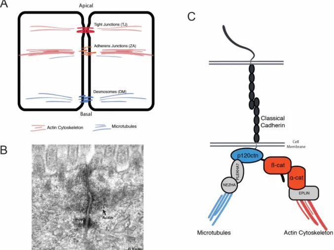
Figure 3. A basic schematic of cell-cell adhesion structures, and structural elements of adherens junctions.
(A) A schematic drawing of adjacent cells, identifying the location of three main cell-cell adhesion structures: tight junctions (TJ), adherens junctions, also known as zonula adherens (ZA), and desmosomes (DM). (B) Electron microscope image of mouse intestinal epithelium cells, indicating the three main structures at cell-cell contacts. The actin filament bundle consisting the ZA is also indicated. (C) A detailed representation of the cadherin-catenin complex including known interacting proteins that are required for the attachment of the complex to the actin cytoskeleton and microtubules.
The image in (B) was obtained from [58]
Adherens junctions and cadherins
The main AJ complex is composed of the adhesion molecule E-cadherin, which mediates strong, calcium-dependent binding between cells, and a catenin complex consisting of a-, b- and p120-catenin, that stabilizes and connects E-cadherin to the actin cytoskeleton (Fig. 3C).
The cadherins are a large family of cell-cell adhesion molecules, and play a role in tissue patterning and maintaining cell adhesion. Classical cadherins are single-pass, type I transmembrane glycoproteins that establish calcium-dependent, homophilic adhesion through binding cadherin molecules on neighbouring cells [59].
The two most well-studied types of classical cadherins are E- and N-cadherin. As its name suggests, epithelial cadherin (E-cadherin) is found in epithelial cells, and contributes to the formation of adherens junctions for tight cell adhesion in epithelial cell layers [60]. Neural (N-cadherin) is most abundant in the nervous system and forms adherens junction-like structures at synapses and other parts of the neuron [61]. There are other forms of classical cadherins that have been identified, such as cadherin-11, but there is still much to understand about the function of these other cadherins due to different patterns of expression across tissue types. A more detailed review about the greater family of cadherins can be found in [62].
Each cadherin molecule is composed of a short C-terminal cytosolic domain, and five “extracellular cadherin” (EC) domains that are essential for calcium-binding and cell-cell adhesion [59]. Upon binding of extracellular calcium ions, the EC-domains adopt a stiff, rod-like structure that extends from the cell membrane to form trans junctional interactions with cadherins on adjacent cells. These trans adhesive structures are incredibly sensitive to fluctuations in extracellular calcium levels, and have been observed to change conformation rapidly in response to removal of extracellular Ca2+. When extracellular concentration of Ca2+ drops below 1 mM, the conformational change of cadherin EC-domains results in detachment from one another, and leads to a rapid loss of cell-cell adhesion [63].
The formation and maintenance of AJ requires the retention of cadherins at the cell membrane, as well as functional attachment of adhesive cadherins to the actin cytoskeleton for mechanical transduction. These two important aspects are regulated through a major family of cadherin interacting proteins known as the catenins, which bind to the cytoplasmic domain of classical cadherins. There is high sequence identity between the cytoplasmic domains of classical cadherins, within which are sites for binding a complex composed of a-, b-, and p120-catenins. Both b-catenin (b-cat) and p120-catenin (p120ctn) are direct interactors of cadherins, binding to different sequences in the cytoplasmic domain through their own highly conserved
armadillo (ARM) repeat domains. However, despite the sequence homology shared between b-cat and p120ctn, they serve functionally different purposes in the cadherin-b-catenin complex.
The linkage of cadherins to the actin cytoskeleton is mediated through b-cat and its association to a-catenin (a-cat). Initially it was believed that b-catenin binding to the distal portion of the cadherin recruits a-catenin, which serves to anchor the cadherin-catenin complex to the actin cytoskeleton either through directly binding F-actin or indirectly through other interacting proteins, such as afadin or epithelial protein lost in neoplasm (EPLIN) [64]. The specific mechanism of interaction between a-cat and F-actin is still under debate. Biochemical studies performed with AJ complexes in mouse and zebrafish indicates that a-cat binding to the AJ complex drastically decreases its affinity for F-actin. However, another study shows that a “minimal cadherin-catenin complex” composed of a E-cadherin/a-cat fusion protein is capable of binding F-actin under force [65, 66]. Regardless, all studies agree that a-cat is a crucial component at AJ to mediate cadherin-actin cytoskeletal interaction. Meanwhile, b-cat appears to serve a supporting role at AJs by bridging cadherin to a-cat, and also harbours a collection of phosphorylation sites that converge from various signalling pathways for fine regulation of AJ. Briefly, phosphorylation of Ser/Thr in the b-cat binding domain of E-cadherin has been shown to strengthen b-cat binding, while phosphorylation of Tyr on both E-cadherin and b-cat reduces their affinity for each other, and promotes the dissociation of the cadherin-catenin complex (Reviewed in [67]).
Independent of a- and b-cat, p120ctn binds cadherins at a separate juxtamembrane domain (JMD) located in a more proximal region of the cadherin cytoplasmic domain and acts as an important regulator of both cadherin stability and its localization to the membrane. Intentional knockdown of p120ctn in a variety of mammalian cell lines results in the destruction of the cadherin-catenin complex at AJ, and a significant reduction in the protein levels of all classical cadherins, and both a- and b-cat [68, 69]. These results suggest that p120ctn plays a pivotal role in the formation and maintenance of the AJ/cadherin-catenin complex even superseding that of a- and b-cat.
The p120-catenin Protein
Structure of p120ctn
Found only in vertebrates, p120ctn is a member of a larger family of ARM-repeat containing proteins including: delta-catenin, p0071, and armadillo-repeat protein deleted in velo-cardio-facial syndrome (ARVCF). Comparing the proteomes across different vertebrate and metazoan species shows that all of these proteins evolved from a d-catenin-like ancestor [70]. Successive evolution has generated multiple p120ctn family proteins in the vertebrate genome, while only one single d-catenin-like protein exists in invertebrates. Interestingly, deletion of p120ctn homologues in invertebrates (p120 in D. melanogaster and JAC-1 in C.
elegans) does not affect survival and only produces minor adhesion phenotypes [71, 72]. In
contrast, p120ctn depletion in vertebrates causes embryonic lethality, while knockdown of other p120ctn members are better tolerated (See Table 1) [73]. This suggests that in vertebrates, p120ctn has evolved into a protein essential for life, while other members of the family appear to play less substantial roles in regulating physiological functions.
The gene coding for p120ctn, ctnnd1, consists of 21 exons encoding a total of 968 amino acids (Fig. 4), with alternative splicing sites occurring at exon 18 (exon A), exon 20 (exon B) which contains a nuclear export signal (NES), and exon 11 (exon C), the presence of which disrupts a nuclear-localization signal (NLS) [74]. In vertebrates, mRNA containing exon A is found to be abundantly expressed, while those with exons B and C are found at much lower copy numbers, with the only exception being in neural tissues where exon C appears to be abundantly expressed [75]. Aside from alternative splicing, the CTNND1 gene contains four unique start codons, and gives rise to four different isoforms, with isoforms 1 and 3 being the most commonly expressed in cells. There are four distinct functional regions to be found in the full-length p120ctn protein, beginning with a coiled-coil domain located near the N-terminal, a phosphorylation or regulatory domain, the ARM domain containing 9 ARM-repeats, and a C-terminal tail [74]. Due to the location of alternative start codons, the coiled-coil domain is only found in isoform 1, and the regulatory domain is present in all but isoform 4. However, the main central ARM-repeat domains critical for the interaction between p120ctn and the proximal region of cadherins remain preserved in all isoforms. However, due to variations in the other domains, there still appears to be functional differences between isoforms. For example,
evidence indicates a stronger expression of isoform 1 in mesenchymal cells expressing N-cadherin, while isoform 3 is preferentially expressed in epithelial cells and interacts more strongly with E-cadherin in these cells [76].
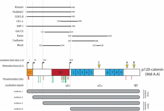
Figure 4. The structural features of p120ctn including main domains, subcellular localization signals, phosphorylation sites, and alternative splicing sites.
The CTNND1 gene for p120ctn codes for a protein up to 968 residues in length. Due to alternative splicing, four different start sites are present to give rise to four different isoforms of varying lengths. Alternatively used exons A, B and C are their locations are indicated. As a member of the armadillo-repeat (ARM) protein family, p120ctn contains 9 ARM-domains indicated from 1-9. The structure of p120ctn contains two nuclear localization signals (NLS) and one nuclear export sequence (NES). All identified Ser, Thr and Tyr phosphorylation sites are identified by S/T/Y labels. The interaction domains of major p120ctn interactors are also indicated.
p120ctn regulation of cell-cell adhesion
Originally discovered as a novel substrate for membrane-associated Src, p120ctn was identified as a protein that plays a role in regulating cell-cell adhesion [77]. Supporting this initial prediction, subsequent studies identified p120ctn to be an important member in the cadherin/catenin complex found at AJ [78].
Similar to b-cat, p120ctn binds to the cytoplasmic tail of classical cadherins but does not play a role in linking cadherins to the actin cytoskeleton. Instead, p120ctn maintains AJ stability by regulating cadherin turnover at the membrane through regulation of cadherin endocytosis [69]. In the absence of p120ctn binding, cadherins become rapidly internalized and are degraded by an endo-lysosomal degradation pathway [79]. Early studies have shown direct interaction between the two proteins: classical cadherin and p120ctn are required for their proper function as a complex at the cell membrane. E-cadherin re-expression in E-cadherin deficient cells causes p120ctn to be recruited to the membrane [80]. Conversely, in E-cadherin expressing cells, downregulation of p120ctn results in a decrease in the total levels of E-cadherin and a drastic decrease in cell-cell adhesion due to loss of AJs [81]. Finally, only re-expression of wild-type p120ctn, and not a mutant incapable of binding cadherin, can rescue cadherin levels in p120ctn-deficient cells [82]. This indicates that p120ctn mediated stabilization of cadherins requires direct interaction between these two proteins.
At a physiological level, the importance of p120ctn in maintaining cell-cell adhesion in vertebrates was studied using tissue-specific knockout models, as whole animal knock-out of p120ctn proved to be embryonic lethal as addressed previously. Descriptions of the effects of conditional p120ctn knockout in various mouse tissues are summarized in Table 1.
Overall, the trend suggests that conditional p120ctn knockouts produce defects in tissue morphogenesis resulting from loss in of cell-cell adhesion. Especially in epithelial tissue, the resulting defects in epithelial barrier function produces an inflammatory phenotype that further drives organ dysfunction (such as in the colon, skin, and salivary glands) and can generate a tumor promoting microenvironment [83]
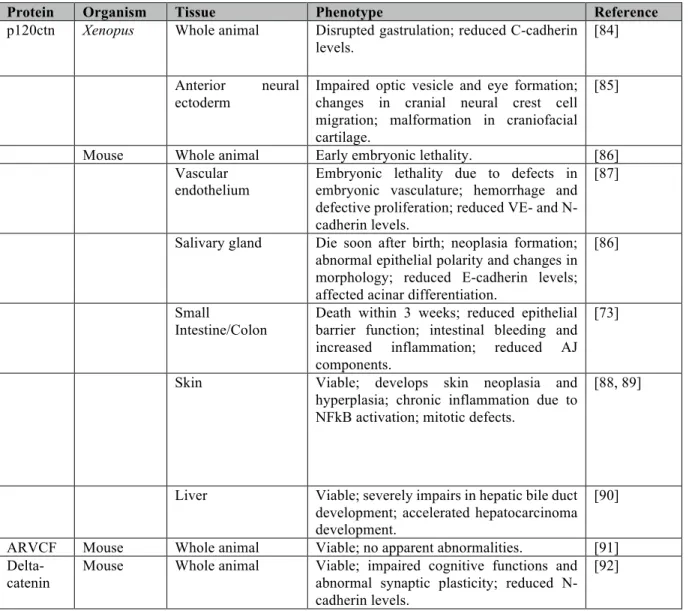
Table 1: Effects of knockout and knockdown of p120ctn and related family members in different species.
Protein Organism Tissue Phenotype Reference
p120ctn Xenopus Whole animal Disrupted gastrulation; reduced C-cadherin levels.
[84] Anterior neural
ectoderm
Impaired optic vesicle and eye formation; changes in cranial neural crest cell migration; malformation in craniofacial cartilage.
[85]
Mouse Whole animal Early embryonic lethality. [86] Vascular
endothelium Embryonic lethality due to defects in embryonic vasculature; hemorrhage and defective proliferation; reduced VE- and N-cadherin levels.
[87]
Salivary gland Die soon after birth; neoplasia formation; abnormal epithelial polarity and changes in morphology; reduced E-cadherin levels; affected acinar differentiation.
[86]
Small
Intestine/Colon
Death within 3 weeks; reduced epithelial barrier function; intestinal bleeding and increased inflammation; reduced AJ components.
[73]
Skin Viable; develops skin neoplasia and hyperplasia; chronic inflammation due to NFkB activation; mitotic defects.
[88, 89]
Liver Viable; severely impairs in hepatic bile duct development; accelerated hepatocarcinoma development.
[90] ARVCF Mouse Whole animal Viable; no apparent abnormalities. [91]
Delta-catenin
Mouse Whole animal Viable; impaired cognitive functions and abnormal synaptic plasticity; reduced N-cadherin levels.
p120ctn and cadherins
As outlined previously, p120ctn plays a significant role in maintaining cadherin expression and stability within cells by directly binding cadherins at its JMD. Crystallographic analysis of the binding interface between the E-cadherin JMD and p120ctn revealed the presence of an endocytic dileucine motif that lies in a region masked by p120ctn [93]. The exposed dileucine motif can be targeted by the adaptor protein AP-2, which promotes the clathrin-dependent endocytosis of cadherins from the membrane [94, 95]. Within the same binding interface also lies two Tyr residues (Y753/754) that, upon phosphorylation by Src, recruits the E3-ubiquitin ligase Hakai. In the absence of p120ctn, Hakai is recruited and promotes the proteasome-dependent degradation of E-cadherin [96]. However, these two mechanisms of cadherin regulation are only present in E-cadherin and are not conserved in other classical cadherins such as N- and P-cadherin.
Recently, a different endocytic DEE-motif was identified that is conserved across all classical cadherins, and re-emphasizes the role of p120ctn as an important regulator of all classical cadherins, not just E-cadherin [97]. This is reinforced by studies showing that p120ctn plays a vital role in regulating neuron growth and polarization through dynamic regulation of N-cadherin [98].
Outside of regulating cadherin dynamics through binding the JMD, a large number of interactors of p120ctn has also been identified that play a role in mediating alternative pathways of cadherin degradation. At AJs, p120ctn can recruit presenilin (PSEN1), a component of the gamma-secretase complex. The consequential assembly of the full complex results in the cleavage of E-cadherin amongst other target proteins, and promotes the disassembly of AJs [99]. Additionally, the C-terminal tail region of p120ctn can also associate with NUMB, and invokes clathrin-dependent endocytosis of the whole AJ complex – potentially for recycling back to the membrane during dynamic changes in cell-cell adhesion [100].
Finally, recent studies have also identified an interaction between the C-terminal domain of p120ctn and EPB41L5, a FERM (Four point one, Ezrin, Radixin and Moesin)-domain containing protein that has been identified to play a role in regulating cell-cell adhesion during EMT. EPB41L5 binding to p120ctn reduces its binding to E-cadherin at AJs and increases the number of E-cadherin positive vesicles detected in cells with EPB41L5 overexpression [101].
Further studies indicate this mechanism may play a significant role in driving the invasion and metastasis of breast cancer cells [102].
p120ctn and RhoGTPases
In addition to membrane localized p120ctn, cytoplasmic and nuclear pools of p120ctn have been observed across various cells, and have been shown to harbour pleiotropic functions to regulate cell adhesion, cytoskeletal remodelling and cell signalling.
Both membrane-bound and cytoplasmic p120ctn have been shown to control small RhoGTPase activity. At the membrane, cadherin-bound p120ctn recruits p190RhoGAP through a RhoGAP binding domain located in its C-terminal tail. The recruitment of p190RhoGAP mediates localized inhibition of RhoA at the cell periphery [103]. Uncoupling of p190RhoGAP from p120ctn in human pulmonary artery endothelial cells caused a drastic reduction in endothelial barrier recovery from disruption by thrombin, and contributes to vascular barrier dysfunction [104].
In contrast, cytoplasmic p120ctn can behave like a rheostat for regulating the balance between RhoA and Rac1 signalling. In the cytoplasm, p120ctn acts as a Rho guanine nucleotide dissociation inhibitor (RhoGDI) by binding RhoA in its inactive, GDP-bound form [105]. It was observed that in an indirect manner, p120ctn can also activate Rac1 by interacting with Vav2, a Rho family guanine exchange factor (GEF). Consistent with these roles, overexpression of p120ctn in NIH-3T3 cells resulted in loss of stress fibres and focal adhesions, while inducing a more dendritic phenotype [106].
However, more recent studies have shown that p120ctn’s effects on RhoGTPase signalling can vary in a tissue and cell-type specific manner. In an invasive lobular carcinoma mouse model, p120ctn may activate Rho/ROCK signalling to promote cell invasion and anchorage independent growth by inhibiting Myosin phosphatase Rho-interacting protein (Mrip) [107]. However, in MDCK cells, cytosolic p120ctn mediates Src- and Rac1-induced anchorage independent growth and anoikis resistance [108].
Collectively, the new emerging effects of p120ctn on Rho and Rac signalling builds a more complex picture of p120ctn’s role as a main regulator of Rho GTPase activity. Depending on the cell type, and biochemical context, cytosolic p120ctn can have a powerful effect on the
dynamics and balance of Rho/Rac signalling, and may significantly affect actin dynamics within epithelial cells.
p120ctn and transcriptional regulation
In addition to localizing to the plasma membrane and the cytoplasm, there are NLS and NES sequences present within p120ctn that enable it to shuttle in and out of the nucleus. Similar to cytoplasmic p120ctn, nuclear p120ctn also appears to be regulated by cadherin levels in cells [109]. Additional mechanisms of p120ctn nuclear translocation appear to exist, implicating p120ctn’s ability to associate with microtubules and its interaction with another transmembrane protein – MUC1 that has been shown to also facilitate the nuclear translocation of b-cat [110, 111].
Upon translocation into the nucleus, p120ctn modulates gene expression by direct interaction with Kaiso, a transcriptional repressor belonging to the Bric à brac/Pox virus and zinc finger (BTB/POZ)-zing finger family transcription factors [112]. p120ctn binding to Kaiso has been shown to relieve Kaiso-mediated transcriptional repression of its target genes. Interestingly, the target genes currently identified in literature controlled by Kaiso includes both tumor suppressor genes such as CDH-1 (E-cadherin) and Rb, as well as known components in canonical and noncanonical Wnt signalling pathways, whose activity has been shown to promote tumor cell metastasis and invasion (Reviewed in [113]). In the context of cancer, the role of Kaiso and p120ctn also appears to be cell type dependent. Evidence of nuclear Kaiso detected in high-grade invasive ducal carcinomas (IDC) and metastatic prostate cancer supports the oncogenic role of Kaiso repression [114, 115]. Conversely, detection of Kaiso in the cytoplasm of non-small-cell lung cancers (NSCLC) and invasive lobular carcinomas (ILC) suggest that Kaiso acts as a tumor suppressor in these cells [115, 116].
From these immunohistological studies, it is clear that localization of Kaiso plays a role in human cancers. However, how p120ctn regulates Kaiso localization remains ambiguous, and requires further research in order to elucidate p120ctn’s exact role in Kaiso dysregulation and cancer.
Regulation of p120ctn by phosphorylation
One of the major methods of regulating p120ctn function is through phosphorylation of residues residing in the “regulatory domain” (RD) (which are present in all isoforms of p120ctn other than isoform 4), and a few in the C-terminal tail of all isoforms. In the last decade, academic efforts have identified many residues on p120ctn located within this region that are targets of post-translational modifications. The sites, as well as the known effects of phosphorylation at these residues, are summarized in Table 2.
Phosphorylation of p120ctn occurs at the membrane, and requires membrane localization of p120ctn rather than cadherin interaction – as it has been shown that simply fusing a CAAX-sequence to p120ctn is capable of inducing p120ctn phosphorylation in cadherin-deficient cell lines [117] This suggests that many, if not all kinases that regulate p120ctn are located at the cell membrane.
The interaction between p120ctn and cadherin is not only crucial to maintain cadherin stability, but also sequesters p120ctn to the cell membrane in epithelial cells and preventing its localization to the cytoplasm and nucleus, where it can exert pleiotropic functions. Hence, it is no surprise that many of these phosphorylation sites serve to alter p120ctn interaction with AJ members, and by connection, its localization into other cellular compartments.
Looking through the data, the broad trend that emerges is that phosphorylation on Tyr residues on p120ctn is required for its supporting role at AJ – serving as docking sites for other proteins to be recruited to act on other components of AJ such as b-cat. In contrast, Ser/Thr phosphorylation modulates p120ctn localization by changing its preference for binding partners away from cadherin. This destabilizes AJ while promoting the other pleiotropic functions of p120ctn such as actin cytoskeletal remodelling and altering gene transcription to promote cell migration.
This trend is supported by data from experiments using site-specific mutants of p120ctn. In Colo205 and A431D cell lines, the expression of 6 S/T phosphorylation-deficient mutants at S252, S268, S288, T310, S312 and T910 on p120ctn exhibited stronger cell-cell adhesion. While a phosphomimetic construct that replaced 4 S/T residues (S268, S288, T310 and S312) with basic, negatively charged glutamic acid greatly reduced cell-cell adhesion [118]. In a follow-up study that was published this year, it was discovered that in conditions of strong
mutated in previous studies, suggesting the presence of other phosphorylation sites that respond to establishment of strong adhesion [119]. In a similar study, replacing 8Y residues (Y96, Y112, Y228, Y257, Y280, Y291, Y296 and Y302) with unphosphorylatable phenylalanines reduces the tumorigenic potential of p120ctn in breast cancer cells [120].
An interesting new insight into the mechanism of tumorigenic tyrosine phosphorylation on p120ctn is the discovery of p120ctn forming two distinct junctional complexes surrounding the ZA belt in MDCK and Caco-2 cells. A complex located just apical to the ZA contains unphosphorylated p120ctn that recruits PLEKHA7 and a microprocessor complex DROSHA and DGCR8 to regulate miR-30b expression in cells suppressing cell transformation markers. On the basolateral side, a population of more Tyr-phosphorylated p120ctn is co-localized with active Src and other proteins that promote cell-cycle progression, endocytosis, and is associated with cell transformation [121]. It is possible that the role of regulating Tyr-phosphorylation on p120ctn impacts the homeostasis between these two complexes, and the imbalance between the two leads to changes in cell phenotype.
Understanding how various cell signalling pathways converge on p120ctn have been an ongoing effort and area of focus in academia, and have been the subject of many reviews in the past decade [122, 123]. Not only is there a large collection of phosphorylation sites present, our understanding of the temporal relationships between these modifications, and how they translate to cellular responses to extracellular signals remain vague. As a major integrator of signals, it has been suggested that not only do individual modification sites matter in regulating p120ctn, but the collective pattern of phosphorylation on p120ctn affects its resulting function.
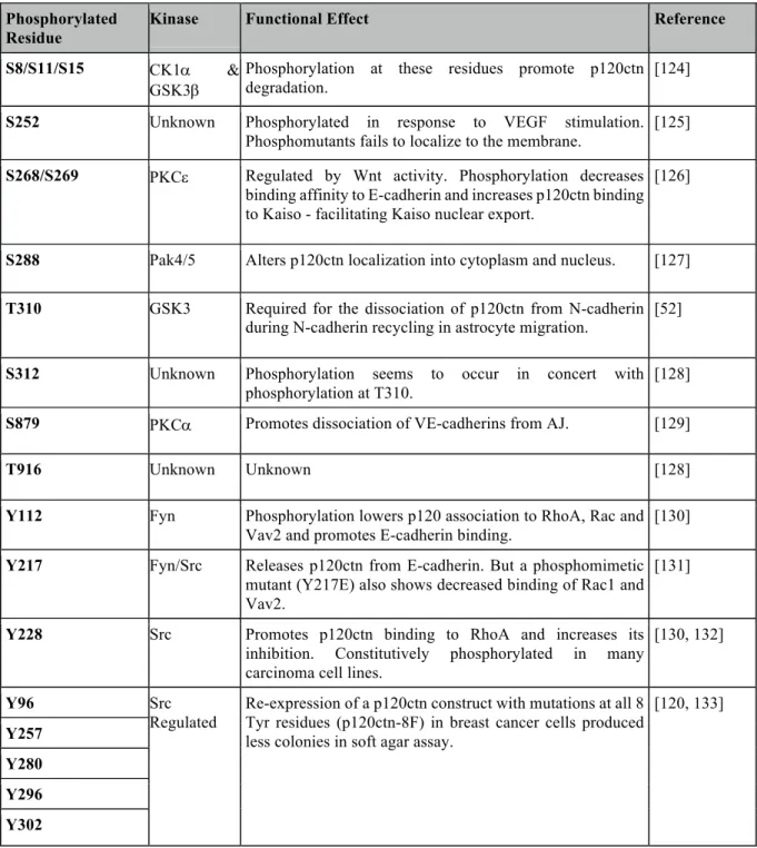
Table 2: Identified phosphorylation sites on p120ctn and the effects of site-specific modifications on p120ctn function.
Phosphorylated Residue
Kinase Functional Effect Reference
S8/S11/S15 CK1a & GSK3b
Phosphorylation at these residues promote p120ctn degradation.
[124]
S252 Unknown Phosphorylated in response to VEGF stimulation. Phosphomutants fails to localize to the membrane.
[125]
S268/S269 PKCe Regulated by Wnt activity. Phosphorylation decreases binding affinity to E-cadherin and increases p120ctn binding to Kaiso - facilitating Kaiso nuclear export.
[126]
S288 Pak4/5 Alters p120ctn localization into cytoplasm and nucleus. [127]
T310 GSK3 Required for the dissociation of p120ctn from N-cadherin during N-cadherin recycling in astrocyte migration.
[52]
S312 Unknown Phosphorylation seems to occur in concert with phosphorylation at T310.
[128]
S879 PKCa Promotes dissociation of VE-cadherins from AJ. [129]
T916 Unknown Unknown [128]
Y112 Fyn Phosphorylation lowers p120 association to RhoA, Rac and Vav2 and promotes E-cadherin binding.
[130]
Y217 Fyn/Src Releases p120ctn from E-cadherin. But a phosphomimetic mutant (Y217E) also shows decreased binding of Rac1 and Vav2.
[131]
Y228 Src Promotes p120ctn binding to RhoA and increases its inhibition. Constitutively phosphorylated in many carcinoma cell lines.
[130, 132]
Y96 Src
Regulated
Re-expression of a p120ctn construct with mutations at all 8 Tyr residues (p120ctn-8F) in breast cancer cells produced less colonies in soft agar assay.
[120, 133]
Y257 Y280 Y296 Y302
p120ctn in cancer
In normal cells, formation of adhesion triggers signalling events that suppress cell growth and migration, but in tumor cells, this regulation is impaired as cells lose adhesive structures, and increases growth and motility. Based on its ability to regulate E-cadherin function, p120ctn was initially identified as a tumor suppressor by maintaining cell-cell adhesion. However, it is also implicated in regulating Rho-GTPase signalling to regulate cell migration and plays a role in Src-mediated cell transformation. Therefore, p120ctn appears to have both tumor suppressing and tumorigenic roles in the cell.
In accordance with its role stabilizing E-cadherin, evidence of p120ctn as a tumor-suppressor is found in studies into p120ctn deletion in vivo using mouse models (See Table 1). In the salivary gland, p120ctn deletion results in severe loss of adhesion due to E-cadherin destabilization, and created neoplastic lesions [86]. Additional studies performed in the oral cavity, esophagus, and forestomach also generated invasive squamous neoplastic lesions, as well as a tumorigenic inflammatory microenvironment, in response to tissue-specific p120ctn deletions [83]. The same deletion and knockdown of p120ctn performed in lung tumor cell lines resulted in loss of E-cadherin, and further promoted cell invasion and metastasis [134]. Immunohistochemical studies have shown that p120ctn is downregulated in certain tumors such as breast, prostate and lung cancers [135-137]. Together, these results suggest that the loss of p120ctn in cells is a significant event in some cancers and leads to increased tumor aggressiveness.
Over the course of cancer development, the loss and downregulation of E-cadherin is observed in many tumors. In response to decreased E-cadherin levels in cells, p120ctn translocation to the cytoplasm is strongly linked to transforming pre-invasive lesions into metastatic tumors in lobular breast carcinoma [138]. This suggests that p120ctn mislocalization to the cytoplasm causes dysfunctional Rho-GTPase signal regulation, and thus driving the development of a metastatic phenotype in tumors. In colon cancer cells undergoing EMT, the loss of E-cadherin coincides with a decrease of RhoA activity and an increase in cytoplasmic p120ctn, which is correlated with increased tumor aggressiveness. Knockdown of p120ctn by RNAi in these cells could restore stress fiber formation, and significantly reduce motility – indicating that cytoplasmic p120ctn is a major driver of cell invasion upon loss of E-cadherin [139]. As addressed in a previous section, cytoplasmic p120ctn is also implicated in driving
invasive lobular breast carcinoma by inhibiting Mrip, and promoting ROCK1-mediated anoikis resistance [107]. Cytoplasmic p120ctn is also observed and correlated with poor prognosis in other cancers, such as pancreatic cancer, and lung squamous cell carcinoma. [137, 140].
In addition to mislocalizing upon E-cadherin loss, p120ctn can drive EMT by playing an essential role in mediating a cadherin switch. Since it is possible for different cadherins to compete for binding with a limited pool of p120ctn in the cell, overexpression of mesenchymal cadherins, such as R- and N-cadherins, can induce E-cadherin destabilization [141]. An isoform switch between isoforms 1 and 3 of p120ctn can also promote the cadherin switch process by selectively stabilizing N- or E-cadherins, respectively. In lung cancer cells, expression of p120ctn-1A promoted EMT and increased N-cadherin expression, while p120ctn-3A inhibited cell invasion and promoted E-cadherin expression [142]. Another isoform-specific tumorigenic function was observed when comparing the expression of isoforms 1 and 4 in renal cancer, where only cytoplasmic isoform 1 was able to effectively inhibit RhoA activity and activate Rac1 due to RhoA binding to the N-terminal domain that is lacking in isoform 4 [143]. Similarly, analysis of p120ctn isoforms in breast cancer cell lines revealed preferential expression of larger isoforms 1 and 2 in more invasive cells [144].
Finally, in some cancers, p120ctn can exert tumorigenic properties even in the presence of E-cadherin. In inflammatory breast cancer, overexpression of the RNA-binding protein eIF4G1 induces an upregulation of p120ctn that further stabilizes E-cadherin. In this specific type of cancer, it appears the stabilization of E-cadherin paradoxically promotes its progression through an unknown mechanism [145]. In squamous cell carcinomas, p120ctn mediated stabilization of E-cadherin is essential for collective cell migration and invasion [146].
Collectively, the data from decades of studying p120ctn in cancer reveals a nebulous number of potential mechanisms by which p120ctn can influence cancer progression. Given the large number of functions and regulatory roles that p120ctn plays within the cell, the mechanism by which it drives cancer development appears to be highly context dependent, and cell-type specific. However, in many of these cases, it appears that regulation affecting p120ctn binding to E-cadherin and localization can significantly alter the tumorigenic roles that p120ctn plays in a context-dependent manner. Studies into the phosphorylation status of p120ctn in cancers may come to reveal new connections by which signals impinging on p120ctn can drive cancer
Rationale and Objectives
Rationale
The Ras/MAPK signalling pathway is responsible for regulating a variety of cellular processes and responses, including regulating cell motility. As a kinase located downstream of the Ras/MAPK pathway, RSK has been shown to play a role in Ras/MAPK-mediated cell spreading and cell motility. However, currently the specific mechanisms by which RSK causes cell-spreading and loss of cell-cell adhesion in cells remain unknown. Our own proteomics studies have identified a new phosphorylation site on p120ctn that is regulated in response to RSK activity.
p120ctn is a crucial component of AJs and its dysregulation and mislocalization away from the plasma membrane is often linked to cancer progression, and more adverse outcomes. As a major stabilizer of classical cadherins in many cell types, its interaction with partner cadherins are crucial to maintaining proper cell function. Previous studies into phosphorylation sites present within the regulatory domain of p120ctn suggests these post-translational modifications (PTM) regulate p120ctn function by affecting its interaction with cadherins. In previous studies conducted by our lab, we have identified a potential novel phosphorylation site – S320 – on p120ctn that is also located within this crucial regulatory region. We hypothesized that phosphorylation of p120ctn at this site by Ras/MAPK activity promotes its dissociation from cadherins, and leads to AJ disassembly and overall loss in cell-cell adhesion (Fig. 1.5).
The main goal of my M.Sc. project is to validate p120ctn as a bona-fide substrate of the Ras/MAPK signalling pathway, and understand how p120ctn is regulated at the functional level by Ras/MAPK activity. Finally, if possible, I wanted to strive to understand how Ras/MAPK mediated regulation of p120ctn affects AJ function and how this translates to impacts on cellular function in the context of cell-cell adhesion and cell migration.
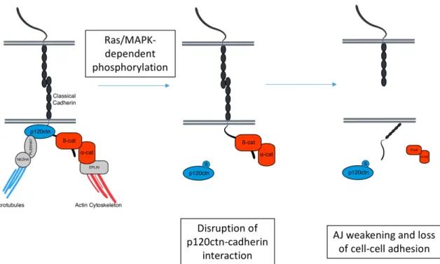
Figure 5. Our proposed model of the effects of Ras/MAPK signalling on p120ctn function and AJ stability.
The location of S320 within the regulatory domain of p120ctn suggests that phosphorylation at this site most likely will affect its binding to cadherins, and induce AJ destabilization. We present this model whereby increased phosphorylation of S320 in response to Ras/MAPK activity induces AJ disassembly by inhibiting p120ctn binding to cadherins at the surface.
Objectives
There are three main objectives to my thesis:
1. Given that we have identified through two separate proteomic screens that p120ctn may be a substrate of RSK, we intend to confirm and demonstrate that the Ras/MAPK pathway, through RSK, regulates phosphorylation at a specific site: S320 on p120ctn 2. As the proposed phosphorylation site, S320, is located within the regulatory domain of
p120ctn, we intend to investigate how Ras/MAPK activity may affect p120ctn function, and how S320 phosphorylation may contribute to the observed effects. 3. Since p120ctn is a major regulatory protein at AJs, we intend to investigate how