HAL Id: tel-01283851
https://tel.archives-ouvertes.fr/tel-01283851
Submitted on 7 Mar 2016HAL is a multi-disciplinary open access
archive for the deposit and dissemination of sci-entific research documents, whether they are pub-lished or not. The documents may come from teaching and research institutions in France or abroad, or from public or private research centers.
L’archive ouverte pluridisciplinaire HAL, est destinée au dépôt et à la diffusion de documents scientifiques de niveau recherche, publiés ou non, émanant des établissements d’enseignement et de recherche français ou étrangers, des laboratoires publics ou privés.
single cell
Daria Bonazzi
To cite this version:
Daria Bonazzi. The developmental polarity and morphogenesis of a single cell. Cellular Biology. Université Sorbonne Paris Cité, 2015. English. �NNT : 2015USPCB010�. �tel-01283851�
Université Paris Descartes
Ecole doctorale interdisciplinaire européenne Frontières du Vivant
Equipe de recherche: Organisation spatiale de la cellule Institut Jacques Monod, 13 Rue Helene Brion 75013 Paris
The developmental polarity and
morphogenesis of a single cell
Par Daria BONAZZI
Thèse de doctorat de biologie cellulaire
Dirigée par Nicolas MINC
Présentée et soutenue publiquement le 06 Mars 2015
Devant un jury composé de :
COUDREUSE, Damien Directeur de recherche Rapporteur WEDLICH SOLDNER, Roland Directeur de recherche Rapporteur BOCELYN-GALLEGO, Evelyne Directeur de recherche Examinateur
PROST, Jacques Directeur de recherche Examinateur
BOUADOUD, Arezki Directeur de recherche Membre invite MINC, Nicolas Directeur de recherche Directeur de these
3
a Marilena
…Anche se non ci sei tu sei sempre con me per antiche abitudini perchè ti rivedrò dovunque tu sia…
5
Acknowledgements
I would like to thank the members of the jury for kindly accepting to evaluate my work: the president Jacques Prost, the examiners Evelyne Bloch-Gallego and Arezki Boudaoud and especially my reviewers Damien Coudreuse and Roland Wedlich-Soldner for revising the manuscript.
I would like to thank Nicolas for having welcomed me in his new-born team: it has been great to work with you, you're a brilliant, efficient and passionate scientist with a never-ending stream of ideas! Under your guidance, I have learned a unique way of doing research and I grew up both professionally and personally. Thank you also for pushing me hard to improve myself: I think you care a lot about people in your team, you want the best for all of them, and that's really special.
I thank all the collaborators who contributed to my PhD work: Arezki Boudaoud and his PhD student Jean-Daniel Julien for building up the mechanical model in the first paper and giving me inspiring suggestions; Maryse Romao, for her expertise in electron microscopy; Rima, for brilliantly starting the spore project before me and for all the professional and personal chats we had along these last three years; finally Delphine Salort, for her mathematical model on cell polarity and its relationship with cell geometry in the second paper.
I would like to thank all the people of the lab for their help and support during my PhD: Hiro, for your naturally wise and calm attitude, with some more extravagant moments in front of a beer (but no ninja ties, I promise!); Armin, for your patience in listening to me sooo many times and for your jokes, that sometimes I don't get but well, that's funny anyway ;). Anaëlle, for your very very very special personality, sometimes a bit scary but well that's Xxẍxxxland, with all its cats and unicorns! Valeria, perché hai illuminato questi ultimi mesi di tesi tesa! E per il tuo singhiozzo che tiene davvero compagnia, e mi mancherà. Moreover, I would like to thank all the people that worked in the Minc's lab in the past: Alexis, I miss your jajaja warm laugh and your jokes about my locura; Yonatan, having you as the first student in my life has been a super
6
enriching and enjoyable experience; Henry and Marguerite, les deux super stagiaires à l'opposé, bref la musique rock contre la classique.
Moreover, I thank Sebastien and his team with whom we pleasantly share the lab space here at the IJM, the colleagues of the third floor, all the people with whom I had nice discussions during common labmeetings, seminars and young scientist clubs; Je voudrais aussi remercier Mireille pour m'avoir aidée à faire face à l'administration francaise, et Patricia pour son travail essentiel de preparation des milieux et des boites. I thank also all the people of the Imagoseine platform, in particular Orestis, France, Xavier and Vincent, for their help with the microscopes and for being always super nice! Finally, I thank Kim and Antonin for taking care of organizing some regular aperos at the IJM, it is always a great opportunity to get to know people from other labs in a relaxed atmosphere.
I need also to thank numerous people in Curie, with whom I shared my daily lab life for about two years: first Matthieu, thanks for accepting my application as a Leonardo da Vinci student, the interdisciplinary atmosphere of your lab and the great research project I had to tackle both contributed to my decision of staying in Paris to start a PhD in cell biology. Next, I thank all the Piel's lab members: Hawa, my big friend in and beyond Curie, you are a perfect mixture of sensitivity and fun, thanks for always encouraging me, for your suggestions (scientific and not) and for all the time we spent together, inside or outside or around the lab: I wish you all the best for your US adventure! Paolo, con annessi Chiara e Mauro e Giovanna, la mia famiglia parigina d'adozione, vi voglio bene! Franzi, the best counselor ever, you really have a special emotional intelligence for both happy and sad things! Emmanuel, it took me some time to get to know you but it was really worth it, you and I are quite similar isn't it? Ewa, we had very funny moments together, I wish you all the best for your future and I hope we will keep in touch! Nico, merci pour être aussi bizarregeek, pour ton aide au labo et pour toutes les soirées chez toi à jouer aux jeux-vidéo, c'était super sympa! Julie, merci pour ton aide avec les microscopes et pour ton soutien pendant les moments plus durs de travail; Mael, tu as été un très bon supervisor pendant mon stage et un collègue excellent après! Yan Jun, thanks for your kindness and your daily smile; Melanie, les pauses cigarettes ne seraient pas étées les même sans ta drôle compagnie! Clotilde, on a pas encore fait de la musique ensemble mais on peut toujours récupérer; Matthew, we just quickly crossed before I moved to IJM but it has been nice to meet you!
7 I would like to thank some other people in Curie with whom I have shared some very nice moments: the fantastic Tran's and Paoletti's team, especially Phong, Anne, Merce, Kathleen, Sergio and Imene, thanks for sharing your pombe's expertise, the strains and the microfabrication hood, and thanks for being always so enjoyable and kind! I thank all the people of the third floor, in particular the Perez's and Houdusse's teams, and the fifth too, with your fantastic smoking balcony. A special thanks goes to Lamine, you are a really great and funny friend! Last but not least, I thank all the people in the Nikon Imaging center who helped me so many times with the microscopes, in particular Lucie and Vincent.
I thank also all the people that contribute to create a passionate and novel working atmosphere in my ED "Frontieres du Vivant", and my TAC supervisors, Phong and Claire, for being very supportive and positive about my work.
This PhD would not have been the same without the very unique experience of the Physiology Course in Woods Hole: two months of hard work in an amazing environment, surrounded by excellent and diverse people! I want to especially thank the three supervisors I've got the chance to work with, Clare, Wallace and Hari, the colleagues (and friends) Tamara, Felix, Alexis and Einat, and Nicolas again for sending me there!
To conclude, I want to thank all the people that encouraged me out of the lab: among my friends in Paris, a special thanks goes to Irene, i compagnoni italiani di lab (in particolare Giulia e Davide!), i mitici pareggiani (in particolare la Michi e Michi) e i membri di Argo, Michele, Matthieu e Rafael. Ringrazio i miei compagni di studi chimici a Bologna, Tea, Magna, Vale, Manu e Tommi, perché anche se siamo un po' sparsi in giro non ci perdiamo mai (troppo) di vista! Ringrazio anche il gruppo degli elettrocomici al Ciamician, in particolare Stefi, con cui mi sono avventurata per la prima volta nel mondo della biologia. E poi salto a Reggio, e allora ringrazio le mie amiche storicissime e insostituibili: Lollipoppy, Laura, Alle, Chiara, Sara ed Elly, é sempre un piacere ritrovarvi quando torno nell'Emilia paranoica! Un grazie speciale va a Luca, per avermi fatto scoprire storie e pensieri attorno alla scienza che restano purtroppo alieni agli scienziati, e per tutto quello che abbiamo condiviso.
Vorrei infine ringraziare la mia fantastica famiglia, per tutto il sostegno e l'affetto che non mi hanno mai fatto mancare: papà, Giuli e Aldo, Tati e Ferni, vi voglio bene!
9
Abstract
The developmental polarity and morphogenesis of a single cell
How cells establish their proper shapes and organization is a fundamental biological problem. In this thesis, I investigated the dynamic development of cellular form and polarity in the rod-shape fission yeast cell. These studies are based on monitoring how small symmetric fission yeast spores grow and self-organize to break symmetry for the definition of their very first polarity axis. In a first part, I studied interplays between surface mechanics of the spore cell wall and the stability of Cdc42-based polarity domains which control spatio-temporal aspects of spore symmetry breaking. In a second part, I studied mechanisms by which these polarity domains control their width and adapt it to cell surface geometry, a process likely relevant to understand how functional cortical domains scale to cell size. Overall these novel investigations focusing on how cells dynamically develop their form and polarity de novo highlight complex feedbacks in morphogenesis that cannot be evidenced by looking at cells at “steady state” or with genetics.
Resumé
Développement de la morphogenèse et de la polarité d’une cellule unique
Comment les cellules établissent leurs formes et organisations internes est un problème biologique fondamental. Au cours de cette thèse, j’ai étudié le développement de la forme cellulaire et de la polarité chez la cellule de levure fissipare. Ces études sont fondées sur l’exploration de la façon dont les petites spores symétriques de levures se développent et s’organisent pour briser la symétrie pour la définition de leur tout premier axe de polarité. Dans une première partie, j’ai étudié les couplages entre la mécanique de surface de la paroi cellulaire des spores et la stabilité de domaines de polarité de Cdc42 qui contrôlent les aspects spatio-temporelles de la brisure de symétrie de ces spores. Dans une seconde partie, j’ai étudié les mécanismes par lesquels ces domaines de polarité contrôlent leur taille et l'adapte à la géométrie
10
de la cellule, un processus vraisemblablement pertinents pour comprendre comment des domaines fonctionnels corticaux s’adaptent à la taille des cellules. Globalement, ces nouvelles recherches focalisant sur la façon dont les cellules développent dynamiquement leur forme et polarité de novo, permettent de mettre en évidence des couplages complexes dans la morphogenèse qui ne peuvent pas être testés en regardant les cellules à « l’état stationnaire» ou avec des outils génétiques.
11
Table of contents
Acknowledgements ... 5 Abstract ... 9 Resumé ... 9 Table of contents ... 11 List of abbreviations ... 15 List of figures ... 17 List of movies ... 21 AIMS ... 25 INTRODUCTION ... 271. PRINCIPLES OF CELL MORPHOGENESIS ... 29
1.1 How to define cell shape? ... 31
1.2 Cell shape and cell function ... 31
1.3 Cell shape and cell polarity ... 33
1.3.1 Cell polarity: an (old) lesson from eggs ... 35
1.3.2 The biochemical revolution: vectorial physiology ... 37
1.3.3 Conserved molecular players for cell polarization ... 38
1.3.3.1 Signal transduction through GPCRs and G proteins ... 39
1.3.3.2 The core proteins of cell polarity: small GTPases ... 40
1.3.3.3 The cytoskeleton ... 41
1.3.3.4 Membrane remodeling: exocytosis and endocytosis ... 44
1.3.3.5 Polarity complexes ... 45
1.3.4 Cell polarization: with or without a cue? ... 47
1.4 Cell shape and cell mechanics ... 48
1.4.1 On growth and form ... 50
1.4.2 Conserved molecular players for cell mechanics ... 51
1.4.2.1 Sensing and transducing a mechanical cue ... 52
1.4.2.2 Structural elements of the cells ... 53
12
1.5 General principles of symmetry breaking ... 57
1.5.1 Reaction-Diffusion systems ... 57
1.5.2 Transport-based positive feedback loop ... 59
1.5.3 Breaking symmetry mechanically ... 59
1.5.4 Mechanochemical patterning ... 60
1.6 Cell shape and cell size ... 62
2. CELL MORPHOGENESIS IN YEAST ... 65
2.1 Why yeast as a model organism? ... 67
2.2. Cell morphogenesis in fission yeast ... 68
2.3 Cell shape and cell polarity in fission yeast ... 70
2.3.1 Molecular players in fission yeast cell polarity ... 71
2.3.1.1 Sensing the partner: GPCRs, G-proteins and mating ... 71
2.3.1.2 Small Rho G-proteins in fission yeast polarized growth ... 72
2.3.1.3 The cytoskeleton: building up polarity domains ... 74
2.3.1.4 Membrane remodeling: polarized transport ... 76
2.3.2 Cell polarization: with or without a cue? ... 78
2.3.3 External control of cell polarity in fission yeast ... 80
2.3.3.1 Manipulation of cell shape ... 80
2.3.3.2 Electrical control of cell polarity ... 81
2.4 Cell shape and cell mechanics in fission yeast ... 82
2.4.1 Cell wall structure ... 82
2.4.2 Cell wall biosynthesis ... 84
2.4.3 Cell wall mechanics ... 86
2.4.4 Turgor pressure ... 87
2.4.5 The mechanics of cell growth ... 89
2.4.6 Another possible force generator: the cytoskeleton ... 91
2.5 Building up a rod-shaped cell ... 91
2.5.1 From round to rod-shaped: spheroplast regeneration ... 94
2.6 Symmetry breaking in yeast models ... 96
2.6.1 Simple Cdc42-based positive feedback ... 97
13
2.6.3 Positive feedback via actin-mediated transport ... 99
2.6.4 Winner-takes all competition ... 101
2.6.5 Coexisting cortical domains ... 102
2.6.6 Oscillations of Rho-GTPases ... 104
2.6.7 Roles of negative feedback ... 107
2.7 Cell size regulation in fission yeast ... 108
3. THE MYSTERIOUS CASE OF SPORES... 111
3.1 The discovery of spores ... 113
3.2 General features of spores ... 114
3.3 The spore wall: composition and biogenesis ... 120
3.4 The spore wall mechanics ... 123
3.5 Spore development ... 125
3.5.1 Sporulation ... 125
3.5.1.1 Meiosis ... 127
3.5.1.2 Forespore membrane assembly ... 128
3.5.1.3 Spore wall assembly ... 130
3.5.2 Spore dormancy ... 132
3.5.3 Spore germination ... 133
3.5.4 Spore outgrowth... 135
3.5.4.1 Guidance by an extrinsic cue ... 135
3.5.4.2 Spontaneous symmetry breaking: the role of polarity ... 138
3.5.4.3 Spontaneous symmetry breaking: the role of mechanics ... 141
3.6 Spore size control ... 145
RESULTS ... 147
Symmetry breaking in spore germination relies on an interplay between polar cap stability and spore wall mechanics ... 149
1.1 Summary ... 149
1.2 Paper ... 151
Actin-based transport adapts polarity domains size to local curvature ... 153
2.1 Summary ... 153
2.2 Paper ... 155
14 CONCLUSION ... 163 METHODS ... 165 1. Photoablation ... 165 2. Death assay ... 165 3. Microchambers setup... 166 4. Microchannels setup ... 166
5. Setups for drug treatment and rinse out ... 167
6. Fluorescence exclusion method ... 167
7. Actin imaging and analysis ... 168
OTHER PUBLICATIONS ... 169
15
List of abbreviations
cAMP/PKA: Cyclic AMP / protein kinase A AP: Anterior-posterior
Arp2/3: Actin related protein 2/3 ATP: Adenosine triphosphate BAR: Bin–Amphiphysin–Rvs CAT: Conidial anastomosis tube
CRIB: Cdc42- and Rac-interactive binding CWI: Cell wall integrity
ECM: Extracellular matrix EF: Electric field
EMT: Epithelial-mesenchymal transition ER: Endoplasmic reticulum
FSM: Forespore membrane GAP: GTPase-activating protein
GDI: Guanine nucleotide-dissociation inhibitor GDP: Guanosine diphosphate
GEF: Guanine nucleotide-exchange factor GFP: Green Fluorescent Protein
GPCR: G protein-coupled receptor GTP: Guanosine triphosphate HOG: High osmolarity glycerol LEP: Leading edge protein
MAPK: Mitogen activated protein kinase MT: Microtubule
MTOC: Microtubule organizing center NDR: Nuclear dbf2-related
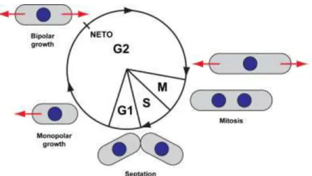
NETO: New end take off
16
NMY-2: Non-muscle myosin II NPF: Nucleation promoting factor OSW: Outer spore wall
PAR: Partitioning-defective PDMS: Polydimethylsiloxane PI: Phosphatidylinositol
PIP: Phosphatidylinositol phosphate PM: Plasma membrane
RD: Reaction diffusion
RGS: Regulator of G protein signaling SASP: Small acid-soluble protein SEM: Scanning electron microscopy SNARE: Soluble NSF Attachment Protein SPB: Spindle pole body
TEM: Transmission electron microscopy WASp: Wiskott–Aldrich syndrome protein
17
List of figures
Figure 1: The complexity of cell shape in an assortment of diverse protists.
Figure 2: Examples of animal cells with unique shapes, directly related to specific cell functions. Figure 3: Diverse polarized cells
Figure 4: Historical diagrams of cell polarity Figure 5: Images of rabbit skeletal muscle Figure 6: A common pathway for cell polarity
Figure 7: Activation of the G alpha subunit of a GPCR Figure 8: The Rho-GTPase cycle
Figure 9: Basic properties of actin filaments and microtubules Figure 10: Establishment of orientated actin arrays
Figure 11: Schematic of the minimal requirements for cell polarization through intracellular trafficking
Figure 12: Interplay between PAR proteins and the polarity machinery
Figure 13: Cue-dependent and random cell polarization in different organisms Figure 14: Integration of mechanical stress in the cell polarity pathway
Figure 15: Examples of cell shapes in protists of the Foraminifera family which can be explained by the law of minimal surface area
Figure 16: Examples of tissue development guided by mechanical cues Figure 17: Scheme for different cases of cortex relaxation in cellular events Figure 18: MT spatial organization in the shoot apical meristem
Figure 19: Pattern formation in chemical systems Figure 20: Actin-dependent positive feedback loop
Figure 21: Analogy of the tension state in an actin gel growing from a bead surface and in the cell cortex
Figure 22: Coupling of mechanical and chemical elements in cell polarization Figure 23: Diverse mechanisms of cell size homeostasis
Figure 24: Yeast as a model system
18
Figure 26: The meiotic cell cycle of fission yeast cells, followed by spore germination and re-entry to the vegetative cell cycle
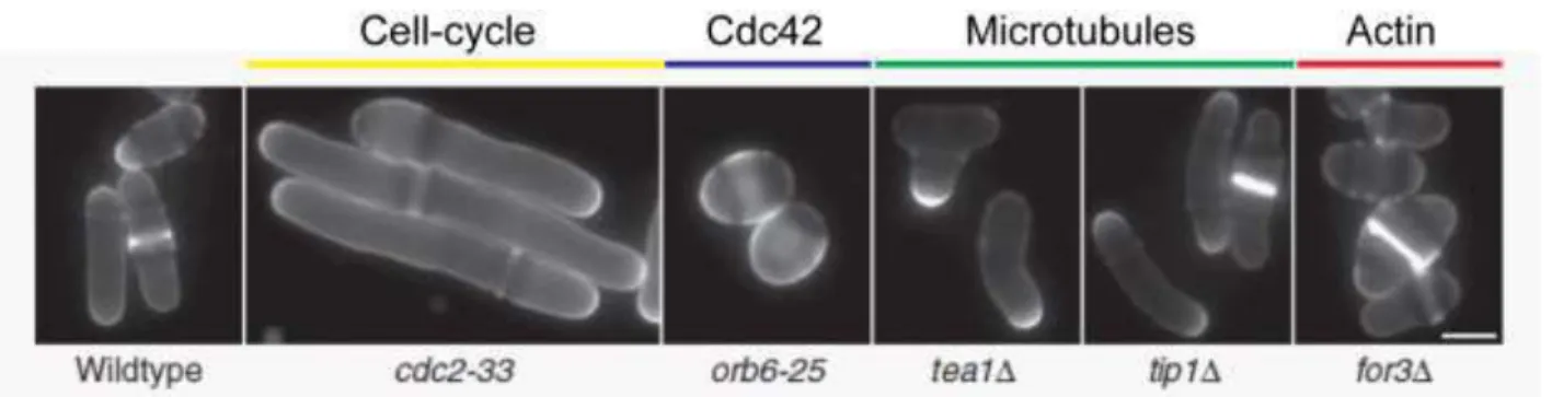
Figure 27: Morphological fission yeast mutants Figure 28: Mating signaling in fission yeast Figure 29: The Cdc42 module
Figure 30: Cytoskeleton organization in fission yeast interphase cells Figure 31: The fission yeast actin cytoskeleton
Figure 32: Microtubule-dependent polarization in fission yeast Figure 33: Polarized secretion in fission yeast
Figure 34: Decision making for polar growth in budding and fission yeast Figure 35: External manipulation of fission yeast cell shape
Figure 36: Electrical regulation of fission yeast polarity Figure 37: Cell wall regeneration in a fission yeast protoplast
Figure 38: Schematic of fission yeast cell wall biosynthesis and structure Figure 39: Illustration of the method used to compute chamber deformation Figure 40: Physical model of cell growth by tip elongation
Figure 41: Comparison of different growth patterns for rod-shaped cells Figure 42: Pathways for control of cell width in fission yeast cells Figure 43: Protein polarization accompanies spheroplast recovery Figure 44: Minimal model of a positive feedback circuit
Figure 45: Cdc42 polarization via reaction-diffusion mechanism
Figure 46: Actin-based mathematical model for the dynamic redistribution of polarized membrane proteins.
Figure 47: A single Cdc42 cluster forms in the simulations with molecular noise Figure 48: Competition and coexistence among GTPase clusters
Figure 49: Cdc42 oscillatory pattern in fission yeast
Figure 50: A spatial gradient links cell size to cell division in fission yeast
Figure 51: Historical drawing of all characterized developmental stages of a single fungal species
Figure 52: Diversity of spore morphology in nature Figure 53: Examples of spores-forming structures
19 Figure 54: Mechanisms of infection by fungal and microsporidian pathogens
Figure 55: The spore coat encases the spore to protect it from hard environmental conditions Figure 56: Example of outgrowing spores
Figure 57: Comparison of the spore wall and the vegetative cell wall Figure 58: Structure of the fission yeast spore wall
Figure 59: Electron microscopy of yeast ascospores and spore walls
Figure 60: A schematic of meiosis and ascospore development in fission yeast
Figure 61: A model for the initiation and development of the FSM on the outer plaque of the SPB
Figure 62: Examples of fission yeast spore wall mutants compared to wild-type
Figure 63: Morphological changes of fission yeast spores during germination and outgrowth Figure 64: Schematic of spore germination and outgrowth in fission yeast
Figure 65: Colocalization of activated GTPases, F-actin and sterol-rich plasma membrane regions constitute sites of polarized growth
Figure 66: Examples of cytoskeleton remodeling during germination and outgrowth Figure 67: Electron micrographs of germinating spores
Figure 68: Outgrowth corresponds to local rupture or dissolution of the outer spore coat Figure 69: Some fungal spores germinate through a germ pore
Figure 70: Schematic of the mechanism for fission yeast spore germination and outgrowth Figure 71: Example of fission yeast spores at different developmental stages
21
List of movies
Movie 1: Phase-contrast time lapse of the development of a wild-type fission yeast spore, Related to Figure 1 of Paper 1. The green outline corresponds to an automatically detected cell contour. Note the change in phase contrast at germination and the elongation of the polar tube at outgrowth. Time is in hr:min.
Movie 2: Phase-contrast time lapse of the development of a wild-type fission yeast spore going through three rounds of cell cycle, related to Figure 1 of Paper 1. Note that the first “mother” cell remains monopolar and grows away from the spore body. Time is in min.
Movie 3: Time-lapse phase-contrast and epifluorescence images of several developing spores expressing the polarized growth marker GFP-bgs4, related to Figure 2 of Paper 1. The signal in the fluorescent channel has been renormalized at each time point, and the scale bars may vary between successive movie. Note the phases of polar cap assembly and disassembly at successive locations and the stabilization at outgrowth. Time is in hr:min.
Movie 4: Time-lapse phase-contrast and epifluorescence images of a developing spore expressing the polarized growth marker GFP-bgs4, related to Figure 2 of Paper 1. Polarity cap drives local sites of polar growth at the spore surface during the wandering phase. GFP-bgs4 cap is indicated by white arrows, and subsequent local growth sites are indicated by red arrows. Time is in hr:min.
Movie 5: Time-lapse phase contrast of a group of four daughter spores attached to each other assuming the shape of the mother ascus, which germinate and outgrow, related to Figure S3 of Paper 1.
Movie 6: In silico simulation of growing spores, related to Figure 4 of Paper 1. The white semidisc represents the polar cap. The colored annulus represents the OSW, and values of strains in the OSW are represented with a color code (increasing from blue to red). When the OSW
22
breaks, the transparent annulus represents the inner cell wall superimposed on the OSW, for simplicity of representation. Note that the OSW ruptures where the strain reaches the red zone.
Movie 7: Time lapse of three spores expressing GFP-bgs4, related to Figure 6 of Paper 1. One spore is ablated at the site of bgs4 cap, another one is ablated away from the polar cap, and the third one is not ablated. The movie starts at the moment of laser ablation. Red arrows and dots point to sites of ablations, and black arrows point at outgrowing tubes. Time is in hr:min.
Movie 8: In silico simulation of growing spores with a hypothesis of positive feedback that biases the stabilization of the polar cap toward sites of maximal surface expansion rates, related to Figure 7 of Paper 1.
Movie 9: In silico simulation of growing spores with isotropic growth, related to Figure 6 of Paper 1. Note how the OSW ruptures at many locations.
Movie 10: In silico simulation of growing spores with no feedback hypothesis between polar cap and wall mechanics or geometry, related to Figure 7 of Paper 1.
Movie 11: In silico simulation of growing spores with a hypothesis of positive feedback that stabilizes the polar cap at sites of minimal surface stress, related to Figure 7 of Paper 1.
Movie 12: In silico simulation of growing spores with a hypothesis of positive feedback that stabilizes the polar cap at sites of maximal curvature, related to Figure 7 of Paper 1.
Movie 13: In silico simulation of growing spores with a hypothesis of positive feedback that stabilizes the polar cap at sites of maximal surface expansion rates.
Movie 14: Confocal images of several developing spores expressing the polarity marker CRIB-GFP, related to Figure 1 of Paper 2. Scale bar, 1m. Note the phases of polar cap assembly and disassembly at successive locations and the corresponding variation in pole cap width. Time frame is 5 min.
23 Movie 15: 3D reconstruction of several developing spores expressing the polarity marker CRIB-GFP, related to Figure 1 and S1 of Paper 2. Confocal z-stacks were acquired with a 0.2m z-step over 5m range. Note the differences in pole cap size for spores with various local radii of curvature.
Movie 16: Maximal z-projections of confocal z-stacks of several developing spores expressing the LifeAct-mCherry, related to Figure 4 of Paper 2. Confocal z-stacks were acquired with a 0.2m z-step over 5m range. Time frame is 30sec. Note that actin structures, both cables and patches, are highly dynamic.
Movie 17: 3D reconstruction of several developing spores expressing CRIB-GFP and LifeAct-mCherry, related to Figure 4 of Paper 2. Confocal z-stacks were acquired with a 0.2m z-step over 5m range. Note the differences in pole cap size for spores with various local radii of curvature, and the corresponding reorganization of the actin cables network.
Movie 18: Phase-contrast time lapse of the development of two bgs4/pbr1–8 fission yeast spores, Related to Discussion. Time is in hr:min.
25
AIMS
Cells come in a wide range of forms and sizes. Cell shape and organization influences many of the basic cellular functions, such as growth, migration or division, and defects in cell morphogenesis can give rise to many diseases.
But how does a cell develop and maintain its shape?
The unicellular fission yeast Schyzosaccharomyces pombe is an excellent model organism to address such kind of questions, as it grows in a simple rod-shape by tip elongation and divides in the middle when it reaches 14 m of length. Moreover, genetic manipulation allows to generate mutants with abnormal shapes, from round to bent or branched ones, hence shedding light on the molecular mechanisms responsible of cell shape regulation. Fission yeast cells grow at cell tips by targeting components such as the Rho-type GTPase Cdc42, and actin nucleators, which promote secretion needed for new membrane addition at cell tips.
In absence of nutrients, fission yeast cells develop to generate spores, as in the case of bacteria, plants, algae and other fungi. Spores are round, symmetric and dormant: moreover, they are encased in a very rigid shell, called outer spore coat, which is responsible of their high resistance to environmental stress, for very long periods of time.
In my PhD thesis, I have investigated how fission yeast spores germinate, grow and regenerate a rod-shaped vegetative cell. In a first part, I have characterized the coordination between polarity cap stabilization, growth and surface mechanics in this symmetry breaking event, in order to specify cell shape. In a second part, I have addressed how polarity domains, built around active-Cdc42, may define their size, and how they may adapt their size with cell size or cell shape.
These studies aim to define general principles for spontaneous polarization in eukaryotes, and how this process may be related to cell mechanics and geometry.
27
29
1.
PRINCIPLES OF CELL MORPHOGENESIS
In this chapter, I will describe some general features of cell morphogenesis. I will start with a short description of how to define cell shape. I will then present some examples to illustrate the relationship between cell form and cell function and its general impact on cell physiology and disease, both at the uni- and multi-cellular level. Hence, I will illustrate in detail the main regulators of cell morphogenesis: first, cell polarity, with a historical introduction of the concept, a description of the main molecular players for eukaryotic cell polarity, and finally a classification of cell polarization processes that can be guided either by extrinsic or intrinsic cues. Second, cell mechanics, with a short historical overview followed by a general description of how forces can shape single cells and tissues. Hence, I will discuss the main sets of principles that have been proposed to generate spontaneous symmetry breaking. To conclude, I will briefly talk about another aspect of cell morphogenesis: cell size regulation, as a result of the balance between growth and division.
1. PRINCIPLES OF CELL MORPHOGENESIS ... 29 1.1 How to define cell shape? ... 31 1.2 Cell shape and cell function ... 31 1.3 Cell shape and cell polarity ... 33 1.3.1 Cell polarity: an (old) lesson from eggs ... 35 1.3.2 The biochemical revolution: vectorial physiology ... 37 1.3.3 Conserved molecular players for cell polarization ... 38
30
1.3.3.1 Signal transduction through GPCRs and G proteins ... 39 1.3.3.2 The core proteins of cell polarity: small GTPases ... 40 1.3.3.3 The cytoskeleton ... 41 1.3.3.4 Membrane remodeling: exocytosis and endocytosis ... 44 1.3.3.5 Polarity complexes ... 45 1.3.4 Cell polarization: with or without a cue? ... 47 1.4 Cell shape and cell mechanics ... 48 1.4.1 On growth and form ... 50 1.4.2 Conserved molecular players for cell mechanics ... 51 1.4.2.1 Sensing and transducing a mechanical cue ... 52 1.4.2.2 Structural elements of the cells ... 53 1.4.2.3 Mechanical forces generated by the cytoskeleton ... 55 1.5 General principles of symmetry breaking ... 57 1.5.1 Reaction-Diffusion systems ... 57 1.5.2 Transport-based positive feedback loop ... 59 1.5.3 Breaking symmetry mechanically ... 59 1.5.4 Mechanochemical patterning ... 60 1.6 Cell shape and cell size ... 62
31
1.1 How to define cell shape?
The term "cell morphogenesis" refers to the processes that generate the forms of cells in the course of their growth, division, or development. The complex structure of a living cell is critical for cellular function. Yet relatively little is known about the mechanisms that produce the complex spatial organization of a living cell. On one hand, cells specify a certain form by integrating signals from the extracellular environment and/or their intracellular spatial organization and establishing a certain polarity axis. On the other hand, cell shape can also be viewed as the result of the mechanical balance of the forces exerted on the cell membrane by intracellular components (in particular, the cytoskeleton in animal cells and the cell wall in plants, fungi and bacteria) and the outside environment. Cell polarity and cell mechanics are highly regulated and interconnected through feedbacks loops to finally achieve a certain shape: in a multi-cellular context, cellular mechanical properties and gene expression/protein activation are coordinated among neighboring cells, to achieve global tissue shaping. It is also to be noted that while many aspects of cell shape and polarity may be determined by external cues, this does not preclude the possibility of self-organizing through spontaneous symmetry breaking. To conclude, another simple and fundamental aspect of cell morphogenesis is size regulation, which results from the balance between cell growth and cell division, and is an important determinant of cellular physiology. From this conceptual landscape, is it possible to identify any general organizational principle in cellular complexity, ranging from free-living cells to cells in tissues?
1.2 Cell shape and cell function
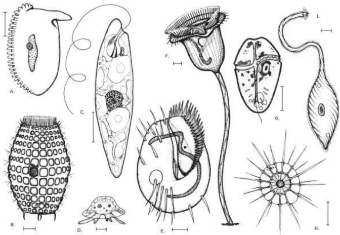
In all living organisms, from prokaryotes to eukaryotes, cells show a high degree of reproducible, non-random geometrical order, together with an extraordinary diversity in cell shapes, the most striking being the elaborate structural specializations of some free-living single-celled organisms. Many of the most complex-looking cells are protists, eukaryotic microorganisms with an elaborate anatomy which includes structures such as sensory bristles, photoreceptors, beating cilia, leg-like appendages, mouth parts, stinging darts, and muscle-like contractile bundles (Figure 1) (Marshall, 2011).
32
Different cell shapes are not just an eccentricity of nature, but are also coupled to specialized cell functions (Figure 2): in higher eukaryotes, muscle cells have a fusiform shape and can stretch and relax, in order to release tension, support high stresses and at the same time maintain contractility; neurons can harbor extensions, reaching lengths of many meters, to communicate between tissues over long distances; epithelial cells possess rectangular shapes to build up barriers that regulate ionic homeostasis between different biological compartments. In case of infection, immune cells efficiently change shape to accomplish specific tasks, in particular finding and eliminating pathogens: for instance, lymphocytes that migrate throughout the body to get to the site of infection can squeeze through narrow pores in order to cross tightly packed tissues such as the endothelium, while neutrophils engulf bacteria and viruses by 'swallowing' them. A very last example of cell shape diversity is given by the two unique players of the fertilization process: on one hand, sperm cells, that are very small, with a long tail-like structure,
Figure 1: The complexity of cell shape in an assortment of diverse protists. The organisms in A, B, E, F,
and I are ciliates; C is a euglenoid; D is an amoeba; G is a dinoflagellate; H is a heliozoan. Scale bars, 10 m. (Sleigh, 1973).
33 called flagellum, to propel themselves and swim over long distances; on the other hand, egg cells, that are round, immobile and big.
As the cell is the functional unit of any living tissue, all shape changes in the organism are driven by events at the cellular level. In combination with cell division, growth and death, changes in individual cell shape are central to tissue morphogenesis and homeostasis. Shape change of individual cells, independent of their neighbors, contributes to different morphogenetic processes in development, such as the migration of single primordial germ cells towards the gonad (Blaser et al., 2006). However, in most morphogenetic events, cell shape change is coordinated amongst hundreds of neighboring cells and drives shrinkage, extension, folding and movement of tissues.
1.3 Cell shape and cell polarity
Today, we know that most cell types of metazoans, but also unicellular organisms, such as yeast and ciliated protozoa can polarize. Even in the case of prokaryotes, the notion of bacterial cells as unorganized bags of proteins has been abandoned, leaving space to the concepts of high spatial organization and asymmetric protein distribution. Cell polarity is essential for a great number of cellular functions, such as differentiation, polarized cell growth, activation of the immune response, directional cell migration, and vectorial transport of molecules across cell layers (Figure 3) (Drubin and Nelson, 1996). For example, the bacterium Caulobacter crescentus shows
Figure 1: Examples of animal cells with unique shapes, directly related to specific cell functions. A brief description of each specific case can be found in the text.
34
polarized distributions of the flagellum and stalk structures, leading to asymmetric cell division; the budding yeast Saccharomyces cerevisiae displays also pronounced cellular asymmetries during its normal growth cycle, that allow to distinguish mother from daughter cells; polarized hippocampal neurons can polarize by growing dendrites at the postsynaptic side and axonal outgrowths at the presynaptic side; cells in epithelial tissues possess a distinctive polarized internal organization, that is essential for the assembly of specific membrane domains, termed apical and basolateral; migrating cells such as T-cells establish a protrusive front, called leading edge, and a contractile rear, to achieve directional movement; finally, some uni- and multi-cellular organisms define an anterior-posterior (or front-back) polarity early during development.
A surprising feature of cell polarity is that it gives rise to this diversity of cell shapes and functions from a basic set of evolutionarily conserved and adapted core mechanisms, including localized assembly of signaling complexes, cytoskeleton remodeling, mobilization of proteins from intracellular pools, and targeted vesicle delivery to sites of membrane growth. (Nelson, 2003).
In tissues, the organization of cells relies on the integration of polarizing signals from different interdependent biological processes (Bryant and Mostov, 2008). First, cells must sense their environment, including where they are in relation to their neighbors, by direct interaction of cells with the extracellular matrix (ECM) through various receptors. Cells can also communicate
Figure 3: Diverse polarized cells. In the neuron picture, F-actin is marked in red and the axonal marker Tau in green; in both the epithelial cell drawing and the migrating T-cell picture, actin is marked in red and microtubules in green; in the C. elegans embryo picture, the early-endosome-associated protein EEA1 is marked in blue and the nonmuscle myosin II NMY-2 is marked in red (Amberg, 1998; Arkowitz and Iglesias, 2008; Thanbichler and Shapiro, 2006).
35 with other cells through adhesion molecules, such as cadherins, and the sensing of diffusible factors, such as morphogens. These combined cues function as instructions for cells to orient and coordinate the asymmetrical distribution of polarity complexes, in order to establish and enforce the generation of an axis of asymmetric organization at the tissue level. Cell polarity is thus crucial for patterning and morphogenesis at the multicellular scale, for development and tissue homeostasis. (Nelson, 2003). Loss of cell polarity can cause developmental disorders and cancer. For example, most human tumours are derived from epithelial tissues, with the common feature of a simultaneous dysregulation of apico-basal cell polarity and cell growth (Wodarz and Nathke, 2007).
1.3.1
Cell polarity: an (old) lesson from eggs
Cell polarity has long been recognized as a fascinating biological phenomenon, related to a number of properties such as cell shape, position, and size; historically, studies in the fields of embryology and developmental biology have been fundamental for the debut of this wide topic. In 1896, in his book "The cell in development and inheritance", Edmund Beecher Wilson
Figure 4: Historical diagrams of cell polarity. A. Schematic of Van Beneden's morphological polarity, based on the definition of an axis passing through nucleus and centrosome. B. C. Schematic of Rabl's and Hatschek's physiological polarity, B. in a gland cell, and C. in a ciliated cell (Wilson, 1896).
36
summarizes the two main currents of thought regarding the concept of cell polarity: one that is based on purely morphological considerations, and another with a more physiological perspective (Wilson, 1896). On the one hand, the Belgian embryologist Edouard Van Beneden conceived cell polarity as a primary morphological attribute of the cell, where the organic axis is identified as a line drawn through the center of the nucleus and the centrosome (Figure 4A) (Beneden, 1883). This view is in agreement with Carl Rabl's theory about nuclear polarity, according to which the chromosome loops converge towards the centrosome and consequently the nuclear axis coincides with the cell axis, giving rise to polarity of the whole egg, an essential factor in development (Rabl, 1885). Moreover, Heidenhain proposed that all the structures of the cell have a definite relation to the primary polarity axis, and that this relation is determined by conditions of tension in the astral rays focused at the centrosome (Heidenhain, 1893). On this basis, he endeavors to explain more mechanistically the position and movements of the nucleus, the succession of division planes and related phenomena. Berthold Hatschek together with Rabl, on the other hand, advanced a quite different hypothesis based on physiological considerations: for these authors, "cell polarity" didn't mean a predetermined morphological arrangement of parts in the cell, but a polar differentiation of the cell substance arising secondarily through adaptation to the cell's environment in tissues, and having not necessary relation with the polarity of Van Beneden (Figure 4, Panels B. and C.) (Hatschek, 1888). This is typically shown in the epithelium, where the free and basal ends of the cells differ widely in relation to the food-supply, and show a corresponding structural differentiation. Although these two conceptions of polarity have entirely different points of departure, Wilson proposed that there may be some cases leading to the same result; in other words, cells where the morphological and physiological polarity axes coincide. In the particular case of epithelial cells, he then suggested then that the position of the centrosome, and hence the direction of the polarity axis, is related to the cell environment.
Altogether, these studies introduce various concepts that have been developed and confirmed in more recent years: first, the definition of cell polarity as the result of a vectorial axis that directs the internal organization of a cell; second, its direct link with cell shape and cell function, in particular in the context of a tissue; finally, the possibility of environmental conditions to be sensed and transduced by the polarity machinery.
37
1.3.2
The biochemical revolution: vectorial physiology
In the second half of the 20th century, advances in the fields of biochemistry had important consequences on the conception of cell polarity. In 1963 Mitchell coined the term "vectorial metabolism" to describe chemical reactions that have a direction in space, and proposed that such reactions underlie many biological transport processes (Mitchell, 1963). The spatial dimension entered into biochemistry not only through biomembranes in the so-called chemiosmotic theory, but also through sliding filaments: in this case, the most familiar example is undoubtedly the contraction of muscle, where actin and myosin filaments run antiparallel while the directional cycling of the myosin heads generate mechanical force upon actin filaments (Figure 5) (Huxley, 1969). Few years later, eukaryotic MT-based structures such as cilia and flagella were identified as another example of the conversion of scalar free energy into vectorial mechanical force. All these cytoskeletal filaments possess an intrinsic polarity, due to the intrinsic asymmetry of each monomer: for instance, MTs elongate and shrink preferentially at the free "plus" end (Kirschner and Mitchison, 1986), while actin filaments grow by monomers addition to the anchored "barbed" end (Pollard and Cooper, 1986). In the same years, the motor proteins that areFigure 5: Images of rabbit skeletal muscle, in a longitudinal (on the left) and cross-section (on the right) view, demonstrating the relative positions of thin and thick filaments in a fixed sarcomere. In this system, the actomyosin contractile force is generated in the region of overlap between thick and thin filaments, leading to muscle contraction (Huxley, 1957).
38
responsible of mediating filaments movement were recognized to possess their own intrinsic vectoriality: in nerve axons, for example, kinesin is the motor for anterograde movement of vesicles (from the cell body towards the growth cone), whereas dynein drives retrograde movement (Vale, 1987; Vallee and Shpetner, 1990). Even if self-assembly from initial monomers is an important feature of cytoskeletal filaments, the cell precisely regulates these structures through a complex signaling network: the way organisms bridge the gap between the molecular and the cellular scale has been defined as "vectorial physiology" (Harold, 1991).
Today, cell polarity is specified by the interplay between the asymmetric accumulation of mobile components (often regulatory molecules) in the cell and the orientated organization of inherently polar cytoskeletal filaments (particularly actin and microtubules) along the cell polarity axis. (Li and Gundersen, 2008).
1.3.3
Conserved molecular players for cell polarization
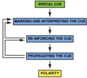
As I have previously described, cell polarity defines the ability of the cell to localize specific proteins to specific regions of the membrane, generating domains. In molecular terms, the orientation of the polarity axis is initially defined by asymmetric cues acting at the cell surface, that can be sensed by a large family of cell-surface receptors, called G-protein-coupled receptors (GPCRs), leading in turn to local activation of a trimeric GTP-binding protein (G protein). Extrinsic and intrinsic cues have then to be recognized and interpreted by signaling molecules (in particular small GTPases and polarity complexes) to be transduced in the cell interior, leading toFigure 6: Schematic representation of the general pathway for the establishment of cell polarity. Adapted from (Arkowitz and Iglesias, 2008; Drubin and Nelson, 1996).
39 the asymmetric activation and/or distribution of downstream effectors. This signaling network results in cytoskeleton rearrangement, polarized vesicles transport and global changes in cell organization. Concomitantly, sorting compartments of the secretory apparatus reorient in the cytoplasm along the axis of polarity relative to the cue. Delivery of newly synthesized proteins to targeting patches at the cell surface reinforces and stabilizes the molecular and structural asymmetry of the cell initiated by the spatial cue. Feedback regulation results in the maintenance of cell polarity (Figure 6) (Drubin and Nelson, 1996).
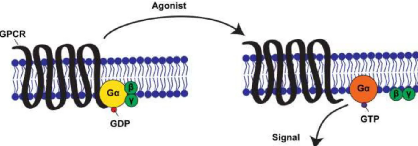
1.3.3.1 Signal transduction through GPCRs and G proteins G-protein-coupled receptors (GPCRs) form the largest family of cell-surface receptors, and mediate most responses to signals from the external world, as well as signals from other cells, including hormones, neurotransmitters, and local mediators (Alberts B., 2002). They are found in all eukaryotes, and also in some unicellular organisms, like the receptors for secreted mating pheromones in yeast. Even if activating signal molecules are chemically and functionally diverse, all GPCRs possess a similar structure, mainly composed of a single polypeptide chain which crosses the lipid bilayer seven times. GPCRs have a characteristic orientation in the plasma membrane, and they all use G proteins to relay the signal into the cell interior. G proteins are composed of three protein subunits - α, , and . In the inactive state, Gα is bound to G dimer and GDP (Figure 7, on the left). Activation by a ligand leads to a GPCR conformational change
Figure 7: Schematic of the activation of the G subunit of a GPCR. In absence of an extracellular cue,
G (yellow circle) interacts with GDP, G (green circles) and a GPCR (black loops). Upon receptor stimulation by a ligand, the receptor's state changes: G dissociates from the GPCR and G , and GTP is exchanged with GDP. As a result, G is activated (orange circle) and in turn leads to the activation of other molecules in the cell. Adapted from (Amberg, 1998; Li et al., 2002).
40
and activation of a G protein by promoting the exchange of GDP/GTP associated with the Gα subunit (Figure 7, on the right). As a result, the G /G dimer dissociates from Gα, and its moieties can act upon their downstream effectors at the cytoplasmic face of the plasma membrane, and thereby initiate specific intracellular signaling responses. GTPase activity of the Gα subunits may also be controlled by regulators of G proteins signaling, called RGS proteins, as well as effectors.
1.3.3.2 The core proteins of cell polarity: small GTPases
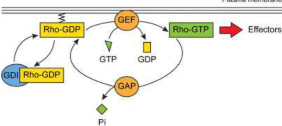
In the context of cell polarity and cell morphogenesis, all signals coming from diverse cell-surface receptors seem to converge on a group of closely related monomeric G proteins that are part of the Rho protein family - Cdc42, Rac, and Rho. Like all GTPases, these proteins act as molecular switches to control cell processes by cycling between an active, GTP-bound state and an inactive, GDP-bound state (Figure 8) (Iden S, 2008). Rho proteins are activated by guanine nucleotide-exchange factors (GEFs) and/or inactivated by GTPase-activating proteins (GAPs). Moreover, the release of GDP from the GTPase is blocked by guanine nucleotide-dissociation inhibitors (GDIs), which can also sequester Rho GTPases in the cytoplasm by masking their C-terminal motifs, required for Rho GTPases to interact with anchoring phospholipids in the plasma membrane. In addition to the high number of regulators, numerous Rho GTPase effector proteins have been described, especially kinases that function as scaffolding proteins to couple the activation of Rho GTPases to downstream signaling pathways.
Moreover, Rho GTPases can regulate cytoskeletal organization, vesicle transport and the
Figure 8: Rho GTPases can cycle between an active, GTP-bound (green rectangle) and an inactive, GDP-bound (yellow rectangle) conformation. This cycle is regulated by activators (GEFs, orange circles) and inhibitors (GAPs, orange circles, and GDIs, blue circles). In the active state, Rho GTPases can interact with various effectors. Adapted from (Etienne-Manneville and Hall, 2002).
41 localization of cytoplasmic proteins, primarily by controlling the phosphorylation of phospholipids, called phosphoinositides, which serve as specific docking sites for proteins at the cell membrane. Finally, they can also regulate each other’s activity through crosstalk. The temporal and spatial balance between the activities of different small GTPases is crucial for many cellular processes, such as cell growth, cell-cell and cell-matrix adhesions, cell migration, cell polarization and epithelial-mesenchymal transition (EMT). Previous studies have identified 20 different Rho-GTPases in mammals, while there are six of them in yeast. In particular, Cdc42, a small GTPase of the Rho family, stands out as key regulator of cell polarity establishment and maintenance in all eukaryotic cells, in particular of polarized growth in yeast (Etienne-Manneville, 2004).
1.3.3.3 The cytoskeleton
Actin filaments and microtubules are polar polymers that are composed, respectively, of globular actin (G-actin) subunits that bind and hydrolyze ATP, and α- and - tubulin heterodimeric subunits that bind and hydrolyze GTP. Polarity results from head-to-tail association of protein subunits, resulting in alignment of the subunits along the polymer lattices and structural differences between the two ends (Figure 9).
Figure 9: Basic properties of actin filaments and microtubules. A. Actin filaments are made of orientated subunits, resulting in a polarized surface polymer, responsible of the motors (myosins) unidirectionality, and two structurally different ends, with distinct nucleotide states and subunits kinetics. B. MTs are composed of - and -tubulin heterodimeric subunits orientated in the same direction, resulting in a polarized surface polymer, responsible of the motors (kinesins and dynein) unidirectionality, and two structurally different ends, with distinct polymerization/depolymerization rates related to the tubulin-bound nucleotide state. Adapted from (Li R, 2008).
42
Actin and microtubules are also dynamic, as they can either polymerize or depolymerize, with different rate constants for growth and shrinkage at the opposite ends. The ability to undergo fast turnover and assembly enables actin and microtubules to reorganize rapidly and locally in response to polarity signals.
A class of cytoskeleton-associated proteins that are particularly important for cell polarity are the motor proteins, characterized by unidirectional movement along actin filaments or microtubules by irreversibly moving from one tightly bound conformation to another using ATP hydrolysis as energy source. Myosins are motor proteins for actin, and most members of this superfamily move towards actin barbed ends, with the exception of myosin-VI. Some myosins, such as myosin-V, display processive movement along actin filaments (that is, they move many consecutive steps before dissociating from the filament) and are thus optimal for transport over long distances. Other myosins, in particular myosin-II, exhibit low processivity but can dimerize to generate contractile forces through the sliding of actin filaments. MT motors encompass kinesins and dynein. Most kinesins move towards MT plus ends with varying degrees of processivity, whereas dynein moves processively towards MT minus ends in the presence of the dynactin complex. In principle, cargo molecules or organelles can be trafficked by motor proteins to specific cellular locations once a cell has established oriented actin or MT arrays.
Cytoskeletal polymers carry out polarized trafficking or localized functions by assembling
Figure 10: Establishment of orientated actin arrays. There are two main classes of actin filament nucleation factors, Arp2/3 complex and formins. Formins promote the assembly of long-straight filaments by remaining continually bound to the elongating barbed end, whereas Arp2/3 complex facilitates the assembly of short-branched filaments by nucleating filaments from the side of a pre-existing filament that are quickly capped. Adapted from (Li R, 2008)
43 actin and MTs into organized arrays. The rate-limiting step for spontaneous actin and MT polymerization is nucleation, which is the formation of small oligomers that can rapidly elongate. In the case of MTs, the alternation between growth and shrinkage, known as dynamic instability, contributes to locally regulate cell polarity.
A key mechanism in the assembly of polarized actin arrays is the activation of actin-nucleation factors, such as the actin-related protein-2/3 (Arp2/3) complex and formin-family proteins, at defined locations (Fig 10). Arp2/3 complex produces branched filaments by anchoring the new one to the pre-existing actin network, in order to push forward the leading edge of motile cells and for endocytosis (Pollard and Borisy, 2003): the free end of the new filament elongates until a capping protein terminates growth. On the other hand, formins produce unbranched filaments for actin bundles found in filopodia and the cytokinetic contractile ring (Wallar and Alberts, 2003). The formin remains associated with the growing end of the filament, providing an anchor and protection against capping. Membrane-bound Rho-family GTPases activate actin-nucleation factors either directly or indirectly through nucleation-promoting factors (NPFs). Through these upstream regulatory factors, Arp2/3 complex- or formin-based actin nucleation occurs maximally near the membrane at sites of Rho GTPase activation (Li and Gundersen, 2008).
Whereas nucleation is the principal mechanism that regulates actin during cell polarization, nucleation of MTs usually occurs near the cell center at the centrosome (or other MT-organizing centers, MTOCs), far from membrane-derived signals that stimulate cell polarity. As a consequence, the initial events that localize and orientate MTs during cell polarization mainly involve factors that regulate the dynamic plus ends. For instance, MTs are frequently captured by cortical factors that increase the stability of the plus ends and/or generate pulling forces: this process increases local MT density and also provides a means to enhance the delivery of cargoes to specific sites. Other cases include alterations in the assembly properties and bundling of MTs.
In animal cells, the actomyosin cytoskeleton plays a central role in the dynamic regulation of cell shape (Salbreux et al., 2012). Actin is also essential for the translocation of secretory vesicles towards the membrane and internalization of endocytic vesicles.
Apart from their contribution to directed vesicle trafficking in large cells, MTs also play a common role in the establishment or maintenance of cortical polarity but are not required for
44
polarity induction per se (Siegrist and Doe, 2007). Similar to the case of migrating cells, in neuronal growth MTs are also essential for positioning the site of protrusion within a growth cone, but not for extension itself (Siegrist and Doe, 2007).
To conclude, interactions between actin and microtubules promote cortical asymmetry to polarize cells for differentiation, division and migration. Some examples of this communication are the establishment of anterior-posterior polarity in the early C. elegans embryo, the mechanisms that polarize Drosophila neuroblasts and mediate spindle positioning, the establishment of polarity in migrating cells and the regulation of cytokinesis by interactions between the mitotic spindle and the cortex (Akhshi et al., 2014).
1.3.3.4 Membrane remodeling: exocytosis and endocytosis Polarized distribution of proteins at the plasma membrane often results from a balance of vesicle delivery and fusion with the plasma membrane (“exocytosis”), diffusion at the plasma membrane, and internalization and membrane recycling (“endocytosis”) (Figure 11). Exocytosis controls the release of molecules into the extracellular space and results in the addition of new membrane material, which essentially leads to cell growth: for secretion to occur over relatively long distances in cells, vesiscle transport relies on the work of molecular motors along cytoskeletal tracks. By restricting secretion to specific sites, growth is polarized and takes place exclusively at these sites. Endocytosis describes the process of internalization of extracellular material and membrane proteins by invagination of the plasma membrane (PM), eventually resulting in the formation of endocytic vesicles and endosomes, that can either be degraded as lysosomes or
45 recycled back to the plasma membrane.
For cell polarity to take place, intrinsic mechanisms sort membrane proteins into different vesicles, and deliver these vesicles to different membrane domains (through cytoskeleton-mediated targeting, vesicle tethering through exocyst complexes and annexins) for membrane fusion (SNARE - Soluble N-ethylmaleimide-sensitive factor Attachment Protein Receptor - proteins). Protein complexes at the plasma membrane (the Par, Crumbs and Scribble complexes) control the identity and distribution of functionally and structurally unique PM domains from the cytosolic face. Moreover, membrane lipid composition regulate secretion and recycling (Orlando and Guo, 2009): for instance, phosphatidylinositol (PI) and phosphatidylinositol phosphates (PIPs) are highly compartmentalized in different organelles, and these local asymmetries in the phospholipid content of plasma membrane domains affect localization of Rho-GTPases such as Cdc42. Finally, extrinsic cues provided by cell adhesion to the ECM and by other cells control the orientation of cell polarity. All these different levels of regulation of cell polarity are integrated into a single network. (Mellman and Nelson, 2008).
1.3.3.5 Polarity complexes
In multicellular tissues there is clearly an increase of complexity, as in the case of epithelial cells, where the fundamental generators of cell polarity have to establish the difference between the apical and basal poles, and they have to do so in a properly oriented way, in accordance with the cell's environment. Cell polarization is achieved by the concerted actions of polarity proteins. These molecules are conserved throughout evolution and can react to extrinsic or intrinsic polarity cues (for example, growth factor gradients or the MT cytoskeleton, respectively). By assembling multiprotein complexes, they induce downstream signalling to trigger the establishment of cellular asymmetry. Of the three described polarity protein complexes - partitioning defective (PAR), Crumbs and Scribble - the PAR complex has the broadest function. In some polarization processes, these complexes cooperate to induce polarity, whereas in other systems they antagonize each other, thereby establishing or maintaining cellular asymmetry.
The crosstalk between polarity proteins and Rho GTPases is essential for the establishment and maintenance of mammalian cell polarization in different cell types, including neuronal, epithelial and T cells (Iden and Collard, 2008): here, I will briefly discuss the case of PAR genes, as they play a fundamental role not only in asymmetric cell division in the early
46
embryo, but in many other processes such as epithelial cell polarity, cell migration, oriented cell division and axon specification (Figure 12A) (Goldstein and Macara, 2007).
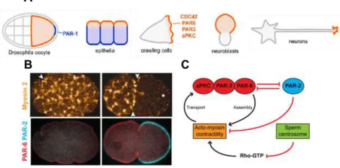
The PAR genes were initially identified by Jim Priess and Ken Kemphues in a screen for maternal-effect genes that are embryonically lethal in the nematode Caenorhabditis elegans (Kemphues et al., 1988): following fertilization, the one-cell embryo polarizes along an anterior– posterior axis to prepare for asymmetric division. PAR-3 and PAR-6 segregate to the anterior domain, whereas PAR-1 and PAR-2 distribute to the posterior domain.
Moreover, the asymmetric distribution of polarity proteins before cell division correlates with local differences in contractility, which is regulated by Rho protein activity: in other words, the initially uniform actomyosin network becomes restricted to the anterior pole, thereby generating a contractile anterior and a non-contractile posterior domain. Sperm entry leads to a local weakening of the contractile network by inhibiting Rho activation: this asymmetry is then amplified through positive feedback loops acting on the actomyosin contractile machinery and thePAR and Rho polarity modules, finally resulting in the establishment of an anterior-posterior
Figure 12: Interplay between PAR proteins and the polarity machinery A. PAR Localization in Multiple Systems. Adapted from (Goldstein and Macara, 2007). B. and C.Symmetry in a C. elegans one-cell embryo. B. Cortical network of myosin-II (on top) and PAR-2/PAR-6 localization (on bottom), before (left) and after (right) symmetry breaking. Arrowheads indicate furrows on the egg surface. An asterisk marks the site of sperm entry. (Goehring et al., 2011; Munro et al., 2004). C. Schematic of the crosstalk between PAR proteins, Rho-GTPases and the cytoskeleton for polarity establishment in C. elegans.
47 polarity axis (Figure 12B - C).
In conclusion, cell polarization is achieved by intricate communication between different classes of proteins, including small GTPases, polarity proteins and cytoskeletal components, which are mutually regulated and differentially distributed within a cell.
1.3.4
Cell polarization: with or without a cue?
As I described in the first section, cell polarity has first been defined as an organizing property of the cell, with an orientation that may be set by a pre-existing cue at the membrane. There are numerous signals that cells can sense and that may compete with each other in vivo (Drubin and Nelson, 1996): these are either localized cues that come from the environment, or spatial landmarks that the system has inherited (Li and Bowerman, 2010). Among the best characterized examples of extracellular signals there are gradients of signaling molecules, sites of cell adhesion, mechanical signals and electric fields. On this basis, the first explanations for the origin of cell polarity had largely focused on the molecular basis of these pre-existing cues and how their signals are propagated.
These extrinsic signals, though important in their physiological contexts, are not always necessary for symmetry breaking: in some cases, asymmetry occurs along random axes even when the cues are removed (Figure 13). For example, cells undergoing chemotaxis such as the social amoeba Dictyostelium discoideum and mammalian neutrophils move in random directions in the presence of a uniform concentration of chemoattractant (Kortholt et al., 2013); budding yeast cells, which naturally bud adjacent to the previous bud scar, are still able to polarize in random orientations when the genes responsible of bud site selection are deleted (Johnson et al., 2011); the zygotes of algae from the genus Fucus undergo an asymmetric first division in the absence of an external signal that normally defines the axis of polarity (Brownlee and Bouget, 1998). Similarly, in the Xenopus laevis egg dorsoventral axis is normally defined by the sperm entry point, but can also be established in eggs that are activated in the absence of sperms (Gerhart et al., 1989). Altogether, these observations suggest that the final asymmetric pattern is entirely self-regulating and that the initial asymmetric cue only provides a bias to orient the pattern correctly with respect to an intrinsic or extrinsic signal.