HAL Id: tel-02117786
https://tel.archives-ouvertes.fr/tel-02117786
Submitted on 2 May 2019HAL is a multi-disciplinary open access archive for the deposit and dissemination of sci-entific research documents, whether they are pub-lished or not. The documents may come from teaching and research institutions in France or abroad, or from public or private research centers.
L’archive ouverte pluridisciplinaire HAL, est destinée au dépôt et à la diffusion de documents scientifiques de niveau recherche, publiés ou non, émanant des établissements d’enseignement et de recherche français ou étrangers, des laboratoires publics ou privés.
Dissection of the role of natural killer cells in
atherosclerosis using selective genetic approaches
Wared Nour Eldine
To cite this version:
Wared Nour Eldine. Dissection of the role of natural killer cells in atherosclerosis using selective genetic approaches. Immunology. Université Sorbonne Paris Cité; Université libanaise, 2017. English. �NNT : 2017USPCB036�. �tel-02117786�
Université Paris V – René Descartes
et
Université Libanaise
THÈSE DE SCIENCES
Pour l’obtention du titre de Docteur
École Doctorale Bio Sorbonne Paris Cité Discipline : Biologie Cellulaire
Dissection of the Role of Natural Killer
Cells in Atherosclerosis Using Selective
Genetic Approaches
Présentée et soutenue publiquement par
Wared Nour ELdine
Le 06 Octobre 2017
Auteur Wared Nour Eldine
Composition du Jury Pr. Jean-Francois Arnal Rapporteur Pr. Aida Habib Abdul Karim Rapporteur Pr. Sophie Ugolini Examinateur Pr. Antonino Nicoletti Examinateur
Projet doctoral dirigé par Dr. Alain TEDGUI et Dr. Kazem ZIBARA
Acknowledgments
I would like first to convey deep thanks to the members of the Jury, Pr. Jean Francois
Arnal and Aida Habib, Pr. Sophie Ugolini and Antonino Nicoletti for participating in the
committee to evaluate my work. Thanks for your time, extreme patience, and intellectual
contribution. I am honoured having each of you as a member in the committee.
To my Lebanese supervisor, Pr. Kazem Zibara, I offer my sincerest gratitude and
appreciation. With you, I stepped to the world of scientific research. It is you, who paved
for me the way, trusted, and encouraged me. With all meanings of thanks, I would thank
…
To Pr. Alain Tedgui, my unique supervisor, no words can describe my gratitude and
affection, I am not fortunate but blessed to be your student, very much touched by your
kindness and generosity before you scientific intelligence, thanks for your guidance
P …
L
spired and will always inspire
…
A particular debt of gratitude is owed to Pr. Ziad Mallat for giving me the opportunity and
welcoming me to be part of his team, where I have learned and grown both scientifically
…
exceptional scientific knowledge and your
uttermost precious contributions whenever needed!
Much thanks goes to our Collaborators in Marseille, the group of Eric Vivier and Sophie
Ugolini, for your expertise in NK cell world, for the valuable discussions we had, and for
P …N
I would like to distribute my thanks to the dream team
“Equipe 5”, for each one of you I
…
Our papy, Bruno, for all the aortas
’
L
organization and rules, Jeremie, a valuable guide, with whom I started my journey, Lynda
and Yacine, my partners in this journey, as well as Marie and Andreas for the French
conversations, helping me conquer my fear of mice :p It would have been impossible
without your hand!
Y
a
“
”
L
naz, Nada joe, Hajer, Anthony,
L
L A L A L
L L
…
My ambitious student, Malak, greetings for your good humour and all those moments you
…
Thanks for our CRs: Hafid who co-supervised me for the first year of my PhD, Soraya and
For Jose, the multitalented guy, o
PA
L ’
impressed by your personality, Special Thanks to you and may your kindness and smile
…
My partners in the office, much Love and Thanks, Francine for your endless news, Isabelle
for your kindness and valuable advices, and Laetitia
…
Miss Sihem, one friend for all, thanks for the early afternoon whispers, coffee breaks, and
…N:
To Sophie, the sweet Greek girl, for the love, soft heart, and warm hugs between the floors
…N
:
To the friends of the third floor: Afef, Caterina, Hira, Anja, Louise, Ikram, Amel, Sylvian
and Ludovic, Nirmine of fondation Carpentier,
N
L
“
”
tells, Mengyao, and Siying of Team 3
…
…
For the Lebanese community in PARCC from the first to the third floor; Hasan, Nancy,
Zainab, Somaya, Waed, Rami, Thanks for all the good moments we shared once!
For the animal house and its people, beginning from the head Elizabeth to all of you,
Dominique, Nicolas, Emma, Corrina, Arex, Thanks for your kindness and taking good care
of my mice!
For the administrative platform; Muriel, the two Martine, Philippe, Bruno and especially
Veronique, Annette
…
L
L
L
…
A
HEGP, Pr.
P
… Finally, Ma
“
”…
…
For all the teams in PARCC, beginning with Team 1; Adele, Marwan, Julliette, Johanne,
Min, Marion, and Xavier, Team 3, Irmine, Team 6, Ivana and Mathilde, Team 10, Thi,
Marie, and Chehrezad
L
’
L
L
for your indispensable cooperation
…
Thanks for the PARCC and its people, who are undoubtedly all kind and nice. I believe that
every one of you contributed to my success with a word
…
For my Lebanese Friends in Paris, with whom I wandered around and discovered Paris
and the world, Lina, Ihsan, Mariam, and Racha, Precious Thanks
…
For my Family, my parents and lovely siblings, the reason behind every success and
L
…
and to you I dedicate this work!
For the One and only One, who gave me these words to thank others, Thanks for everything
I have, your messages, your blessings, your givings and gifts
“A , a a
y l ,
a a ”, It is but you, the origin, the roots, the beginning and the
infinity of everything beautiful, In your name I begin and I close this speech and every
stage in my life!
i
Table of Contents
List of Abbreviations ... iv
List of Figures and Tables ...x
I. INTRODUCTION and LITERATURE REVIEW ...1
-Chapter 1-Atherosclerosis-A Cardiovascular Disease ...2
A. Historical Perspective ...3
B. Epidemiology and Risk Factors ...4
1. Hypercholesterolemia ...4
2. Hypertension ...5
3. Obesity ...5
4. Diabetes...6
5. Autoimmune/Autoinflammatory diseases ...6
C. Sites of Atherosclerosis Development ...7
D. Classification of Atherosclerotic plaques ...9
1. Stages of evolution of atherosclerotic plaques ...10
a. Intimal Xanthoma...10
b. Intimal Thickenings ...11
c. Fibrous Cap Atheromata...11
d. Thin Fibrous Cap Atheromata ...11
2. Lesions associated with clinical complications ...11
a. Lesions associated with presence of thrombus ...11
i. Plaque rutpure ...11
ii. Plaque erosion ...12
iii. Calcified nodules ...12
b. Lesions not associated with presence of thrombus ...12
i. Fibrocalcific plaques ...12
ii. Intraplaque Hemorrhage ...13
3. Healed Ruptures/erosions ...14
ii
E. Animal Models of Atherosclerosis ...15
1. ApoE deficient mice ...15
2. LDL receptor deficient mice ...16
F. Atherosclerosis-a chronic inflammatory disease ...17
1. Changes in the vessel wall associated with development of lesions ...17
2. Initiation of Atherosclerosis ...18
3. Endothelial cell activation and Leukocyte recruitment ...19
4. Amplification of the inflammatory response and lesion progression ...20
5. Role of innate and adaptive immune cells in atherosclerosis ...22
a. Innate Immune cells ...22
b. Adaptive Immune cells ...27
-Chapter 2- Natural Killer Cells ...33
A. What Natural Killer Cells are? ...36
B. Origin, Development, and Maturation from HSCs ...37
1. Generation of NK cell progenitors from HSCs...37
2. Developmental stages from NKP to mature NK cells ...38
C. NK cell Trafficking and Tissue Distribution ...41
D. NK cell Receptors and Ligands ...42
1. Immunoglobulin Superfamily ...43
a. Killer immunoglobulin receptors (KIR) family ...43
b. Leukocyte Ig-like inhibitory receptors ...44
c. CD16a (FcRIIIa) ...45
d. DNAM-1 (CD226) ...45
e. Natural cytotoxicity receptors (NCRs) ...46
2. C-type lectin superfamily ...48
a. Ly49 family receptors ...48
b. NKR-P1 receptors ...48
c. CD94-NKG2 heterodimer receptors ...48
d. NKG2D receptor ...49
3. Toll-like receptors (TLRs) ...50
iii
E. NK cell Effector Function ...51
1. Granule exocytosis pathway ...51
2. Death receptor pathway ...52
3. Activation by-and production of cytokines...52
4. Antibody dependent cell mediated cytotoxicity ...55
F. Regulation of NK cell Activity ...55
1. Missing self-recognition by NK cells ...55
2. DCs-mediated IL-15 trans-presentation ...58
3. NK-Treg cross talk ...60
G. NK cell Memory ...61
H. Physiological Functions of NK cells ...63
1. NK cells as regulatory cells ...63
2. Anti-tumoral and anti-viral activities of NK cells ...64
3. NK cells and inflammatory diseases ...64
-Chapter 3- NK cells and Cardiovascular Diseases ...66
II. OBJECTIVES ...70
III. RESULTS ...72
IV. GENERAL DISCUSSION and CONCLUSION ...113
iv
List of Abbreviations:
A
ADCC Antibody dependent cell mediated cytotoxicity AGE Advanced glycation end
AHA American heart association
AIDS Acquired immune deficiency syndrome AML Acute myeloid Leukemia
Ang II Angiotensin II
APC Antigen presenting cell ApoE Apolipoprotein E
B
BAT3 Antigen-B-associated transcript 3 Bcl-2 B-cell lymphoma 2
BM Bone marrow
C
CAD Coronary artery disease CBV Coxsackie virus B CCL Chemokine ligand CCR C-C chemokine receptor CD Cluster of differentiation CLP Common lymphoid progenitor CRP C-reactive protein
CSF Colony stimulating factor CTL Cytotoxic T lymphocyte CVD Cardiovascular disease CXC C-X-C motif Chemokine
v
D
DC Dendritic Cell
DNAM-1 DNAX accessory molecule-1 dsRNA Double-stranded RNA
E
EAE Experimental autoimmune encephalomyelitis
EC Endothelial cell
eNOS endothelial Nitric oxide synthase ER Endoplasmic reticulum
ERK Extracellular signal-regulated kinase ERT EAT-2 related transducer
ESL1 E-selectin ligand 1
F
FcR Fc receptor
G
GM-CSF Granulocyte-macrophage colony-stimulating factor GPI Glycoprotein I Grz Granzyme H HA Hemagglutinin HCMV Human cytomegalovirus HDL High-density lipoprotein
HLA-DR Human leukocyte antigen – antigen D related HMG-CoA Hydroxymethyl glutaryl-coenzyme A
HSC Hematopoietic stem cell HSP Heat-shock protein
vi
I
ICAM-1 Intracellular adhesion molecule 1 IDL Intermediate-density lipoprotein IDO Indoleamine 2,3-dioxygenase IEL Intestinal epithelial lymphocyte IgM Immunoglobulin M
IL Interleukin
ILT LIRIg-like transcript iNKT Invariant natural killer T iNOS inducible Nitric oxide synthase IP-10 Inducible protein-10
IRF3 Interferon regulatory factor 3
IS Immunological synapse
ITAM Immunoreceptor tyrosine-based activating motif ITIM Immunoreceptor tyrosine-based inhibitory motif
J
Jak3 Janus kinase 3
JNK Janus kinase
K
KIR Killer immunoglobulin receptors
L
LAMP-1 Lysosome associated membrane protein-1
LDL Low-density lipoproteins Ldlr Low density lipoprotein recptor
LFA-1 Lymphocyte function-associated antigen-1 LIR Leukocyte immunoglobulin inhibitory receptor
LN Lymphe node
vii
LPS Lipopolysaccharide
LTi Lymphoid tissue inducer Ly6c Lymphocyte antigen 6 complex Ly49 Lectin like receptors
M
MAPK Mitogen activated protein kinase MCP-1 Monocyte chemoattractant protein-1
MDA5 Melanoma differentiation-associated protein-5
MHC Major Histocompatibility complex
MI Myocardial infarction
MIC Class I chain–related molecules MMP Matrix metalloproteinase
MULT-1 Murine UL 16-binding protein like transcript 1
N
NADPH Nicotinamide adenine dinucleotide phosphate NCR Natural cytotoxicity receptor
NF-kB Nuclear factor kB NK Natural killer NKC NK gene complex NO Nitric oxide O OP Osteoporotic
oxLDL Oxidized low density lipoprotein PAI-I Plasminogen activator inhibitor-1 PAMP Pathogen associated molecular pattern PCNA Proliferating cell nuclear antigen PD-1 Programmed cell death protein 1 pDC Plasmacytoid Dendritic cell
viii
PDGF Platelet derived-growth factor PGE 2 Prostaglandin E2
PKC Protein kinase C
PSGL1 P-selectin glycoprotein ligand 1
R
RA Rheumatoid arthritis
RAET-1 Retinoic acid early transcript-1 RAG Recombination-activating gene RAGE AGE receptor
ROS Reactive oxygen species
S
SAP Serum amyloid P component SHP1 SRC homology phosphatase 1 SLE Systemic lupus erythematosus SMC Smooth muscle cell
SR Scavenger receptor
SREC1 Scavenger receptor class F member 1
STAT5a Signal transducer and activator of transcription 5A
T
TCR T cell receptor
TGF- Transforming growth factor- TH1 T helper type 1
TICAM-1 Toll-IL-1 receptor domain containing adaptor molecule-1 TIR Toll-interleukin 1 receptor
TLR Toll like Receptors TNF Tumor necrosis factor
ix
U
ULBP UL-16 binding protein
V
VCAM-1 Vascular cell adhesion molecule 1 VEGF Vascular endothelial growth factor VLA-4 Very late antigen-4
VLDL Very low density lipoprotein VSMC Vascular smooth muscle cell
W
WHO World health organization
WT Wild type
Z
x
List of Figures and Tables
Figure 1: Sites of atherosclerotic lesion formation in humans and mouse ...8
Figure 2: Simplified scheme of the classification of lesions modified by Virmani after AHA Recommendation. ...10
Figure 3: Main lesion types of atherosclerosis and proposed sequence of their development ....13
Figure 4: Human coronary artery with compensatory enlargement ...14
Figure 5: Stages in the development of atherosclerotic lesions ...21
Figure 6: Immune components of the atherosclerotic plaque ...24
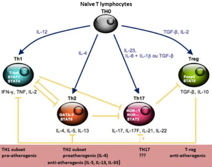
Figure 7: Role of different CD4 T cell subsets ...28
Figure 8: Phenotypic markers of developing NK cells in the mouse ...39
Figure 9: NK cell receptors ...43
Figure 10: Structure of Natural Killer cytotoxicity receptors ...46
Figure 11: Signaling pathways leading to NK cell activation ...54
Figure 12: Balance between inhibitory and activating signals governs NK cell function. ...55
Figure 13: Models proposed for NK cell education. ...57
Figure 14: Priming NK cells in vivo ...59
Figure 15: IL-2 dependent Treg -NK cell cross talk ...60
Figure 16: Three paths towards becoming a memory NK cell ...62
Figure 17: Proposed mechanism of NK-cell enhanced atherogenecity in an inflammatory context ...117
List of Tables
Table 1: Histological classification of the stages of atherosclerosis proposed by AHA ...9Table 2: Role of different innate and adaptive immune cells in atherosclerosis development ...34
1
I.
INTRODUCTION
&
2
-CHAPTER 1-
Atherosclerosis- A
Cardiovascular
3
A. Historical perspective
Even though atherosclerosis is reaching epidemic proportions nowadays, it is not in anyway a disease specific to modern days. It is rather an ancient disease with a fascinating history, firstly characterized in medical works of ancient Egyptians, Greeks, and Romans [1], [2]. Nowadays, atherosclerosis is recognized as a chronic inflammatory disease of large and medium sized arteries, characterized by the accumulation of low-density lipoproteins (LDL) and the recruitment of circulating monocytes and T cells in the intima, leading to the formation of macrophage derived foam-cells, as well as the proliferation of smooth muscle cells (SMCs), and accumulation of collagen and proteoglycans in the fibrous cap [3], [4]. However, its recognition and characterization progressed over centuries. In the latter half of last millennium (1575), the anatomist Fallopius wrote of “degeneration of arteries into
bone”. Later on, in 1740, the German physician Krell described for the first time “calcium concretions” in the arterial wall of aged subjects, which he called “bony plates” [5].
The term “atheroma” was first introduced in 1755 by Albrecht von Haller to designate the plaque deposited on the innermost layer of systemic artery walls. Then in 1904, Felix Marchand suggested the word “atherosclerosis” should be better used to refer to the degenerative process of the intimal layer of arteries, which derives from athera, meaning “porridge” or “gruel” in ancient Greek, and “skleros” signifying “hard” in Greek.
Later, in 1815, Hodgson proposed that inflammation is the underlying cause of atheromatous arteries. However, the first mechanistic assessment of atherosclerosis was initiated in 1908 by the Russian scientist Alexander Ignatowski who demonstrated that rabbits fed a diet of milk and egg yolk develop atherosclerosis. Subsequently, in 1913, Anitschkov and Chalatov reproduced experimental atherosclerosis by adding pure cholesterol (“cholesterin” as it called at that time) to rabbit food [6]. Since then, the lipid theory of atherosclerosis was established and predominated for most of the 20th century [1].
Soon after, this concept was replaced by the “response to injury hypothesis” which dates back to the pioneering work of Virchow and Carl von Rokitansky who discovered inflammatory cells in the developing lesions. This theory was later developed by Russel Ross (1976), who proposed that endothelial injury leads to platelet adhesion and smooth muscle cell proliferation [7].
At that time, it was believed that SMC proliferation, drived by platelet derived-growth factor (PDGF) secreted from platelets, is the principal factor occluding the artery. However, it was later established that SMC proliferation is rather modest and it seems to be beneficial by
4
contributing to plaque stabilization. Later, when Poole and Florey observed monocytes adherence and migration to the endothelium after feeding cholesterol to rabbits, Ross revisited his “response to injury theory” and published in the New England Journal of Medicine a remarkable review entitled: “Atherosclerosis: a chronic inflammatory disease” [8].
The precise identification of cell components of human atherosclerotic plaques using modern immunohistochemical techniques by Göran Hansson and his colleagues was instrumental in the change of opinion regarding the role of inflammation and immunity, rather than SMC proliferation, in the pathogenesis of atherosclerosis. Histologically, the lipid laden foam cells originate from macrophages and large amount of T cells, about 20%, surround the plaque and are present in the fibrous cap, pointing to a role of immunity in atherosclerosis [9],[1].
It is now widely accepted that atherosclerosis is a chronic inflammatory disease of the artery wall where immune reposnes are involved at all stages of the disease.
B. Epidemiology and Risk Factors
According to the World Health Organization (WHO), cardiovascular diseases (CVD) are the leading cause of mortality worldwide accounting for 17.7 million victims in 2015 (WHO report 2017), which represents 30% of all global deaths, mainly from ischemic heart disease and stroke. If the current trend continues, this number is predicted to increase to 26 million deaths in 2030 [10].
Although CVD and related chronic diseases were considered to be diseases of industrialized countries, WHO reported that 80% of the 17.7 million cardiovascular deaths in 2015 took place in low and middle-income countries (WHO report 2017).
The likelihood of occurrence of CVD increases in the presence of multiple risk factors for atherosclerosis. These are modifiable or non-modifiable. The non-modifiable are: age, gender, and family history. The modifiable risk factors are hypercholesterolemia, hypertension, smoking, diabetes mellitus, obesity, and sedentary lifestyle [11].
1. Hypercholesterolemia
Among the many cardiovascular risk factors, elevated plasma cholesterol level is probably unique in being sufficient for the development of atherosclerosis, even in the absence of other known risk factors. If all adults had plasma cholesterol levels <150 mg/dl, symptomatic disease would be rare [12].
5
accumulating in the intima can be oxidized and initiate inflammation by increasing the expression of adhesion molecules by the endothelium, and chemokines and cytokines by macrophages. The five single-gene disorders that result in elevated LDL levels are associated with premature coronary atherosclerosis [13].
In addition, lipid-lowering agents, such as statins (HMG-CoA reductase-inhibitors) significantly reduce cardiovascular mortality, both in primary or secondary prevention [14]. Recent clinical trials have shown that reduction in the rate of coronary events is directly related to the magnitude of reduction in LDL cholesterol levels [13].
2. Hypertension
Epidemiological studies clearly pointed out that arterial hypertension is a major risk factor for CVD, especially stroke. Elevated blood pressue levels have been highly predictive of atherosclerosis associated-cardiovascular events. In human subjects, carotid artery intima-media thickness is highly correlated with blood pressure levels, and accurately reflects CV risk. This is supported by experimental evidence demonstrating that hypertension increases the rate of atherosclerotic plaque development in hypercholesterolemic rabbits, monkeys, and mice.
Hypertension presents in several forms, some linked to activation of the renin-angiiotensin system and elevated circulating Angiotensin II (Ang II), and some with normal Ang II levels. A study by Mazzoli et al reported that hypercholesterolemic mice (Apoe-/-) with high circulating or normal Ang II levels exhibited similar atherosclerosis plaque size, but those of hypertensive animals had signs of instability, as well as enhanced systemic inflammation [15].
Several mechanisms can account for hypertension-induced atherosclerosis. Firstly, the pressure-induced stretch of the wall increases endothelial permeability to low-density lipoprotein and their subsequent retention ton the intima. Also, hypertension aggravates endothelial dysfunction, enhances monocyte adhesion, and activates the transcription of inflammatory genes [16]. All these factors are central to the development of atherosclerosis and will be discussed through the chapter.
3. Obesity
Several clinical and subclinical abnormalities are found in obese patients, such as insulin resistance, atherogenic dyslipidemia, prothrombotic and proinflammatory state, all of which contribute to the development of atherosclerotic plaques and their complications [17].
6
protein (CRP), which reflects high cytokine levels secreted by excess adipose tissue. This may render stable atherosclerotic plaques vulnerable to rupture [18], [19]. In addition, increased production of plasminogen activator inhibitor-1 (PAI-1) by adipose tissue favors a prothrombotic state [20]. Circulating Tumor necrosis factor- (TNF-) is markedly increased in obese patients [21]. Moreover, leptin, synthesized by adipose tissue, plays a role in obesity-induced endothelial dysfunction. It increases oxida0tive stress in endothelial cells, promotes vascular cell calcification, and smooth muscle cell proliferation and migration [22].
4. Diabetes
Diabetes is defined as fasting blood glucose of 126 mg/dl or greater. Persons with type 1 and type 2 diabetes are at increased risk for coronary artery disease (CAD).
For example, in a Finnish population-based study, the seven-year incidence of myocardial infarction among 1373 non-diabetic subjects with and without pre-myocardial infarction at baseline was 18.8% and 3.5% respectively. However, in 1059 persons with type 2 diabetes, the incidence rates of myocardial infarction increased to 45% and 20%, respectively [13]. Hyperglycemia induces the formation of reactive oxygen species (ROS) and advanced glycation end products (AGE), which plays a major role in the pathogenesis of diabetes, mainly through endothelial dysfucntion. AGEs exert their pathogenic effects by engaging binding sites/receptors. They have been shown to activate macrophages in an NF-kB-dependent fashion, leading to induction of proinflammatory cytokines, such as IL-1 and TNF-. In addition, their binding to the endothelial receptor for AGE (RAGE) results in depletion of cellular anti-oxidant defense mechanisms (glutathione, vitamin C) and the generation of ROS. Increased cellular oxidative stress induces the endothelium to express the procoagulant tissue factor and adhesion molecules such as E-selectin, intracellular adhesion molecule 1 (ICAM-1), and vascular cell adhesion molecule 1 (VCAM-1).
In addition, hyperglycemia increases the matrix metallopeptidase 9 (MMP-9) activity, partly due to increased transcription of MMP-9 through redox-sensitive mechanisms. MMPs are involved in monocyte invasion, the vascular smooth muscle cell (VSMC) migration and collagen degradation; its dysregulation is a critical factor in development of vascular lesionsand plaque instabilty [23].
5. Autoimmune/Autoinflammatory diseases
Chronic infections [24] and autoimmune diseases, specifically rheumatoid arthritis (RA) [25] and systemic lupus erythematosus (SLE) [26], also represent a risk factor for atherosclerosis.
7
Patients with autoimmune diseases, such as SLE or RA, are at particularly high risk of CVD. These patients benefit from treatment with antibodies directed against the B-cell receptor CD20.
The chronic systemic inflammation that occurs in RA can contribute to endothelial dysfunction and oxidative stress to promote atherosclerosis [27]. In patients with RA, anti-TNF- therapy reduced inflammation, thrombotic risk, and the incidence of cardiovascular events.
Accelerated atherosclerosis is a major source of morbidity in SLE. CD4+ T cells from SLE-susceptible mice transferred into Low density lipoprotein receptor (Ldlr-/-) deficient mice increased atherosclerosis [28].
C. Sites of Atherosclerosis development
Atherosclerosis localizes to reproducible sites of the vascular tree. Lesions are found primarily in large and medium sized muscular arteries [29].
Regions of arterial tree with laminar shear stress are resistant to the development of atherosclerosis due to orderly blood flow. However, branch points of arteries, which experience low shear stress, turbulence and oscillating flow, are predisposed to lesion formation [30]. These areas of arterial branches, bifurcations, and curvatures may be located in the coronary, cerebral, carotid, femoral arteries, and in the aorta [31], [32] (Figure 1A). Before development of atherosclerosis, the predilection sites are characterized by changes in endothelial turnover and gene expression, [33] presence of subendothelial dendritic cells and in humans, by the presence of adaptive intimal thickening [34].
Fluid mechanical forces can directly influence endothelial cell structure and function [35]. Endothelial cells align with in the direction of laminar flow. In areas of disturbed flow, this alignment is abolished.
Endothelial cells express genes, which are differentially regulated by blood flow rate. These include cell surface adhesion molecules (VCAM-1), intracellular adhesion molecules (ICAM-1), pro-oxidant enzymes (lipoxygenases, NADPH oxidases) and antioxidant enzymes (NO synthase, superoxide dismutase) genes [36]. Oscillatory shear stress substantially upregulates VCAM-1, ICAM-1, and E-selectin in cultured human endothelial cells [37]. Nitric oxide (NO), the potent vasodilator with anti-inflammatory activities, is the end product of conversion of L-arginine to L-citrulline by the endothelial nitric oxide synthase (eNOS). eNOS is induced in a dose-dependent manner by laminar shear stress and downregulated by oscillatory shear stress [38]. One of the early responses to hypercholesterolemia is the
8
attenuation of endothelium dependent production of NO. In addition to decreased vasodilation, deficiency in NO enhances SMC proliferation and platelet aggregation and adhesion [39], [40].
Figure 1: Sites of atherosclerotic lesion formation in humans and mouse (A) Shcematic shows the common sites of atherosclerosis formation in human (B) Schematic showing the major arterial vasculature distribution of atherosclerosis (gray shading) in Ldlr-/- mice fed a high atherogenic
diet. 1: indicates aortic sinus; 2: ascending aorta; 3: lesser curvature of aortic arch; 4: greater curvature of aortic arch; 5: innominate artery; 6: right common carotid artery; 7: left common carotid artery; 8: left subclavian artery; 9: thoracic aorta; 10: renal artery; 11: abdominal aorta; 12: iliac artery. (adapted from Vanderlaan et al, ATVB-2004 and De Bakey, 1985)
Accumulating evidence shows that blood flow affects whether a plaque progresses into a vulnerable or stable plaque [29].
Similarly in mice, the development of atherosclerosis occurs at reproducible sites predetermined by the forces experienced by the endothelium. This vascular distribution is depicted in Figure 1B. These sites are precisely where turbulent, pulsatile, and non-laminar flow forces predominate [36].
9
D. Classification of Atherosclerotic Plaques
The pathogenesis of atherosclerotic lesions has been inferred from microscopic analysis of arteries in different age groups.
The classification of atherosclerotic lesions was initially proposed by Stary and his colleagues (1995), then adopted by the American heart association (AHA) based on the histological composition and structure of the plaque. This classification comprises seven stages of chronological evolution and increasing gravity [41], [42] (Table 1).
Table 1: Histological classification of the stages of atherosclerosis proposed by the AHA,
(adapted from stary ey al, Circulation, 1995)
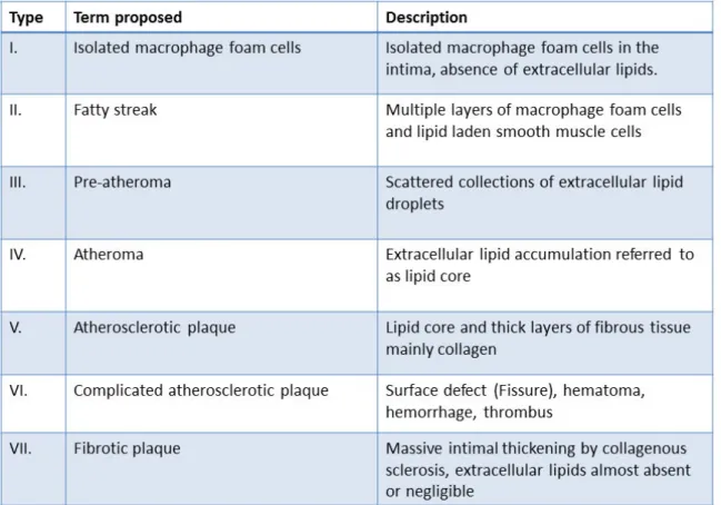
Later in 2000, Renu Virmani and Stephen Schwartz introduced an alternative and simpler classification, which emphasizes the link between lesion morphology and clinical disease. Lesions types recognized in this classification are displayed in Figure 2 [43].
10
Figure 2: Simplified scheme of the classification of lesions modified by Virmani after AHA recommendation, The boxed areas represent the seven types of lesions. Dashed lines were used for two boxes because there is controversy about their roles in the initial phase of lesion formation and both lesions can exist without progression to a fibrous cap atheroma. The processes leading to lesion progression are listed between categories. Lines depict current concepts of how one category may progress to another, (adapted from Virmani et al, 2000, ATVB)
1. Stages of evolution of atherosclerotic plaques
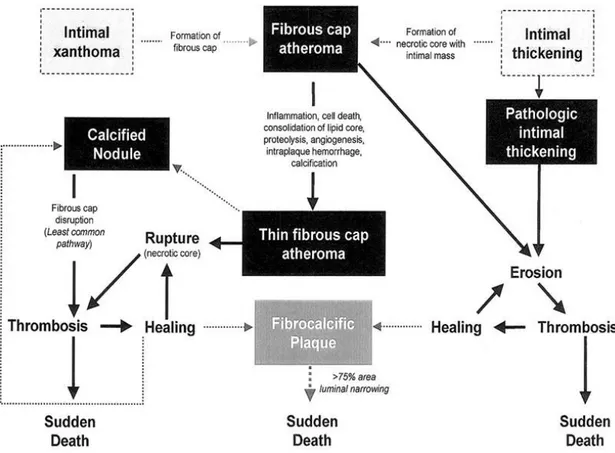
The new scheme by Virmani et al [43] defines seven categories considering the pathophysiological progression, including accumulation of lipids, formation of cap and necrotic core, remodeling of the fibrous plaque and thrombosis. The main lesions of atherosclerosis and the proposed cellular and morphological sequential changes are represented in Figure 3 [44].
a. Intimal Xanthoma
This term was proposed instead of type I lesion, “fatty streak” or initial lesion in the AHA’s scheme. Xanthoma is a general pathological term that describes focal accumulations of lipid laden-macrophages. In humans, intimal xanthomas present in children, probably reflecting risk factors of the mother, but appear to regress with age since they are rarely found in
11
elderly [45]. Xanthomas are harmless and fully reversible if the stimuli that caused their formation dissipate [44].
b. Intimal Thickenings
Although the authors did not exclude the fact that some human lesions may begin as intimal xanthomata, they suggested that most adult human lesions originate as preexisting intimal masses. Indeed, the distribution of these intimal thickenings observed in children is correlated with the usual distribution of lesions of the adult [46].
c. Fibrous Cap Atheromata
It is defined as a distinct layer of connective tissue completely covering the lipid core. It consists purely of SMCs in a collagenous proteoglycan matrix, with varying degrees of infiltration by macrophages and lymphocytes. Thus, the fibrous cap atheroma may have thick or thin cap overlying a lipid rich core.
d. Thin fibrous Cap Atheromata
The aim behind adding this type of lesion, which was not recognized by the AHA classification, is that lesions with thin fibrous caps are most likely prone to rupture. Thin fibrous cap is defined as one which is <65 µm thick. This definition is after morphometric examination of a series of 41 ruptured plaques in which 95% of the caps measured <64 µm thick. Its content is distinguished from earlier fibrous cap lesions by loss of SMCs, extracellular matrix, and inflammatory infiltrate [47]. However, the thickness of the cap is not sufficient to allow prediction of plaque rupture. The latter necessitates other relevant features such as the extent of inflammation in the cap, fissuring, calcification, and intraplaque hemorrhage.
2. Lesions associated with clinical complications
Literature review revealed that plaque rupture is responsible for 76% of all heart attacks caused by coronary thrombosis worldwide. The remaining 24% are caused by plaque erosion and other less well defined mechanisms [12].
Many factors determine whether a thrombus occurs or not. Autopsy studies show that vulnerability is a function of increased number of macrophages, increased expression of tissue factor, reduced number of SMCs, large lipid core and thin plaque cap [48].
a. Lesions associated with presence of thrombus i. Plaque rupture
12
In plaque rupture, a structural defect -gap- in the fibrous cap exposes the contents of necrotic core to circulating blood immediately causing more or less atherothrombotic process. Plaque rupture occurs where the cap is the thinnest and most infiltrated by foam cells [44].
Ruptured plaques are characterized by a large necrotic core and disrupted fibrous cap infiltrated by macrophages and lymphocytes, with a sparse SMC content. Ruptures are observed in 60% of individuals dying suddenly with luminal thrombi and are the most frequent cause of death in young men (<50 years) and old women (>50 years) [49].
ii. Plaque erosion
Virmani uncovered another mechanism of coronary thrombosis occurring in unruptured non-inflammatory plaques, described as plaque erosion [1]. Eroded plaques differ from ruptured plaques in that they have a base rich in proteoglycans and smooth muscle cells.
Plaque erosion accounts for 20% of all sudden deaths or 40% of coronary thrombi in patients dying suddenly from coronary artery atherosclerosis. It affects mainly young individuals and women before menopause and is associated with smoking [50], [51].
They are associated with less luminal narrowing and less calcification, and they are less likely to have foci of macrophages and T cells compared with ruptured plaque. Recently studies provided experimental evidence that endothelial apoptosis might be a major determinant of plaque erosion [52].
iii. Calcified nodule
The least frequent lesion of thrombosis is referred to as calcified nodule. The luminal region of the plaque consists of breaks in the calcified plates, bone formation, and interspersed fibrin with a disruptive surface fibrous cap and an overlying thrombus. These lesions are found in midright coronary artery and might be related to the frequent occurrence of plaque hemorrhage [43].
b. Lesions not associated with presence of thrombus i. Fibrocalcific plaques
These plaques are characterized by thick, fibrous caps overlying extensive accumulations of calcium in the intima close to the media. They are referred to as fibrocalcific due to the small lipid-laden necrotic core, if present. One hypothesis is that these plaques result from healing of an atheromatous plaque following incomplete rupture or erosion [53].
13
ii. Intraplaque hemorrhage
The pathogenesis of intraplaque hemorrhage is controversial and has been discussed for years. Constantinides originally suggested that hemorrhage originates from cracks in the luminal surface [54]. Later, Davis proposed that hemorrhage initiates within the shoulder region, the thinnest portion of fibrous cap, allowing entry of blood from the lumen into the necrotic core, which then comes into contact with collagen fibrils and tissue factor [48]. The most recent hypothesis “Paterson’s” supports the rupture of vasavasorum within advanced lesions. This hypothesis was confirmed by Virmani’s observations in a series of sudden coronary death cases, where hemorrhage was mostly frequent in ruptured plaques.
Figure 3: Main lesion types of atherosclerosis and proposed sequence of their development, Movat pentachrome staining of coronary plaques shows collagen and reticular fibers (yellow), fibrin (bright red), muscle (red), elastic fibers (black to blue). A: Adaptive intimal thickening characterized by accumulation of SMCs in the intima. B: Xanthoma corresponds to accumulation of foam cells in the intima, C: Pathological intimal thickening denotes the accumulation of extracellular lipid pools in the absence of apparent necrosis, D: Fibroatheroma indicating the presence of necrotic core, E: Fibrocalcific plaque which results from clacification of the necrotic core and the surrounding tissue. (adapted from Fog Bentzon et al, Circ. Res. 2014)
14
3. Healed Ruptures/Erosions
Fractured or eroded plaques may heal in the absence of occlusive thrombus and clinical manifestation.
Healed ruptures are characterized by a disrupted fibrous cap filled in by SMCs, proteoglycans, and collagen. The collagen newly synthesized by SMCs is type III replaces the original collagen type I. Lesions may exhibit multilayering of lipid and necrotic core, suggestive of previous episodes of thrombosis [48]. Others show no evidence of a preexisting rupture of the fibrous cap, but instead distinct layers of dense collagen interspersed with SMCs and proteoglycans containing fibrin and platelets.
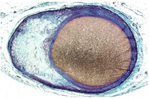
4. Compensatory enlargement
Seymour Glagov showed that in response to plaque growth, the arterial wall can remodel itself by increasing its external diameter to accomodate the plaque without narrowing of the lumen [55]. (Figure 4)
Figure 4: Human Coronary Artery with Compensatory Enlargment, (adapted from S.
15
E. Animal Models of Atherosclerosis
Early observations of atherosclerotic lesions in animals date back to more than a century, when Ignatowski found arterial thickening in rabbits fed the lipid rich animal proteins. Since then several animal species were used for atherosclerosis studies; monkey, pig, rabbit, and mice. The most widely used model is the mouse because of the ease of manipulating their genome.
C57BL/6 strain is the most susceptible strain to atherogenic diet, compared to other strains such as BALB/c, SWR, and NZB [56], [57]. However, C57BL/6 mice have low plasma cholesterol levels, mainly in the high-density lipoprotein (HDL), even under high fat diet. So they develop only small lesions when fed an atherogenic diet over a long period [58]. Therefore, transgenic mice on a C57BL/6 background were generated. Among these, the Apoe-/- and Ldlr-/- mice are the most commonly used mouse models.
1. ApoE deficient mice
ApoE is a 34 KDa glycoprotein synthesized in liver, brain, intestine, lung, and macrophages in both humans and mice [59]. It is a structural component of all lipoprotein particles except low-density lipoproteins (LDL). ApoE functions as a ligand for cell surface lipoprotein receptors that clear chylomicrons and very low- density lipoprotein (VLDL) remnants by the liver [60].
Mice deficient for this glycoprotein (Apoe-/- mice) have increased plasma levels of total cholesterol (300 to 500 mg/dl), mostly in the VLDL and chylomicron fractions [61]. Apoe -/-mice are hyperlipidemic even when fed a chow diet [62]. Under normal dietary conditions, Apoe-/- mice have already very high cholesterol levels and develop lipid streaks with foam cells and SMC within 6-8 weeks and more advanced plaques at 20 weeks with necrotic core and fibrous cap [63]. This process can be exacerbated under the influence of western diet (consisting of 21% fat and 1.5% cholesterol, similar of everyday diet of western countries) with female mice more sensitive than male mice [64].
The use of Apoe-/- mice has some attendant disadvantages. ApoE has several functions in addition to its indispensable role in lipoprotein metabolism. It is implicated in macrophage biology, immune functions, and adipose tissue biology. Morevoer, it is expressed by bonemarrow derived cells which prevents the transfer of Apoe+/+ bonemarrow into Apoe -/-mice because of the correction of cholesterol efflux and normalization of plasma cholesterol concentrations [65].
16
2. LDL receptor deficient mice
LDL receptor is a membrane receptor of a molecular weight of 160 KDa, which mediates endocytosis of cholesterol rich LDL, and thus maintains plasma level of LDL. It also facilitates the uptake of ApoB- and ApoE- containing lipoproteins [66], [67].
In contrast to Apoe-/- mice, Ldlr-/- mice display modestly elevated plasma cholesterol levels and develop no or only mild atherosclerosis when fed a normal diet. Therefore, these mice are fed western diet to accelerate atherosclerosis. Ldlr-/- mice have increased IDL and LDL sized particles, whereas HDL and triglycerides remain unaffected [68].
Ldlr-/- mice plaques are the same as those seen in Apoe-/- mice. The plaque development occurs in a time dependent manner, initially in proximal aorta spreading towards distal aorta. A western diet induces larger and more advanced lesions, with collagen rich fibrous cap, and a necrotic core containing cholesterol and cellular enrichment adjacent to the lumen [69], [70].
The Ldlr-/- mouse model has some advantages compared to the Apoe-/- mice. In Ldlr-/- mice, plasma cholesterol is mostly carried by LDL particles, which is similar to human lipid profile. Also, the absence of LDL receptor does not have impact on inflammation as compared to ApoE deficiency. Thus atherosclerotic plaque development in this mouse model is due to elevated plasma lipid levels solely and not caused by functions linked to the receptor itself [71]. Moreover, the Ldlr-/- mouse model shares the characteristics observed in human familial hypercholesterolemia, which is caused by absence of functional LDL receptor [72], [73], [74]. Finally, Ldlr-positive donors can be used in bonemarrow transplantation experiments, because the receptor rapidly becomes downregulated, even in expressing cells, as plasma cholesterol concentrations increase [65].
In our laboratory, we use in common practice the irradiation/transplantation method starting form Ldlr-/- mice, making it possible to obtain the so-called chimeric mice. This technique entails the lethal irradiation (9.5 Gy) of atherosclerosis prone mice to cause medullary aplasia, followed by their reconstitution with the bone marrow (BM) cells of donor mice, which usually present invalidation or overexpression of the gene of interest. This method allows to study specifically the role of a molecule expressed by cells derived from the bone marrow of which leukocytes are a major part. In our studies, the group of mice irradiated and reconstituted with the bone marrow deficient for the gene of interest is always compared to another control group irradiated and reconstituted with controls’ bone marrow. This way we would exclude the effect of irradiation on atherosclerosis development, but may not eliminate
17 the possible interaction between the gene of interest and the “irradiation factor”.
F. Atherosclerosis-a chronic inflammatory disease
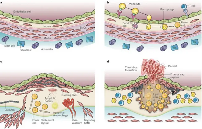
1. Changes in vessel morphology associated with development of atherosclerotic lesions
The normal arteries consist of three major layers: tunica intima, media, and adventitia respectively from inside to outside (Figure 5, A). The tunica intima is lined by a monolayer of endothelial cells that is in contact with blood overlying a basement membrane. It is defined as the layer starting from the endothelium and extending to the luminal margin of the media.
Endothelial cells and SMCs are the principal cellular components of the human arterial intima. Isolated macrophages are also present, in addition to sparse mast cells [75].
The endothelium serves as a barrier between circulating molecules and cells in blood. It is a major regulator of vascular homeostasis, by synthesizing signaling molecule, it inhibits platelet aggregation and adhesion, SMC proliferation, and leukocyte adhesion and migration. Structural and functional changes in endothelial cells contribute to the pathogenesis of atherosclerosis.
The sub-endothelial layer of the intima consists of two layers. The inner layer, a proteoglycan layer mainly contains non-fibrous connective tissue, small amounts of elastic fibers, and SMCs of synthetic and contractile phenotype. The musculoelastic layer is a thicker layer, which underlies the proteoglycan layer, contains a large number of SMCs of the contractile phenotype and elastic fibers [75], [76].
In response to growth regulatory molecules such as growth factors, SMC number would increase in the developing intima by mitosis. Originally, it was thought that SMCs in atherosclerotic lesions are solely derived from the media. But, recently, it has been suggested that bone marrow progenitor cells infiltrate the intima and probably differentiate in vivo to form SMCs [77]. Under pro-atherogenic conditions, SMC differentiate in macrophage-like capable of internalizing lipids and transforming into foam cells [78].
Collagen maintains endothelial cell integrity by anchoring endothelial cells to subendothelial matrix. The major types of collagen in the artery wall are the two interstitial collagens, types I and III.
Type III collagen is localized to the subendothelial space of the intima; it could be synthesized by the endothelium. Increased amounts of type I may reflect the metabolic properties of an increased number of SMCs present in the intima, because SMCs in culture
18 synthesize both type I and III collagen [75].
The internal elastic lamina, generally considered part of media, denotes the border between intima and media. In areas of vascular transitions, the internal elastic lamina is partly or completely absent and the intima and media may appear as a single unit.
The middle layer, tunica media, is the thickest layer of normal vessels. It contains 20% of SMCs both contractile and synthetic phenotype and 60% collagen and elastin, which provides contractile ability for the vessel [79]. By proliferating and migrating to the subendothelial intima, medial SMCs can contribute to the development of atherosclerosis. The outermost layer, tunica adventitia, contains sparse collagen fibrils, nerve endings, and its own nutrient arterial supply calledvasa vasorum. It is separated from the media by the external elastic lamina. The major cell types in the adventitia are sparse fibroblasts and mast cells [75]. This layer can change function and contribute to the development of atherosclerosis. Lymphoid organ like structure are found in the adventitia of human plaques; these are mainly composed of B and T cells [80].
2. Initiation of atherosclerosis
In 1976, Ross proposed the response to injury hypothesis, which states that atherosclerotic lesions result as a response to some form of injury to the endothelial cells that result in their desquamation. The loss of the endothelium exposes the underlying collagen layer and SMCs to platelet derived growth factor from platelets that adhere to the exposed subendothelial connective tissue. Infiltration of platelets derived factors such as lipoproteins and hormones would lead to migration and proliferation of SMCs, which form new connective tissue and results in intracellular and extracellular lipid deposition [81].
While the response to injury hypothesis supposed that endothelial desquamation is an essential event in atherogenesis, it was later demonstrated that the developing atheromatous lesions are covered by an intact endothelial layer throughout most stages of lesion progression. Thus in 1995, Kevin Williams and Ira Tabas formulated the “response to retention” hypothesis, which states that extracellular trapping of cholesterol-rich lipoprotein within the arterial intima is sufficient for initiation of atherosclerosis [82].
According to this hypothesis, hypercholesterolemia induces rapid LDL retention followed by their firm adhesion to proteoglycans of extracellular matrix, which prevents their return to the blood stream. Then, LDL are susceptible to enzymatic and non-enzymatic oxidation, as well as modification by extracellular proteins in the subendothelial space [83], [84]. A number of potential oxidant-generating systems have been identified to target lipid and apolipoprotein B
19
component of LDL. In addition to metal ions and oxidative radicals, these include: myeloperoxidases, inducible nitric oxide synthase, and 15-lipoxygenase (15-LO) [85]. Once oxidized, LDL exerts various pro-inflammatory biological effects leading to the development of atherosclerotic plaques. The “retention hypothesis” and “inflammatory theory” are complementary, LDL accumation and modifications in the intima being the primary trigger of vascular inflammation.
3. Endothelial cell activation and Leukocyte recruitment
In lesion prone areas, atherosclerotic lesions begin to develop under an intact but activated and dysfunctional endothelium [12].
Soon after initiating an atherogenic diet, light microscopy reveals attachment of blood leukocytes to the endothelial cells that line the intima [86], [87].
The normal arterial endothelium does not support the adherence of leukocytes. However, when subjected to irritative stimuli (such as dyslipidemia, hypertension, or pro-inflammatory cytokines), endothelial cells express adhesion molecules that capture leukocytes on their surfaces [76].
Oxidised LDL (oxLDL) activates endothelial cells leading to expression of VCAM-1 and ICAM-1, P- and E- selectins [88].
The recruitment of circulating leukocytes occurs via a tightly regulated multi-step process mediated by a combination of cell surface adhesion molecules.
Initially, activated endothelial cells express E-selectin (CD62E) and P-selectin (CD62P), which mediates the tethering and rolling of the circulating monocytes. selectin binds to P-selectin glycoprotein ligand-1 (PSGL1). Although PSGL1 is constitutively expressed by leukocytes, it is functional only when glycosylated correctly. In addition to PSGL-1, E-selectin also binds to glycosylated CD44 and E-E-selectin ligand 1 (ESL1) [89].
Besides selectins, VCAM-1, a member of immunoglobulin-like superfamily of adhesion molecules participates in the slow rolling and early adhesion of mononuclear leukocytes to the arterial wall by binding to its ligand, Very late antigen-4 (VLA-4), also known as 41 integrin.
VCAM-1 is not routinely expressed under physiological conditions, but is rather induced by cytokine-stimulated endothelium such as Interleukin-1 or tumor-necrosis factor- (TNF-).
The functional importance of VCAM-1 expression in atherosclerotic lesions is supported by studies demonstrating that monocyte adhesion to endothelial cells of carotid arteries from
20
Apoe-/- mice was significantly inhibited by antibody blockade of VLA-4 or VCAM-1 [89], [87].
Firm adhesion to the activated endothelium is followed by leukocyte diapedesis between intact endothelial cells and their junctions to penetrate the tunica intima. This migration is directed by a chemoattractant gradient (Figure 5, B).
Several families of chemoattractant cytokines (chemokines) participate in recruiting leukocytes into the arterial intima. Of major importance is the monocyte chemoattractant protein-1 (MCP-1/CCL2), which interacts with its receptor CCR2 (C-C chemokine receptor 2) on monocytes/macrophages. Studies using compound mutant mice lacking CCR2 or MCP-1/CCL2 and susceptible to atherosclerosis (Ldlr-/- or ApoE-/-) have shown striking decreases in mononuclear phagocyte accumulation and local lipid levels [87], [85]. Cytokines, such as IL-8, may also play a role in macrophage/monocyte trafficking. Also, chemokines, which include trio-IFN--inducible chemokines of the CXC (C-X-C motif Chemokine) family; monokine induced by IFN- (Mig/CXCL9), inducible protein-10 (IP-10/CXCL10), and inducible T-cell a chemoattractant (I-TAC/CXCL11) are selective for lymphocyte recruitment. These bind to the chemokine receptor CXCR3 expressed by T cells in the atherosclerotic lesion [90].
4. Amplification of the inflammatory response and lesion progression
Once resident in the arterial intima, monocytes acquire characteristics of tissue macrophages [76]. Monocytes increase the expression of scavenger receptors such as scavenger receptor A and CD36, and then internalize lipoprotein particles modified by glycation or oxidation [87]. This gives rise to arterial foam cell, the hallmark of early atheromatous precursor, which results from accumulation of lipid droplets within the cytoplasm [91].
Oxidized LDLs are internalized by macrophages. In contrast to the LDL receptor whose expression is down-regulated when intracellular cholesterol content increases, scavenger receptor expression is not downregulated in response to increased modified LDL. Therefore, continuous uptake of oxidized LDL accounts for foam cell formation [92], [93].
Within the intima, the foam cell serves many functions related to complication of atherosclerosis. Notably, foam cells secrete proinflammatory cytokines and reactive oxygen species that amplify local inflammatory response in the lesion.
Lesion progression results from accumaltion of macrophages and is accompanied by migration of SMCs from the media to the intima. Intimal SMCs as well as the medial derived SMCs would proliferate, some of them taking up modified lipoproteins, contribute to foam
21
cell formation and synthesize extracellular matrix proteins such as collagen, elastin, and proteoglycans that lead to the development of fibrous cap [8]. Significant distinctions in gene expression and modes of lipid loading between macrophages and SMCs exist. VSMC derived-macrophage-like cells have reduced phagocytic capacity and thus reduced ability to clear lipids, dying cells, and necrotic debris, which would exacerbate inflammation [94], [78]. Plaque macrophages die in advancing lesions, some by apoptosis. Extracellular lipids derived from dead and dying cells accumulate in the central region of a plaque, often denoted the lipid or necrotic core, contributing to the growth of the atherosclerotic plaque [76] (Figure 5, C).
Figure 5: Stages in the development of atherosclerotic lesions, (A) The normal artery contains 3 layers: tunica intima, media, adventitia. (B) The initial steps of atherosclerosis development include: adhesion of leukocytes to the activated endothelial monolayer, migration into intima, maturation into macrophages and their uptake of foam cells. (C) Lesion progression marked by migration and proliferation of SMCs. (D) Thrombosis, or lesion complication. (adapted from
Libby, 2011, Nature)
In addition to secreting growth factors for SMCs, inflammatory cells residing in the plaque including macrophages, elaborate MMPs, and produce angiogenic mediators such as acidic
22
and basic fibroblast growth factor. Microvessels in plaques may not only serve as site for haemorrhage but may also perform nutritive function promoting plaque growth.
If inflammatory conditions prevail and risk factors such as dyslipidemia persist, the lipid core can grow and MMPs secreted by activate macrophages and vascular cells can degrade the extracellular matrix, while proinflammatory cytokines such as IFN- secreted can limit the synthesis of new collagen. These changes contribute to thinning of the fibrous cap and render it friable and susceptible to rupture. When the plaque ruptures, blood coming in contact with the Tissue factor expressed by macrophages and leukocyte-derived microparticles in the plaque coagulates.
Platelets activated by thrombin generated from the coagulation cascade and by contact with the intimal compartment instigate thrombus formation. If the thrombus occludes the vessel persistently, an acute myocardial infarction can result [87] (Figure 5, D).
5. Role of innate and adaptive immune cells in atherosclerosis
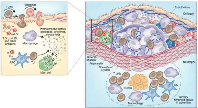
The defense of the normal artery depends on innate immune responses mounted by the endothelial cells (ECs) and after inflammatory challenge by macrophages and other immune cells recruited to the artery wall [95]. Although components of the adaptive immunity are not required for initiation, adaptive immune cells have a major role in progression and complications of the disease. The role of most cells of the innate and adaptive immunity is discussed below in details (Figure 6).
a. Innate immune cells
Monocytes
Under homeostatic conditions, monocytes circulate in blood, bone marrow and spleen. These cells are short lived, and do not proliferate in blood. They are phagocytic and can develop into macrophages and dendritic cells (DCs) in vivo and in vitro.
During inflammation, blood monocytes migrate to lymphoid and non-lymphoid tissues, in response to tissue-derived signals such as infection or tissue damage [96].
During the earliest phase of atherogenesis, blood derived monocytes home to the intima and subintima where they differentiate into macrophages or even DCs.
Monocytes are already visible in the fatty streak, the earliest lesion in human and experimental atherosclerosis [97]. Systemic depletion of circulating monocytes by clodronate significantly reduces plaque formation, pointing to their importance in atherogenesis [98]. Also, Osteopetrotic mice (op/op), deficient in circulating monocytes, tissue macrophages and
23
osteoclasts, are highly protected from atherosclerosis in the setting of hypercholesterolemia [99].
In mice, monocytes can be defined by their expression of Ly6C, an epitope of Gr-1. Classical Ly6ChighGr-1+ monocytes are CCR2highCX3CR1low, extravasate to tissues and mediate inflammation, phagocytosis, and proteolysis. However, non-classical Ly6ClowGr-1 -monocytes defined as CCR2lowCX3CR1high, mediate wound repair, tissue remodeling and expression of chemokines [100].
Under basal conditions, normal mice have few circulating monocytes evenly distributed between Ly6Chi and Ly6Clo populations. Hypercholesterolemia induces monocytosis in the bonemarrow, blood, and spleen with a preferential and dramatic increase in Ly6chigh monocytes. In Apoe-/- mice, the numbers of circulating monocytes is 50% higher than in wild type mice [101], [102]. One mechanism could be that cholesterol enrichment of hematopoeitic stem and progenitor cells increases their expression of IL-3 and GM-CSF receptor, and thus hematopoietic stem cell (HSC) proliferation [103].
Ly6Chigh monocytes home to atherosclerotic lesions in a manner dependent on CCR2 and CX3CR1, while Ly6clowmonocytes infiltrate lesions via CCR5.
Our laboratory has shown that combined inhibition of CCL2, CX3CR1, and CCR5 in Apoe -/-mice abrogates bone marrow monocytosis and reduces circulating monocytes despite persistent hypercholesterolemia. These effects were associated with marked additive reduction in atherosclerosis, and interestingly, the lesion size was highly correlated witht the number of circulating monocytes [104].
Ly6Chigh subpopulation exhibits series of functions, which render them particularly pathogenic in the context of atherosclerosis. For example, they bind with high avidity to endothelial monolayers. As mentioned earlier, PSGL-1 mediates adhesion and transmigration of monocytes through binding to E- and P-selectins of the activated endothelium. It is worth noting that Ly6Chigh monocytes express higher levels of PSGL-1 than Ly6Clow, which suggests its selective recruitment to the developing lesion [100].
Also, Ly6Chigh monocytes express high amounts of pro-inflammatory cytokines and proteases implicated in the pathogenesis of atherosclerosis than their Ly6Clow counterparts. Ly6Chigh monocytosis is induced not only in peripheral blood but also in spleen. Pro-inflammatory monocytes of splenic origin comprise up to a quarter of the mononuclear phagocytes in mouse atheromata [105], [106].
However, it remains unknown whether foam cells arise from either differentiated Ly6ChighGr-1+ or Ly6ClowGr-1- monocytes.
24
Figure 6: Immune components of the atherosclerotic plaque, Several types of cells of the immune response are present in the atheroma; these include: macrophages, T cells, dendritic cells, and mast cells. Cells may accumulate in the adventitia outside the atheroma where they develop into tertiary lymphoid structures with germinal centers. (adapted from Hannson and Hermanson, 2011,
Nature)
Macrophages
The first demonstration of a major role of macrophages in atherosclerosis was the study whereby macrophage colony-stimulating factor (op) and apolipoprotein E deficient mice had significant decrease in plaque size compared to control Apoe-/- despite high cholesterol levels [99].
Once in the arterial wall, monocytes differentiate into macrophage under the action of colony stimulating factor (M-CSF) produced by activated endothelial cells and SMCs. Intimal macrophages take up lipids through their scavenger receptors. Numerous scavenger receptor family members including SR-A1, SR-A2, SR-B1, CD36, LOX-1, and SREC1 can bind oxidized LDL and promote lipid-laden foam cells formation, the hallmark of atheromata [107].
SR-A1 and CD36 receptors mediate 75-90% of the degradation of acetylated or oxidized LDL in vitro. Combined deficiency of SR-A1 and CD36 reduced atherosclerosis in Apoe-/- mice. Plaques in these mice have reduced inflammation, macrophage apoptosis, and secondary necrosis, which suggests that these mice have roles beyond lipid uptake [108], [109].