SYNTHÈSE
REVUES
médecine/sciences 2018 ; 34 (focus issue, F1) : 99-104
médecine/sciences
UNBS5162
inhibits colon
cancer growth via
suppression of
PI3K/Akt signaling
pathway
Fan Zhang1, Hui-zeng Lv2, Ji-ming Liu2, Xiao-yong Ye2, Cun-chuan Wang1*
>
Colon cancer is a common cause of
cancer-related death worldwide. However, the underlying
mechanism of tumor progression of colon cancer
remains far from being elucidated. In the present
study, we report the role of UNBS5162 in colon
cancer. UNBS5162 is a naphthalimide that can
intercalate into DNA and suppress the expression
level of CXCL chemokines. Here, we investigated
its effect on cell proliferation, mobility and
apop-tosis in HCT116 cells, and explored the underlying
mechanism. A CCK8 assay revealed that UNBS5162
can block the proliferation of colon cancer cells.
Base on a Transwell assay, we showed that cell
migration and invasion ability of HCT116 cells
are inhibited by UNBS5162. In addition, Annexin
V-FITC/PI assay and Western blot analysis were
performed to detect whether UNBS5162 could
induce cell apoptosis. The results indicated that
UNBS5162 increases the number of apoptotic cells
remarkably. Furthermore, Western blot analysis
demonstrated that UNBS5162 down-regulates the
expression level of Bcl2, and up-regulates that of
Bax as well as the level of activated Caspase-3.
Moreover, we examined the impact of UNBS5162
on PI3K/Akt signaling pathway. UNBS5162
subs-tantially inhibited the phosphorylation of Akt and
its downstream effector mTOR, and reduced the
expression of p-70. Taken together, these results
suggest that UNBS5162 should be considered as a
potent therapeutic anticancer agent that targets
the PI3K/AKT signaling pathway. <
Key words: apoptosis, invasion, migration, PI3K,
proliferation, UNBS5162.
environment pollution and other carcinogenic factors (age, smoking,
alcohol, and digestive disorders). Colon cancer is one of the most common malignant tumors with the highest incidence in the 40 to 50-year-old population [1]. Globally, there are more than 1.4 million new cases in 2016 accounting for 10-15% of all malignancies [2]. In China, the incidence of colon cancer has increased every year. The death rate of colon cancer rates is second only to that of lung cancer, while liver cancer, accounts for the third place in China [3].
The phosphatidylinositol-3-kinase (PI3K)/Akt/mammalian target of rapamycin (mTOR) signaling pathway is an essential signaling pathway regulating cell survival and growth, proliferation, metabolism and genomic stability in eukaryotes cells [4, 5]. The PI3K/Akt pathway can be activated by a series of signals including growth factors, cytokines, hormones and extracellular matrix. Activation of Akt phosphorylates TSC2 (S939, S981, S1130, S1132, T1462) leading to TSC1/TSC2 complex dissociation and inhibition of TSC2 GAP ability [6-8], resulting in Rheb-GTP levels increase and downstream mTOR activity [9-12]. The over-activationof PI3K/Akt signaling pathway and of its downstream molecule mTOR are widely observed in multiple types of cancers. In tumor cells, the PI3K/Akt activity initiates a signal transduction cascade that promotes cancer cell growth and survival via protecting cancer cells from undergoing apoptosis [13, 14]. The presence of mutations in PIK3CA, that encodes the p110alpha catalytic subunit of PI3K, causes chronic activation of Akt [15]. Furthermore, loss-of-function mutations or silencing expression of phosphatase
1Gastrointestinal Surgery
Department, the First Affiliated Hospital of Jinan University, 613 Huangpu Avenue West, Guangzhou, 510630 PR China.
2Department 1 of General
Surgery, the 5th Affiliated Hospital, Guangzhou Medical University, Guangzhou, 510700, PR China.
Co-first authors: Fan Zhang, Hui-Zeng Lv
* corresponding author: Cun-Chuan Wang, MD wangcunchuan1@163.com
Introduction
Colon cancer is a malignant epithelial tumor which develops due to several factors, including heredity,
obtained from AMRESCO (Solon, OH, USA). RIPA Lysis Buffer, Protease Inhibitor Cocktail and BCA Protein Assay Kit were purchased from ComWin Biotech (Beijing, China). Pre-stained protein marker was obtained from Thermo Fisher Scientific. LDS Sample Buffer was obtai-ned from Invitrogen (Carlsbad, CA, USA). ECL Substrate Kit was purchased from Proteintech Group (Chicago, IL, USA). Matrigel was obtained from BD Biosciences (Bedford, MA, USA). Transwell inserts were obtained from Millipore (Billerica, MA, USA). Annexin V-FITC/PI Apoptosis Detection Kit was obtained from 4A Biotech (Nanjing, China).
Antibodies against Akt, mTOR, p-Akt, and p-mTOR were purchased from Cell Signaling Technology (Danvers, MA, USA),. Anti-Bax, -Bcl-2, -P-70, -Active-caspase3, -GAPDH and HRP-conjugated secondary antibodies (goat anti-rabbit or anti-mouse) were purchased from Proteintech Group (Wuhan, China). The antibodies were diluted at 1:1,000 except for anti-GAPDH (1:5,000) and HRP-conjugated secondary antibodies (1:5,000).
Cell culture
The human colon carcinoma cell line HCT116 was obtai-ned from the Cell Bank of the Chinese Academy of Sciences (Shanghai, PR China). HCT116 cells were cultu-red in RPMI 1640 medium at 37 °C in an incubator with a 5% CO2 humid atmosphere. The RPMI 1640 medium was
supplemented with 10% FBS, penicillin (100 U/mL) and streptomycin (0.1 mg/mL) (= complete medium). When adherent HCT116 cells entered into the growth logarith-mic phase, they were washed with PBS 3 times, and then digested with trypsin for 3 min until the cell morphology changed to a round shape. Complete medium was added to block further trypsin digestion. After single cell suspension, HCT116 cells were then seeded into 6-well plates and cultured in plates until they reached to 80% confluency. Cells were then treated with UNBS5162 (10 µM) for 24 h. Control cells were treated with DMSO.
Western blot assay
HCT116 cells were cultured in 6-well plates and treated with UNBS5162 or DMSO for 24 h. After stimulation, the cells were washed with cold PBS and lysed with a RIPA buffer (50 mM Tris-HCI pH 7.4, 150 mM NaCl, 0.5% sodium deoxycholate, 1 mM EDTA, 1% NP-40, 0.1% SDS) for 15 min on ice. The RIPA buffer contained a protease inhibitor cocktail. HCT116 cells were then harvested and centrifuged at 12,000 rpm for 15 min at 4°C. Pro-tein concentration was determined using a BCA assay and then mixed with 5× protein loading buffer. Protein samples were boiled at 95 °C for 5 min and stored at -80 °C for further analysis.
and tensin homolog (PTEN) due to epigenetic alterations lead to the PI3K pathway signaling hyper-activation [16]. In addition, another main function of activated Akt is is to block apoptosis. Akt can acti-vate NF-κB signaling pathway via regulating IκB kinase (IKK), pro-moting transcription of pro-survival genes [17]. Furthermore, Akt can phosphorylate pro-apoptotic proteins, including Bcl-2, Bax, BAD and Bim, to inhibit apoptosis [18]. Owing to the fact that Akt can promote cancer cell growth and protect cells from apoptosis, it has become an attractive target for drug development in cancer therapy.
Increasing reports suggest that there is a positive correlation between PI3K/Akt activation and colon tumorigenesis. Hyper-activated Akt signaling and decreased expression of PTEN have been found in 60-70% of human colon cancer patients [19]. Therefore, PI3K/Akt has been suggested as a potential target for colon cancer therapy. In recent years, the dysfunction of mTOR pathway has been also found in colon cancer. Based on the important role of PI3K/Akt/mTOR signaling pathway in colon tumorigenesis and progression, a series of chemical inhibitors which target various components of this pathway have been reported for colon cancer therapeutics. LY294002 inhibitor blocks PI3K signaling pathway, inhibits cell proliferation and induces a pro-grammed cell death in colon cancer cell lines [20]. Studies show that Buparlisib, a pan-PI3K inhibitor, is a safe drug that has shown antitu-mor activity in a phase I clinical trial [21]. Recent studies have also shown that MK-2206, a new oral-specific Akt1/2/3 inhibitor, provides antitumor activity in vitro and in vivo [22]. Rapamycin, a specific inhibitor of mTOR, can inhibit the tumor growth in APCmin/- mice [23].
Dual PI3K/mTOR inhibitors are under development and have theoreti-cal advantage. These dual inhibitors can completely suppress PI3K/Akt signaling pathway caused by negative feedback of mTORC1. BEZ235 is currently being assessed in early clinical trials for the therapy of colon cancer patients [24].
Thus, there is an urgent need to develop novel and more effective PI3K/Akt signaling pathway inhibitors for colon cancer therapy. In our present study, we studied the effects of UNBS5162, which inhibited the proliferation significantly as well as the invasion and migration ability of HCT116 cells. Further, our data showed that UNBS5162 up-regulated pro-apoptotic gene BAX expression and decreased anti-apoptotic gene Bcl2 expression, inducing then HCT116 cells apoptosis. Moreo-ver, it has been suggested that UNBS5162 specifically inhibits the phosphorylation of Akt and its downstream effector mTOR and p-70. Overall, these findings highlight a new PI3K/Akt signaling pathway inhibitor UNBS5162 and its anti-cancer activity.
Materials and Methods
Reagents and antibodies
RPMI 1640 medium was purchased from Hyclone (Thermo Fisher Scien-tific, Waltham, MA, USA). Fetal bovine serum (FBS) was obtained from Gibco (Carlsbad, CA, USA). Penicillin and Streptomycin were purchased from Sigma-Aldrich (St. Louis, MO, USA). Trypsin was obtained from Solarbio Science & Technology Company (Beijing, China). UNBS5162 was purchased from MedChem Express (Princeton, NJ, USA). DMSO was
SYNTHÈSE
REVUES
photographed and counted. The migration assay was the same as invasion assay, except that the top cham-bers were not coated with Matrigel.
Statistical analysis
All experimental data were analyzed using the SPSS Sta-tistics 18.0 software. The data were presented as mean ± SD. Comparison of means between different groups were carried out by Student’s unpaired t-test. A P-value less than 0.05 was considered statistically significant.
Results
UNBS5162 inhibits proliferation of HCT116 cells
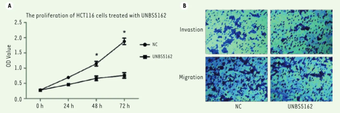
To confirm the effect of UNBS5162 on colon cancer progression, HCT116 cells were treated with UNBS5162 (10 µM) or DMSO, and then CCK-8 assay was performed to examine the changes in cell proliferation. Prolif-eration rate was measured dayly during the subse-quent 72 hours. UNBS5162 effectively suppressed the proliferation of HCT116 cells, while DMSO as a negative control had no effect. The proliferation at 48 hours and 72 hours was markedly inhibited (Figure 1A, P<0.05).
UNBS5162 suppresses migration and invasion of HCT116 cells
In addition to proliferation, we evaluated the effects of UNBS5162 on cell migration and invasion. A Transwell assay was carried out to examine its ability to inhibit migration and invasion of HCT116 cells. As shown in Figure 1B, the number of cells invading through the matrigel-coated membranes was decreased signifi-cantly in presence of UNBS5162, as well as the number of cells migrating through non-coated membranes Protein samples were separated on SDS-PAGE gels and transferred to
PVDF membranes. PVDF membranes were blocked with 5% of non-fat milk-TBST for 1 h at room temperature, and then incubated overnight with primary antibodies at 4 °C. HRP-coupled secondary antibodies were added to the membranes for 1 h and the immune-reactive bands were then visualized with ECL reagent. GAPDH was used as a protein control. Subsequently, the bands were scanned and the greyscale images were analyzed using the QUANTITY ONE software.
Cell Counting Kit-8 assay
HCT116 cells were seeded into 96-well plates at a density of 1×10³ cells
(100 µL) per well. UNBS5162 (10 µM) was added to UNBS5162-group and DMSO was added to the negative control group at the relevant concentration. Cells were grown at 37 °C in a 5% CO2 humid
atmos-phere incubator. CCK8 solution (10 µM) was added to each well daily, followed by incubation for another 1.5 h before cell number evaluation with the CCK-8 assay. Proliferative indexes were measured at 450 nm using a microplate reader to detect OD values. Calculation of mean values was used for plotting the proliferative curves.
Transwell migration and invasion assay
The Matrigel was thawed on ice overnight and diluted with pre-cooled serum-free medium. Subsequently, the diluted Matrigel (100 µL) was dispensed and added into the upper surface of the chambers for 4-6 h at 37 °C in a 5% CO2 humid atmosphere incubator. Then, the
serum-free medium (500 µL) was added into the bottom of the 24-well plate to hydrate the lower surface of chambers, replacing the serum-free medium with complete medium after 30 min. HCT116 cells were resus-pended with serum-free medium after treatment with UNBS5162 or DMSO for 24 h. 1×10⁵ cells (100 µL) were seeded to the top chamber for
overnight culture. The cells present on the upper surface of the cham-bers were swabbed with wet cotton swabs. Those on the lower surface of the chamber were fixed and stained with 0.1% crystal violet. Five random fields per sample from three independent experiments were
A B 2.5 2.0 1.5 1.0 0.5 0.0 OD V alue
The proliferation of HCT116 cells treated with UNBS5162
NC UNBS5162
0 h 24 h 48 h 72 h NC UNBS5162
Migration Invastion
Figure 1. UNBS5162 inhibits the proliferation and mobility of HCT116 cells. A. Proliferation of HCT116 cells treated with UNBS5162 or DMSO (NC)
for 72 h. P-value was calculated by an independent t-test (P<0.05). B. Transwell assay of HCT116 cell migration and invasion ability in presence of
UNBS5162 inhibits the activation of PI3K/Akt signaling pathway in HCT116 cells
Finally, we measured the involvement of PI3K/Akt signa-ling pathway in UNBS5162-induced suppression of HCT116 cells. PI3K/Akt signaling pathway plays an important role in the progression of tumors. Cell proliferation, aggressi-veness, metastasis and apoptosis are related to PI3K/Akt signaling pathway. In the present study, we explored whe-ther the inhibitory effects of UNBS5162 were associated with the modulation of the PI3K/Akt signaling pathway in HCT116 cells. Three key components, mTOR, Akt and P-70, were analyzed by Western-blot. Previous studies have shown that mTOR, Akt and P-70 play an important role in cell proliferation and metastasis. Figure 3A and B show that UNBS5162 inhibits the phosphorylation of Akt and mTOR proteins in HCT116 cells treated with UNBS5162, but in control groups. In addition, the expression of P-70 protein was decreased simultaneously.
These results further confirms the impact of UNBS5162 on PI3K/Akt signaling pathway in HCT116 cells. Thus, UNBS5162 represents a potential drug for colon cancer, which may provide novel insights into the understanding of the mechanism of the antitumor effects and should be further investigated.
(P<0.05). DMSO as a control had negligible effects on cell migration and invasion. Thus, these in vitro results suggest that the metastasis ability of HCT116 cells could be inhibited by UNBS5162 in vivo.
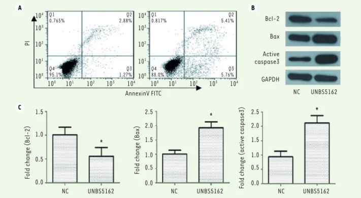
UNBS5162 induces apoptosis of HCT116 cells
We then explored whether UNBS5162 can impact apoptosis of HCT116 cells. HCT116 cells were first cultured with serum-free medium and then treated with UNBS5162 or DMSO, respectively. Cells were double-stai-ned with Annexin V-FITC and Propidium Iodide and analyzed using flow cytometry analysis. Compared with control group, HCT116 cell apoptosis was remarkably promoted by treatment with UNBS5162. As shown in Figure 2A, the percentage of apoptotic cells in control group was only 5.0%, while 22.7% of cells treated with UNBS5162 were apoptotic. To further investigate the effect of UNBS5162 on cell apoptosis, Wes-tern-blot analysis was performed to characterize the expression of Bcl-2, Bax and active caspase-3, three apoptosis-related proteins. Bcl-2 is an anti-apoptotic protein that protects cells against apopto-tic stimuli. Bax is a pro-apoptoapopto-tic protein involved in the induction of cell apoptosis. Bcl-2 and Bax are Bcl-family proteins, while caspase-3 belongs to the caspase family members. Activation of caspase-3 leads to cell apoptosis. In our study, as demonstrated in Figure 2B and 2C, UNBS5162 obviously inhibited the expression of Bcl-2 and promoted the expression of Bax. Activation of caspase-3 was also increased in cells treated with UNBS5162.
C PI AnnexinV FITC Bcl-2 Bax GAPDH Active caspase3 NC UNBS5162 NC UNBS5162 NC UNBS5162 NC UNBS5162 1.5 1.0 0.5 0.0 Fold change (Bcl-2) 2.5 2.0 1.5 1.0 0.5 0.0
Fold change (Bax)
2.5 2.0 1.5 1.0 0.5 0.0
Fold change (active caspase3)
Q1 0.765% Q4 95.1% Q2 2.88% Q3 1.27% Q1 0.817% Q4 88.0% Q2 5.41% Q3 5.76% 104 103 102 101 100 100 101 102 103 104 104 103 102 101 100 100 101 102 103 104
Figure 2. UNBS5162 promotes HCT116 cells apoptosis. A. HCT116 cells cultured in presence of UNBS5162 or DMSO were labeled with Annexin V/FITC
and PI solution. Results from flow cytometry analysis (Flowjo software) showed that the percentage of apoptotic cells was 22.7%, while only 5.03%
in the control group (NC). B. The expression levels of Bcl-2, Bax and active caspase-3 proteins were measured using Western-blot analyses after
incubation with UNBS5162 or DMSO for 24 h.C. Three experiments were repeated to confirm the results, P<0.05 compared with the control group
SYNTHÈSE
REVUES
expression in HCT116 cells (Figure 3A and B). All these results indicated that UNBS5162 blocks the growth, pro-liferation, migration, and invasion of HCT116 cells and promotes apoptosis via a marked inhibition of the PI3K/ Akt/mTOR signaling cascade.
In summary, we have identified a new inhibitor, UNBS5162, for the PI3K/Akt/mTOR signaling pathway, which could suppress proliferation of HCT116 cells and promote cell apoptosis via the up-regulation of the pro-apoptotic protein Bax and down-regulation of the anti-apoptotic protein Bcl-2. In addition, UNBS5162 treatment inhibited HCT116 cells migration and inva-sion. Moreover, our study suggested that the PI3K/Akt/ mTOR signaling pathway is involved in the inhibitory effects triggered by UNBS5162 on colon cancer cells. These results indicate that UNBS5162 is a poten-tial agent for the treatment of colon patients, likely through its ability to impact the PI3K/Akt/mTOR signa-ling pathway. ‡
CONFLICT OF INTEREST
The authors declare that they have no conflict of interests. REFERENCES
1. Haggar FA, Boushey RP. Colorectal cancer epidemiology: Incidence, mortality, survival, and risk factors. Clin Colon Rectal Surg 2009;22:191-197.
2. Arnold M, Sierra MS, Laversanne M, Soerjomataram I, Jemal A, Bray F. Global patterns and trends in colorectal cancer incidence and mortality. Gut 2017;66:683-691.
3. Gonzalez M, Robert JH, Halkic N, Mentha G, Roth A, Perneger T, Ris HB, Gervaz P. Survival after lung metastasectomy in colorectal cancer patients with previously resected liver metastases. World J Surg 2012;36:386-391.
Discussion
It is well known that PI3K/Akt signaling pathway can be induced by different extracellular signals, including hormones, growth factors and components of the extracellular matrix [25]. Activated Akt can regulate cancer cell growth, differentiation, motility, survival and apoptosis. Previous research show that mutations in PIK3CA or PTEN, both of which result in abnormal activation of PI3K signaling pathway, are commonly found in many cancer patients [26]. Thus, this signaling pathway is a promising target for colon cancer therapy.
Our results showed that UNBS5162, a naphthalimide that can interca-late into DNA and suppress the expression level of CXCL chemokines [27], significantly decreases cell viability and inhibits HCT116 cell proliferation (Figure 1A). Furthermore, UNBS5162 also inhibited the migration and invasion ability of HCT116 cells (Figure 1B). Most anti-cancer agents in current use induce apoptosis of anti-cancer cells. Our pre-sent study shows that a higher percentage of apoptotic cells occurred in the UNBS5162 group compared with the control group (Figure 2A). Western-blot results indicated that UNBS5162 reduces the expression of the anti-apoptotic protein Bcl-2 and increases that of the pro-apoptotic protein Bax. Active caspase-3 level was also up-regulated in HCT116 cells by UNBS5162 treatment in parallel (Figure 2B). Altogether, these results suggest that HCT116 is a potential anticancer agent for colon cancer.
Based on the relationship between PI3K/Akt signaling pathway, cell proliferation and cell apoptosis, we inferred that UNBS5162 may tar-get PI3K-related molecules and could be involved in the regulation of apoptosis. Our results demonstrated the ability of UNBS5162 to decrease the phosphorylation of Akt and its substrate mTOR in HCT116 cells (Figure 3A and B). UNBS5162 treatment also down-regulated P-70
A B AKT P-AKT mTOR P-mTOR p70 GAPDH
NC UNBS5162 Fold change (AKT) NC UNBS5162 NC UNBS5162 1.5 1.0 0.5 0.0 Fold change (mT OR) 1.5 1.0 0.5 0.0 NC UNBS5162
Fold change (GAPDH)
1.5 1.0 0.5 0.0
NC UNBS5162 NC UNBS5162
Fold change (P-AKT)
1.5 1.0 0.5 0.0 Fold change (P-mT OR) 1.5 1.0 0.5 0.0 NC UNBS5162 Fold change (p70) 1.5 1.0 0.5 0.0
Figure 3. UNBS5162 inhibits the activation of the PI3K/Akt signaling pathway in HCT116 cells. A. Western-blot analysis of proteins involved in the
PI3K/Akt signaling pathway in HCT116 cells cultured in presence of UNBS5162 or DMSO (NC). B. The expression levels of proteins (indicated in the
ordinate legends) detected by Western-blot experiments performed in triplicates were analyzed using the QUANTITY ONE software, P<0.05 compared with the control group (NC).
apoptosis. Leukemia 2001;15:515-522.
19. Carnero A, Blanco-Aparicio C, Renner O, Link W, Leal JF. The pten/pi3k/akt signalling pathway in cancer, therapeutic implications. Curr Cancer Drug Targets 2008;8:187-198.
20. McNamara CR, Degterev A. Small-molecule inhibitors of the pi3k signaling network. Future Med Chem 2011;3:549-565.
21. Geuna E, Milani A, Martinello R, Aversa C, Valabrega G, Scaltriti M, Montemurro F. Buparlisib, an oral pan-pi3k inhibitor for the treatment of breast cancer. Expert Opin Investig Drugs 2015;24:421-431.
22. Malkomes P, Lunger I, Luetticke A, Oppermann E, Haetscher N, Serve H, Holzer K, Bechstein WO, Rieger MA. Selective akt inhibition by mk-2206 represses colorectal cancer-initiating stem cells. Ann Surg Oncol 2016;23:2849-2857.
23. Hurez V, Dao V, Liu A, Pandeswara S, Gelfond J, Sun L, Bergman M, Orihuela CJ, Galvan V, Padron A, Drerup J, Liu Y, Hasty P, Sharp ZD, Curiel TJ. Chronic mtor inhibition in mice with rapamycin alters t, b, myeloid, and innate lymphoid cells and gut flora and prolongs life of immune-deficient mice. Aging Cell 2015;14:945-956.
24. Wang H, Zhang L, Yang X, Jin Y, Pei S, Zhang D, Zhang H, Zhou B, Zhang Y, Lin D. Puma mediates the combinational therapy of 5-fu and nvp-bez235 in colon cancer. Oncotarget 2015;6:14385-14398.
25. Zhu L, Ding X, Zhu X, Meng S, Wang J, Zhou H, Duan Q, Tao J, Schifferli DM, Zhu G. Biphasic activation of pi3k/akt and mapk/erk1/2 signaling pathways in bovine herpesvirus type 1 infection of mdbk cells. Vet Res 2011;42:57.
26. Martelli AM, Evangelisti C, Follo MY, Ramazzotti G, Fini M, Giardino R, Manzoli L, McCubrey JA, Cocco L. Targeting the phosphatidylinositol 3-kinase/akt/ mammalian target of rapamycin signaling network in cancer stem cells. Curr Med Chem 2011;18:2715-2726.
27. Mijatovic T, Mahieu T, Bruyere C, De Neve N, Dewelle J, Simon G, Dehoux MJ, van der Aar E, Haibe-Kains B, Bontempi G, Decaestecker C, Van Quaquebeke E, Darro F, Kiss R. Unbs5162, a novel naphthalimide that decreases cxcl chemokine expression in experimental prostate cancers. Neoplasia 2008;10:573-586.
REFERENCES
4. Beck JT, Ismail A, Tolomeo C. Targeting the phosphatidylinositol 3-kinase (pi3k)/akt/mammalian target of rapamycin (mtor) pathway: An emerging treatment strategy for squamous cell lung carcinoma. Cancer Treat Rev 2014;40:980-989.
5. Papadimitrakopoulou V. Development of pi3k/akt/mtor pathway inhibitors and their application in personalized therapy for non-small-cell lung cancer. J Thorac Oncol 2012;7:1315-1326.
6. Huang J, Manning BD. The tsc1-tsc2 complex: A molecular switchboard controlling cell growth. Biochem J 2008;412:179-190.
7. Hoogeveen-Westerveld M, van Unen L, van den Ouweland A, Halley D, Hoogeveen A, Nellist M. The tsc1-tsc2 complex consists of multiple tsc1 and tsc2 subunits. BMC Biochem 2012;13:18.
8. Li Y, Inoki K, Vikis H, Guan KL. Measurements of tsc2 gap activity toward rheb. Methods Enzymol 2006;407:46-54.
9. Li Y, Inoki K, Guan KL. Biochemical and functional characterizations of small gtpase rheb and tsc2 gap activity. Mol Cell Biol 2004;24:7965-7975.
10. Inoki K, Li Y, Xu T, Guan KL. Rheb gtpase is a direct target of tsc2 gap activity and regulates mtor signaling. Genes Dev 2003;17:1829-1834.
11. Parmar N, Tamanoi F. Rheb g-proteins and the activation of mtorc1. Enzymes 2010;27:39-56.
12. Sato T, Nakashima A, Guo L, Tamanoi F. Specific activation of mtorc1 by rheb g-protein in vitro involves enhanced recruitment of its substrate protein. J Biol Chem 2009;284:12783-12791.
13. Lee JT, Lehmann BD, Terrian DM, Chappell WH, Stivala F, Libra M, Martelli AM, Steelman LS, McCubrey JA. Targeting prostate cancer based on signal transduction and cell cycle pathways. Cell Cycle 2008;7:1745-1762.
14. Chang F, Lee JT, Navolanic PM, Steelman LS, Shelton JG, Blalock WL, Franklin RA, McCubrey JA. Involvement of pi3k/akt pathway in cell cycle progression, apoptosis, and neoplastic transformation: A target for cancer chemotherapy. Leukemia 2003;17:590-603.
15. Miled N, Yan Y, Hon WC, Perisic O, Zvelebil M, Inbar Y, Schneidman-Duhovny D, Wolfson HJ, Backer JM, Williams RL. Mechanism of two classes of cancer mutations in the phosphoinositide 3-kinase catalytic subunit. Science 2007;317:239-242.
16. Song MS, Salmena L, Pandolfi PP. The functions and regulation of the pten tumour suppressor. Nat Rev Mol Cell Biol 2012;13:283-296.
17. Huang WC, Hung MC. Beyond nf-kappab activation: Nuclear functions of ikappab kinase alpha. J Biomed Sci 2013;20:3.