ADAPTATION DE STREPTOMYCES SCABIES AU PÉRIDERME DE LA POMME DE TERRE
par
Mario Khalil Habeeb Khalil
Thèse présentée au Département de biologie en vue de l’obtention du grade de docteur ès sciences (Ph.D.)
FACULTÉ DES SCIENCES UNIVERSITÉ DE SHERBROOKE
ADAPTATION OF STREPTOMYCES SCABIES TO POTATO PERIDERM
by
Mario Khalil Habeeb Khalil
Thesis submitted to the Department of biology to obtain the degree of doctor of philosophy in science (Ph.D.)
FACULTY OF SCIENCE UNIVERSITY OF SHERBROOKE
Le 3 Décembre 2019
le jury a accepté la thèse de Monsieur Mario Khalil Habeeb Khalil dans sa version finale.
Membres du jury
Professeure Carole Beaulieu Directrice de recherche
Département de Biologie, Université de Sherbrooke
Professeure Nathalie Beaudoin Codirectrice de recherche
Département de Biologie, Université de Sherbrooke
Docteure Tanya Arseneault Évaluatrice externe
Agriculture et Agroalimentaire Canada
Docteur Benoît Leblanc Évaluateur interne
Département de Biologie, Université de Sherbrooke
Professeur Sébastien Roy Président-rapporteur
v SOMMAIRE
Le périderme de la pomme de terre est composé principalement de subérine. La subérine est composée de deux domaines, les domaines polyaliphatique et polyaromatique. Le domaine aliphatique de la subérine est constitué de polyesters d’acides gras avec des acides féruliques estérifiés. L'acide férulique est supposé se lier transversalement à la partie aromatique de la subérine. La subérine aromatique est composée principalement d’acides férulique et coumarique. Des recherches antérieures ont montré l’importance des esters férulés dans le maintien de l’intégrité du périderme, qui agit comme une barrière contre l’entrée de pathogènes. Il est de plus en plus évident que Streptomyces scabies, une bactérie causant la gale commune de la pomme de terre, peut dégrader la partie aliphatique de la subérine, mais son aptitude à dégrader le domaine aromatique n’a pas été étudiée.
Dans la première partie de cette thèse, une surexpression hétérologue du gène sub1 a été faite dans une souche de E. coli. Ensuite, la protéine codée par ce gène a été purifiée. L’activité de l’enzyme Sub1 sur divers substrats dont un polymère synthétique (PET) et des biopolymères comme la subérine et la cutine a été testé. Notre étude démontre que Sub1 est bel et bien une cutinase/subérinase puisque des acides gras sont libérés de ces substrats en présence de l’enzyme. Sub1 hydrolysait aussi le polyester synthétique PET et pourrait donc être intéressante dans des applications biotechnologiques.
Dans la deuxième partie, une étude chronologique de la déplétion en acides trans-féruliques et en acides p-coumariques a été réalisée à l'aide de la chromatographie en phase liquide à haute performance (HPLC) en échantillonnant périodiquement les
vi
surnageants de culture de S. scabies souches EF-35 et 87.22 cultivées en présence de ces composés aromatiques. Cette étude a révélé que S. scabies peut dégrader les acides trans-férulique et p-coumarique. Une analyse protéomique de S. scabies 87.22 cultivé en présence d'acide trans-férulique a permis d'identifier certaines protéines qui pourraient être impliquées dans le catabolisme du ferulate. De plus, l'expression des gènes codant pour les protéines identifiées a été déterminée par qRT-PCR en présence des acides trans-férulique et des acides p-coumarique. Les gènes testés étaient fortement exprimés en présence des deux composés. Les données suggèrent que le catabolisme des acides trans-férulique et des acides p-coumarique s'est produit via la voie du β-cétoadipate.
Dans la troisième partie, nous avons comparé les réponses de S. scabies 87.22, S. acidiscabies ATCC 49003 et S. turgidiscabies Car8 lors de leur interaction avec le périderme de la pomme de terre enrichi en subérine. S. scabies 87.22 et les espèces émergentes induisant la gale commune (Streptomyces acidiscabies ATCC 49003 et Streptomyces turgidiscabies Car8) ont été comparées à la fois pour leur capacité à se développer sur le périderme de la pomme de terre enrichi en subérine et à récupérer les nutriments de ce substrat. Les résultats ont révélé que, contrairement à S. scabies 87.22, S. acidiscabies ATCC 49003 et S. turgidiscabies Car8 présentaient une capacité d'utilisation médiocre vis-à-vis des deux constituants principaux de la fraction aromatique de la subérine (acides trans-férulique et p-coumarique). S. scabies 87.22 a également montré une croissance supérieure à celle des autres espèces induisant la gale commune lorsqu'elle était cultivée en culture pure ou en co-culture avec S. acidiscabies ATCC 49003 et S. turgidiscabies Car8 dans un milieu contenant de la subérine. De plus, S. scabies 87.22 semble mieux adapté que les deux espèces de Streptomyces à dégrader le matériau cellulosique incrusté dans les parois cellulaires subérisées du périderme de pomme de terre.
vii
Les résultats présentés dans cette thèse visaient à démontrer pour la première fois la capacité de S. scabies à dégrader non seulement la partie aliphatique de la subérine, mais également le domaine aromatique. Il a également été démontré que S. scabies 87.22 était bien mieux adapté à la plante hôte que les agents pathogènes émergents (S. acidiscabies ATCC 49003 et S. turgidiscabies Car8). Toutes ces propriétés pourraient conférer un avantage à S. scabies 87.22 en tant que saprophyte par rapport aux autres espèces induisant la gale commune et en tant que parasite, car la dégradation de la subérine peut faciliter à la fois la colonisation de la pomme de terre et les processus infectieux.
Mots clés: Sub1, espèces induisant la gale commune, hydroxycinnamates, acide trans-férulique, subérine, Streptomyces scabies.
viii SUMMARY
Potato periderm is composed mainly of suberin. Suberin is composed of two domains (polyaliphatic and polyaromatic domains). The aliphatic domain of suberin consists of fatty acid polyesters with esterified ferulic acids. Ferulic acid is believed to cross‐link to the aromatic moiety of suberin. The aromatic suberin is composed mainly of ferulic and coumaric acids. Previous research reported the importance of ferulate esters in maintaining the integrity of the periderm which acts as a barrier against pathogens entry. Evidence accumulates that Streptomyces scabies, a potato common scab-inducing bacterium, can degrade the aliphatic part of suberin but its ability to degrade the aromatic domain has not been studied.
In the first part of this thesis, a heterologous overexpression of the sub1 gene was made in an E. coli strain. Then, the protein encoded by this gene was purified. The activity of the enzyme Sub1 on various substrates including the synthetic polymer polyethylene terephthalate (PET) and the natural polymers such as suberin and cutin has been tested. Our study demonstrates that Sub1 is a cutinase/suberinase since fatty acids are released from these substrates in the presence of this enzyme. Sub1 also hydrolyzed the synthetic polyester polyethylene terephthalate (PET) and could therefore be of interest in biotechnological applications.
In the second part, a time course study of both trans-ferulic and p-coumaric acids depletion was carried out using High Performance Liquid Chromatography (HPLC) by periodically sampling culture supernatants of S. scabies strains EF-35 and 87.22 grown in the presence of these aromatic compounds. This study revealed that S. scabies can degrade both trans-ferulic and p-coumaric acids. A proteomic analysis of
ix
S. scabies 87.22 in the presence of trans-ferulic acid allowed the identification of some proteins which could be involved in the catabolism of ferulate. Moreover, the expression of the genes encoding the identified proteins was determined by qRT-PCR in the presence of both trans-ferulic and p-coumaric acids. The tested genes were highly expressed in the presence of both compounds. The data suggest that trans-ferulic and p-coumaric acids catabolism occurred via the β-ketoadipate pathway.
In the third part, we compared S. scabies 87.22, Streptomyces acidiscabies ATCC 49003 and Streptomyces turgidiscabies Car8 in their interaction with suberin-enriched potato periderm. S. scabies 87.22 and the emerging common scab-inducing species (Streptomyces acidiscabies ATCC 49003 and Streptomyces turgidiscabies Car8) were compared for both their ability to grow on suberin-enriched potato periderm and their retrieval of nutrients from this substrate. Results revealed that, in contrast to S. scabies 87.22, the other two common scab-inducing species S. acidiscabies ATCC 49003 and S. turgidiscabies Car8 exhibited poor utilization of the two main constituents of the aromatic moiety of suberin (trans-ferulic and p-coumaric acids). S. scabies 87.22 also showed higher growth rates than the other common scab-inducing species when grown in pure culture, or in co-culture, with S. acidiscabies ATCC 49003 and S. turgidiscabies Car8 in suberin-containing medium. Furthermore, this study also demonstrated that S. scabies 87.22 is better adapted than the two other Streptomyces species to degrade the cellulosic material embedded in the suberized cell walls of potato periderm.
The results presented in this thesis demonstrated for the first time the ability of S. scabies to degrade not only the aliphatic part of suberin but also the aromatic domain. S. scabies was shown also to be better adapted to its host plant compared to the emerging pathogens S. acidiscabies ATCC 49003 and S. turgidiscabies Car8. All
x
these properties could give an advantage to S. scabies 87.22 over the other common scab-inducing species as both a saprophyte and as a parasite since suberin degradation may facilitate both the potato tuber colonization and the infection processes.
Key words:Sub1,common scab-inducing species, hydroxycinnamates, trans-ferulic acid, suberin, Streptomyces scabies.
xi
ACKNOWLEDGEMENTS
I would like to express my deepest gratitude to my supervisor, professor Carole Beaulieu for her continuous help, support and advice throughout my studies. Also, I would like to thank my co-supervisor professor Nathalie Beaudoin and my advisors professor Sébastien Roy and professor Benoît Leblanc for their constructive comments and suggestions.
I would like to thank all the members of the laboratory for their kind help and advice.
I would like to thank the Natural Sciences and Engineering Research Council of Canada (NSERC), Centre SÈVE, QC, Canada and Ministry of Higher Education, Egypt for their financial support to this project.
xii TABLE OF CONTENTS SOMMAIRE ... v SUMMARY ... viii ACKNOWLEDGEMENTS ... xi LIST OF ABBREVIATIONS ... xv
LIST OF TABLES ... xviii
LIST OF FIGURES ... xx
CHAPTER 1 ... 1
GENERAL INTRODUCTION ... 1
1.1. The potato ... 1
1.2. The potato in Canada ... 1
1.3. Potato tubers ... 2
1.4. Potato tuber cells ... 3
1.5. Chemical composition of suberin ... 5
1.6. Common scab of potato ... 8
1.6.1. The main common scab-inducing species ... 9
1.6.2. Streptomyces scabies ... 10
1.6.2.1. Disease cycle of Streptomyces scabies ... 11
1.6.2.2. Virulence factors of S. scabies ... 13
1.6.2.2.1. Factors involved in external colonization ... 13
1.6.2.2.2. Factors involved in internal colonization ... 14
1.6.2.2.2.1. nec1 gene ... 14
1.6.2.2.2.2. tomA gene ... 15
1.6.2.2.2.3. Indole acetic acid ... 16
1.6.2.2.3. Toxins ... 17
xiii
1.6.2.2.3.2. Concanamycins ... 23
1.6.2.2.3.3. Coronafacoyl phytotoxins ... 24
1.6.2.2.4. Degradation of periderm constituents by S. scabies .... 25
1.6.3. Other common scab-inducing species ... 27
1.7. Degradation of suberin ... 29
1.7.1. Degradation of the aliphatic moiety of suberin ... 30
1.7.2. Degradation of the aromatic moiety of suberin ... 35
1.8. Objective of this work ... 37
CHAPTER 2 ... 38
ENZYMATIC DEGRADATION OF P-NITOPHENYL ESTERS, POLYETHYLENE TEREPHTHALATE, CUTIN, AND SUBERIN BY SUB1, A SUBERINASE ENCODED BY THE PLANT PATHOGEN STREPTOMYCES SCABIES ... 38
2.1. Preface ... 38
2.2. Manuscript of the article: Enzymatic degradation of p-nitrophenyl esters, polyethylene terephthalate, cutin, and suberin by Sub1, a suberinase encoded by the plant pathogen Streptomyces scabies ... 40
CHAPTER 3 ... 67
THE PLANT PATHOGENIC BACTERIUM STREPTOMYCES SCABIES DEGRADES THE AROMATIC COMPONENTS OF POTATO PERIDERM VIA THE β-KETOADIPATE PATHWAY ... 67
3.1. Preface ... 67
3.2. Manuscript of the article: The plant pathogenic bacterium Streptomyces scabies degrades the aromatic components of potato periderm via the β-ketoadipate pathway ... 69
CHAPTER 4 ... 134
GROWTH AND ENZYMATIC ABILITY OF COMMON SCAB-INDUCING SPECIES ON CHEMICAL CONSTITUENTS OF POTATO PERIDERM ... 134
xiv
4.2. Manuscript of the article: Growth and enzymatic ability of common
scab-inducing species on chemical constituents of potato periderm ... 136
CHAPTER 5 ... 202
GENERAL DISCUSSION AND CONCLUSION ... 202
xv LIST OF ABBREVIATIONS % Percentage °C Degree Celsius μg Microgram μL Microliter μm Micrometer μM Micromolar
BLAST Basic Local Alignment Search Tool
C Cytosine
cDNA Complementary Deoxyribonucleic acid Ceb Cellobiose binding protein
CebR Cellulose utilization repressor
cfu Colony-forming unit
CM Control medium
CM+C Control medium supplemented with p-coumaric acid CM+F Control medium supplemented with trans-ferulic acid CM+S Control medium supplemented with suberin
COG Clusters of Orthologous Groups of proteins
CRISPR Clustered Regularly Interspaced Short Palindromic Repeats
Da Dalton
DNA Deoxyribonucleic acid
EDTA Ethylenediaminetetraacetic acid
Fae Feruloyl esterases
FDR False discovery rate
xvi
G Guanine
h Hour
HPLC High Performance Liquid Chromatography
IAA Indole-3-acetic acid
IaaH Indole-3-acetamide hydrolase
IaaM Tryptophan monooxygenase
IAM Indole-3acetamide
IPTG Isopropyl β-D-1-thiogalactopyranoside
kb Kilobase
KEGG Kyoto Encyclopedia of Genes and Genomes
kg Kilograms
kV Kilovoltage
L Liter
LSD Least Significant Difference
M Mega m/z Mass-to-charge ratio min Minutes mL Milliliter mmol Millimoles MS Mass spectrometer
NCBI National Center for Biotechnology Information
ng Nanogram
NPB Nitrophenyl butyrate
nm Nanometer
NOS Nitric oxide synthetase
NSAF Normalized Spectral abundance factor
NSpC Normalized Spectral Count
PAHs Polycyclic aromatic hydrocarbons
xvii
PCR Polymerase Chain Reaction
PET Polyethylene terephthalate
pH Potential hydrogen
PW Primary wall
qRT-PCR Quantitative real time polymerase chain reaction
Rf Retention factor
RNA Ribonucleic acid
rpm Rotations per minute
s Second
SD Standard deviation
SDS-PAGE Sodium Dodecyl Sulfate Polyacrylamide gel electrophoresis
SpC Number of Spectral counts
SW Secondary wall
TA Terephthalic acid
TLC Thin layer chromatography
TW Tertiary wall
v/v Volume per volume
Vmax The maximal velocity
w/v Weight per volume
xviii
LIST OF TABLES
CHAPTER 3 p.
Table 3.1. Bacterial strains and plasmids used in this study 74
Table 3.2. Primers used in the gene expression assay 78
Table 3.3. Proteins detected only in Streptomyces scabies 87.22 proteome in control medium supplemented with ferulic acid
85
Table S1 Supplementary Table. Proteins produced by Streptomyces scabies 87.22 during growth in CM supplemented or not with trans-ferulic acid
109
CHAPTER 4
Table 4.1. Synthetic sequences used in this study 143
Table 4.2. Primers used in this study 144
Table 4.3. Predicted extracellular proteins distributed within functional groups of Streptomyces species
156
Table 4.4. Predicted extracellular proteins involved in polysaccharides catabolism of Streptomyces species
xix
Table 4.5. Predicted extracellular proteins involved in lipid metabolism of Streptomyces species
161
Table 4.6. Relative expression level of targeted cellulase genes 163
Table S2 Supplementary Table. Proteins produced by S. scabies 87.22 grown in CM+suberin
178
Table S3 Supplementary Table. Proteins produced by S. acidiscabies ATCC 49003 grown in CM+suberin
189
Table S4 Supplementary Table. Proteins produced by S. turgidiscabies Car8 grown in CM+suberin
xx
LIST OF FIGURES
CHAPTER 1 p.
Figure 1.1. Diagrammatic representation of a cross section through a potato tuber
3
Figure 1.2. Plant cell wall structure 4
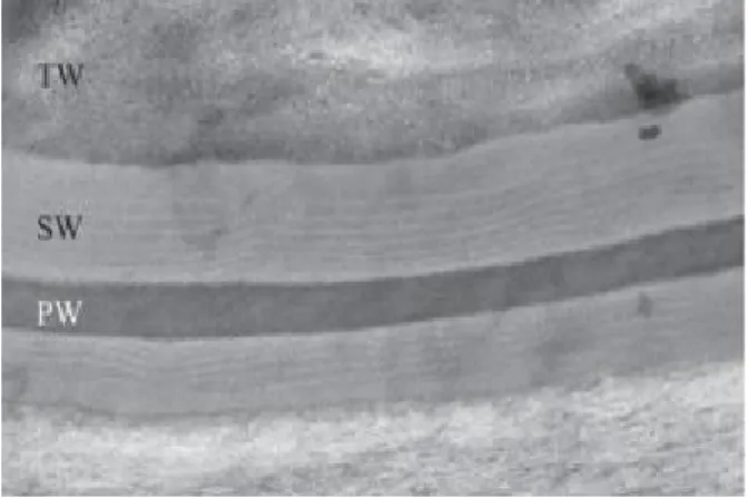
Figure 1.3. Cell wall structure of the potato phellem, showing suberized secondary wall (SW), primary wall (PW) and tertiary wall (TW)
5
Figure 1.4. Tentative model for the structure of potato suberin 6
Figure 1.5. Aliphatic precursors of suberized tissues 7
Figure 1.6. Phenolic precursors of suberized tissues 7
Figure 1.7. Lesions of common scab on potato tubers 9
Figure 1.8. Electron micrograph of Streptomyces scabies EF-35 10
Figure 1.9. Electron micrograph of filamentous mycelium of Streptomyces scabies
11
xxi
Figure 1.11. Colonization and penetration of potato by S. scabies 14
Figure 1.12. Structural formula of the thaxtomins produced by phytopathogenic species of Streptomyces
18
Figure 1.13. (A) thaxtomin biosynthetic gene cluster in S. scabies 87.22. (B) thaxtomin A biosynthetic pathway in Streptomyces
20
Figure 1.14. Involvement of CebR in the control of expression of the thaxtomin biosynthetic and regulatory genes
21
Figure 1.15. Hypothetical mechanism of action of suberin and cellulose on the production of thaxtomin in S. scabies
23
Figure 1.16. Concanamycin A structure 24
Figure 1.17. Schematic diagram showing the genetic organization of the pathogenicity islands from Streptomyces turgidiscabies Car8, Streptomyces scabies 87.22 and Streptomyces acidiscabies 84.104
29
Figure 1.18. Photos of suberin after treatment with cutinase CcCUT1 obtained by fluorescence microscopy
30
Figure 1.19. Electron micrographs of suberin-enriched potato periderm incubated with S. scabies EF-35 for 0 to 60 d
xxii
Figure 1.20. Esterase activity of culture supernatants of S. scabies EF-35 grown in minimal medium supplemented with 0.5% suberin, 0.5% suberin + 0.5% starch, and 0.5% starch
34
CHAPTER 2
Figure 2.1. Three-dimensional structural model of the Sub1 protein 44
Figure 2.2. SDS-PAGE gel of the cytoplasmic extract obtained from pET-transformed Escherichia coli strain SHuffle T7, without (E. coli SHuffle T7-pET) or with (E. coli SHuffle T7-pET-sub1) the insert of the sub1 gene, after induction with different concentrations of IPTG
51
Figure 2.3. Esterase activity of cytoplamic extracts from Escherichia coli SHuffle T7 transformed with plasmid pET without (E. coli SHuffle T7-pET) or with (E. coli SHuffle T7-pET-sub1) the sub1 insert and exposed to various concentrations of IPTG
52
Figure 2.4. SDS–PAGE gel of the cytoplasmic soluble proteins obtained from Escherichia coli transformed with SHuffle T7-pET-sub1, after fractionation on the affinity column (IMAC)
53
Figure 2.5. Esterase activity of the purified Sub1 enzyme using p-nitrophenyl substrates of different carbon chain sizes (C4, C8, C10 and C12) in the absence or presence of Triton X-100 (0.5%)
xxiii
Figure 2.6. Effect of the substrate (p-NPB) concentration on the initial speed (V0) of the hydrolysis reaction of the esterase Sub1
55
Figure 2.7. Degradation of cutin and suberin by enzyme Sub1 at room temperature over a 20-day period, as expressed by the release of fatty acids in the incubation medium
56
Figure 2.8. Concentrations of terephthalic acid (TA) released following the hydrolysis of ground particles of polyethylene terephthalate by 3 µg of Sub1 enzyme
57
CHAPTER 3
Figure 3.1. Kinetics of trans-ferulic and p-coumaric acids utilization in Streptomyces scabies strains EF-35 (A) and 87.22 (B)
81
Figure 3.2. Quantitation of the utilization of trans-ferulic acid and production of vanillic acid over time in the presence of (A) S. scabies 87.22. (B) S. scabies com15301. (C) SCAB_15301
83
Figure 3.3. Catabolism of vanillate in Streptomyces scabies strains 87.22, SCAB_15301 and com15301
84
Figure 3.4. Relative expression levels (Error bars represent standard deviations) of targeted genes involved in trans-ferulic and p-coumaric acids degradation from Streptomyces scabies 87.22 grown in control medium (CM) alone, CM
xxiv
supplemented with trans-ferulic acid (CM+F, gray bars) and CM supplemented with p-coumaric acid (CM+C, black bars)
Figure 3.5. Hypothetic degradation pathway of trans-ferulic and p-coumaric acids in Strepotmyces scabies 87.22
91
Figure 3.6. Relative expression levels (Error bars represent standard deviations) of targeted genes involved in trans-ferulic and p-coumaric acids degradation from S. scabies 87.22 grown in control medium (CM) alone and control medium (CM) supplemented with suberin (CM+S, black bars)
93
Figure 3.7. (A) Vanillate accumulation from suberin-enriched potato periderm in SCAB_15301 (no accumulation was detected with the wild-type or the complemented strain). (B) Utilization of trans-ferulic acid in the SCAB_15301 when grown and in the presence of trans-ferulic acid. (C) Utilization of trans-ferulic acid in the SCAB_15301 when grown in the presence of both trans-ferulic acid and vanillate
95
CHAPTER 4
Figure 4.1. Growth of three common scab-inducing species estimated by the number of genomic DNA copies
152
Figure 4.2. Growth inhibition of S. turgidiscabies Car8 on YME medium by S. acidiscabies ATCC 49003 (colony at center)
xxv
Figure 4.3. Transmission electron micrographs of potato periderm incubated in the presence of common scab-inducing strains
164
Figure 4.4.
Figure F1
Kinetics of trans-ferulic and p-coumaric acids utilization in S. acidiscabies ATCC49003 (A) and S. turgidiscabies Car8 (B)
Supplementary Figure. Fragmentation patterns of ions at 25 eV with nitrogen collision gas using a collision-induced dissociation cell
166
1 CHAPTER 1
GENERAL INTRODUCTION
1.1. The potato
The potato plant, Solanum tuberosum L., is an annual perennial plant belonging to the Solanaceae family. The potato is characterized by its tubers which are very rich in starch. The potato originates from the Andes region of South America and was introduced to Europe by Spaniards in the 16th century and was subsequently distributed all over the world (Brown, 1993). The outbreak of late blight in Ireland in 1840 caused a great famine and led to significant emigration to Canada and the United States. The potato is one of the most important food crops around the world and is grown in more than 150 countries. The potato comes in the fourth position of the most important food crops after maize, wheat and rice (Ezekiel et al., 2013). Potato tubers are rich in carbohydrates, vitamins, minerals, fibers and proteins and can be transformed into several products, such as chips, french fries, flours, etc.
1.2. The potato in Canada
Canada is the 13th largest potato producing country in the world and the second in the Americas (after the United States). In Canada, potato farmers planted 347,416 acres (140, 594 hectares) of potato in 2018. Potato is grown in all canadian provinces but mostly in Prince Edward Island, New Brunswick, Manitoba, Alberta and British Columbia. Prince Edward Island accounted for 24.2% of the total potato planted area in 2018, followed by Manitoba (18.5%) and Alberta (15.9%) (Government of Canada,
2
2018). In 2017, at 57.9 kg/person per year, potatoes accounted for 35% of all vegetables consumption in Canada and were far ahead of the tomato (24.8 kg/person, 15%) (Ministry of Agriculture, Fisheries and Food, Québec). Québec is the 5th largest potato producing province in Canada. In 2017, more than 600 companies in Québec produced 590,000 tonnes of potato on an area of 17,400 hectares, generating sales of $173 million (Ministry of Agriculture,Fisheries and Food, Québec).
1.3. Potato tubers
The underground part of the potato plant has fibrous roots and stolons (underground stems). The fibrous roots allow the plant to withdraw the essential elements and nutrients from the soil for its growth. Stolons act as the plant reserve organ carrying the tubers at their ends. Potato tubers represent about 75 to 85% of the total dry matter of the plant. An early event in potato tuber formation is the replacement of the epidermis of the expanding stolon tip by periderm. The periderm originates from cell division in the epidermis and hypodermis. The periderm is composed of three types of tissues; phellem, phellogen and phelloderm (Figure 1.1). The phellogen layer in the periderm form several layers of suberized cells (phellem) to the outside and the phelloderm to the inside. The phellem cells are rectangular and embedded with suberin to form a protective layer which is impermeable to water and gases and resists infection by micro-organisms (Tyner et al., 1997)(Figure 1.1). Lenticels are formed in the periderm from divisions of cells below the stomata of the original epidermis. These divisions form a tissue of rounded cells with intercellular spaces opening to the exterior to allow the gas exchange through periderm (Tyner et al., 1997). These lenticels can act as an entrance for several plant pathogens (Scott et al., 1996).
3
Figure 1.1. Diagrammatic representation of a cross section through a potato tuber (Tyner et al., 1997).
1.4. Potato tuber cells
Like all plant cells, the cell wall of potato cells is composed of three parts: 1) the middle lamella, which is the outer cell wall layer consisting mainly of polysaccharides called pectins (Jarrige et al., 1995), 2) the primary cell wall, composed of cellulose microfibrils contained within a gel-like matrix of hemicellulose fibers and pectin polysaccharides, and 3) the secondary cell wall, a layer formed between the primary cell wall and the plasma membrane. Once cells have stopped dividing and growing, the primary cell wall may thicken to form a secondary cell wall. This rigid layer is composed of cellulose and hemicellulose to strengthen and supports the cell and can be enriched in phenolic compounds (Figure 1.2). Potato cells contain amyloplasts instead of chloroplasts which are specialized in starch storage.
4 Figure 1.2. Plant cell wall structure.
(https://www.thoughtco.com/cell-wall-373613)
The cell walls of potato cells, especially those located at the surface of tubers, act as the first barrier to prevent the pathogens entry. As most cells, phellem cells possess a primary cell wall, a secondary cell wall but also a tertiary wall (the innermost layer of the secondary wall) (Figure 1.3) (Beaulieu et al., 2016). When the cells of the phellem develop, they will become suberized and die (because the suberin deposited in the cells prevents any cellular exchange), to form a protective layer (Sabba and Lulai, 2002) against pathogens entry. Suberin found in phellem cells can play a protective role against pathogens (Kolattukudy, 1984; Lulai and Corsini, 1998) by forming a biochemical barrier due to the lipidic and phenolic compounds found in the polymer (Kolattukudy, 1984). The periderm becomes mature with complete lipid coverage two or three weeks after harvest of the potatoes (Neubauer et al., 2013).
5
Figure 1.3. Cell wall structure of the potato phellem, showing suberized secondary wall (SW), primary wall (PW) and tertiary wall (TW) (Beaulieu et al., 2016).
1.5. Chemical composition of suberin
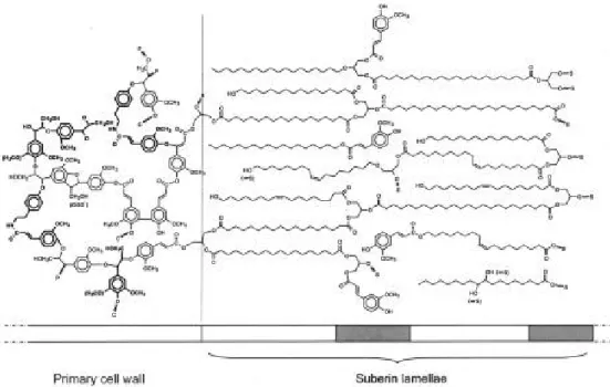
Suberin is a waxy substance with a complex structure. It is composed mainly of two domains, a glycerol-based polyaliphatic domain located between the primary cell wall and the plasma membrane which is covalently linked to a hydroxycinnamic acid-monolignol polyphenolic domain embedded in the primary cell wall (Figure 1.4) (Bernards, 2002).
6
Figure 1.4. Tentative model for the structure of potato suberin (Bernards, 2002).
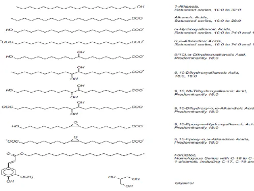
The structure of the aliphatic part of suberin was elucidated based on the analysis of the degradation products obtained by depolymerization through cleavage of the ester bonds using the alkaline hydrolysis or methanolysis (Gandini et al., 2006). The obtained monomers of the aliphatic moiety of suberin are mainly long chains of aliphatic dicarboxylic acids (16-24 carbon atoms), ω-hydroxy fatty acids (from 20 to 30 carbon atoms), very long chain fatty acids (≥ 30 carbon atoms), fatty alcohols and glycerols (Bernards 2002; Gandini et al., 2006) (Figure 1.5). The composition of suberin varies according to the plant species and tissues (Graça and Santos, 2007). Cutin, which is another plant polymer and the main component of the plant cuticle, covers all aerial surfaces of plant. It is very similar to the aliphatic domain of suberin. However, unlike suberin, cutin rarely contains molecules with chains containing more than 20 carbon atoms (Beisson et al., 2012).
7
Figure 1.5. Aliphatic precursors of suberized tissues (Bernards, 2002).
The polyaromatic domain of suberin is similar to lignin (Bernards, 2002). Upon depolymerization of suberin, a mixture of hydroxycinnamic acids (particularly ferulic acid) is released (Figure 1.6). Although both suberin and lignin contain monolignols, certain hydroxycinnamic acids and their derivatives, such as feruloyltyramine, are found only in suberin (Bernards et al., 1995).
8
Serra et al. (2010) suggested that ferulic acid plays an important role in the crosslinking of the two suberin domains. When the gene encoding a fatty omega-hydroxyacid/fatty alcohol hydroxycinnamoyl transferase (FHT) was silenced in potato, the resulted tubers contained much smaller amounts of ferulate esters. FHT enzyme is known to conjugate ferulic acid with omega-hydroxyacid and fatty alcohols. Moreover, the maturation was prevented, and the water loss increased greatly in the potato tubers with the knocked out gene. This suggests that ferulic acids play important role in periderm integrity (Serra et al., 2010).
1.6. Common scab of potato
Potato diseases can reduce the yield, quality and marketability of the tubers. Potato can be infected by different pathogens including fungi, oomycetes, bacteria, viroids, viruses, protozoa and nematodes (Hooker, 1981) and these pathogens can affect the plant during all stages of development, and even during storage.
Common scab is a potato disease characterized by superficial or deep corky lesions on tubers (Lerat et al., 2009) (Figure 1.7) or on the roots of various vegetables such as beet, radish, and parsnip (Goyer and Beaulieu, 1997). The size and color of the common scab lesions are quite variable (Beauséjour et al., 2003). Although the disease does not affect crop yield (unless in severe cases) and the infected tubers are safe for human consumption, these infections reduce the marketability of infected tubers causing significant economic losses. For example, in Canada, the total economic loss from potato common scab was estimated to be between 15.3 to 17.3 million Canadian dollars for the year 2002 (Hill and Lazarovits, 2005). Common scab of potato generates economic losses of about 15% in Quebec (Hill and Lazarovits, 2005).
9
Figure 1.7. Lesions of common scab on potato tubers (Lerat et al., 2009).
1.6.1. The main common scab-inducing species
Common scab is caused by pathogenic streptomycetes including Streptomyces scabies (Streptomyces scabiei), Streptomyces acidiscabies, Streptomyces turgidiscabies, Streptomyces stelliscabiei and Streptomyces europaeiscabiei (Healy et al., 2000; Loria et al., 1997; Miyajima et al., 1998; Pasco et al., 2005). Pathogenic streptomycetes acquire their pathogenicity through the presence of pathogenicity islands (PAIs) on their chromosomes. These islands encode different virulence factors and are absent from non-pathogenic strains of the same or closely related species (Gal-Mor and Finlay, 2006).
Streptomycetes produce about 70% of clinically useful antibiotics discovered to date (Bérdy, 2005). They are responsible for the characteristic smell of soil, which is related to the production of the volatile compound, geosmin (Bear and Thomas, 1964). The genus Streptomyces is a Gram-positive bacterium, strictly aerobic and belongs to the order Actinobacteria. Streptomycetes have a high guanine and cytosine (G+C) content and can undergo a complex morphological and physiological differentiation process during their life cycles (Burger and Eichenlaub, 2003). Streptomyces live in
10
soil mostly as saprophytes and produce extracellular hydrolytic enzymes. These extracellular enzymes break down structural polymers such as lignin and cellulose to provide nutrients (Crawford and Crawford, 1980). Streptomyces (Figure 1.8) form sporogenic aerial hyphae (Stackebrandt et al., 1997) and its spores are resistant to desiccation and can germinate and develop into branched filamentous hyphae when conditions become favorable. Streptomycetes play an important role in the decomposition and mineralization of organic matter through the production of extracellular enzymes, thus bringing an important contribution to the carbon cycle and nutrient recycling in the environment (Lewin et al., 2016).
Figure 1.8. Electron micrograph of Streptomyces scabies EF-35 (Lerat et al., 2012).
1.6.2. Streptomyces scabies
Streptomyces scabies is the main causal agent of potato scab (Goyer et al., 1996). It was first described by Thaxter in 1890, but a new description of the species was later reported by Lambert and Loria (1989b). It is known for its ability to produce a melanoid pigment.The species is characterized by its smooth, grey spores which are carried on
11
spiral chains. These spiral chains are mycelial filaments approximately 1 μm in diameter which carry spores that are between 0.5 and 1 μm in diameter (Figure 1.9). S. scabies growth is inhibited on solid medium when the pH is below 5 (Lambert and Loria, 1989b). The fully sequenced genome of S. scabies strain 87.22 is composed of approximately 10.1 M base pairs with a G+C content of 71.45%.
Figure 1.9. Electron micrograph of filamentous mycelium of Streptomyces scabies (Lerat et al., 2009).
1.6.2.1. Disease cycle of Streptomyces scabies
S. scabies can survive during winter in soil and on the surface of tubers and decaying plant material. In the spring, the pathogen is spread from one location to another by splashing water (irrigation or rain) and wind, and on seed tubers and farm equipment with leftover soil residue. When the spores encounter the potato, they will germinate, and the infection process begins. The pathogen invades the periderm of the tuber by entering mainly by lenticels or wounds (Adams and Lapwood, 1978), during the first five weeks of the development of tubers. Direct penetration has also been reported (Loria et al., 2003). After penetration, the pathogen can grow through up to three
12
peridermal cell layers, causing the death of these cells. The bacterium then feeds on these cells saprophytically. The pathogen also secretes a toxic substance (thaxtomin). This causes the tuber cells to divide rapidly leading to suberization of cells which will be converted into cork (suberized) cells that isolate the bacterium and the surrounding tuber cells. As the tuber cells above this suberized layer die, the pathogen feeds on them. As the suberized layers are pushed out and sloughed off, the pathogen grows and multiplies in the additional dead cells. This growth cycle may occur several times throughout the growing season, enlarging the lesion. Lesion size will also vary depending on when infection occurs. Generally, the earlier a tuber becomes infected, the larger the lesion. Figure 1.10 summarizes the disease cycle of the disease (Agrios, 2005).
Figure 1.10. Disease cycle of potato common scab caused by S. scabies (Agrios, 2005).
13 1.6.2.2. Virulence factors of S. scabies
1.6.2.2.1. Factors involved in external colonization
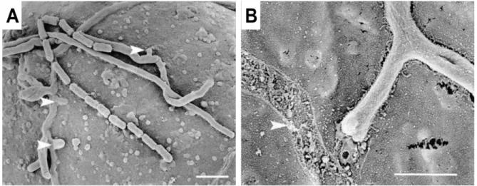
Pathogenicity is defined as ability of an organism to cause disease or damage to its host while virulence refers to the degree of pathology caused by the organism. Successful colonization of a host plant requires that the pathogen has effective mechanisms for penetrating the plant tissues. S. scabies can infect tissues of potato tuber and develops intercellularly after its entry through lenticels or wounds (Agrios, 2005). S. scabies can also use the direct penetration through the mechanical force (Figure 1.11A) (Loria et al., 2003). Thaxtomin produced by plant pathogenic streptomycetes was also shown to aid penetration of developing plant tissues by inhibiting primary cell wall development (Loria et al., 2003), therefore contributing to virulence. The filamentous nature of S. scabies allows it to colonize aggressively the tuber surface of the potato. Hydrophobin-like compounds are thought to play a role in host-pathogen interactions since an attachment matrix at the interface of potato tuber cells and S. scabies hyphae colonizing those cells has been detected (Figure 1.11B) (Loria et al., 2003). Hydrophobins are small, secreted hydrophobic proteins that contribute to the pathogenicity of some fungi. The direct penetration is likely to be accompanied by secretion of hydrolytic enzymes (Komeil et al., 2014) that would facilitate the breakdown of the surficial barrier and the degradation of walls, as suggested by microscopic observations of infected tissue (Błaszczak et al., 2005).
14
Figure 1.11. Colonization and penetration of potato by S. scabies.
(A) S. scabies directly penetrates the cells of potato by using short branches emerging from the main hyphae. The short hyphae are perpendicular to the primary hyphae and penetrate within a very short distance from the branch point, indicating that these secondary hyphae are in fact penetration structures (arrow heads). (B) an attachment matrix at the interface of potato tuber cells and colonizing hyphae of S. scabies. The arrow shows a track left by the removal of a Streptomyces hypha, suggesting bacterial attachment (Loria et al., 2003).
1.6.2.2.2. Factors involved in internal colonization
1.6.2.2.2.1. nec1 gene
The nec1 gene is responsible for the synthesis of a necrosis factor (Bukhalid and Loria, 1997). S. scabies, as most potato common scab-inducing species, possess nec1 (Bukhalid et al., 1998). When a fragment of 9.4 kb containing the nec1 region was transferred to S. lividans, a non-pathogenic Streptomyces species, the
15
recombinant strain was able to cause necrosis and colonization of the potato slices (Bukhalid and Loria, 1997). It was proposed that nec1protein is a virulence factor which is secreted at the beginning of the infection (Joshi et al., 2007a). However, nec1 is not necessary for pathogenicity. It would be useful for the colonization of the host plant or for the suppression of defense responses of the host plant. Radish plants inoculated with the S. turgidiscabies mutant nec1 are still infected, but the mutant failed to colonize the meristem of radish root (Joshi et al., 2007a).
1.6.2.2.2.2. tomA gene
The tomA gene is an orthologous gene to genes encoding tomatinases in pathogenic fungi of tomato. This gene was first discovered in the pathogenicity island of S. turgidiscabies Car8 (Kers et al., 2005) but was also identified in the genome of S. scabies 87.22 (Seipke and Loria, 2008). Tomatinases belong to a class of secreted enzymes, called saponinases (Kers et al., 2005). Saponinases are known to detoxify the phytoanticipins which are antimicrobial compounds produced by plants to defend themselves against pathogens (Bouarab et al., 2002). The severities of disease for tomato seedlings affected by S. scabies 87.22 wild-type and tomA strains were indistinguishable, suggesting that tomatinase is not important in pathogenicity on tomato plants. However, presence of tomA in several pathogenic species of Streptomyces suggests their involvement in the suppression of plant defense mechanisms to facilitate internal colonization (Seipke and Loria, 2008).
16 1.6.2.2.2.3. Indole acetic acid
The ability of S. scabies to produce indole-3-acetic acid (IAA), a plant auxin, is well known (Manulis et al., 1994). S. scabies was found to produce indole-3-acetic acid (IAA) through the Indole-3-acetamide (IAM) pathway. Two enzymes, tryptophan monoxygenase (IaaM) and indole-3-acetamide hydrolase (IaaH), participate in IAA synthesis via the IAM pathway. The orthologous of those two genes were identified in the genome of S. scabies 87.22 based on amino acid sequences homology. The identified genes in S. scabies were named as (SCAB_75511) for tryptophan monoxygenase and (SCAB_75501) for indole-3-acetamide hydrolase (patten et al., 2013). When deletion mutants of these genes were generated, the IAA production by all the three generated mutant strains was lower than the wild type strain (87.22) (Hsu, 2010). When inoculated onto radish seedlings, all three mutant strains were reduced in virulence relative to the wild type strain. Genetic complementation of the deletion strains partially restored both IAA production and virulence on radish seedlings suggesting that both (SCAB75511) and (SCAB75501) genes contribute to the virulence of S. scabies through the synthesis of IAA which is considered as a virulence factor (Hsu, 2010). The exact role of IAA in S. scabies pathogenicity is unknown but asin Pseudomonas syringae, IAA is believed to stimulate the release of saccharides from the wall of plant cells, which could provide a source of nutrients for microorganisms and facilitate pathogens colonization (Bender et al., 1999).
17 1.6.2.2.3. Toxins
1.6.2.2.3.1. Thaxtomins
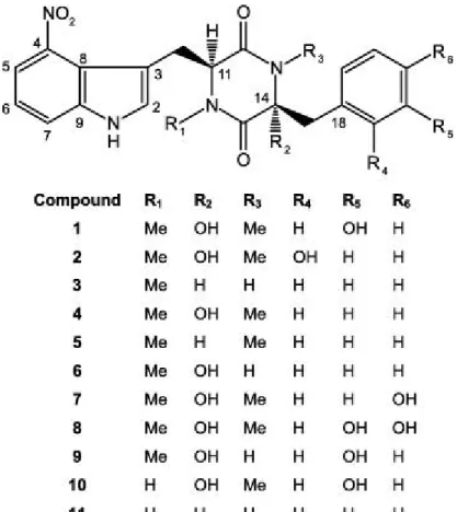
All common scab-inducing species produce a family of phytotoxic secondary metabolites known as thaxtomins. The thaxtomins are cyclic dipeptides (2,5-diketopiperazines) formed from the condensation of two aromatic amino acids, L-phenylalanine and L-tryptophan containing a 4-nitroindole group which is essential for the phytotoxicity of these compounds (King and Calhoun, 2009). Eleven members of this family have been isolated and characterized (Figure 1.12). These members can be distinguished by the presence or absence of N-methyl or hydroxyl groups on the basic structure. When the 4-nitro group is either removed, or shifted to the 5, 6 or 7 positions of the indole ring; or if the phenyl side chain is replaced; or conversion to the D,L configuration instead of the L,L configuration of the compound will result in a total loss of compound toxicity (King et al., 1992).
18
Figure 1.12. Structural formula of the thaxtomins produced by phytopathogenic species of Streptomyces. Compound 1 represents thaxtomin A and compound 4 corresponds to thaxtomin B (King and Calhoun, 2009).
Thaxtomin A is the main phytotoxin produced by pathogenic S. scabies (Healy et al., 2000). Thaxtomins are yellowish secondary metabolites secreted during the transition from vegetative phase to stationary phase. Thaxtomins induce phenotypic changes in the host plant, such as cell hypertrophy, stunted and seedling growth retardation (Leiner et al., 1996), tissue necrosis and inhibition of cellulose synthesis (Scheible et al., 2003), alteration in the influx of Ca2+ (Errakhi et al., 2008) and H+ ions (Tegg et al., 2005) in plant cells and programmed cell death (Duval et al., 2005).
19
The genes needed to synthesize thaxtomins are present on a single locus (txt) on the chromosome of the pathogenic species (Figure 1.13A). Two non-ribosomal peptide synthetases, TxtA and TxtB, are required to form the cyclic diketopiperizine moiety from the amino acids phenylalanine and tryptophan (Healy et al., 2000). Nitric oxide synthetase, TxtD/NOS, is responsible for the nitration of the indole group of L-tryptophan (Johnson et al., 2009; Kers et al., 2004). The txtD gene encodes a nitric oxide synthetase (NOS) that converts L-arginine to nitric oxide (Figure 1.13B). The deletion of the txtD gene in S. turgidiscabies drastically decreases the production of thaxtomin A (Barry et al., 2012). In addition, another enzyme appears to be involved in the nitration reaction, cytochrome P450 monooxygenase, encoded by the txtE gene (Bignell et al., 2010). Following cyclization, the compound is hydroxylated by cytochrome P450 monooxygenase (TxtC) (Healy et al., 2000). Sequence analysis of the thaxtomin biosynthetic cluster in S. acidiscabies previously revealed the presence of a small open reading frame (designated ORFX) that is located immediately downstream of the txtB gene (Healy et al., 2000). This gene is conserved in the S. turgidiscabies and S. scabies thaxtomin biosynthetic clusters and is predicted to encode a protein belonging to the MbtH-like superfamily. This family, named after the MbtH protein from the mycobactin biosynthetic cluster in Mycobacterium tuberculosis, consists of small proteins (about 70 amino acids) frequently associated with the gene clusters for non-ribosomal biosynthesis of peptide antibiotics and siderophores (Baltz, 2011). Although the function of these proteins is unknown, recent studies suggest that they are required for the biosynthesis of some non-ribosomally-synthesized secondary metabolites. Transcriptional analysis of the mbtH-like gene in the S. scabies thaxtomin biosynthetic cluster (txtH; Figure 1.13A) indicated that the gene is expressed under thaxtomin-inducing conditions and that expression is dependent on txtR (Figure 1.13A) (Bignell et al., 2010).
20
Thaxtomin is produced in vitro only when pathogenic bacteria grow in presence of plant extracts (Beauséjour et al., 1999). In vitro, biosynthesis of the toxin occurs during secondary metabolism (Lerat et al., 2010) and is favored by the presence of cellobiose, a disaccharide derived from the degradation of cellulose. TxtR is an AraC/XylS family protein regulated by cellobiose and transcription of txtR and thaxtomin biosynthetic genes was upregulated in response to cellobiose (positive transcription regulator) (Joshi et al., 2007b).
Figure 1.13. (A) thaxtomin biosynthetic gene cluster in S. scabies 87.22. (B) thaxtomin A biosynthetic pathway in Streptomyces (Bignell et al., 2010).
21
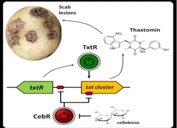
A more recent study identified cellulose utilization repressor (CebR) in the thaxtomin biosynthetic cluster (Francis et al., 2015). CebR is known to be involved in the primary metabolism and nutritional functions in non-pathogenic streptomycetes. However, In S. scabies, cellobiose and cellotriose inhibited the ability of CebR to bind to DNA, leading to an increased expression of the thaxtomin biosynthetic and regulatory genes txtA, txtB, and txtR. Deletion of CebR results in constitutive thaxtomin A production and hypervirulence of S. scabies. Thus, the pathogenicity of S. scabies is under dual control, under the control of CebR and TxtR proteins. CebR is a cellobiose sensor, and the key to block the expression of the txtR gene. On the other hand, the txtR gene is key to unlock the production of thaxtomin (Figure 1.14) (Francis et al., 2015). CebR-binding sites also found in S. acidiscabies and S. turgidiscabies suggesting that CebR is most likely an important regulator of virulence in these species as well.
Figure 1.14. Involvement of CebR in the control of expression of the thaxtomin biosynthetic and regulatory genes.
(http://www.reflexions.uliege.be/cms/c_386474/en/thaxtomin-a-next-generation-weed-killer?portal=j_55&printView=true)
Cellobiose was identified as an inducer of thaxtomin A synthesis in S. scabies (Johnson et al., 2007). However, when the bacterium was grown in the presence of cellobiose as the sole carbon source, there was a weak induction of txt genes
22
expression which are responsible for the production of thaxtomins. Similarly, the addition of suberin to minimal medium induced a low production of thaxtomin A (Beauséjour et al., 1999). However, when suberin and cellobiose are combined in the culture medium, the synthesis of thaxtomin A increased considerably and the expression of the thaxtomin biosynthetic genes (txtA, txtB, txtD/nos and txtC) increased significantly. This suggests that S. scabies needs both molecules to maximize thaxtomins production. Therefore, it is possible that the ability of S. scabies to produce glycosyl hydrolases, including cellulases, is correlated with the pathogenicity of this bacterium, because it is closely related to the degradation of cellulose to cellobiose.
Padilla-Reynaud et al. (2015) proposed a model that could explain the effect of suberin and cellobiose on thaxtomin production in S. scabies. In this model, the constituents of the potato periderm promote the onset of S. scabies’ virulence mechanisms specifically, the major periderm constituents, cellulose and suberin, both play a role in triggering the production of thaxtomins. In the presence of cellulose only (Figure 1.15A), a low amount of cellulases are produced allowing the release of cellobiose, the inducer of thaxtomin biosynthetic genes. However, cellobiose was shown to lock morphogenesis and secondary metabolism (Lerat et al., 2010) by allowing the production of a subtilase-like protease inhibitor and, thus, limiting the production of secondary metabolites such as thaxtomins. Cellulases were produced in the presence of suberin only (Figure 1.15C), whereas cellobiose was not produced in the absence of accessible cellulose. Since inducers of thaxtomin biosynthetic genes are lacking, the biosynthesis of thaxtomins is low, depending only on signals promoting morphogenesis and secondary metabolism. In the presence of both suberin and cellulose (Figure 1.15B), suberin may have a dual function in the virulence of S. scabies. It may first stimulate the production of cellobiose, the transcriptional inducer of thaxtomin biosynthetic genes, by stimulating cellulase activity and the consequently
23
the release of cellobiose from cellulose. It may then inhibit the effects of cellobiose on secondary metabolism by acting as a signal molecule for morphogenesis, thereby promoting the production of secondary metabolites such as thaxtomins (Padilla-Reynaud et al., 2015).
Figure 1.15. Hypothetical mechanism of action of suberin and cellulose on the production of thaxtomin in S. scabies (Padilla-Reynaud et al., 2015).
1.6.2.2.3.2. Concanamycins
S. scabies produces phytotoxins other than thaxtomins, namely, concanamycins A (Figure 1.16) and B (Natsume et al., 1996). Analyses of four taxonomically different Streptomyces species that induce common scab of potato showed that only S. scabies produces concanamycins, while other species produce only thaxtomins (Natsume et al., 1996). Concanamycins are macrolide secondary metabolites that are produced by S. scabies and other Streptomyces spp. and have been shown to display phytotoxic activity (Haydock et al., 2005; Natsume et al., 1996). A recent study showed necrosis-inducing activity of concanamycin A and its synergistic action with thaxtomin A in a potato tuber slice assay (Natsume et al., 2017). Concanamycins A and B were also detected in the tubers inoculated with S. scabies in pot experiments and in field-grown diseased potatoes (Natsume et al., 2017). The gene cluster responsible for
24
concanamycin biosynthesis has already been identified (Haydock et al., 2005). The concanamycins exhibit a wide range of important biological activities: antiviral, antiprotozoal and antineoplastic. These activities are thought to be due to their potent inhibition of the vacuolar (V-type) H+-ATPase (Muroi et al., 1993). However, the role of concanamycins in the plant-pathogen interaction is still not fully understood.
Figure 1.16. Concanamycin A structure (Haydock et al., 2005).
1.6.2.2.3.3. Coronafacoyl phytotoxins
The biosynthetic gene cluster predicted to produce coronafacoyl phytotoxins was identified in the genome sequence of S. scabies 87.22. Coronafacoyl phytotoxins are non-host-specific phytotoxins that are produced by different plant-pathogenic bacteria (Bignell et al., 2018). These metabolites are composed of the bicyclic hydrindane ring-based polyketide coronafacic acid and an amino acid or amino acid derivative linked via an amide bond. Coronatine is the predominant family member of coronafacoyl phytotoxins and the most toxic form (Bignell et al., 2018). Coronatine exhibits a wide range of biological activities in plant tissues such as induction of tissue hypertrophy, stimulation of ethylene production, anthocyanin accumulation and inhibition of root elongation. The role of coronafacoyl phytotoxin production in common scab disease development remains unclear. S. scabies is the only plant-pathogenic Streptomyces
25
sp. that is able to produce this family of phytotoxins. Although coronafacoyl phytotoxin are not essential for the pathogenicity of S. scabies however, it is believed that additional roles for these metabolites remain to be discovered (Bignell et al., 2018).
1.6.2.2.4. Degradation of periderm constituents by S. scabies
Glycoside hydrolases (also called glycosidases or glycosyl hydrolases) catalyze the hydrolysis of glycosidic bonds in complex sugars present in potato periderm such as cellulose.Glycoside hydrolases can be classified in several ways and the simplest way is based on their substrate specificities. Together with glycosyltransferases, glycosidases form the major catalytic machinery for the synthesis and breakage of glycosidic bonds. The classification based on substrate specificity could be problematic for enzymes that act on multiple substrates. This is particularly true for glycoside hydrolases, which often act on very complex polysaccharides and have a broad spectrum of substrates. For example, endoglucanases, typically considered as cellulases but may also be active on other substrates such as xylan, β-glucan and chitosan, etc. to varying degrees (Henrissat and Davies, 1997). Glycosyl hydrolases may be also classified based on their mode of action on a polysaccharide or based on sequence comparison since there is a direct relationship between sequence similarity and three-dimensional folding and therefore the structure of the protein. The hydrolysis of the β-1,4 glycosidic linkages of the cellulose is carried out by mixtures of hydrolytic enzymes collectively called cellulases. These cellulases include enzymes that synergistically hydrolyze internal linkages (endoglucanases) and external linkages (cellobiohydrolases) (Dashtban et al., 2009). The products resulting from these hydrolyses are cellobiose and cellodextrin which inhibit endoglucanase and cellobiohydrolase activity. Thus, the effective hydrolysis of cellulose requires the presence of β-glucosidases to break down the final glycosidic linkages and produce glucose monomers (Maki et al., 2009).
26
In addition to cellulases, S. scabies produces many other enzymes that degrade cell wall components such as mannosidases, xylanases/cellulases and cellobioses hydrolases (Joshi et al., 2010). Komeil et al. (2014) also demonstrated that suberin-enriched potato periderm induced the production of a large amount of glycosyl hydrolases such as cellulases, xylanases and licheninases. This result was unexpected because the substrate contained very few contaminating polysaccharides that act as inducers for the glycosyl hydrolases.
Glycosyl hydrolases, and especially cellulases of S. scabies, are of great interest because they may be possibly involved in the pathogenesis. Glycosyl hydrolases would be able to hydrolyze the β bonds of the polysaccharides present in the plant walls. The suberin which was purified from polysaccharides was found to act as an inducer for cellulases better than cellulose itself in S. scabies (Komeil et al., 2013). Another study by Padilla-Reynaud et al. (2015) demonstrated that suberin promoted glycosyl hydrolase activity when added to cellulose-, xylan-, or lichenin-containing media. Also this study showed that the addition of suberin to a cellulose-containing medium increased the production of glycosyl hydrolases (Padilla-Reynaud et al., 2015).
27 1.6.3. Other common scab-inducing species
Potato common scab may be also caused by other species of Streptomyces, aside from S. scabies, such as S. acidiscabies (Lambert and Loria, 1989a), S. turgidiscabies (Miyajima et al., 1998), S. stelliscabiei and S. europaeiscabiei (Pasco et al., 2005). Both S. acidiscabies and S. turgidiscabies produce symptoms like those of S. scabies, however, they have a more limited geographical distribution than that of S. scabies. S. acidiscabies has occasionally been reported in the United States and Canada in low pH soils (Faucher et al., 1992; Lambert and Loria, 1989a). S. acidiscabies causes a scab disease of potato in soils with pH values below 5.2. S. acidiscabies is distinct from S. scabies in its production of a red or yellow, pH-sensitive diffusible pigment, in the place of melanin. It also grows on agar media at pH 4.0 (versus pH 5.0 for S. scabies) and does not use raffinose as a carbon source (Lambert and Loria, 1989a).
S. turgidiscabies was isolated from potato grown in eastern Hokkaido, Japan. It is distinct from other scab-inducing species by having flexuous spore chains and grey mass color. It does not grow on agar media at pH 4.0 or 37 °C and does not produce melanin or other diffusible pigments. It can utilize raffinose and inulin as a carbon source (Miyajima et al., 1998). Both S. scabies and S. turgidiscabies can be found in the same potato cultivars, fields, tubers and also in the same scab lesion (Lehtonen et al., 2004).
A large pathogenicity island has been identified and partially sequenced in the genome of S. turgidiscabies Car8 (Kers et al., 2005). This island was used to identify virulence regions conserved in the S. scabies 87.22 genome. The genes associated with the pathogenicity island of S. turgidiscabies are found in two remote regions of the genome of S. scabies 87.22 (Lerat et al., 2009). These two regions are: 1)
28
toxicogenic region (estimated coordinates c. 3596–3653 kb; G+C content of 68%). All genes shown to be involved in thaxtomin biosynthesis are found in this region. These genes are txtAB (Healy et al., 2000), txtC (Healy et al., 2000), nos (Kers et al., 2004) and txtR (Joshi et al. 2007b). 2) The second segment of the pathogenicity island (PAI), which is called the colonization region contains more genes (estimated coordinates c. 8471–8581 kb; G+C content of 68.5%). This chromosomal region contains genes such as nec1 and tomA which are not essential to pathogenicity but play a very important role in virulence. It is believed that these two genes could play a role in the infection process by suppressing the plant defense mechanism (Joshi et al., 2007a; Seipke and Loria, 2008). In S. acidiscabies, the virulence genes found on the pathogenicity island are localized in two separate regions of the chromosome, called the toxicogenic region and the colonization region like S. scabies. The S. scabies toxicogenic region (TR) can be further divided into two sub-regions (TR1 and TR2) that are flanked by two attachment (att) sites and are separated by an internal att site. TR1, which is 20 kb in size, contains the entire thaxtomin gene cluster, and TR2, which is 157 kb, includes putative integrative and conjugative elements (Chapleau et al., 2016). It is believed that TR2 is required for mobilization, while TR1 is required for pathogenicity, and both are required for the emergence of new plant-pathogenic species (Zhang et al., 2016). The genetic organization of the plant-pathogenicity islands from three common scab-inducing species is illustrated in Figure 1.17.
29
Figure 1.17. Schematic diagram showing the genetic organization of the pathogenicity islands from Streptomyces turgidiscabies Car8, Streptomyces scabies 87.22 and Streptomyces acidiscabies 84.104 (Li et al., 2019).
1.7. Degradation of suberin
Suberin degradation is a complex process which is not fully understood and broadly attributed to fungi. Suberin is known to be recalcitrant to microbial degradation (Kontkanen et al., 2009). This property explains why cork, essentially made of suberin, is the preferred material for wine bottle stoppers.
30
1.7.1. Degradation of the aliphatic moiety of suberin
Several studies were conducted to characterize the enzymes produced by fungi which are involved in suberin degradation. Degradation of suberin has been shown in some plant pathogenic fungi (Hynes et al., 2006) and saprophytic fungi. For example, when Aspergillus nidulans was grown in a medium supplemented with suberin, it was shown to utilize suberin as carbon source (Martins et al., 2014). The suberin-grown fungus Fusarium solani f. sp pisi was also found able to generate a cutinase-like esterase which depolymerizes the aliphatic components of suberin (Fernando et al., 1984). Raspberry suberin was shown also to be degraded by the two fungal genera Fusarium solani f. sp. pisi and Armillaria mellea (Zimmermann and Eeemüller, 1984). Coprinopsis cinerea secreted a cutinase (CcCUT1) that is able to hydrolyze suberin (Kontkanen et al., 2009) (Figure 1.18).
Figure 1.18. Photos of suberin after treatment with cutinase CcCUT1 obtained by fluorescence microscopy. (A) the suberin reference, and (B) suberin treated with CcCUT1 (Kontkanen et al., 2009).
31
A study by Ofong and Pearce (1994) showed the release of extractable suberin monomers from plant tissues (Salix repens rhytidome or purified potato periderms) following prolonged incubation (5 months) with the fungus Rosellinia desmazieresii. In their study, long-chain aliphatic monomers were detected using gas liquid chromatography, in culture filtrates of R. desmazieresii grown on S. repens rhytidome. Very low concentrations of C14, C16, C18 and C20 compounds were present, and traces of longer chain molecules were detectable which were absent in filtrates from uninoculated flasks. These compounds were present in the chemical depolymerisation products from suberin. These results confirm the degradation of suberin. Similarly, when cultures of Fusarium solani f. sp. pisi and Armillaria mellea were grown in a medium supplemented with 0.5 % suberin isolated from raspberry periderm, the culture supernatant of both fungi were found to contain suberin monomers like fatty alcohols and acids with chain-lengths from C16 to C26, as well as C16 and C18 ω-hydroxyacids which could be considered as suberin degradation products (Zimmermann and Eeemüller, 1984).
The degradation of suberin could be explained by the ability of these fungi to synthesize esterases which are able to degrade the aliphatic part of suberin. Their presence in the culture medium was detected by measuring esterase activity. The fungus Rosellinia desmazieresii, causing of a ring-dying disease of Salix repens was found to secrete esterases in culture medium supplemented with S. repens rhytidome (2.0 g) and potato suberin (0.5 g) (Ofong and Pearce, 1994). The extracellular esterases from culture filtrates and cell-bound esterases from mycelial washings were assayed using p-nitrophenyl butyrate as a chromogenic substrate. Cell-wall-degrading enzymes, especially esterases capable of degrading suberised phellem tissues, are likely to be important for infection in those fungi that can directly penetrate and colonize the bark of woody plants. R. desmazieresii is one of the relatively few fungi that are known to infect in this manner (Ofong and Pearce, 1994). Martins et al. (2014)
32
reported that during the fungal growth of saprophytic fungus Aspergillus nidulans on suberin, cutinase 1 and some lipases (e.g. AN8046) acted as the major suberin degrading enzymes. Esterases that degrade suberin belong to cutinases and some cutinases were shown to degrade both suberin and cutin such as CcCUT1 which is a cutinase produced by the fungus Coprinopsis cinerea and is able to degrade suberin (Kontkanen et al., 2009).
The plant pathogenic bacterium, S. scabies was shown to be involved in the degradation of aliphatic part of suberin (Beaulieu et al., 2016). This study showed also physical, chemical and proteomic evidence of degradation of suberin by S. scabies strain EF-35 and suberin degradation was observed under electron microscopy (Beaulieu et al., 2016) (Figure 1.19).
33
Figure 1.19. Electron micrographs of suberin-enriched potato periderm incubated with S. scabies EF-35 for 0 to 60 d. PW: primary cell wall; SW: suberized secondary cell wall; TW: tertiary cell wall. * shows the zone of secondary wall detachment. White arrows show suberin lamellae detached from the secondary wall. Black arrows show broken suberin lamellae (Beaulieu et al., 2016).