HAL Id: dumas-01540808
https://dumas.ccsd.cnrs.fr/dumas-01540808
Submitted on 16 Jun 2017HAL is a multi-disciplinary open access archive for the deposit and dissemination of sci-entific research documents, whether they are pub-lished or not. The documents may come from teaching and research institutions in France or abroad, or from public or private research centers.
L’archive ouverte pluridisciplinaire HAL, est destinée au dépôt et à la diffusion de documents scientifiques de niveau recherche, publiés ou non, émanant des établissements d’enseignement et de recherche français ou étrangers, des laboratoires publics ou privés.
Long-term outcome of dumbbell neuroblastoma in
children under one year old: results of the INES-FU-SCI
study
Claire Freycon
To cite this version:
Claire Freycon. Long-term outcome of dumbbell neuroblastoma in children under one year old: results of the INES-FU-SCI study. Human health and pathology. 2016. �dumas-01540808�
AVERTISSEMENT
Ce document est le fruit d'un long travail approuvé par le
jury de soutenance et mis à disposition de l'ensemble de la
communauté universitaire élargie.
Il n’a pas été réévalué depuis la date de soutenance.
Il est soumis à la propriété intellectuelle de l'auteur. Ceci
implique une obligation de citation et de référencement
lors de l’utilisation de ce document.
D’autre part, toute contrefaçon, plagiat, reproduction illicite
encourt une poursuite pénale.
Contact au SID de Grenoble :
bump-theses@univ-grenoble-alpes.fr
LIENS
LIENS
Code de la Propriété Intellectuelle. articles L 122. 4
Code de la Propriété Intellectuelle. articles L 335.2- L 335.10
http://www.cfcopies.com/juridique/droit-auteur
1
UNIVERSITE GRENOBLE ALPES FACULTE DE MEDECINE DE GRENOBLE
Année 2016 N°
LONG-TERM OUTCOME OF DUMBBELL
NEUROBLASTOMA IN CHILDREN UNDER ONE YEAR
OLD: RESULTS OF THE INES-FU-SCI STUDY
THESE
PRESENTEE POUR L’OBTENTION DU DOCTORAT EN MEDECINE DIPLÔME D’ETAT
FREYCON Claire,
THESE SOUTENUE PUBLIQUEMENT A LA FACULTE DE MEDECINE DE GRENOBLE* Le 16 décembre 2016
DEVANT LE JURY COMPOSE DE Président du jury :
Mr le Professeur PLANTAZ, directeur de thèse Membres
Mr le Professeur DEBILLON Mr le Professeur GRIFFET
Mr le Docteur SELEK
Mme le Docteur DUBOIS-TEKLALI
*La Faculté de Médecine de Grenoble n’entend donner aucune approbation ni improbation aux opinions émises dans les thèses ; ces opinions sont considérées comme propres à leurs auteurs.
6 REMERCIEMENTS
A Monsieur le Pr Plantaz, vous me faites l’honneur de diriger et présider cette thèse. Je vous remercie de m’avoir permis de réaliser ce travail. Merci pour votre disponibilité, votre encadrement et votre patience pendant les 6 années de mon internat.
A Monsieur le Pr Debillon, merci pour la qualité de votre enseignement. Solliciter vos
compétences et votre expérience pour juger ce travail m’a semblé une évidence. Merci d’avoir accepté.
A Monsieur le Pr Griffet, merci d’avoir accepté de prendre le temps d’évaluer mon travail et de faire partie de ce jury.
A Monsieur le Docteur Selek, vous me faites l'honneur de participer au jury de ma thèse. Je vous remercie de l'intérêt que vous avez porté à mon travail avec votre regard de
neurochirurgien.
A Madame le Docteur Dubois-Teklali, tu me fais l’honneur d’apporter ton expérience à la critique de ce travail en siégeant dans mon jury de thèse. Merci d’avoir accepté.
A toutes les équipes d’oncologie pédiatrique de Grenoble et de l’IHOP pour m’avoir donné goût à cette spécialité dans une ambiance agréable, en particulier Pat, Nini et Pauline. A toutes les équipes de néonatologie de Grenoble et Chambéry, de réanimation pédiatrique pour votre accueil, vous m’avez conforté dans mon choix de faire de la pédiatrie.
A mes cointernes : Camille, Suzanne, Marine, Audrey, Cécile pour m’avoir initié à la néonat…et à la piscine, Gaëlle pour ta bonne humeur et le récit de tes débuts en parapente, Elise, Célia pour m’avoir fait aimer un stage lyonnais et pour m’avoir accueilli l’automne dernier.
A tous les copains chambériens du 1er semestre : Elodie et Camille, merci de rester à Grenoble pour toutes ces sorties, cinés, randos, gouter, escalade et bien d’autres encore. Marion pour me faire confiance pour garder Nina (elle est restée en sécurité) et être les 2 dernières à passer notre thèse, Jany pour m’avoir transmis la passion de Catane et d’avoir pu un jour gagner ex-aequo, et Nina. Claire et Claire-Lise pour toutes ces belles vacances passées et il nous reste encore pleins de sentiers à arpenter, et toujours Claire pour avoir pris le temps de corriger cette thèse. Suzanne et Julien. Gaëlle, Benoit et Agathe. Anne-Laure et Béné. Tiss, David et
7 Célian. Merci pour ce premier semestre inoubliable et tous pour ces moments passés
ensemble et à venir.
A Hélène pour ce soutien mutuel en Master 2, les petits pique-niques au soleil et les petites pauses de 10h-midi-16h.
A Elise pour ce semestre en réa néonat agrémentés de M&M’s, d’avoir supporté mes bavardages, et pour poursuivre cette amitié sur les gardes, les sentiers et bientôt les pistes. A mes parents pour votre soutien durant toutes ses années d’études, de m’avoir aidé dans mon choix de médecine avant même que je n’en sois sûre moi-même. Merci d’être là.
A ma petite sœur, Juliette. A Jean-Marie et Carine.
A mes grands-parents pour m’avoir transmis votre passion de la médecine et surtout de la pédiatrie, et pour toutes ces soirées passées chez vous pendant tout mon externat.
8 TABLE OF CONTENTS ABSTRACT ... 9 RESUME ... 10 INTRODUCTION ... 12 METHOD ... 13 Population ... 13 Data collection ... 14
Definition of late effects ... 15
Statistical analysis ... 15
RESULTS ... 16
Patients characteristics ... 16
Initial symptoms ... 19
Treatment of epidural compression and extraspinal tumor ... 20
Event-free and overall survivals ... 21
Long-term outcome ... 23 DISCUSSION ... 26 CONCLUSION ... 28 ACKNOWLEDGMENTS ... 30 REFERENCES ... 31 APPENDIX ... 34
9 ABSTRACT
Purpose – The aim of this study was to evaluate the prevalence and type of late effects according to therapeutic approach and initial presentation in infants with neuroblastoma with spinal canal involvement (SCI).
Methods – This is a multicentric European cohort follow-up study including infants with dumbbell neuroblastoma treated according to INES protocol between 1999 and 2004. One hundred patients with SCI were included initially and a follow-up form based on the last examination of the patients had to be returned by institutions from 2014.
Results – A total of 63 patients were enrolled in INES-FU-SCI. Main initial symptoms at diagnosis included motor deficit (52%), neurovegetative dysfunctions (27%), bladder (21%) and bowel dysfunctions (14%) and pain (17%). The median interval between occurrence of first symptoms and diagnosis was 14 days (0-290 days). Initial treatment was chemotherapy in 51 cases and neurosurgery ± chemotherapy in 12 cases. After a median follow-up of 11.9 years (2.7-14.7 years), 31 patients (51%) had one or more sequelae, including motor deficit (34%), sphincter dysfunction (30%) and spinal deformities (33%). No significant difference between first-line two treatments on long-term outcome was found. The severity of motor deficit and the presence of sphincter dysfunction at diagnosis were correlated with the occurrence of late effects, except for spine deformities. The estimate of overall survival at 5 and 10 years was 96.8% (CI95, 87.4%-99.2%) and the event free survival was 95.0% (CI95, 85.3%-98.4%).
Conclusion – Fifty-one percent of infants with neuroblastoma with SCI developed late effects, without significant difference between first-line neurosurgery and chemotherapy. A prospective study is necessary and has recently developed to clarify the incidence and severity of sequelae and facilitate treatment data collection.
10 RESUME
Propos – Le but de cette étude était d’évaluer la prévalence et le type de complications à long terme d’enfants de moins de 1 an ayant eu un neuroblastome avec compression médullaire selon leur traitement de première ligne et les symptômes initiaux.
Méthodes – Il s’agit d’une étude de cohorte de suivi multicentrique européenne incluant des enfants de moins de 1 an avec un neuroblastome en sablier, traités selon le protocole INES, entre 1999 et 2004. Cent patients avec compression médullaire ont été inclus et des formulaires de suivi basés sur la dernière visite médicale des patients devaient être renvoyés par les centres de traitement à partir de 2014.
Résultats – Soixante-trois patients ont été inclus dans l’étude INES-FU-SCI. Les principaux symptômes initiaux étaient des déficits moteurs (52%), troubles neuro-végétatifs (27%), troubles sphinctériens vésicaux (21%), douleur (17%) et troubles sphinctériens rectaux (14%). La durée médiane entre la survenue des premiers symptômes et le diagnostic était de 14 jours (0-290 jours). Le traitement initial était une chimiothérapie pour 51 patients et une neurochirurgie ± chimiothérapie pour 12 patients. Après un délai médian de suivi de 11.9 ans (2.7-14.7 ans), 31 patients (51%) présentaient au moins une complication, comprenant des déficits moteurs (34%), troubles sphinctériens (30%) et déformations vertébrales (33%). Aucun avantage significatif sur le devenir à long terme n’est retrouvé entre les deux traitements initiaux. La sévérité du déficit moteur et la présence d’un trouble sphinctérien au diagnostic sont corrélées avec la survenue de complications à long terme, en dehors des déformations vertébrales. La survie globale à 5 et 10 ans est estimé à 96.8% (IC95%, 87.4%-99.2%) et la survie sans événement à 95.0% (IC95%, 85.3%-98.4%).
Conclusion – Cinquante et un pour cent des enfants de moins de 1 an ayant eu un neuroblastome avec compression médullaire ont développé des complications à long terme, sans avantage significatif entre les deux traitements de première ligne. Une étude prospective
11 est nécessaire et a été récemment mis en place pour préciser l’incidence et la sévérité de ces complications et faciliter le recueil des données de traitement.
12 INTRODUCTION
Neuroblastoma is the most common extracranial solid tumor in childhood. It is a malignant embryonal tumor of the sympathetic nervous system derived from neural crest cells. Between 10 to 15 percent of children with neuroblastoma present a spinal canal involvement (SCI a.k.a. dumbbell neuroblastoma), by infiltration of the intervertebral foramina. This kind of neuroblastoma is more common in children with non-metastatic, biologically favorable disease and has an excellent overall survival (1–7). The most common primary tumor site is thoracic (4). Patients presenting dumbbell neuroblastoma are frequently younger than patients presenting neuroblastoma without SCI. Approximately 60% of dumbbell neuroblastoma are diagnosed during the first year of life (1–4). Fifty to sixty-five percent of these patients develop acute as well as long-term neurologic symptoms related to epidural compression, such as motor deficit, sphincter dysfunction or pain. Therefore, the incidence of symptomatic dumbbell neuroblastoma in general population is about 5% (1,3–5) but it is higher in infancy, around 18% (8). These symptoms of SCI are difficult to detect in an early phase, especially among infants. This may be the cause of a frequent delay in diagnosis and lead to the development of irreversible neurological impairment.
Early decompression of the spinal canal is an emergency and may prevent long term neurological deficit. The treatment includes mainly chemotherapy and/or neurosurgery. These therapeutics may have late effects (including spine deformities for neurosurgery). Although chemotherapy seems to be safer and more effective, the treatment strategy remains controversial and varies among institutions.
Moreover, infants appear more vulnerable to developing late effects of initial tumor and/or treatment, which could be visible only when children have grown up (9).
The SIOPEN (International Society of Paediatric Oncology Europe Neuroblastoma) Infant Neuroblastoma European Follow-Up Study (INES-FU) was designed to assess the long-term
13 outcome of the infants treated according to the SIOPEN INES studies (10–12), with a particular focus on patients presenting a SCI. The INES-FU-SCI study includes children treated for a neuroblastoma with SCI in infancy and assess health status, prevalence and type of late effects according to therapeutic approach and initial presentation as well as long-term event free and overall survival.
METHOD
Population
This is a multicentric European cohort follow-up study aiming to evaluate survival and long-term effects in infants (0-12 months) with neuroblastoma treated according to SIOPEN INES protocol. The inclusion criteria of INES-FU was all patients included in the INES studies (age <1 year old and histologically proven neuroblastoma as per International Neuroblastoma Staging System (INSS) definition (13)). For our analysis, only patients with SCI documented by computed tomography (CT) and/or magnetic resonance imaging (MRI), with symptomatic or asymptomatic epidural spinal compression were included. Patients with diagnosis of ganglioneuroma or parental refusal were excluded.
In Europe, the INES protocol was active between 1999 and 2004 to treat children younger than 1 year old with neuroblastoma, consisting in four trials and one study. The median age at onset was about 6 months. Seven hundred and fifty-one patients were included in INES protocol among which 100 (13%) had a SCI. All these patients have been treated very early with chemotherapy and/or surgery and/or high dose chemotherapy and/or radiotherapy according to INES recommendations. This protocol included specific guidelines for dumbbell neuroblastoma. Infants with symptomatic epidural compression received initial chemotherapy, consisting of 2 courses of the association of carboplatin (6.6 mg/kg days 1 through 3) and etoposide (5 mg/kg days 1 through 3; CarboVP) followed by 2 courses of
14 cyclophosphamide (10 mg/kg days 1 through 5), doxorubicin (1 mg/kg days 4 and 5) and vincristine (0.05 mg/kg days 1 and 5; CAdO) if unresectable extraspinal tumor or none symptomatic response of neurologic deficit. Neurosurgical decompression (laminectomy or laminotomy) was performed in case of rapid neurologic deterioration (1-2 days) or neurologic symptoms worsening in patient undergoing chemotherapy. A delayed resection of extraspinal tumor was achieved if possible and a delayed neurosurgery was performed if progressive deficit, severe deficit or large size of tumor. Concomitant administration of dexamethasone consisted in a bolus of 0.5 mg/kg followed by 0.2 mg/kg/day. Early discussion including pediatric oncologist, radiologist and neurosurgeon was recommended.
Data collection
The INES-FU protocol and a case report form (CRF) (Appendix 1) were sent to all national coordinators. They collected data in all institutions that enrolled patients in the INES studies. The institutions were invited to send a follow-up form based on the last examination of all the patients alive or lost to follow-up at last collection. No supplementary investigation was required. The beginning of the data collection was April 2014. Data were censored on March 31, 2016.
Source data were contained in source documents: patient’s medical records and provided information on clinical features at diagnosis, treatment of epidural compression and extraspinal tumor and long-term sequelae (database of INES in Toulouse, France).
This study was to be conducted according to international standards of Good Clinical Practice (International Conference on Harmonization guidelines), applicable government regulations and Institutional research policies and procedures. This protocol was an updating of INES CRF. All subjects for this study have been provided a consent form describing the study and providing information for them to make an informed decision about their participation in this study.
15 Definition of late effects
Motor deficit was graded using the ASIA (American Spinal Injury Association) Impairment Scale (Table 1) (14). The other main presenting symptoms, that were pain, bladder and bowel dysfunctions were reported as either being present or absent. Neurovegetative dysfunctions included pallor, marbled skin, hiccup, regurgitation, tremor and clonus (present or absent). The spinal abnormalities were graded according to the Common Terminology Criteria for Adverse Events (CTCAE) version 4.2 (15).
Table 1 : Grading of motor deficit
ASIA Impairment Scale for motor deficit (modified) Grade 0 Normal
Grade 1 Mild hyposthenia, movements possible against gravity
Grade 2 Moderate hyposthenia, movements possible but not against gravity Grade 3 Severe hyposthenia with no spontaneous movements
ASIA, American Spinal InjuryAssociation
Statistical analysis
Descriptive statistics were performed in terms of absolute frequencies and percentages for qualitative data and the Pearson’s chi-square test or Fisher’s exact test, if appropriate, was applied to compare proportions. Means, standard deviations and medians were used to describe quantitative variables. Continuous variables were stratified into categorical variables using different criteria if necessary. Event free survival (EFS) and overall survival (OS) were calculated according to the Kaplan-Meier method. Survival estimate referred to 5 and 10 years from diagnosis of neuroblastoma and the related 95% confidence intervals (95% CI) were calculated. OS time was calculated as the time from diagnosis to death or to last visit if the patient was still alive. EFS time was calculated as the time elapsed from diagnosis to events or to last visit if the patient had no event. Disease progression, relapse and death as a result of any reason were considered as events in the EFS analysis. All statistical tests were
16 two-sided and P-values < 0.05 were considered statistically significant. All statistical analyses were performed using the software STATA (version 14).
RESULTS
Patients characteristics
Between 2004 and 2009, 751 patients were registered in INES protocol among which 100 had a SCI. A total of 63 evaluable patients treated for neuroblastoma with SCI were enrolled in INES-FU-SCI (response rate of 61%) among which 3 were not described originally as dumbbell neuroblastoma.
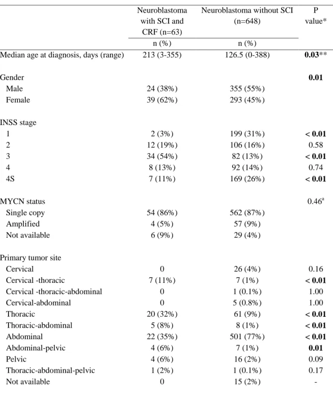
The patient characteristics are summarized in Table 2. The median age at diagnosis was 7 months (range, 0-11.6). Male/female sex-ratio was 0.62. Thirty-four (54%) infants were classified as stage 3, 12 (19%) as stage 2, 8 (13%) as stage 4, 7 (11%) as stage 4S and 2 (3%) as stage 1, according to INSS criteria (13). Four patients (6%) had MYCN amplification. The most common primary tumor sites were abdominal (35%) and thoracic (32%) but thoracic was the most frequent when we considered multilevel locations.
When compared to non-dumbbell neuroblastoma included in INES protocol, the median age at diagnosis was significantly higher in dumbbell neuroblastoma infants (213 days vs 126.5 days, respectively; p=0.03). The gender distribution was different with an excess of female in neuroblastoma with SCI than without SCI (62% vs 45%, respectively; p=0.01). The proportion of stage 3 was significantly higher in dumbbell neuroblastoma group than in neuroblastoma without SCI group (54% vs 13% respectively; p<0.01) and the proportion of stage 1 and 4S was significantly lower. We did not observe a significant difference in MYCN status between the two groups. Overall, patients with SCI were more likely to have a thoracic primary tumor (32% vs 9%, p<0.01) and less likely to have an abdominal primary tumor site (77% vs 35%, p<0.01) than patients without SCI.
17 The global risk profile of the 40 patients treated for neuroblastoma with SCI and no questionnaire returned was not different from that of the 63 patients included with respect to age at diagnosis (p=0.67), stage (p=0.90), MYCN status (p=1.00), gender (p=0.65) and primary tumor site.
18
Table 2 : Patients characteristics at diagnosis.
Neuroblastoma with SCI and
CRF (n=63)
Neuroblastoma without SCI (n=648)
P value*
n (%) n (%)
Median age at diagnosis, days (range) 213 (3-355) 126.5 (0-388) 0.03**
Gender 0.01 Male 24 (38%) 355 (55%) Female 39 (62%) 293 (45%) INSS stage 1 2 (3%) 199 (31%) < 0.01 2 12 (19%) 106 (16%) 0.58 3 34 (54%) 82 (13%) < 0.01 4 8 (13%) 92 (14%) 0.74 4S 7 (11%) 169 (26%) < 0.01 MYCN status 0.46a Single copy 54 (86%) 562 (87%) Amplified 4 (5%) 57 (9%) Not available 6 (9%) 29 (4%)
Primary tumor site
Cervical 0 26 (4%) 0.16 Cervical -thoracic 7 (11%) 7 (1%) < 0.01 Cervical -thoracic-abdominal 0 1 (0.1%) 1.00 Cervical-abdominal 0 5 (0.8%) 1.00 Thoracic 20 (32%) 61 (9%) < 0.01 Thoracic-abdominal 5 (8%) 8 (1%) < 0.01 Abdominal 22 (35%) 501 (77%) < 0.01 Abdominal-pelvic 4 (6%) 7 (1%) 0.01 Pelvic 4 (6%) 16 (2%) 0.09 Thoracic-abdominal-pelvic 1 (2%) 1 (0.1%) 0.17 Not available 0 15 (2%) -
*P value test comparing the distribution with Pearson’s chi-square or Fisher’s exact test. ** P value test comparing the median age at diagnosis with Mann-Whitney’s test. a Comparing patients with MYCN status available only.
19 Initial symptoms (Table 3)
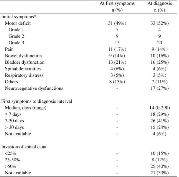
At diagnosis, the main symptom of epidural compression was motor deficit (33 patients; 52%) of grade 1 in 4 patients (12%), grade 2 in 9 (27%) and grade 3 in 20 (61%). It involved the upper extremities in one patient, the lower extremities in 26 and both extremities in 6 patients. Neurovegetative dysfunctions (mainly pallor and marbled skin) were reported in 27% of patients, bladder dysfunction in 25%, bowel dysfunction in 16% and pain in 14%. Twelve patients (19%) had no symptoms of SCI.
The median interval between occurrence of first symptoms and diagnosis was 14 days, with a range from 0 to 290 days, and was less than 8 days in 18 patients (29%), between 8-30 days in 26 (41%) and more than 30 days in 15 (24%). During this interval, the number of patient with motor deficit increased from 31 to 33 (from 15 to 20 with grade 3) and the number of patients with bladder dysfunction from 13 to 16.
The degree of invasion of the spinal canal by the tumor was available for 43/63 patients (68%) and was less than 25% in 10/43 patients (23%), between 25-50% in 8/43 (19%) and more than 50% in 25/43 patients (58%). The mean number of vertebrae involved was 5.8 ± 3.4 (range, 2-19).
20
Table 3: Features of epidural compression at first visit and diagnosis.
At first symptoms At diagnosis
n (%) n (%) Initial symptoms* Motor deficit 31 (49%) 33 (52%) Grade 1 7 4 Grade 2 9 9 Grade 3 15 20 Pain 11 (17%) 9 (14%) Bowel dysfunction 9 (14%) 10 (16%) Bladder dysfunction 13 (21%) 16 (25%) Spinal deformities 4 (6%) 4 (6%) Respiratory distress 3 (5%) 3 (5%) Others 8 (13%) 7 (11%) Neurovegetative dysfunctions - 17 (27%)
First symptoms to diagnosis interval
Median, days (range) - 14 (0-290)
≤ 7 days - 18 (29%)
7-30 days - 26 (41%)
> 30 days - 15 (24%)
Not available - 4 (6%)
Invasion of spinal canal
<25% - 10 (15%)
25-50% - 8 (12%)
>50% - 25 (40%)
Not available - 21 (33%)
*Patients may have more than one symptom
Treatment of epidural compression and extraspinal tumor
Twelve of the 63 patients underwent neurosurgery within 12 days after diagnosis. Six patients underwent laminectomy and 6 laminotomy. Complete intraspinal tumor resection was achieved in 3 patients. Symptoms improved after neurosurgery in 8 patients (67%) within 2 weeks. The majority of children treated by neurosurgery at diagnosis also received chemotherapy (10/12; 83%) among which 6 underwent neurosurgery for progressive neurologic deficit after 1-12 days of chemotherapy.
21 Chemotherapy as initial therapeutic approach was given to 45 patients (71%), according to INES protocol. Symptoms improved in 17 patients (33%) within 2 weeks.
Forty-four patients underwent delayed resection of the extraspinal tumor among which 21 had complete resection. A delayed neurosurgery was performed in 6 patients for progressive deficit or size of the tumor.
Corticosteroid therapy was given to 22 patients (35%) within 17 days after diagnosis, according to INES recommendations, among which 2 with primary neurosurgery.
Overall treatment consisted in chemotherapy alone in 11 patients, chemotherapy and surgery in 43 patients (27 with resection of the extraspinal tumor only, 1 with neurosurgery only and 15 with resection of the extraspinal tumor and neurosurgery) and surgery alone in 6 patients (resection of the extraspinal tumor and/or neurosurgery). Overall, neurosurgery was performed in 17 patients (11 initial, 5 delayed and 1 patient with initial and delayed neurosurgery). No patient had radiation therapy. Treatment data were not available for 2 patients.
Event-free and overall survivals
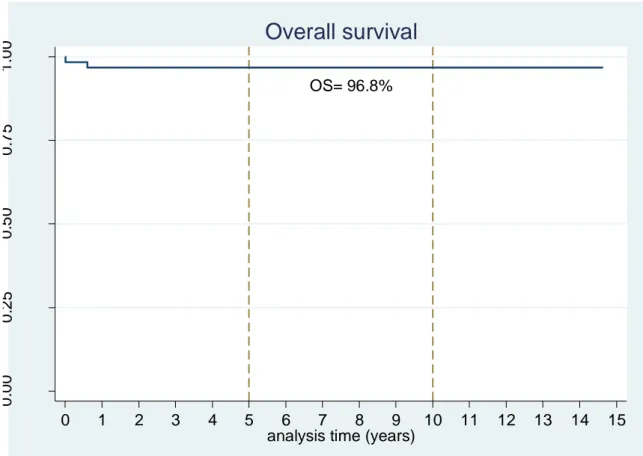
At the time of the last follow-up, 61 children were alive. The median follow-up was 11.9 years (range, 2.7-14.7 years). The Kaplan-Meier estimate of overall survival (OS) at 5 and 10 years was 96.8% (95% CI, 87.4%-99.2%) (Fig.1). Fifty-two patients had a complete response, 7 were alive with minimal residue free-progression and 1 with progression, 2 were dead and data on disease status were not available for the remaining 2 (they were alive). The event-free survival (EFS) at 5 and 10 years was 95.0% (95% CI, 85.3%-98.4%) (Fig.2). One patient with INSS stage 4S and invasion of the spinal canal > 50% was deceased of original disease, 7 months after diagnosis, after initial treatment by chemotherapy followed by surgery and secondary neurosurgery. The other patient was deceased 5 days after diagnosis after chemotherapy and corticoid therapy due to worsening of symptoms.
22
Figure 1: Kaplan-Meier curve for the overall survival after a dumbbell neuroblastoma in infancy.
Figure 2: Kaplan-Meier curve for the event free survival after a dumbbell neuroblastoma in infancy.
OS= 96.8% 0 .0 0 0 .2 5 0 .5 0 0 .7 5 1 .0 0 0 1 2 3 4 5 6 7 8 9 10 11 12 13 14 15
analysis time (years)
Overall survival
EFS = 95.0 % 0 .0 0 0 .2 5 0 .5 0 0 .7 5 1 .0 0 0 1 2 3 4 5 6 7 8 9 10 11 12 13 14 15analysis time (years)
23 Long-term outcome
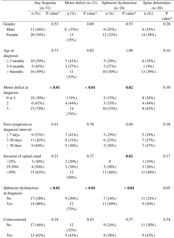
By the time of the last visit, 31 patients (51%) had one or more sequelae (Table 4). The mean number of sequelae by patient was 0.9 ± 1.1. Late effects appeared to be more frequent in neurosurgery ± chemotherapy group compared to chemotherapy group (67% vs 44%) but this difference was not significant (p=0.21). Sequelae were more frequent in patients with severe motor deficit at diagnosis (p < 0.01) or sphincter dysfunction at diagnosis (88% vs 38% without sphincter dysfunction, respectively; p < 0.01) (Table 5).
Motor deficit was documented in 21 patients (34%) including 9 with grade 1, 9 with grade 2 and 3 with grade 3. We did not observe any significant difference in the proportion of motor deficit between the two initial types of treatment but only a trend of more severe motor deficit with neurosurgery compared to chemotherapy (50% vs 33%; p=0.29). Severe motor deficit at diagnosis (grade 2 or 3) and sphincter dysfunction at diagnosis were significantly associated with the presence of motor deficit at the last follow-up. Among 20 patients with grade 3 motor deficit at diagnosis, 6 (30%) did not have late effects. Three underwent chemotherapy as initial treatment and 3 neurosurgery associated with chemotherapy.
Sphincter dysfunctions were detected in 18 patients (30%) including 10 with bladder dysfunction, 2 with bowel dysfunction and 6 with both dysfunctions. There were more sphincter dysfunctions in patients treated by neurosurgery than in those treated by chemotherapy alone but this difference was not significant (42% vs 24%, respectively; p=0.24). Sphincter dysfunctions were significantly more frequent in children who had presented these symptoms at diagnosis (p < 0.01), with severe motor deficit at diagnosis (p = 0.02) or with invasion of the spinal canal more than 25% (p = 0.02).
Spinal deformities were reported in 20 patients (33%) including 15 with only scoliosis, 1 with only kyphosis, 3 with kyphoscoliosis and 1 with kyphoscoliosis and lordosis. Among 19 patients with scoliosis, 7 patients (37%) had grade 1, 11 (58%) grade 2 and 1 (5%) grade 3.
24 Spinal deformities were more frequent but not significant in patients treated by neurosurgery (3 laminotomy and 3 laminectomy) ± chemotherapy compared to those undergoing chemotherapy alone (50% vs 27%, respectively; p=0.12).
Gender, age at diagnosis, first symptoms to diagnosis interval and corticoid therapy were not significantly associated with the presence of sequelae.
Table 4 : Long-term sequelae according to initial treatment.
Patients Initial treatment P value**
Neurosurgery ± Chemotherapy (n=12*) Chemotherapy (n=45*) n (%) n (%) n (%) Any sequelae 31 (51%) 8 (67%) 20 (44%) 0.207 Motor deficit 21 (34%) 6 (50%) 15 (33%) 0.288 Grade 1 9 2 7 Grade 2 9 2 7 Grade 3 3 2 1 Sphincter dysfunction 18 (30%) 5 (42%) 11 (24%) 0.238 Bladder 16 (26%) 4 10 Bowel 8 (13%) 2 5 Spinal deformities 20 (33%) 6 (50%) 12 (27%) 0.122 Grade 1 8 3 4 Grade 2 11 3 7 Grade 3 1 0 1 Scoliosis 19 (31%) 6 (50%) 11 (24%) 0.086 Kyphosis 5 (8%) 0 5 Lordosis 1 (2%) 0 1
*4 patients underwent an initial resection of extraspinal tumor (no initial symptom) and 2 patients had no treatment data
**P value test comparing the distribution with Pearson’s chi-square or Fisher’s exact test. Patients may have more than one sequelae.
25
Table 5 : Long-term sequelae in relation to clinical or treatment factors.
Any Sequelae (n=31)
Motor deficit (n=21) Sphincter dysfunction (n=18)
Spine deformities (n=20)
n (%) P value* n (%) P value* n (%) P value* n (%) P
value* Gender 0.53 0.89 0.53 0.28 Male 11 (46%) 8 (33%) 6 (25%) 6 (25%) Female 20 (54%) 13 (35%) 12 (32%) 14 (38%) Age at diagnosis 0.73 0.82 1.00 0.16 ≤ 3 months 10 (59%) 7 (41%) 5 (29%) 6 (35%) 3-6 months 5 (45%) 3 (27%) 3 (27%) 1 (9%) > 6months 16 (49%) 11 (33%) 10 (30%) 13 (39%) Motor deficit at diagnosis < 0.01 < 0.01 0.02 0.30 0 or 1 10 (30%) 3 (9%) 5 (15%) 8 (24%) 2 6 (67%) 4 (44%) 3 (33%) 4 (44%) 3 15 (79%) 14 (74%) 10 (53%) 8 (42%) First symptoms to diagnosis interval 0.41 0.78 0.69 0.38 ≤ 7 days 9 (53%) 7 (41%) 5 (29%) 5 (29%) 7-30 days 11 (42%) 8 (31%) 6 (23%) 7 (27%) > 30 days 9 (64%) 5 (36%) 5 (36%) 7 (47%)
Invasion of spinal canal 0.21 0.27 0.02 0.17
<25% 3 (30%) 2 (20%) 0 1 (10%) 25-50% 4 (50%) 3 (38%) 3 (38%) 3 (38%) >50% 15 (63%) 12 (50%) 11 (46%) 11 (46%) Sphincter dysfunction at diagnosis < 0.01 < 0.01 < 0.01 0.05 No 17 (38%) 9 (20%) 7 (16%) 11 (24%) Yes 14 (88%) 12 (75%) 11 (69%) 9 (56%) Corticosteroid 0.24 0.43 0.27 0.34 No 17 (46%) 12 (32%) 9 (24%) 11 (30%) Yes 13 (62%) 9 (43%) 8 (38%) 9 (43%)
26 DISCUSSION
This analysis of a multicentric european cohort follow-up study shows the clinical presentation of neuroblastoma with SCI, its treatment and the late effects in infants with or without initial neurologic symptoms. These results confirm the main features of dumbbell neuroblastoma compared to non-dumbbell neuroblastoma.
Overall, the treatment of neuroblastoma with SCI in this study followed the INES recommendations. Chemotherapy represents the first initial treatment for the majority of patients (71%). Thirty-seven percent of patients presenting with paraplegia were treated by initial neurosurgery. No patient had radiation therapy. The exception was for corticosteroid used only for 35% of patients included in this study and half of patients with severe motor deficit (grade 2-3). Corticoid treatment is described to improve early symptom relief but seems to have no impact on long-term outcome, in the previous studies (4). Chemotherapy alone can cure some patients. Among 9 patients treated only by chemotherapy, 7 (78%) had a complete response.
The median interval between occurrence of first symptoms and diagnosis was 14 days, similar to previous studies (12 days in the experience of AIEOP –Italian Association for Pediatric Hematology-Oncology (6) and German neuroblastoma trials (4)) but shorter than another Franco-Italian study of 2011 (23 days) (16) and a recent Dutch publication (6 weeks) (17). This interval had no clear impact on the occurrence of late effects, which looked comparable to previously published series (1,3–5). Moreover, sphincter dysfunction and motor deficit at diagnosis may be underestimated in infants, in whom sphincter control and walking have not yet been achieved. Both parental and medical information and education should be heightened to decrease this interval.
While long-term overall survival was excellent (97% to 10 years), long-term functional outcome was poor. Fifty-one percent of patients of our study had one or more sequelae at the
27 time of their last follow-up. This percentage was lower than in recent previous publications (68% for 34 infants treated between 2000 and 2011 (6), 58% for 98 children treated between 1979 and 2002 (16), 71% for 99 children treated between 1989 and 2008 (4) and 95% for 19 children treated between 1980 and 2007 (17)). However, their cohort’s patients were often older and only the Italian cohort of AEIOP concerned infants (6). The severity of motor deficit at presentation was the strongest predictor of occurrence of late effects in our study, except for spine deformities, which is comparable to previous studies (1–6,16).
No significant difference between first-line neurosurgery and chemotherapy on long-term outcome was found. The number of late effects after initial neurosurgery was higher but these differences were not statistically significant. Previously published studies have also reported the lack of significant difference between either treatment (4,6), unlike other studies that found that neurosurgery leads significantly to more long-term orthopedic consequences (16). Spinal deformity following intradural spinal tumor resection has been well described by several authors. Pre-operative scoliotic deformity, an age less than 13 years old and an increasing number of resections increased the risk for progressive spinal deformity (18–20). The two decompression modalities have different indications; neurosurgery is limited to rapid neurological deterioration (1-2 days) or a poor response to chemotherapy and may have benefits in this specific population (1,5,16), whereas chemotherapy may be indicated for the remaining patients. Moreover, severely affected patients received more aggressive treatment, often including neurosurgery.
Few studies took interested in long-term outcome of children treated before one year old (the recent AIEOP studied 34 infants (6)). Therefore, the historical comparisons are not relevant to evaluate the change in management strategies in infants and the occurrence of late effects. The strength of our study is the prospective collection of initial tumor data within the INES protocol. Moreover, the data of follow-up are based on the last physical examination of the
28 medical record, and not on self-questionnaire or telephone conversation. The median follow-up was 11.9 years, longer than the majority of other studies.
Our study is limited by the size of our cohort which lowers the statistical power of analyses. The response rate is limited (61%). However, this is the largest cohort of dumbbell neuroblastoma survivors diagnosed before one year old.
Prospective studies for neuroblastoma with SCI are necessary to clarify the incidence and severity of sequelae and facilitate treatment data collection. The SIOPEN network is implementing an international, observational, prospective registry of peripheral neuroblastic tumors presenting with spinal canal involvement to assess the impact of different risk factors on neurologic and orthopedic outcomes and to optimize treatment guidelines for the management of symptomatic SCI (and the issue of intraspinal residue). An early and prolonged rehabilitation care and the interest of neurosensory explorations could also be considered.
CONCLUSION
In conclusion, fifty-one percent of infants with SCI developed late effects, without significant difference between first-line neurosurgery and chemotherapy. A good knowledge of these late effects is important for the implementation of appropriate long-term follow-up to improve prevention, early detection and optimal management of these sequelae to limit their incidence and severity.
31 ACKNOWLEDGMENTS
Coordinators of INES-FU study: Caroline Munzer (Hôpital des enfants, Toulouse, France) and Alberto Garaventa (Instituto Giannina Gaslini, Genova, Italia)
Study centers of countries involved: France (Besançon, Bordeaux, Clermont-Ferrand,
Grenoble, Institut Curie, Institut Gustave Roussy, Lille, Lyon, Marseille, Montpellier, Rennes, Rouen, Saint-Etienne, Strasbourg, Toulouse, Tours), Italy (Calgari, Ferrara, Genova, Milano, Modena, Napoli, Padova, Palermo), Spain (Bilbao, Murcia, Valencia), United Kingdom (Oxford), Belgium (UCL Saint Luc), Austria, Portugal, and Swiss (Lausanne).
International Society of Pediatric Oncology, European Neuroblastoma Group (SIOPEN).
Manuscript in preparation for submission to the Journal of Clinical Oncology
REFERENCES
1. Plantaz D, Rubie H, Michon J, Mechinaud F, Coze C, Chastagner P, et al. The treatment of neuroblastoma with intraspinal extension with chemotherapy followed by surgical removal of residual disease. A prospective study of 42 patients--results of the NBL 90 Study of the French Society of Pediatric Oncology. Cancer. 1996; 78:311‑9.
2. Hoover M, Bowman LC, Crawford SE, Stack C, Donaldson JS, Grayhack JJ, et al. Long-term outcome of patients with intraspinal neuroblastoma. Med Pediatr Oncol. 1999; 32:353‑9.
3. De Bernardi B null, Pianca C, Pistamiglio P, Veneselli E, Viscardi E, Pession A, et al. Neuroblastoma with symptomatic spinal cord compression at diagnosis: treatment and results with 76 cases. J Clin Oncol Off J Am Soc Clin Oncol. 2001; 19:183‑90.
4. Simon T, Niemann CA, Hero B, Henze G, Suttorp M, Schilling FH, et al. Short- and long-term outcome of patients with symptoms of spinal cord compression by neuroblastoma. Dev Med Child Neurol. 2012; 54:347‑52.
5. Katzenstein HM, Kent PM, London WB, Cohn SL. Treatment and outcome of 83 children with intraspinal neuroblastoma: the Pediatric Oncology Group experience. J Clin Oncol. 2001; 19:1047‑55.
32 6. De Bernardi B, Quaglietta L, Haupt R, Castellano A, Tirtei E, Luksch R, et al.
Neuroblastoma with symptomatic epidural compression in the infant: the AIEOP experience. Pediatr Blood Cancer. 2014; 61:1369‑75.
7. De Bernardi B, Balwierz W, Bejent J, Cohn SL, Garrè ML, Iehara T, et al. Epidural compression in neuroblastoma: Diagnostic and therapeutic aspects. Cancer Lett. 2005; 228:283‑99.
8. Moppett J, Haddadin I, Foot AB. Neonatal neuroblastoma. Arch Dis Child Fetal Neonatal Ed. 1999; 81:F134-137.
9. Pintér AB, Hock A, Kajtár P, Dóber I. Long-term follow-up of cancer in neonates and infants: a national survey of 142 patients. Pediatr Surg Int. 2003; 19:233‑9.
10. Rubie H, De Bernardi B, Gerrard M, Canete A, Ladenstein R, Couturier J, et al. Excellent outcome with reduced treatment in infants with nonmetastatic and unresectable
neuroblastoma without MYCN amplification: results of the prospective INES 99.1. J Clin Oncol. 2011; 29:449‑55.
11. Canete A, Gerrard M, Rubie H, Castel V, Di Cataldo A, Munzer C, et al. Poor survival for infants with MYCN-amplified metastatic neuroblastoma despite intensified treatment: the International Society of Paediatric Oncology European Neuroblastoma Experience. J Clin Oncol. 2009; 27:1014‑9.
12. De Bernardi B, Gerrard M, Boni L, Rubie H, Cañete A, Di Cataldo A, et al. Excellent outcome with reduced treatment for infants with disseminated neuroblastoma without MYCN gene amplification. J Clin Oncol. 2009; 27:1034‑40.
13. Brodeur GM, Pritchard J, Berthold F, Carlsen NL, Castel V, Castelberry RP, et al. Revisions of the international criteria for neuroblastoma diagnosis, staging, and response to treatment. J Clin Oncol. 1993; 11:1466‑77.
14. Rosman N, Gilmore H. Spinal cord injury. In: Swaiman KF, Ashwal S, Pediatric Neurology : Principals and practice. 3e ed. St Louis, Mosby; 1999. p. 954‑66.
15. U.S.DEPARTMENT OF HEALTH AND HUMAN SERVICES. Common Terminology Criteria for Adverse Events (CTCAE) Version 4.0. National Cancer Institute; 2009. 16. Angelini P, Plantaz D, De Bernardi B, Passagia J-G, Rubie H, Pastore G. Late sequelae of
symptomatic epidural compression in children with localized neuroblastoma. Pediatr Blood Cancer. 2011; 57:473‑80.
17. Kraal K, Blom T, Tytgat L, van Santen H, van Noesel M, Smets A, et al. Neuroblastoma With Intraspinal Extension: Health Problems in Long-Term Survivors. Pediatr Blood Cancer. 2016; 63:990‑6.
18. Yao KC, McGirt MJ, Chaichana KL, Constantini S, Jallo GI. Risk factors for progressive spinal deformity following resection of intramedullary spinal cord tumors in children: an analysis of 161 consecutive cases. J Neurosurg. 2007; 107:463‑8.
33 19. McGirt MJ, Garcés-Ambrossi GL, Parker SL, Sciubba DM, Bydon A, Wolinksy J-P, et al.
Short-term progressive spinal deformity following laminoplasty versus laminectomy for resection of intradural spinal tumors: analysis of 238 patients. Neurosurgery. 2010; 66:1005‑12.
20. McGirt MJ, Chaichana KL, Atiba A, Bydon A, Witham TF, Yao KC, et al. Incidence of spinal deformity after resection of intramedullary spinal cord tumors in children who underwent laminectomy compared with laminoplasty. J Neurosurg Pediatr. 2008; 1:57‑ 62.
34 APPENDIX
43 Appendix 2
Neuroblastome avec extension intra-rachidienne : état des connaissances et intérêt d’un registre prospectif international.
Spinal canal involvement in neuroblastoma: state of the knowledge and time for an international prospective registry.
AUTEURS
C Freycon1, I Schiff1, L Selek2, C Piolat3, C Durand4, D Plantaz1.
AFFILIATION:
1: Clinique Universitaire de Pédiatrie. CHU de Grenoble. CS 10217 38043 Grenoble cedex. 2: Clinique Universitaire de Neurochirurgie. CHU de Grenoble. CS 10217 38043 Grenoble cedex
3: Clinique Universitaire de Chirurgie Pédiatrique. CHU de Grenoble. CS 10217 38043 Grenoble cedex
4: Clinique Universitaire d’Imagerie Pédiatrique. CHU de Grenoble. CS 10217 38043 Grenoble cedex
Correspondance: D Plantaz Clinique Universitaire de Pédiatrie. CHU de Grenoble. CS 10217 38043 Grenoble cedex: +33 4 76 76 54 69 (DPlantaz@chu-grenoble.fr)
Résumé
Dix à quinze pour cent des enfants porteurs d’un neuroblastome présentent une extension de leur tumeur primitive dans le canal rachidien, à travers un ou plusieurs foramen
intervertébraux. Soixante pour cent d’entre eux ont des signes de compression médullaire lors du diagnostic (douleurs dorsales, déficit moteur, troubles de l’équilibre, faiblesse des
membres inférieurs, parapaparésie ou paraplégie, cyphoscoliose, troubles sphinctériens vésico rectaux, troubles sensoriels, incluant des douleurs neuropathiques). Il s’agit le plus souvent d’enfants jeunes, au cours des deux premières années de vie, qui sont porteurs de tumeurs plus souvent localisées et biologiquement favorables, justifiant un traitement peu agressif sur le plan oncologique, du fait d’un pronostic vital favorable. Il faut souligner la difficulté du diagnostic des signes neurologiques chez les jeunes nourrissons. Bien que les traitements de décompression (chimiothérapie, neurochirurgie) offrent des chances d’amélioration, voire de récupération complète, ces patients sont globalement exposés à d’importants risques de séquelles neurologiques à long terme (déficit moteur, trouble sphinctérien) ainsi qu’à des
44 séquelles orthopédiques ; ceci souligne l’importance d’études sur ce groupe de patients pour mieux connaitre les facteurs liés à la maladie et à son traitement qui permettraient d’améliorer leur prise en charge. C’est dans cet objectif que le réseau SIOPEN (European Neuroblastoma Research Network) a mis en place un registre international, multi-centrique prospectif des tumeurs neuroblastiques avec extension intra-rachidienne, dont l’objectif principal est de collecter prospectivement les données afin d’évaluer les facteurs de risque sur l’évolution neurologique et orthopédique à court et long terme. Ceci devrait permettre d’améliorer les recommandations de prise en charge de tels patients.
Mots-Clés : neuroblastome, sablier, compression médullaire, laminectomie, séquelles tardives Abstract
Up to 10-15% children with neuroblastoma have direct tumour extension through the intervertebral foramen into the spinal canal. Sixty percent have symptoms of spinal cord compression (back pain, motor dysfunction, gait disturbance, limb weakness, paralysis, kyphoscoliosis, bladder and bowel and sensory disturbance including pain) at presentation. These patients are generally young, within the first 2 years of life, have biologically
favourable disease and would often otherwise require minimal therapy for their
neuroblastoma and generally have excellent survival. Symptoms and signs of spinal canal compression may be under recognised in young infants. Although many patients experience symptom improvement with effective therapy (chemotherapy or decompressive
neurosurgery), patients with neuroblastoma and SCI (Spinal Cord Involvement) are at risk of long standing neurologic (motor, bowel, bladder, sensory and pain) and orthopaedic
consequences. Recent retrospective studies have shown unacceptably high rates (60-75%) of long term neurologic and orthopaedic disability emphasising the importance of focusing on this subgroup of patients to better identify important patient and treatment related factors to improve current management strategies. To address this need the European Neuroblastoma Research Network (SIOPEN) has developed an international, multi-centre prospective registry study of peripheral neuroblastic tumours presenting with spinal canal involvement. The primary objective is to prospectively collect data on patients with neuroblastoma and SCI to evaluate the combined effects of different risk factors on the eventual neurologic and orthopaedic outcomes. It is hoped that this will lead to better treatment guidelines for the management of symptomatic SCI with reduced long term functional sequelae.
45 Le contexte: incidence, présentation, traitement du neuroblastome.
Le neuroblastome affecte de manière prédominante le jeune enfant avec un diagnostic pour la majorité des patients fait au cours de la première décade. L’indicence globale est de 12.6 cas par million d’enfants par an avec une majorité des cas survenant chez le jeune enfant avec une diminution rapide de l’incidence parallèle à l’augmentation de l’âge (1). Le neuroblastome est une tumeur embryonnaire ayant pour origine le système nerveux sympathique et pouvant se développer à n’importe quel niveau le long de l’axe adréno-sympathique (glandes surrénales, nerfs et ganglions sympathiques). Le traitement des patients atteints de neuroblastome est basé sur un ensemble de critères comportant l’évaluation clinique, le bilan d’extension, le type anatomo-pathologique, les facteurs biologiques qui permettent d’estimer la réponse au traitement, le risque de récidive et les chances de guérison. Le neuroblastome biologiquement favorable (low and intermediate risk) présente un potentiel élevé de régression spontanée et/ou de maturation tumorale lors d’une simple observation ou sous traitement d’intensité modérée (2). Les patients avec des neuroblastomes de risque faible ou intermédiaire sont, soit des enfants âgés de moins de 18 mois (incluant certaines formes métastatiques), soit des enfants plus âgés avec des tumeurs localisées mais aux caractéristiques biopathologiques favorables, en particulier l’absence d’amplification de NMYC (3). Les essais et les études cliniques ont confirmé la possibilité d’une désescalade thérapeutique associée au maintien d’une excellente survie pour les enfants atteints de neuroblastomes de risque faible ou intermédiaire (4-9). Au contraire, les patients avec un neuroblastome de haut risque sont définis par un âge supérieur à 12 pour une maladie étendue ou pour tout âge et tout stade (à l’exception des stades L1 INSS 1) en cas d’amplification de l’oncogène NMYC. Les taux de guérison se sont améliorés au cours des deux dernières décennies mais le risque de récidive reste de l’ordre de 50% associé à un risque élevé de séquelles à long terme pour les
survivants.
Outre l’augmentation des taux de survie, l’objectif actuel reste donc la limitation des séquelles à long terme. Un groupe spécifique de neuroblastomes présente un risque élevé d’altération de la qualité de vie à long terme, en rapport direct avec la tumeur et/ou son traitement : ce sont les tumeurs présentant une extension intra-canalaire dits neuroblastome en sablier.
Extension intra-rachidienne et compression spinale dans le neuroblastome. Une extension intra-canalaire est présente chez 10 à 15% des enfants atteints de
neuroblastome. L’extension intra-canalaire est directement secondaire à une extension de la tumeur primitive intra-rachidienne à travers le ou les foramen(s) intervertebral(aux)
aboutissant à une compression médullaire extra-durale symptomatique ou non (figure 1). Entre 50% et 65% des patients porteurs de neuroblastome en sablier présentent une forme symptomatique avec un déficit neurologique au moment de la présentation initiale de telle sorte que l’incidence globale des neuroblastomes en sablier symptomatiques est de l’ordre de 5% (10-13), mais elle est plus élevée chez les nouveau-nés, de l’ordre de 18% (14-15). Le diagnostic différentiel d’une compression médullaire d’origine tumorale inclut d’autres tumeurs, sarcomes (sarcome d’Ewing, rhabdomyosarcome, ostéosarcome, tumeur rhabdoïde),
46 lymphomes, leucémies et histiocytose langerhansienne, ainsi que les tumeurs spinales (16-17). De plus, différentes tumeurs bénignes, comme les tératomes, la neurofibromatose, les méningiomes, peuvent se présenter avec des symptômes de compression médullaire. Présentation Clinique.
Les neuroblastomes en sablier sont observés chez des enfants statistiquement plus jeunes que les formes sans extension intra-rachidienne, avec une médiane d’âge de survenue inférieure à 2 ans (10-12, 18). Des cas symptomatiques congénitaux, voire en anténataux ont été rapportés (19); mais aussi à l’inverse chez des adolescents et des adultes (10, 12, 18). La localisation de la tumeur primitive est plus fréquemment thoracique (10-13). Le contingent intra-canalaire peut s’étendre sur plusieurs étages vertébraux et le degré d’envahissement du canal peut atteindre 100% de sa largeur (10-13, 18).
Les signes cliniques en rapport avec l’envahissement intra-canalaire d’un neuroblastome comportent un ensemble très polymorphe de symptômes incluant une douleur radiculaire ou neuropathique, des douleurs dorsales, une régression motrice, des troubles de la marche, la faiblesse d’un membre ou sa paralysie (incluant une monoplégie ou une paraplégie), une cyphoscoliose, un trouble sphinctérien (vésical et/ou anal), et des signes sensoriels dont la douleur (10-13).
Les symptômes d’un neuroblastome en sablier peuvent être difficiles à reconnaitre en particulier chez les nourrissons; l’hypomobilité des membres inférieurs peut être un piège avant l’âge de la marche, surtout lorsqu’elle n’est pas révélée à l’examen. Cela explique que la durée des symptômes consécutifs à l’extension intra-rachidienne soit très variable, pouvant même durer plusieurs mois à partir du premier signe (10-13). Il a été montré, en particulier chez les nourrissons âgés de moins de 1 an, une augmentation du risque de déficit moteur et de vessie neurologique corrélée à l’augmentation du délai entre les premiers signes et le diagnostic (20).
Traitement et évolution des neuroblastomes avec extension intra-canalaire symptomatique.
Historiquement, une décompression neurochirurgicale ou par radiothérapie ont été utilisées dans les cas de compression médullaire des neuroblastomes en sablier, jusqu’à la mise en évidence de l’efficacité de la chimiothérapie dans les années 1980 (21-22). Depuis, le recours à la radiothérapie en traitement initial de la compression médullaire a disparu, en particulier du fait des risques à long terme chez ces très jeunes enfants (10-13).
Les recommandations actuelles pour les neuroblastomes avec extension intra-canalaire soulignent l’importance d’un diagnostic précoce et de la prise en charge urgente par une équipe multidisciplinaire incluant pédiatre oncologue, neurochirurgien, neuropédiatre,
orthopédiste pédiatrique, radiopédiatre, pour réaliser en urgence le bilan et le staging adaptés et réaliser le traitement approprié. Il est crucial d’exclure un autre type de tumeur et d’obtenir un diagnostic anatomo-pathologique, ainsi que du tissu tumoral pour les études biologiques déterminantes pour le choix thérapeutique. Alors qu’une chimiothérapie initiale est le plus
47 souvent réalisée (association carboplatine vépéside), une décompression neurochirurgicale doit être envisagée pour les patients présentant une paralysie sévère d’évolution rapide ou une mauvaise réponse à la chimiothérapie (10, 13, 15). Bien que des recommandations aient été établies dans certains protocoles, on ignore comment elles ont été appliquées individuellement et leur impact sur le devenir des patients.
Traitement initial et réponse précoce au traitement de la compression médullaire Entre 60 et 80% des patients symptomatiques vont présenter une amélioration neurologique sous traitement. Même si un diagnostic tardif n’a pas pu être associé dans toutes les études avec un pronostic fonctionnel plus péjoratif, ceci a été montré récemment (20). Bien que la sévérité initiale d’une compression médullaire puisse laisser craindre une mauvaise évolution neurologique, des récupérations partielles et des récupérations complètes parfois surprenantes peuvent s’observer chez des patients gravement déficitaires (figure 2), soulignant
l’importance d’un traitement adapté rapide (10-13). La corticothérapie est systématiquement associée avec une amélioration précoce des symptômes, mais un impact sur l’évolution à long terme qui n’est pas démontré (12). La décompression chirurgicale et la chimiothérapie
auraient un taux de récupération précoce équivalent (65-70%) mais il persiste des controverses sur le taux de handicap à long terme (60-70%) (23). La décompression chirurgicale pourrait avoir un avantage chez certains enfants avec déficit rapidement
progressif (10, 13). De plus, les patients présentant un neuroblastome mature sont susceptibles d’une moins bonne réponse à la chimiothérapie et pourrait bénéficier d’une approche
chirurgicale (13).
Evolution à long terme.
La survie à long terme des enfants atteints de neuroblastomes en sablier est bonne reflétant, l’âge inférieur, la prépondérance des formes localisées et une biologie tumorale plus favorable (10-13).
Les connaissances sur le devenir fonctionnel de ces patients sont issues d’études
rétrospectives monocentriques, d’études de registre et d’essais thérapeutiques (10-13, 23-24). Alors que ces études confirment la gravité du problème fonctionnel des neuroblastomes en sablier, leurs limites sont liées au petit nombre de patients, au suivi hétérogène, aux
différences méthodologiques dans les analyses rétrospectives ne permettant pas d’apporter des réponses définitives aux questions en suspens.
Un déficit fonctionnel sévère au diagnostic est associé à un risque élevé de séquelles à long terme (10-11). Des études récentes ont montré un taux élevé (60-70%) de séquelles tardives après un suivi médian de plus de 5 ans, incluant un déficit moteur, une spasticité, une
cyphoscoliose, une vessie neurologique, une dysfonction ano-rectale, des troubles sensitifs, un retard de croissance, un syndrome douloureux chronique, suggérant que les conséquences fonctionnelles aient pu être jusque-là sous-estimées et qu’un suivi spécifique à long terme évalué soit nécessaire pour de tels patients (24). Une étude est actuellement en cours sur les enfants traités avant l’âge de 1 an en Europe entre 1999 et 2004 (étude INES FU).
48 Questions non résolues.
Les questions qui restent à résoudre portent sur l’impact du délai entre les premiers signes et la mise en route du traitement, le bénéfice de la corticothérapie, et surtout le traitement initial de décompression le plus approprié (chimiothérapie versus neurochirurgie) et les critères de choix entre ces deux approches. Une meilleure connaissance du devenir fonctionnel à long terme est également nécessaire, ainsi que des modalités de prise en charge de séquelles. Techniques neurochirurgicales et complications à long terme.
Les conséquences orthopédiques tardives (cyphoscoliose) des interventions de décompression neurochirurgicale ont été rapportées chez l’enfant. Il existe de multiples facteurs pouvant influer sur la survenue des séquelles orthopédiques, incluant l’âge des patients (risque plus élevé avant 13 ans) (25), l’existence d’une déformation rachidienne préopératoire (26) et l’étendue de la chirurgie (risque plus élevé en cas d’intervention sur plus de 4 étages vertébraux) (27). L’impact de la technique décompressive (laminoplastie versus
laminectomie) n’a pas été définitivement tranché. Certains ont pu suggérer un risque plus élevé après une laminectomie (28), mais une méta-analyse n’a pas démontré d’augmentation du risque de cyphoscoliose après laminectomie plutôt que laminoplastie (29). Une large étude n’a pas montré de différence sur l’incidence des déformations radiologiques à long terme (27), alors que des études à plus court terme suggéraient un avantage à la laminoplastie (30). La fixation rachidienne post-opératoire a été défendue mais son efficacité reste incertaine, pouvant peut-être plus retarder la déformation rachidienne que réduire son incidence (31). En conséquence, le risque de séquelles orthopédiques à long terme et les résultats équivalents de la chimiothérapie et de la neurochirurgie décompressive suggèrent le recours à une
intervention chirurgicale de décompression uniquement chez certains patients, comme ceux porteurs de déficit neurologiques rapidement progressifs, et en cas d’échec de la
chimiothérapie ou de tumeur neuroblastique mature (tableau 1) Nécessité d’une étude prospective.
Il existe une collaboration internationale pour rassembler de manière prospective des informations à large échelle concernant la présentation, la prise en charge et le devenir des enfants atteints de neuroblastome en sablier, symptomatique ou non. Le réseau de recherche européen sur le neuroblatome (SIOPEN) coordonne un registre observationnel international prospectif des patients de moins de 18 ans avec un neuroblastome présentant une extension spinale. La coordination internationale de l’étude est assurée par l’AIEOP (Associazione Italiana Ematologia-Oncologia Pediatrica), Gaslini Children’s Hospital à Gênes -Italie (Dr Haupt). En France, la coordination est assurée par le CHU de Grenoble (Pr Plantaz). Dans ce registre, les modalités de prise en charge ne font pas l’objet de recommandations mais sont déterminées par le protocole thérapeutique le plus adapté à chaque patient selon l’équipe pluridisciplinaire référente en charge de l’enfant. Le registre des neuroblastomes en sablier va enregistrer des données initiales incluant les caractéristiques démographiques, les signes cliniques, la durée des symptômes, la présence et l’intensité des signes neurologiques et orthopédiques, les données neuroradiologiques, les caractéristiques bio-pathologiques ainsi
49 que le traitement initial (observation, chimiothérapie, neurochirurgie). Des échelles
standardisées seront utilisées pour le grading des symptômes et le retentissement fonctionnel incluant l’échelle CTCAE (Common Toxicity Criteria for Adverse Events), l’échelle de douleur FLACC (Face, Legs, Activity, Cry, Consolability) et l’échelle de séquelle ASIA (American Spinal Injury Association). La réponse précoce au traitement sera évaluée à 72 heures, 1 semaine, 2 semaines, 4 semaines, et 2 mois, par le médecin oncopédiatre, avec l’aide éventuelle d’un médecin rééducateur ou d’un kinésithérapeute Les traitements successifs seront collectés, ainsi que l’évolution clinique et la meilleure réponse radiologique. Un suivi prospectif standardisé sera réalisé à 6 mois du diagnostic, à un an puis annuellement jusqu’à 5 ans, et à 10 ans, pour documenter le devenir fonctionnel à long terme.
On peut espérer que cet enregistrement prospectif de données standardisées permette d’établir des corrélations entre les signes cliniques, les données pathologiques et biologiques, le type de traitement, la réponse précoce et les séquelles tardives; ceci devrait permettre d’établir des recommandations solides pour la prise en charge des enfants avec un neuroblastome en sablier dans l’objectif de limiter leurs séquelles.
Remerciements Enfant et Cancer (association Hubert Gouin) pour le soutien financier pour le Registre international des neuroblastomes en sablier (SIOPEN)
Aucun des auteurs n’a de conflit d’intérêt.
Références
1. Lacour B. Incidence des cancers de l'enfant. in Epidémiologie des tumeurs de l'enfant. Springer-Verlag. France 2009. p 56-65.
2. Monclair T, Brodeur GM, Ambros PF, et al. The International Neuroblastoma Risk Group (INRG) staging system: an INRG Task Force report. J Clin Oncol. 2009; 27:298-303 3. Schleiermacher G, Mosseri V, London WB, Maris JM, et al. Segmental chromosomal alterations have prognostic impact in neuroblastoma: a report from the INRG project. Br J Cancer. 2012; 107: 1418-22.
4. Hero B, Simon T, Spitz R, Ernestus K, et al. Localized infant neuroblastomas often show spontaneous regression: results of the prospective trials NB95-S and NB97. J Clin Oncol. 2008; 26: 1504-10
5. De Bernardi B, Gerrard M, Boni L, Rubie H, et al. Excellent outcome with reduced treatment for infants with disseminated neuroblastoma without MYCN gene amplification. J Clin Oncol. 2009; 27: 1034-40
50 6. Rubie H, De Bernardi B, Gerrard M, Canete A, et al. Excellent outcome with reduced treatment in infants with nonmetastatic and unresectable neuroblastoma without MYCN amplification: results of the prospective INES 99.1. J Clin Oncol. 2011; 29:449-55.
7. Strother DR, London WB, Schmidt ML, Brodeur GM, et al. Outcome after surgery alone or with restricted use of chemotherapy for patients with low-risk neuroblastoma: results of Children's Oncology Group study P9641. Journal of clinical oncology : J Clin Oncol. 2012; 30(15) :1842-8.
8. Nuchtern JG, London WB, Barnewolt CE, Naranjo A, McGrady PW, Geiger JD, et al. A prospective study of expectant observation as primary therapy for neuroblastoma in young infants: a Children's Oncology Group study. Annals of surgery. 2012; 256 (4):573-580. 9. Kohler JA, Rubie H, Castel V, Beiske K, et al. Treatment of children over the age of one year with unresectable localised neuroblastoma without MYCN amplification: results of the SIOPEN study. Eur J Cancer. 2013; 49: 3671-9
10. Plantaz D, Rubie H, Michon J, Mechinaud F, et al. The treatment of neuroblastoma with intraspinal extension with chemotherapy followed by surgical removal of residual disease. A prospective study of 42 patients--results of the NBL 90 Study of the French Society of Pediatric Oncology. Cancer. 1996;78: 311-9.
11. De Bernardi B, Pianca C, Pistamiglio P, Veneselli E, et al. Neuroblastoma with symptomatic spinal cord compression at diagnosis: treatment and results with 76 cases. J Clin Oncol. 2001; 19: 183-90.
12. Simon T, Niemann CA, Hero B, Henze G, et al. Short- and long-term outcome of patients with symptoms of spinal cord compression by neuroblastoma. Dev Med Child Neurol. 2012; 54: 347-52.
13. Katzenstein HM, Kent PM, London WB, Cohn SL. Treatment and outcome of 83 children with intraspinal neuroblastoma: the Pediatric Oncology Group experience. J Clin Oncol. 2001; 19: 1047-55.
14. Michalowski MB1, Rubie H, Michon J, Montamat S, et al. Neonatal localized neuroblastoma: 52 cases treated from 1990 to 1999. Arch Pediatr. 2004; 11: 782-8 15. Moppett J, Haddadin I, Foot AB. Neonatal neuroblastoma. Arch Dis Child Fetal Neonatal 1999; 81: F134-7.
16. Pollono D, Tomarchia S, Drut R, et al. Spinal cord compression: a review of 70 pediatric patients. Pediatr Hematol Oncol. 2003; 20: 457-66.
17. Hesketh E, Eden OB, Gattamaneni HR, Campbell RH, et al. Spinal cord compression--do we miss it? Acta Paediatr. 1998; 87: 452-4.
51 18. Hoover M, Bowman LC, Crawford SE, Stack C, et al. Long-term outcome of patients with intraspinal neuroblastoma. Med Pediatr Oncol. 1999; 32 :353-9.
19. Delahaye S, Doz F, Sonigo P, Saada et al. Prenatal diagnosis of dumbbell neuroblastoma. Ultrasound Obstet Gynecol. 2008; 31: 92-5.
20. De Bernardi B, Quaglietta L, Haupt R, Castellano A, et al. Neuroblastoma with symptomatic epidural compression in the infant: the AIEOP experience. Pediatr Blood Cancer. 2014; 61: 1369-75
21. Hayes FA, Thompson EI, Hvizdala E, O'Connor D, Green AA. Chemotherapy as an alternative to laminectomy and radiation in the management of epidural tumor. J Pediatr. 1984; 104: 221-4.
22. Hayes FA, Green AA, O'Connor DM. Chemotherapeutic management of epidural neuroblastoma. Med Pediatr Oncol. 1989; 17:6-8
23. De Bernardi B, Balwierz W, Bejent J, Cohn SL, Garre ML, Iehara T, et al. Epidural compression in neuroblastoma: Diagnostic and therapeutic aspects. Cancer letters. 2005; 228: 283-299.
24. Angelini P, Plantaz D, De Bernardi B, Passagia JG, Rubie H, Pastore G. Late sequelae of symptomatic epidural compression in children with localized neuroblastoma. Pediatr Blood Cancer. 2011; 57: 473-80.
25. Yao KC, McGirt MJ, Chaichana KL, Constantini S, Jallo GI. Risk factors for progressive spinal deformity following resection of intramedullary spinal cord tumors in children: an analysis of 161 consecutive cases. J Neurosurg. 2007; 107:463-8.
26. Kaptain GJ, Simmons NE, Replogle RE, Pobereskin L. Incidence and outcome of kyphotic deformity following laminectomy for cervical spondylotic myelopathy. J Neurosurg. 2000;93: 199-204.
27. McGirt MJ, Garces-Ambrossi GL, Parker SL, et al. Short-term progressive spinal deformity following laminoplasty versus laminectomy for resection of intradural spinal tumors: analysis of 238 patients. Neurosurgery. 2010; 66: 1005-12
28. Joaquim AF, Cheng I, Patel AA. Postoperative spinal deformity after treatment of intracanal spine lesions. Spine J. 2012; 12:1067-74.
29 Ratliff JK, Cooper PR. Cervical laminoplasty: a critical review. J Neurosurg. 2003; 98: 230-8
30. McGirt MJ, Chaichana KL, Atiba A, et al. Incidence of spinal deformity after resection of intramedullary spinal cord tumors in children who underwent laminectomy compared with laminoplasty. J Neurosurg Pediatr. 2008; 1: 57-62