OATAO is an open access repository that collects the work of Toulouse
researchers and makes it freely available over the web where possible
Any correspondence concerning this service should be sent
to the repository administrator:
tech-oatao@listes-diff.inp-toulouse.fr
This is an author’s version published in:
https://oatao.univ-toulouse.fr/
26018
To cite this version:
Abou al Fadil, Taissir and Jauneau, Alain and Martinez, Yves and Rickauer, Martina and Dechamp-Guillaume, Grégory Characterisation of sunflower root colonisation by Phoma macdonaldii. (2009) European Journal of Plant Pathology, 124 (1). 93-103. ISSN 0929-1873.
Official URL:
https://doi.org/10.1007/s10658-008-9396-9
Characterisation
of sunflower root colonisation
by
Phoma macdonaldii
Taissir Abou Al Fadil & Alain Jauneau & Yves Martinez & Martina Rickauer & Grégory Dechamp-Guillaume
Abstract Phoma macdonaldii, the causal agent of black stem disease of sunflower (Helianthus annuus), also attacks roots and collars of the plants, resulting in early death. Totally resistant lines do not exist for infection of the aerial parts, but tolerant lines have been characterised. This paper presents a study on colonisation of a partially resistant and a susceptible sunflower line by P. macdonaldii. The fungus was transformed with a constitutively expressed reporter gene encoding the jellyfish green fluorescent protein via Agrobacterium tumefaciens, and colonisation of sunflower roots by this transformed strain was studied by various microscopy techniques including confocal and scanning electron microscopy. The results show that penetration of the fungus into the root occurred through natural fissures or through the epidermis and was similar in both lines. In contrast, the colonisation rate of the stele was reduced in the partially resistant
line, and the morphology of the fungal hyphae was also affected. The effect on hyphal morphology was strongest in the stele, indicating a localised production of defence compounds in this line.
Keywords Black stem disease .
Green fluorescent protein . Leptosphaeria lindquistii
Introduction
Phoma macdonaldii (teleomorph Leptosphaeria lind quistii) is the causal agent of black stem disease of sunflower (Helianthus annuus). This disease occurs worldwide and is one of the most serious threats to sunflower culture in France (Peres and Lefol 1996). Primary inoculum originates from overwintering fungal structures (perithecia, pycnidia, and mycelia) in crop debris, which are spread to healthy plants by splashing rain, and by windblown ascospores and conidia produced during wet weather. The most notable symptom of infection is the formation of a typical black lesion on the stem at the base of the petiole, but lesions also occur on leaves, the collar and the plant head. Direct contact of the stem-base or roots with infected residues is supposed to favour infection of the collar and root system of plants, leading to a girdling lesion at the soil level. The resulting symptoms are stunted plants with thin stems, smaller heads, blackened pith and premature death, referred to as ‘early death’ or ‘premature ripening’ T. A. Al Fadil
:
G. Dechamp Guillaume (*)Université de Toulouse; INPT; UNR AGIR; ENSAT, 31320 Castanet Tolosan, France
e mail: dechamp@ensat.fr A. Jauneau
:
Y. Martinez IFR 40,31320 Castanet Tolosan, France M. Rickauer
Université de Toulouse; INPT;
Symbiose et Pathologie des Plantes; ENSAT, 31320 Castanet Tolosan, France
(Donald et al.1987, Peres et al.2000). Yield losses of 30% may occur in Europe (Maric et al.1987) and up to 70% losses have been reported in USA (Smolik et al.1983).
To date, sunflower genotypes with partial resis-tance to P. macdonaldii have been described, but no fully resistant genotypes are available. Previous work showed that partial resistance to black stem disease is conferred by more than one gene with additive effects (Roustaee et al. 2000a). A more recent study with nine French isolates of P. macdonaldii and four sunflower genotypes suggested the presence of tissue-specific resistance genes (Abou Al Fadil 2006) Phoma macdonaldii penetrates into the plants either directly via enzymatic degradation of the plant cell wall or by mechanical pressure, or indirectly through wounds and natural openings such as lenticels and stomata (Isaac 1992, Roustaee et al. 2000b). On the same host plants, different tissues may be penetrated differently by the same fungus. A first small-scale transcriptomic study has shown differential expression of defence-related genes in petioles of susceptible and resistant lines (Alignan et al.2006).
However, to date only little data about root infection by the fungus are available. In order to investigate the colonisation process of sunflower roots with P. macdonaldii, we used the GFP marker gene. This gene has first been cloned from the jellyfish Aequoria victoria in 1992, and a gfp variant is now widely used in filamentous fungi (Lorang et al.2001). Expression of this gene under the control of consti-tutive promoters has allowed the study of colonisation of various host plants by pathogenic fungi, such as colonisation of tomato roots by Fusarium oxysporum (Olivain et al. 2006), or colonisation of Arabidopsis leaves by Colletotrichum destructivum (O’Connell et al.2004). A major advantage of the method is that the green autofluorescence can be detected by epifluorescent or confocal microscopy needing neither addition of a substrate nor destruction of the tissue. Although transformation of Phoma medicaginis, a pathogen of Medicago, with the GFP gene has been reported recently (Dhulipala and Marek 2007), no study of the colonisation of a host plant by GFP-expressing Phoma has been published to date. Due to the ease of the GFP strategy, P. macdonaldii was transformed with the gfp gene, and the transgenic strain producing the green fluorescent protein was used for inoculation of a susceptible and a tolerant
line of sunflower. The colonisation of the two sunflower lines was studied with various microscopic methods.
Materials and methods
Genetic transformation of the fungus
Phoma macdonaldii strain TA4 was obtained previ-ously from infected sunflower fields in central France (Abou Al Fadil 2006); it was transformed with the GFP marker gene by Agrobacterium tumefaciens — mediated transformation (ATMT) essentially as de-scribed by Mullins et al. (2001). The binary vector pBin-GFP-hph (O'Connell et al.2004) which contains a hygromycin resistance marker and the GFP gene under control of the constitutive glyceraldehyde-3-phosphate dehydrogenase promoter of Aspergillus nidulans was introduced into A. tumefaciens strain AGL-1 by standard electroporation methods and trans-formed bacteria were grown on LB medium containing rifampicine 50 µg ml 1, ampicilline 50 µg ml 1 and kanamycine 50 µg ml 1.
Briefly, P. macdonaldii monosporic strain TA4 (Abou Al Fadil et al. 2007b) was grown on PDA at 24°C with a photoperiod of 12 h (37 µE m 2s 1). After 2 weeks of culture pycniospores were collected by flooding the Petri dish with sterile water and counted under a microscope with a Mallassez counting chamber, and spore concentrations were adjusted to 107 spores ml 1. Equal volumes of a fresh pycnio-spore suspension and a suspension of A. tumefaciens AGL-1 carrying pBin-GFP-hph at a OD600 of two were mixed, and 200 µl of the mixture were spread on co-cultivation medium GI covered with a cellophane membrane and containing 200 µM Acetosyringone. After 2 days of co-cultivation at 22°C in the dark, allowing gene transfer between the bacterium and the fungus, the membrane was transferred to selective PDA medium containing 50 µg ml 1hygromycine for transformant selection, and 50 µg ml 1 spectinomy-cine and 50 µg ml 1cefotaximine for elimination of the bacterium. After 3 days of culture at 24°C the membrane was peeled off and discarded, and the Petri dishes were further incubated at 24°C. Isolated fungal colonies appeared after a further 3 days of culture and were transferred to fresh selective PDA medium. They were checked for fluorescence under the
microscope, and after production of pycniospores from fluorescent mycelium grown on selective PDA medium containing 50 µg ml 1hygromycine, single spores were isolated and transferred to fresh selective medium. Plant growth and root inoculation
Sunflower (H. annuus) lines C150 and C137 are F9 recombinant inbred lines (RILs) from a cross between PAC2 and RHA266 (Abou Al Fadil et al. 2007a). QTL of sunflower partial resistance to Phoma basal stem rot and root necrosis have been characterised recently in our laboratory, by analysing this RILs population. C150 and C137 are respectively suscep-tible and tolerant to Phoma root necrosis (Abou Al Fadil et al. 2007a). After surface-sterilisation with a 6% sodium hypochlorite solution and three washes in sterile water, seeds were laid on slanted basal MS medium containing 0.3% phytagel, in vertically positioned square Petri dishes (15 x 15 cm). Plants were grown axenically in a phytotron at 25°C with a 14 h photoperiod and light intensity of 200 µEm 2s 1.
Roots of 10 day-old plants (2-leaf stage), grown as described above, were inoculated with 2 µl of a spore suspension (106 spores ml1) from GFP-expressing strain TA4-E. The spores were obtained from 2 week-old cultures on PDA as described above for the initial strain TA-4 and were deposited with a pipette at the centre of the root. The Petri dish was kept in horizontal position for 30 min in order to keep the inoculum in place. The Petri dishes were then incubated in the phytotron under the same conditions as before. Three independent inoculations were analysed, with 2 to 3 roots for each time point, by collecting root fragments 2 to 7 days after inoculation (dai).
Sample preparations for microscopy
For optical microscopy, sections of 80-100 µm in thickness were produced from fresh root samples embedded in agarose (5% w/v of low melting point agarose) using a vibratome (Microcut H1250; Energy Beam Science Inc., St Louis MO,USA) and were mounted on a glass slide in a drop of distilled water. Some sections were counterstained with 0.5% Evans blue.
For transmission electron microscopy (TEM), sam-ples (three independent inoculations, 2 to 3 roots each experiment) were fixed with 2.5% glutaraldehyde in
50 mM sodium cacodylate buffer (pH 7.2) for 24 h at 4°C. Samples were rinsed in the buffer without glutaraldehyde, dehydrated in a series of aqueous solutions of increasing ethanol concentrations (10, 30, 50, 70, 80, 95, 100%, 2 h each) and then infiltrated step-wise (25, 50, 75 and 100%, in ethanol, 12 h each at 4°C) with London Resin White (Oxford Agar, Oxford, UK). Infiltrated samples were embedded in capsules and allowed to polymerise for 24 h at 70°C. Semi-thin (1 µm thickness) and ultra-thin (80–90 nm thickness) sections were prepared using an UltraCut E ultramicrotome (Reichert-Leica Germany). Semi-thin sections were mounted on glass slides and stained with toluidine blue (0.5%, w/v in an aqueous solution of 2.5% sodium carbonate, pH 11). Ultra-thin sections were collected on gold grids and submitted to the periodic acid-thiocarbohydrazide-silver proteinate re-action (PATAg). For PATAg staining, sections were floated on a 1% (w/v) aqueous solution of periodic acid for 30 min at room temperature, rinsed twice in distilled water for 15 min and treated overnight at 4°C with a 20% aqueous solution of acetic acid containing 0.2% thiocarbohydrazide. Sections were washed in solutions of decreasing concentrations of acetic acid and finally in water, floated in a 1% (w/v) aqueous solution of silver proteinate for 30 min in the dark, washed in water and air-dried before observation.
For scanning electron microscopy (SEM), small pieces of samples were fixed and dehydrated as indicated above. They were then critical-point dried with CO2as a transitional fluid. The samples were sputter-coated with gold palladium with a JEOL JFC 1100.
Imaging techniques
An inverted microscope (DMIRBE, Leica, Rueil-Malmaison, France) was used to obtain images in bright field and fluorescence from fresh samples. The micro-scope was equipped with epifluorescence illumination (excitation filter, BP 450–490 nm, suppression filter LP 515). Images were acquired using a CCD camera (colour Coolview, Photonic Science, Robertsbridge, UK) and treated by image analysis (Image Pro-Plus, Media Cybernetics, Silver Spring, MD, USA).
Confocal images were acquired with a spectral confocal laser scanning system (SP2 SE, Leica, Germany) equipped with an upright microscope (DM 6000, Leica, Germany). Observations were made using 10× (HC PL Fluotar, N.A. 0.3) and 40×
(HCX PL APO, N.A. 0.8) dry and water immersion objectives, respectively. The 488 nm ray line of an argon laser was used to detect the GFP fluorescence emission collected in the range between 490 and 540 nm. Two diode lasers were used to collect the autofluorescence and thus depict the general feature of the tissue. The emitted autofluorescence was collected in the range between 410 and 470 nm (with the 405 nm blue diode laser) and in the range between 570 and 630 nm (with the diode laser at 561 nm). When samples were counterstained with 0.5% Evans blue, only the diode laser at 561 was used and the emitted fluorescence collected in the range between 570 and 650 nm. The pictures were computed by projection of 20 to 30 plan-confocal images acquired in z dimension with 0.5 to 1 increment between two focal planes.
For TEM, micrographs were performed using a Hitachi-H600 (Japan) transmission electron micro-scope operating at 100 kV and taken on Kodak-Electron films (Kodak France). For SEM, specimens were examined with a Hitachi C450 SEM at 15 kV and photographs taken on Illford 125 ISO film.
Results
Transformation of P. macdonaldii
Phoma macdonaldii strain TA4 was isolated from the collar of black stem-diseased sunflower from central France, and was described previously as being able to infect sunflower roots. We transformed this fungal strain with the gfp marker gene by means of A. tumefaciens — mediated transformation (ATMT). Two bacterial concentrations of OD600 0.2 and 2, were tested in combination with fungal spore concentrations of 106 ml 1 and 107 ml1. Only the higher bacterial density yielded hygromycin-resistant fungal colonies. The higher spore concentration also proved to be more efficient, with yields of 18 transformants per million spores, against 1 transformant per million obtained with 106 spores ml1. As expected, the presence of acetosyringone during co-cultivation was essential for successful transformation. All hygromycine-resistant colonies were fluorescent and released fluorescent spores. Single-spore isolates were prepared from the transformants with strongest fluorescence. One strain, called TA4-E, was then used for all inoculation experiments. This transformed strain had similar mean
aggressivity as the wild-type assessed by a symptom scale of root necrosis described previously (Abou Al Fadil et al.2007a).
Colonisation of sunflower roots by P. macdonaldii Two sunflower RILs, C150 and C137, respectively susceptible and tolerant to Phoma root necrosis (Abou Al Fadil et al.2007a), were used in order to analyse root colonisation by P. macdonaldii. Roots of 10 day-old plantlets were infected with spores of the GFP-expressing strain TA4-E. Two dai, a thick layer of mycelium was observed at the inoculation point from which the fungus colonised the root surface. The fungus was positioned against the root surface, showing long hyphae with numerous ramifications; the density of the fungal sheath decreased 1 and 2 cm away from the inoculation point (data not shown).
Transverse sections of infected roots were observed using bright field and epifluorescence microscopy. Figure1 shows pictures of susceptible C150 roots, 3 and 4 dai. Three dai, the fungus had penetrated into the root taking advantage of natural fissures at the site of emergence of lateral roots (Fig.1A and B). In front of the penetrating fungus, the plant cells exhibited a brown colour (Fig. 1A) and yellow fluorescence (Fig. 1B). Although the sites of lateral root fissures were the preferred penetration sites, infection also occurred at the epidermal layer around the whole root (Fig. 1A–D). The cortical cells below the infected epidermal cell were invaded and the yellow autofluor-escence was observed to be restricted to the cortical cell walls surrounding the infected root cells (Fig. 1D). After one more day, the epidermis and the first layers of the cortical cells were totally colonised by the fungus, the infected brownish tissues being underlined with a yellow bright area of plant cell autofluorescence (data not shown). These first steps of penetration and cortex colonisation were similar in roots of both susceptible C150 and tolerant C137 plants (data not shown).
In order to gain information on the penetration mechanisms of the fungus, root sections were ana-lysed by SEM. To penetrate into the epidermal cells the fungus did not show a preference for any particular region of the host cell wall (i.e., tangential cell wall or epidermal cell junction). No specialised penetration structures such as appressoria or hyphal swellings were observed; the hyphae penetrated directly into the cells (Fig.2E–G). The root epidermal
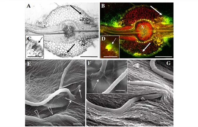
Fig. 1 Penetration of P. macdonaldii strain TA4 E into roots of CI :>0 sunflower. A and C, bright field, B, D, epifluorescence microscopy of transverse sections; E, F, G SEM of infected roots 3 dai. The fu.ngus is penetrating the root mainly at sites of lateral root emergence (A and B). Note the brown colouration in front of the penetration of the fungus (A, C) which is yellow fluorescent under epifluorescence illumination. The bright yellow autofluorescent spots in the root pericycle (B) corre spond to the Gasparian band. The black and white arrows cell wall did not exhibit major alterations at sites where penetration of the fungus was observed, and in some cases it seemed to be invaginated where pushed by the penetrating hypha (Fig. 10). Hyphae were also observed to grow embedded inside the cell wall matrix (Fig. 2E). Again, observations were identical in roots of both susceptible C l 50 and tolerant C137 sunflower.
Observations by confocal microscopy were per formed in order to better visualise the fungus within the plant tissues at Jater colonisation stages. Five dai, all root tissues were invaded in the susceptible C150 Iine (Fig. 2A and C), and large areas of the cortical parenchyma were damaged (arrowheads). Within the stele, fungal hyphae were observed in phloem and xylem cells. As expected, the hyphae grew from the outer part towards the inner part of the root but also
indicate the brown colouration and fluorescence in front of the invaded root tisS\les. Detailed view of direct penetration of hyphae into root epidermal cells (E to G). Dashed arrows show
sites of penetration (E, F), the white arrow shows a tip emerging from the plant cell wall matrix, and open arrow heads show hyphae embedded in the plant cell wall matrix (E).
ln G the hypha seems to invaginate the cell wall. $cale bars: A, 8=300 µm and C, 0=150 µm, E, F and G=S µm
according to the longitudinal axis of the organ (Fig 2C). Two days Jater, a massive colonisation of the phloem and xylem was observed (Fig. 2D).
ln roots of tolerant C137 plants, the fungus was mainly restricted to the cortical tissue 5 dai (Fig. 2B). After 7 days however, the phloem tissue was invaded (Fig. 2E) and a deeper penetration into the stele was observed at sites of emergence of Jateral roots (Fig. 2F). Later, both phloem and xylem were equally colonised ( data not shown). ln our observations on root sections of two plants from three independent inocu lation experiments, the tolerant Iine C137 showed consistently a delayed colonisation of the stele.
Within the root tissues, the hyphae were growing inter- and intracellularly (Fig. 20, H). Observations on semi-thin sections of C150 under bright light illustrated that the hyphae were present within the
cortical parenchyma, in the cell, between two adjacent cells and in the intercellular space (Fig. 21). In the xylem, the fungus was able to colonise neighbouring xylem vessels, penetrating through pores between lignin appositions (Fig. 2J).
Fungal morphology in colonised roots
Further observations of infected sunflower root tissue under bright field rnicroscopy showed that hyphae underwent morphological changes in the tolerant line
<1111 Fig. 2 Laser scanning confocal microscopy of sunflower root colonisation by P. macdonaldii strain TA4 E. Transverse
sections of CJ50 (A, D) and CJ37 (B, E) and longitudinal sections of Cl 50 (C) and Cl 37 (F), 5 (A to C) and 7 (D to F) dai. Wïthin the cortical tissue, significant damage (arrowheads) was observed either in transverse (A) or longitudinal (C) sections. The arrows show hyphae in xylem vessels (D) and phloem cells (E). Detailed view of location of the fungus inside the CJ50 root using laser scanning confocal microscopy (G H) or bright field microscopy (1 J), at 5 dai. Hyphae were located inside the cell in the cortical parenchyma (CP) and between cells (arrows in H and I). ln the xylem tissue, hyphae were obseived inside the vessels, between vessels (dashed arrow) and penetrating from one vesse! to another (arrow in J). The white autofluorescent spots in the root pericycle (A, B) correspond to the Gasparian band. $cale bars A, B, C, F= 250 µm; D and E=IO0 µm; G=200 µm; H=50 µm; 1 and J= 150 µm
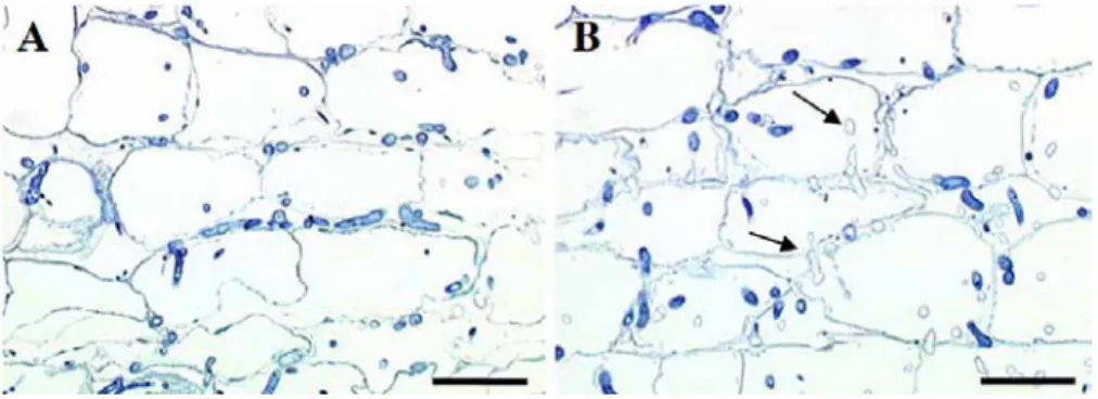
C137. Hyphae in roofs ofC150 were intensely stained
with toluidine blue (Fig. 3A), whereas nwnerous empty hyphae were observed within the root tissues of C137
plants (Fig. 3B), 7 dai. This suggested that the fungus was affected in the C137 plant tissues, and encouraged us to further analyse the appearance of the hyphae in
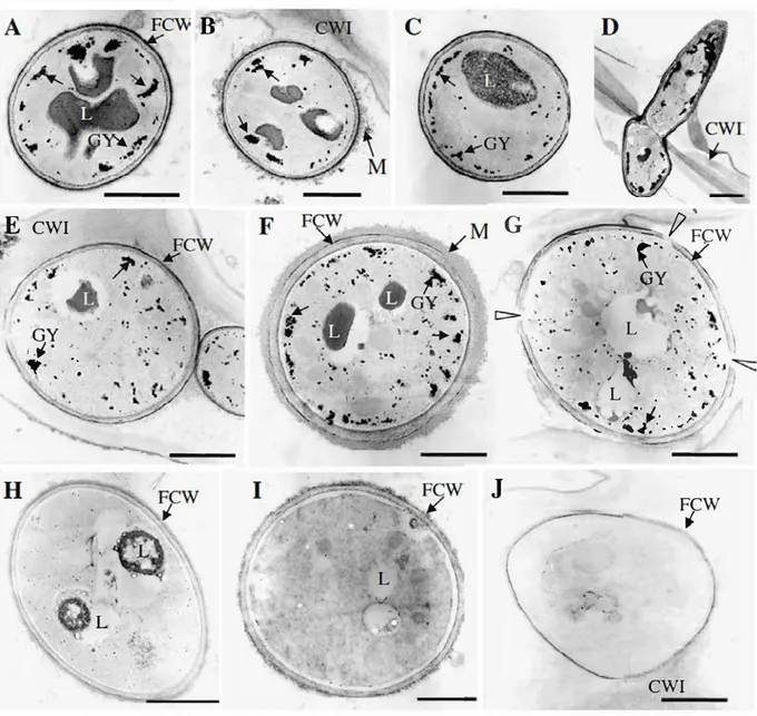
the root cells at higher magnification using TEM 7 dai. Independent of the localisation of the fungus within the C150 root tissues, most of the hyphae exhibited a similar profile (from the epidermal cell layer through the cortex to the xylem vesse!, Fig. 4A-D). Hyphae were surrounded by a cell wall heavily stained with PATAg in the outer layer; a diffuse extracellular matrix was rarely observed around the fungus (Fig. 4B). The cytoplasm of the hyphae contained nwnerous granules of glycogen and large lipid droplefs.
In the root tissues oftolerant C137 (Fig. 4E-J), the morphology of the hyphae exhibited significant changes according to the localisation of the fungus
Fig. 3 Fungal growth of P. macdonaldii strain TA4 E in infected sunflower roots. Semi thin longitudinal sections of Cl 50 (A) and Cl 37 (B) root outer cortical parenchyma, 7 dai.
within the root. In the epidermis and the first cortical cell layer, most of the hyphae had a similar appearance to those in C150, although the lipid bodies seemed to be partially lysed and were reduced in size (Fig. 4E). In the outer cortical layer, additional types ofhyphae were easily observed, such as hyphae
surrounded by a thick extracellular matrix in close contact with the fungal cell wall (Fig. 4F), or exhibiting an incomplete cell wall (arrowheads, Fig. 40). Hyphae contained numerous glycogen granules and partially lysed lipid bodies (Fig. 4F), or lipid bodies were not visible and appeared as empty vacuoles and the glycogen granules were reduced in size (Fig. 40). In the inner part of the cortex (including the endodermis) and the stele tissues, the cytoplasm of the hyphae was devoid of glycogen granules and the lipid bodies were Iargely, if not totally, lysed (Fig. 4H,I). Most of the hyphae in the inner cortex and the stele tissues appeared devoid of subcellular structures (Fig.4J). Counting of toluidine blue-stained sections revealed that 40--45% of the hyphae in the inner cortex (n=300) and 80-85% in the stele (n=70) were empty.
Empty hyphae were also observed in the Cl 50 root tissues but in a much lower proportion ( only 14-18% of the hyphae were empty within the inner cortical and stele tissues). Overall, fungal hyphae became more affected as they penetrated into the centre of roofs of the tolerant Iine, whereas their appearance did not significantly change in the susceptible Iine.
Discussion
Partial resistance against root necrosis caused by P
macdonaldii bas been described recently and
under-Fungal hyphae appear stained with toluidine blue; note the numerous empty hyphae in the Cl 37 root tissues (black arrows). $cale bars=50 µm
E
CWI
GY
J. , ', ,,,,..
.. ..
1 • •B
--...,
_
____
Fig. 4 Profiles of P. macdonaldii strain TA4 E hyphae in roots of susceptible and tolerant sunflower lines. Transmission electron micrographs of PATAg stained hyphae within the root tissues of Cl 50 (A to D) and CJ3 7 (E to J) plants 7 dai. Note the different appearance of hyphae in the epidennal layer (A lying QTL have been characterised (Abou Al Fadil et al. 2007a), using a F9 RILs population and several fungal strains. For the present study, we selected two RlLs which are susceptible and tolerant towards strain TA4, and investiigated the steps of fungal penetration and root colonisation.
The GFP reporter gene is now widely used in fungi (Lorang et al. 2001). It is a powerful tool to study plant-pathogen interactions, notably colonisation of the host plant (Bottin et al. 1999, Bolwerk et al. 2005,
J
p
'
G �-,,
. .. t·
FCW
/ .•1 • 1 ••'
. ·...
n.y
'
..
'....
L
. .. .
.
�L . .
'\
.
..
,,. . . ,�
.,
-...:·'
·1
.·.,..,, rFCW
'
CWI
and E), the outer cortical parenchyma (B, F, G), the inner cortical parenchyma (C, H, I), the phloem (J) an d the xylem (D). FCW, fungal cell wall; CWI, primary plant cell wall; CWH, secondaiy plant cell wall; GY, glycogen granules; L, lipid bodies; M, extracellular matrix. $cale bars= 1 µm
Olivain et al. 2006) and related gene expression patterns (Dumas et al. 1999). The discovery that filarnentous fungi can be transformed via A. tumefaciens (de Groot et al. 1998) bas made genetic transformation easier and more efficient. When we used this technique in order to obtain a strain which constituti vely expressed the GFP gene, we obser ved that high concentrations of both spore and bacterial suspensions gave higher yields of transformants. This might be correlated to the fact that P macdonaldii is a rather
slow-growing fungus, and illustrates that the success of transformation depends in part on the balance between fungal and bacterial growth, as reported for other fungal species (de Groot et al.1998).
High transformation efficiencies such as 100 to 500 transformants per million spores have been reported for other fungi (de Groot et al.1998, Mullins et al.2001), which is much higher than our yield with P. macdonaldii. To our knowledge Leptosphaeria maculans (anamorph Phoma lingam) is the sole related species for which ATMT has been reported (Eckert et al. 2005), and transformation efficiencies were comparable to our results. For the purpose of this work it was not necessary to increase the yield of transformants, but we noted that one of the trans-formants was strongly affected in its virulence (data not shown). The ease and efficiency of this method makes ATMT a good tool for insertional mutagenesis in order to study fungal development or pathogenicity (Blaise et al. 2007). The characterisation of the P. macdonaldi transformant which exhibited reduced pathogenicity, might lead to insights into the mecha-nisms of pathogenicity or virulence of this fungus and the interaction with its host plant.
As previously described for infection of petioles and stems (Roustaee et al. 2000b), P. macdonaldii penetrated directly into the root tissue without differentiation of infection structures, taking advantage of the natural fissures at sites of lateral root formation. The accumulation of brown compounds with a yellow fluorescence under UV light was observed at distance from invading hyphae in the fissures, and was restricted to cells adjacent to infected cells in the case of penetration of the epidermis. This might indicate induction by a diffusible factor released from infected cells. Accumulation of autofluorescent phenolic com-pounds is a well-known defence reaction in plants, and scopoletin accumulation has been characterised in sunflower stems infected by Phoma (Alignan 2006). However, the spectrum of the yellow fluorescent compound analysed under the microscope did not correspond to scopoletin or other standards available in our laboratory (data not shown). In addition, the tolerant and susceptible sunflower line exhibited similar levels of accumulation, indicating that this compound was not involved in tolerance against the fungus.
Penetration into the root epidermis and colonisation of the outer cortex occurred at similar rates in both
lines, but at later stages colonisation of the tolerant line was significantly delayed. Whereas the stele of the susceptible line was readily invaded, the fungus seemed to be inhibited in crossing the endodermis of the tolerant line. This cannot be explained by structural or morphological differences between the two lines, since both have a similar thickness of the endodermis and the same number of cellular layers in the cortex (data not shown). Resistance taking place in the stele has been described for vascular wilt fungi such as Fusarium oxysporum f.sp. lycopersici in tomato, where expression of the I 2 resistance gene co-localises with sites of containment of the fungus (Mes et al. 2000) However, P. macdonaldii is not a vascular fungus and stele colonisation was not prevented in the tolerant line. Interestingly, in the interaction of sunflower with Sclerotinia sclerotio rum, two levels of resistance have been described, one against penetration of the fungus and one against colonisation once the pathogen has penetrated (Castano et al. 1993). Resistance against colonisation by Sclerotinia also involves organ-specific genes (Robert et al.1987), similar to the interaction of sunflower and P. macdonaldii (Abou Al Fadil et al.2007a). However, in the interaction with P. macdonaldii resistance against penetration has never been observed or reported. Whatever the organ or developmental stage of the plant, tolerant lines always showed reduced rates of colonisation by the fungus (unpublished results).
The most surprising observation in our study was that the fungal hyphae were seemingly affected in their morphology in the tolerant root tissues, indicating a strongly lowered viability of the fungus in the stele. However, this did not prevent symptom development on inoculated plants. Fungal biomass does not seem to play a significant role in symptom development, which is in agreement with the observation that toxins (such as zinniol in Phoma) and lytic enzymes are considered to be the main pathogenicity factors in necrotrophic fungi. It will be interesting to identify the plant defence compounds that had such a strong effect on fungal development; according to the gradient of hyphal infection they should be produced in the centre of the root and diffuse towards the outer tissues. Efforts to study defence by transcriptomics in the sunflower— P. macdonaldii interaction have been made (Alignan et al. 2006), and defence-related genes induced in inoculated cotyledons have been identified. However,
the present observations show that the situation in planta can be more complex, and important genes might be missed when whole organs are studied. The recently developed technique of laser-assisted microdissection allows the targeting of specific tissues and has been used successfully for transcriptomic studies at a cellular level in plant — pathogen interactions (Tang et al. 2006). This approach should enable us to identify the defence genes involved in tolerance against P. macdonaldii, and to understand the mechanisms of resistance against colonisation. Such genes might also be of use as markers for the selection of new resistant cultivars. Acknowledgements The authors thank Dr. Bernard Dumas (UMR5546 CNRS UPS) for the gift of plasmid pBin GFP hph.
References
Abou Al Fadil, T. (2006). Déterminisme de la tolérance du tournesol à Phoma macdonaldii au collet et sur racines : approches génétiques et histologiques. PhD dissertation, Institut National Polytechnique de Toulouse.
Abou Al Fadil, T., Poormohammad Kiani, S., Dechamp Guillaume, G., Gentzbittel, L., & Sarrafi, A. (2007a). QTL mapping of partial resistance to Phoma basal stem and root necrosis in sunflower (Helianthus annuus L.). Plant Science, 172, 815 823.
Abou Al Fadil, T., Dechamp Guillaume, G., Darvishzadeh, R., & Sarrafi, A. (2007b). Genetic control of partial resistance to ‘collar’ and ‘root’ isolates of Phoma macdonaldii in sunflow er. European Journal of Plant Pathology, 117, 341 346. Alignan, M. (2006). Phoma du tournesol : déterminisme de la
tolérance de l'hôte à la maladie. PhD dissertation, Institut National Polytechnique de Toulouse.
Alignan, M., Hewezi, T., Petitprez, M., Dechamp Guillaume, G., & Gentzbittel, L. (2006). A cDNA microarray approach to decipher sunflower (Helianthus annuus) responses to the necrotrophic fungus Phoma macdonaldii. New Phytologist, 170, 523 536.
Blaise, F., Rémy, E., Meyer, M., Zhou, L., Narcy, J. P., Roux, J., et al. (2007). A critical assessment of Agrobacterium tumefaciens mediated transformation as a tool for patho genicity gene discovery in the phytopathogenic fungus Leptosphaeria maculans. Fungal Genetics Biology, 44, 123 138.
Bottin, A., Larche, L., Villalba, F., Gaulin, E., Esquerré Tugayé, M. T., & Rickauer, M. (1999). Green fluorescent protein (GFP) as gene expression reporter and vital marker for studying development and plant microbe interaction in the tobacco pathogen Phytophthora parasitica var. nicotianae. FEMS Microbiology Letters, 176, 51 56.
Bolwerk, A., Lagopodi, A. L., Lugtenberg, B. J. J., & Bloemberg, G. V. (2005). Visualisation of interactions between a pathogenic and a beneficial Fusarium strain during biocontrol of tomato foot and root rot. Molecular Plant Microbe Interactions, 18, 710 721.
Castaño, F., Vear, F., & Tourvieille de Labrouhe, D. (1993). Resistance of sunflower inbred lines to various forms of atttack by Sclerotinia sclerotiorum and relations with some morphological characters. Euphytica, 68, 85 98. De Groot, M. J. A., Bundock, P., Hooykaas, P. J. J., &
Beijersbergen, A. G. M. (1998). Agrobacterium tumefaciens mediated transformation of filamentous fungi. Nature Biotechnology, 16, 839 842.
Donald, P. A., Venette, J. R., & Gulya, T. J. (1987). Relationship between Phoma macdonaldii and premature death of sunflower. Plant Disease, 71, 466 468. Dhulipala, M., & Marek, S. M. (2007). Phoma medicaginis as a
model pathosystem for Medicago. Phytopathology, 97, S28. Dumas, B., Centis, S., Sarrazin, N., & Esquerré Tugayé, M. T. (1999). Use of Green fluorescent Protein to detect expression of an endopolygalacturonase gene of Colleto trichum lindemuthianum during bean infection. Applied and Environmental Microbiology, 65, 1769 1771. Eckert, M., Maguire, K., Urban, M., Foster, S., Fitt, B., Lucas,
J., et al. (2005). Agrobacterium tumefaciens mediated transformation of Leptosphaeria spp. and Oculimacula spp. with the reef coral gene DsRed and the jellyfish gene gfp. FEMS Microbiology Letters, 253, 67 74.
Isaac, S. (1992). Fungal Plant Interactions. London: Chapman and Hall.
Lorang, J. M., Tuoru, R. P., Martinez, J. P., Sawyer, T. L., Redman, R. S., Rollins, J. A., et al. (2001). Green Fluorescent Protein is lighting up fungal biology. Applied and Environmental Microbiology, 67, 1987 1994. Maric, A., Camprag, D. & Masirevic, S. (1987). La tacheture noire
du tournesol (Phoma macdonaldii Boerema ; synonymes : Phoma oleracea var. helianthi tuberosi Sacc. Stade terminal : Leptospaeria lindquisti Frezzi). (in Serbo Croatian), Bolesti i stetocine suncokretai njihovo suzbijanje, 37 45. Mes, J. J., Van Doorn, A. A., Wijbrandi, J., Simons, G.,
Cornelissen, B. J. C., & Haring, M. A. (2000). Expression of the Fusarium resistance gene I 2 colocalizes with the site of fungal containment. Plant Journal, 23, 183 193. Mullins, E. D., Chen, X., Romaine, P., Raina, R , Geiser, D. M., &
Kang, S. (2001). Agrobacterium mediated Transformation of Fusarium oxysporum: an efficient tool for insertional mutagenesis and gene transfer. Phytopathology, 91, 173 180. O'Connell, R. J., Herbert, C., Sreenivasaprasad, S., Khatib, M., Esquerré Tugayé, M. T., & Dumas, B. (2004). A novel Arabidopsis Colletotrichum pathosytem for the molec ular dissection of plant fungal interactions. Molecular Plant Microbe Interactions, 17, 272 282.
Olivain, C., Humbert, C., Nahalkova, J., Fatehi, J., L’Haridon, F., & Alabouvette, C. (2006). Colonization of tomato root by pathogenic and nonpathogenic Fusarium oxysporum strains inoculated together or separately into the soil. Applied and Environmental Microbiology, 72, 497 510. Pérès, A., & Lefol, C. (1996). Phoma macdonaldii Boerema:
élément de biologie et mise au point d’une méthode de contamination artificielles en conditions controlées. (Paper presented at the 14th International Sunflower Conference, Beijing).
Pérès, A., Poisson, B. & Drolon, G. (2000). Le syndrome “Pieds secs” du tournesol : étude des causes et approche de la nuisibilité. (Paper presented at the 15th International Sunflower Conference, Toulouse).
Robert, N., Vear, F., & Tourvieille de Labrouhe, D. (1987). L’hérédité de la résistance au Sclerotinia sclerotiorum (Lib.) de Bary chez le tournesol. I. Etude des réactions à deux tests mycéliens. Agronomie, 7, 423 429.
Roustaee, A., Barrault, G., Dechamp Guillaume, G., Lesigne, P., & Sarrafi, A. (2000a). Inheritance of partial resistance to black stem (Phoma macdonaldii) in sunflower. Plant Pathology, 49, 396 401.
Roustaee, A., Dechamp Guillaume, G., Gelie, B., Savy, C., Dargent, R., & Barrault, G. (2000b). Ultrastructural studies of the mode of penetration by Phoma macdon
aldii in sunflower seedlings. Phytopathology, 90, 915 920.
Smolik, J. D., Walgenbach, D. D. & Carson, M. L. (1983). Initial evaluations of early dying of sunflower in South Dakota. Pages 24 25 In: Proceedings of the Sunflower Research Workshop, Fargo.
Tang, W., Coughlan, S., Crane, E., Beatty, M., & Duvick, J. (2006). The application of laser microdissection to in planta gene expression profiling of the maize anthracnose stalk rot fungus Colletotrichum graminicola. Molecular Plant Microbe Interactions, 19, 1240 1250.