Université de Montréal
Étude des déterminants génétiques et
moléculaires de la scoliose idiopathique
Par
Dina Nada
Programme de Sciences biomédicales
Faculté de médecine
Thèse présentée à la Faculté des Études Supérieures
en vue de l’obtention du grade de Philosophiæ Doctor (Ph.D.)
en Sciences biomédicales
Option Générale
Université de Montréal
Faculté des études supérieures
Cette thèse intitulée:
Étude des déterminants génétiques et
moléculaires de la scoliose idiopathique
Présentée par :
Dina Nada
a été évaluée par un jury composé des personnes suivantes :
Dr. Stéphane Roy, président-rapporteur
Dr. Alain Moreau, directeur de recherche
Dr. Mark E Samuels, co-directeur de recherche
Dr. Gerardo Ferbeyre, membre du jury
Dr. Matthew B. Dobbs, examinateur externe
Résumé
La scoliose idiopathique (SI) est une maladie complexe de la colonne vertébrale. Elle survient principalement à l'adolescence et affecte ~ 1-4% de la population mondiale pédiatrique avec une prévalence plus élevée chez les femmes. Dans la plupart des cas, la cause sous-jacente de la SI est inconnue, bien qu'une composante génétique soit bien reconnue. Un certain nombre de gènes et de loci candidats ont été proposés, mais peu ont été répliqués avec succès dans de multiples études. Les études d'association génomique (GWAS) ont identifié plusieurs gènes candidats prédisposant à la SI. Parmi ceux-ci, le locus LBX1 est de loin celui qui a été répliqué avec le plus de succès dans différentes populations, bien que jusqu'à maintenant il n'ait pas été testé dans la population québécoise.
L’objectif principal de cette thèse, a été l'étude des déterminants génétiques de la SI par des approches complémentaires. Dans la première étude, de nouveaux gènes enrichis de variants rares, pouvant contribuer à la maladie ont été identifiés par séquençage d’exomes entiers (WES) dans une cohorte de patients québécois atteints de SI, suivi d'une deuxième phase de séquençage ciblé des 24 meilleurs gènes candidats dans une seconde cohorte indépendante. Parallèlement, nous avons effectué une approche par WES dans une famille multiplex unique constituée de trois sœurs affectées avec des parents sains. Nos résultats impliquent un nouveau gène, FAT3, non précédemment associé à la SI, comme un gène candidat d’importance pour cette condition. Dans la deuxième étude, nous avons effectué un GWAS pour tester les marqueurs génétiques associés à la dans la population québécoise. Les
l'analyse actuelle était de tester l'association du locus LBX1. Nos résultats appuient une association avec la région proche du gène LBX1 dans notre population. Dans la troisième étude, notre approche est une approche de gène candidat. Nous avons essayé d'élucider les corrélations génétiques et biochimiques des niveaux circulants en YKL-40 avec le risque de progression de la maladie. Une étude antérieure dans notre laboratoire a démontré que les patients atteints de SI ont un dysfonctionnement distinctif de la signalisation des récepteurs couplés aux protéines Gi inhibitrices (Gi), permettant leur classification en trois endophénotypes (FG1, FG2 et FG3). L'étude des profils d'expression de ces endophénotypes a montré une élévation significative de l'expression du gène CHI3L1, codant pour la glycoprotéine sécrétée YKL-40, chez les patients classés dans l’endophénotype FG1 par rapport à ceux classés dans les endophénotypes FG2 et FG3 qui sont plus enclins à développer une scoliose sévère. Nous avons démontré que les garçons du groupe FG1 présentent des niveaux plasmatiques significativement plus élevés en YKL-40 que les autres groupes tout comme les patients présentant une scoliose non-sévère. Les SNP dans le gène CH13L1 ont montré une association significative avec les niveaux plasmatiques d’YKL-40 dans des cas non-sévère. L'analyse fonctionnelle in vitro a révélé que la glycoprotéine YKL-40 pourrait jouer un rôle protecteur dans le contexte de la SI en bloquant le défaut de signalisation Gi induit par l'élévation de l'ostéopontine.
En résumé, nous proposons une nouvelle association du gène FAT3 avec la SI, répliquons l'association du gène LBX1 dans une nouvelle cohorte et proposons un nouveau marqueur biochimique, YKL-40, associé à des formes non sévères de la SI.
Abstract
Idiopathic Scoliosis (IS) is a complex disorder of the spine. It mostly occurs between 10 and 15 years old and affects ~1-4% of the global pediatric population with a much higher prevalence in females. In most cases the underlying cause of IS is unknown, although a genetic component is well recognized. A number of candidate genes and loci have been suggested, but few have been successfully replicated in multiple studies. Genome wide association studies (GWAS) have identified several candidate genes for IS susceptibility. Among these the LBX1 locus is by far the most successfully replicated locus in different populations, although until now it has not been tested in the French-Canadian population. Few studies have attempted to detect rare causal variants in IS and this field of research is still in its infancy.
The primary goal of this thesis was to investigate the genetic component of IS through complementary approaches. In the first study, we aimed to find new genes enriched with rare variants, which might contribute to the disease. Hence, we performed whole exome sequencing (WES) in a French-Canadian IS cohort, followed by a second phase of targeted sequencing of the 24 best candidate genes in a replication cohort. In parallel, we performed WES in a unique multiplex family of three affected sisters with healthy parents. Our results implicate a novel gene, FAT3, not previously associated with IS, as a strong candidate for this condition. In the second study we performed a GWAS to test for genetic markers associated with IS in our French-Canadian cohort. The complete results of the GWAS analysis are
LBX1 gene in our French-Canadian population. In the third study, our approach is a candidate gene approach. We attempted to elucidate the genetic and biochemical correlates of circulating YKL-40 levels with the risk of spinal deformity progression in the context of IS. Our prior works have demonstrated that IS patients exhibit a distinctive G inhibitory (Gi) protein-coupled receptor signaling dysfunction, which enabled their classification into three distinct biological endophenotypes (FG1, FG2 or FG3). Previous microarray analysis revealed a significant elevation in the expression of CHI3L1 gene, encoding for the secreted glycoprotein YKL-40, in IS patients classified in FG1 endophenotype when compared to FG2 and FG3 ones, which are more prone to develop a severe scoliosis. In this study, we demonstrated that IS males classified in FG1 endophenotype exhibit significant higher plasma YKL-40 levels than controls and other IS endophenotypes. Furthermore, the non-severe scoliosis group showed significant higher levels of YKL-40 than controls. SNPs in CHI3L1 gene showed significant association with YKL-40 plasma levels in non-severe cases. Functional in vitro analysis showed that YKL-40 could play a protective role in the context of IS by altering the Gi-signaling dysfunction induced by the elevation of osteopontin in IS.
In sum, we propose a novel association of FAT3 gene with IS, replicated the association of LBX1 gene with IS in a new cohort, and propose a new biochemical marker, YKL-40, associated with non-severe forms of IS and has a plausible protective role in IS.
Key words: Idiopathic scoliosis, whole exome sequencing, rare variants, GWAS, FAT3, LBX1, CHI3L1, YKL-40, endophenotypes, French-Canadian population
Materials table
Résumé ... i
Abstract ... iii
Materials table ... v
List of tables... viii
List of figures ... ix
Abbreviations list ... x
Acknowledgements ... xv
CHAPTER I: INTRODUCTION ... 1
1. Studying genetics of complex diseases ... 2
1.1 Human genome ... 2
1.2 Human genomic variation ... 2
1.3 Simple versus complex diseases ... 5
1.4 Approaches to studying genetics of diseases ... 8
1.4.1 Family-based approaches (Linkage studies) ... 8
1.4.2 Population-based approaches (Association studies) ... 9
1.4.3 Sequencing approaches ... 11
2. Idiopathic Scoliosis (IS) ... 14
2.1 Definition ... 14
2.2 Classification... 15
2.3 Epidemiology ... 16
2.4 Diagnosis... 18
2.4.3 Physical examination and imaging ... 19
2.5 Management ... 20
2.5.1 Physiotherapy ... 21
2.5.2 Bracing ... 21
2.5.3 Surgical treatment ... 22
2.6 Etiopathogenesis and different hypotheses ... 23
2.6.1 Neuromuscular ... 23
2.6.2 Biomechanics ... 24
2.6.3 Biochemistry ... 25
2.6.4 Genetics... 26
2.7 Biological endophenotypes associated with IS ... 26
3. Genetic studies in Idiopathic Scoliosis ... 27
3.1 Family based studies and suggested modes of inheritance ... 28
3.1.1 Genome wide linkage studies ... 28
3.1.2 Candidate gene linkage studies ... 29
3.2 Population based studies ... 30
3.2.1 Genome wide association studies ... 30
3.2.2 Candidate genes association studies ... 31
3.3 Sequencing studies ... 33
3.4 Genome wide expression profiling studies ... 34
3.5 Summary ... 35
4. Objectives ... 36
CHAPTER II: RESULTS ... 37 1. Article 1: Rare variants in the FAT3 gene are associated with familial and isolated
2. Article 2: A Replication Study for Association of LBX1 locus with Adolescent Idiopathic
Scoliosis in French-Canadian Population ... 77
3. Article 3: Association of Circulating YKL-40 Levels and CHI3L1 Gene Variants with Idiopathic Scoliosis and the Risk of Spinal Deformity Progression ... 100
CHAPTER III: DISCUSSION... 125
CHAPTER IV: FUTURE PERSPECTIVES AND CONCLUSIONS ... 136
REFERENCE LIST ... 142
ANNEX I : REVIEW ARTICLE ... i
Biochemistry of Idiopathic Scoliosis From Discovery to Diagnostic Biomarkers.. ... ii
List of tables
Table I: Types of common variation in the Genome ... 4
Table II: Estimated heritability for some complex diseases ... 7
Table III: Genomic regions that have been linked to Idiopathic Scoliosis ... 29
Table IV: The most important GWAS results for Idiopathic Scoliosis ... 31
Table V: Genes which have been reported to be associated with Idiopathic scoliosis ... 32
List of figures
Figure 1: Demonstrating different types of common variation in the Genome ... 4 Figure 2: Different approaches to studying disease-associated variants ... 8 Figure 3: Different steps for targeted sequencing ... 13 Figure 4: The Cobb angle used for measurement of the degree of curvature of
the spine ... 15 Figure 5: Prevalence of AIS worldwide ... 17 Figure 6: a) Signs of Scoliosis b) Adam’s forward bending test using a
scoliometer for measuring the trunk deformity ... 19 Figure 7: Planar Cell Polarity pathway might be involved in the
Abbreviations list
ABI ACAN AD AFLPs AIS AKAP2 AR bps BNC2 BRCA1 BRCA2 C17orf67 CDCV CDH7 CDRV CFH CHI3L1 CNVs COL1A1 COL1A2 COL2A1 COMP CTLSO CTS 3D DOT1L Applied Biosystems Aggrecan Autosomal dominantAmplified fragment length polymorphisms Adolescent idiopathic scoliosis
A-kinase anchoring protein 2 Autosomal recessive
Base pairs Basonuclin2 Breast cancer 1 Breast cancer 2
Chromosome 17 open reading frame 67 Common disease common variant Cadherin 7
Common disease rare variant Complement factor H
Chitinase 3-like 1 Copy number variants Collagen type I alpha1 Collagen type I alpha2 Collagen type II alpha1
Cartilage oligomeric matrix protein Cervico-thoraco-lumbo-sacral orthosis Computed tomography scan
Three-dimensional
DZ E EpiG ESR1 ESR2 ELN FAM101A FAT3 FBN1 FBN2 FG1 FG2 FG3 G Gi Giα Gi PCR GPER GPR126 GWAS HOXB8 HOXB7 HOXA13 HOXA10 HSPG2 IGF1 IL6 IL-17RC Indels Dizygotic twins Environment Epigenetics Estrogen receptor1 Estrogen receptor2 Elastin
Family with sequence similarity 101, member A FAT Atypical Cadherin 3
Fibrillin-1 Fibrillin-2 Functional group 1 Functional group 2 Functional group 3 Genes G inhibitory G inhibitory alpha
G inhibitory protein-coupled receptor G protein-coupled estrogen receptor G protein-coupled receptor 126 Genome-wide association study Homeobox B8
Homeobox B7 Homeobox A13 Homeobox A10
Heparan sulfate proteoglycan 2 Insulin like growth factor 1 Interleukin-6
Interleukin 17 receptor C Insertion-deletions
Kb KCNJ2 LA LBX1 LD LDL LOD MAF MATN1 MB MiRNA MMP3 MRI MTNR1A MTNR1B MZ NGS P PAX1 PCP PCR PITX1 POC5 PTK7 RFLPs RNA SOLiD SOX9 SNPs Kilobase pair
Potassium voltage-gated channel subfamily J member2 Linkage analysis
Lady bird homeobox-1 Linkage disequilibrium Low-density lipoprotein Logarithm of the odds Minor allele frequency
Matrilin 1, cartilage Matrix Protein Megabase pair
Micro ribonucleic acid Matrix metallopeptidase 3 Magnetic resonance imaging Melatonin receptor 1A Melatonin receptor 1B Monozygotic
Next generation sequencing Phenotype
Paired box 1 Planar cell polarity
Polymerase chain reaction Paired-like homeodomain 1 POC5 centriolar protein Protein tyrosine kinase 7
Restriction fragment length polymorphisms Ribonucleic acid
Sequencing by oligonucleotide ligation and detection SRY-Box 9
STRs TGFB1 TLSO TPH1 VANGL1 VDR WES WGS XLD ZIC2
Short tandem repeats
Transforming growth factor beta 1 Thoraco lumbo-sacral orthosis Tryptophan hydroxylase 1
VANGL Planar Cell Polarity Protein 1 Vitamin D receptor
Whole exome sequencing Whole genome sequencing X-linked dominant
This work is dedicated to my family
.
“Pursue one great Decisive aim with force and determination” Carl Von Clausewitz
“There is no substitute for hard work” Thomas A. Edison
Acknowledgements
I would first like to express my deep appreciation to my supervisor Dr. Alain Moreau, who gave me the chance to join his lab and grow as a research scientist. My sincere thanks for your incredible support both at the academic and personal levels, for your continuous encouragement and for believing in me.
I would like to express my sincere gratitude and deep thanks to my co-supervisor Dr. Mark Samuels. Your passion for research has inspired me and taught me how to develop my research capabilities. My deep thanks for your wise guidance and valuable discussions which helped to deepen my scientific knowledge. Your advice and support have been invaluable.
I am so grateful to my thesis committee members Dr. Carolina Alfieri and Dr. Zoha Kibar for their valuable advices and support.
I sincerely thank all the jury members for kindly accepting to evaluate my thesis: Dr. Stéphane Roy, Dr. Alain Moreau, Dr. Mark E Samuels, Dr. Gerardo Ferbeyre, Dr. Matthew B. Dobbs and Dr. Hubert Labelle.
I would also like to sincerely thank all my colleagues in the lab:
Dr. Cédric Julien for his incredible help and support. Anita Franco and Saadallah Bouhanik for their sincere help and support both on the personal and professional levels. My dear friends Lakshmi Suvarnan, Nancy Karam, Mohamed Elbakry and Niaz Oliazadeh for their nice company and agreeable time we spent together in the lab. Dr. Kristen Fay Gorman and Dr.
Lacroix and Qilin Tang for their kindness and help. Dr. Maryam Taheri for her kindness, help and support.
Special thanks to Mr. Pierre Rompré for his kind assistance in the statistical analyses, Mme Sandy Lalonde and Mme Dominika Kozubska from the admission department in the research center of Sanite-Justine Hospital for their kind help, and Mme France Fauteux from the department of Biomedical Sciences for her kind assistance.
Also I would like to acknowledge my sources of funding: CHU Sainte-Justine Foundation and the Stars Foundation, and the department of Biomedical Sciences, Faculty of Medicine, University of Montreal.
I deeply thank my beloved family. To the soul of my father, your wish that I attain this goal in my life was the secret behind my strength and persistence to reach this goal, hope you are proud of me. My mom, thank you for your unconditional love, support and sacrifices. My beloved husband, thank you for your love, patience, encouragement and support, you are my backbone. To the three pieces of my heart, my kids, thank you for being there in my life and for being my motivation to face the challenges and to pursue my goals in life.
Finally, I would not have achieved this accomplishment without the continuous help and support from the people around me.
CHAPTER I
INTRODUCTION
1. Studying genetics of complex diseases
1.1 Human genome
The human genome is the hereditary material, which consists of the chemical deoxy ribonucleic acid (DNA). DNA contains the genetic information needed for a human being to be a functional organism. Most of the human genome resides within the cellular nuclei, and a small part is contained within the mitochondria in the cytoplasm. Each nucleated cell in the body has its own copy of the human genome, which includes from 20,000 to 50,000 genes according to the definition of the term. The DNA is composed of around 3.3 billion base pairs, however less than 1.5% of it encodes for proteins. Approximately 50% of the linear length of the genome is composed of single copy DNA sequences. The other 50% are repetitive DNA sequences [1].
1.2 Human genomic variation
Understanding genetic and genomic variation is the key for understanding genetics of human diseases. The DNA sequence is roughly 99.5% identical between any two unrelated individuals [1]. Some of the DNA sequence variations among individuals are responsible for their phenotypic differences and quantitative trait variations as well as disease susceptibility. Currently, many tens of millions of single variants and more than a million complex variants are discovered and reported [1]. A variant is sometimes defined as a polymorphism, when its frequency is more than 1% in a population. Common variants are often defined as those with a minor allele frequency > 5%, while uncommon variants have frequency 1-5% and rare
field of human genetics regarding the precise definitions of these terms; however, for this thesis these definitions will be employed.
The different types of variants or polymorphisms in the human genome include single nucleotide polymorphisms (SNPs), insertion- deletion (indels) polymorphisms, copy number variants (CNVs), inversion polymorphisms and large chromosomal rearrangements (see Figure 1 and Table I).
SNPs are the most common type of variation in the human genome. This type of variation includes only a single base of DNA. On average a SNP occurs once every 1000 bps (base pairs) in the genome [1]. The SNP consortium was organized in 1999 in order to create a public resource of information on SNPs [2]. This was followed by the International HapMap project and the 1000 genomes project, with a goal of identifying and characterizing all common SNPs and SNP haplotypes in the world human population [3-6]. Most of the SNPs identified by these projects lie in the non-protein-coding regions. Nonetheless, more than 100,000 exonic SNPs have been identified and reported to date [1].
Insertion-Deletion (Indels) polymorphism or variation can be due to insertion or deletion either of a single bp or up to around 1000 bps. More than one million indels have been reported.
Copy number variants (CNVs) are similar to indels but there is variation in the number of copies of bigger segments of the genome which can reach up to hundreds of thousands bps. Minimum size of CNVs is not determined, but is typically defined as 1000 bp (1 kb). CNVs that include exons can influence gene dosage and might have significant role in diseases.
Inversions are rearrangements of the DNA sequence where a specific chromosomal region is reversed. Inversion can differ in size from few bps up to many mega bps.
Figure 1: Demonstrating different types of common variation in the Genome. Table I: Types of common variation in the Genome (modified from[1]).
Type of variation
Size (approximately)
Number of alleles
SNPs 1bp 2 (most of the times)
Indels From 1bp to more than 100 bps 2, in case of microsatellites: 5 or more
CNVs From 1kb to more than 1Mb 2 or more
1.3 Simple versus complex diseases
Most diseases are presumed to involve interplay between genetic and environmental factors. Monogenic diseases or Mendelian diseases are those with a very high genetic component where the role of environmental factors is minor. This class of diseases is usually caused by variation in one single locus or gene. With the presence of an informative pedigree, the mode of inheritance of monogenic diseases can be easily identified. The mode of inheritance can be either autosomal dominant or co-dominant where the presence of one variant allele causes the disease to manifest while the presence of two variant alleles leads to a more severe form of the disease, or recessive where two variant alleles should be present in trans for the disease phenotype to be manifested. Other modes of inheritance include x-linked or y-linked and mitochondrial patterns. Most simple Mendelian diseases are rare with a low prevalence among populations, examples include Marfan syndrome [7], Huntington disease [8], cystic fibrosis [9], and fragile X syndrome [10].
On the other hand, in complex diseases, no single variant is responsible for the disease phenotype to manifest, but rather a complex interplay between multiple variants and environmental factors are responsible for diseases manifestation. Most major diseases affecting humans belong to this category including Alzheimer’s disease, asthma, Parkinson’s disease, and autoimmune diseases [11].
The difference between monogenic and complex diseases is not always obvious. The role of environmental factors could be very important in some monogenic diseases, for example phenylketonuria occurs in patients who carry the disease-causing gene only if they
monogenic disorders, cystic fibrosis and sickle cell anemia, may be modulated by other genes [12, 13]. Also single genes were reported to be responsible for the manifestation of complex diseases in some families e.g. BRCA1 [14] and BRCA2 [15] in breast cancer.
Most of the known clinically important genetic diseases and traits are catalogued on the Online Mendelian Inheritance in Man database (OMIM) (www.omim.org).
Penetrance represents the proportion of individuals who carry a specific genotype and manifest the associated phenotype. Penetrance varies in different diseases from approaching 100% in monogenic diseases to much lower penetrance in complex diseases.
Heritability is a measure of the genotypic variance that explains the phenotypic variance of a trait or a disease. High scores of heritability indicate strong genetic contribution and low heritability scores implies more effect of environmental factors. Heritability of different diseases is typically estimated by family and twin studies [16, 17]. A difference in the prevalence of the disease among relatives of an affected individual and its prevalence among relatives of a healthy individual indicates high heritability and more genetic influence on the disease. In twin studies, the prevalence of the disease in monozygotic twins (MZ) and dizygotic twins (DZ) is compared. MZ twins are genetically identical, while DZ twins share 50% of their DNA and both types of twins share many early and late environmental factors [18]. A higher concordance rate of the diseases in MZ twins compared to DZ twins implies high genetic impact and gives higher heritability scores. Table II summarizes the estimated heritability for some complex diseases together with the proportion of this heritability explained by known susceptibility variants for each trait.
Table II: Estimated heritability for some complex diseases, modified from [19].
Disease
Heritability (%)
Proportion of heritability explainedIdiopathic Scoliosis 38 [17] <2 [20]
Alzheimer’s disease 79 23.22
Bipolar disorder 77 2.77
Breast cancer 53 12.52
Coronary artery disease 49 25.15
Crohn’s disease 55 13.43 Prostate cancer 50 31.16 Schizophrenia 81 0.39 Systemic lupus 66 13.2 Type 1 diabetes 80 13.63 Type 2 diabetes 42 27.93
1.4
Approaches to studying genetics of diseases
Figure 2:Different approaches to studying disease-associated variants (adapted from [21]).
1.4.1 Family-based approaches (Linkage studies)
Linkage analysis (LA) studies are based on the investigation of an informative family pedigree with the aim of identifying a chromosomal region, which is shared among affected family members and different from unaffected family members. Many types of genetic markers have been employed for linkage map construction. In early studies Restriction Fragment Length Polymorphism (RFLPs) and Amplified Fragment Length Polymorphisms (AFLPs) were more used [22, 23]. RFLPs are differences in length between different DNA segments arising from different positions of restriction enzyme sites caused by different types
added adaptors to the end of digested fragments followed by amplification. Microsatellites or Short Tandem Repeats (STRs) have also been used as markers for LA studies. These are present as multiallelic due to different numbers of repeated DNA sequence which is composed of 2-4 nucleotides [1]. Microsatellites are PCR-based, polymorphic and highly reproducible which made them good markers for LA studies. Currently, high density SNP panels for high resolution map for LA are being employed [24]. The ability to detect genomic loci harboring causative variants is highly affected by the density of SNP panels, knowledge of the amount of Linkage Disequilibrium (LD) in the genome and frequency of the haplotypes [25]. LD is a measure of co-inheritance of alleles of two different loci (usually close to each other on a given chromosome). The higher the LD the more chance that alleles of the two loci are co-inherited [1]. LA has proved to be powerful and successful in identifying genomic regions harboring causal mutations for monogenic diseases segregating in individual families [26, 27]. On the other hand, LA methods have limited success in mapping genomic risk regions for complex diseases. The reason behind this might be due to the presence of multiple variants of low or moderate effects within multiplex families, such that the power of LA algorithms is not enough to detect linkage [28]. Rischet al suggested that even studies which include 2000 sib pairs are unable to detect loci with a moderate effect[29].
1.4.2 Population-based approaches (Association studies)
The concept of association studies is to compare the frequency of genomic variants in cases versus controls to detect a statistical association between alleles of any of these variants
identified genomic region to be linked with the disease. With the advances of SNP genotyping technologies and the availability of high-density polymorphism databases, association studies were extended to study hundreds of thousands or millions of SNPs throughout the whole genome in an unbiased approach (GWAS: Genome wide association study). SNP associations together with large haplotype blocks involving the associated SNPs can be detected which implicates a disease susceptibility locus. Candidate genes association studies suffered limited success in replication studies. GWAS on the other hand proved their effectiveness in identifying many common variants to be associated with many complex diseases [30, 31]. GWAS became available for investigators after the completion of the Human Genome and the HapMap projects in 2003 and 2005 respectively [32, 33].
The first GWAS was published in 2005, which reported an association between a polymorphism in the complement factor H gene (CFH) and age-related macular degeneration [34]. This was followed by many successful GWAS published in 2006-2007 where many common variants were reported to be associated with several complex diseases such as Crohn’s disease [35, 36], type I and II diabetes [36-38], obesity [39], coronary artery disease and rheumatoid arthritis [36]. As of now, hundreds of GWAS have been performed and contributed in the identification of thousands of variants associated with many complex diseases [31, 40-44]. However, care has to be taken in GWAS analysis and correction for population stratification should be done. It has been shown that population stratification can lead to false positive results where the differences in allele frequencies could be unrelated to the tested disease [45, 46]. In general, many reported GWAS associations have proved difficult to replicate, either due to differences in the study populations, definitions of the
significant proportion of GWAS loci have been replicated in multiple studies; the most obvious being the major histocompatibility (HLA) locus on chromosome 6, which is universally observed to be strongly associated with many different autoimmune disorders.
1.4.3 Sequencing approaches
The main aim of the sequencing approach is to identify rare variants. In fact, sequencing techniques have the potential to identify both rare and common variants where the most comprehensive design to study variation in any disease would be to sequence the whole genome in a large population [47], however for studying common variants, dense SNP genotyping arrays remain for the moment less expensive than whole genome sequencing. That said, sequencing approaches designed to detect rare variants would detect common ones as well obviously.
Previously, DNA was sequenced by the Sanger method [48], a technology which was although a breakthrough in the field of molecular genetics, yet was very expensive for sequencing large number of individuals for research laboratories. More recently, large-scale sequencing has been universally performed using the so-called Next Generation Sequencing (NGS) technologies. NGS refers to sequencing approaches where massive sequencing reactions are carried out in parallel, typically without the need for electrophoresis. The 454 pyrosequencing method was introduced in 2004 [49] and is now commercialized via Roche. The Solexa reversible termination method was introduced in 2006 [50] and is now commercialized by Illumina. Sequencing by oligonucleotide ligation and detection (SOLiD) method was introduced in 2007 by ABI (Applied Biosystems) [51], now commercialized by
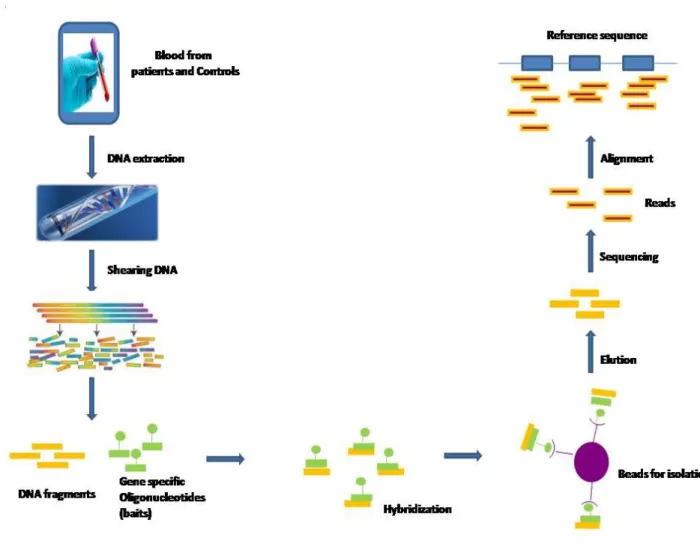
chemistry method, and Life Technologies introduced Ion Torrent techniques [52] to replace SOLiD. NGS technologies lead to drastic reduction in the cost and time of the sequencing approaches, which consequently dramatically affected the field of study of genetics of complex diseases. However, whole genomic sequencing (WGS) is still expensive for laboratory research. In 2007, a sequence capture technology was introduced by Nimblegen whereby microarrays were used to capture specific sites of the genome to be sequenced (typically protein-coding exons) [53]. More recently sequence capture has been adapted to solution hybridization using biotin-labelled RNA oligonucleotide probes to target desired genomic content to be studied (Figure 3). Targeted sequencing gave researchers a less expensive alternative to WGS in terms of total cost, although the cost per base pair is higher due to the targeting and capture steps in the protocol. Targeted sequencing may focus on subsets of genes belonging to biological pathways of interest, or loci previously linked or associated with a disease. More generally, targeting is used to capture and analyze all known genomic protein coding regions, which comprise about1.5 % of the genome and have the potential of harboring many or most of high penetrance disease causing variants (whole exome sequencing, WES) [54, 55]. Sequencing approaches can be done for families or case/ control populations; where either sequencing multiply affected families (multiplex) searching for rare variants segregating with the disease or sequencing a population of cases and healthy controls and looking for genes enriched with rare variants in cases versus controls. It is important to note that targeted sequencing approaches have more potential for artifacts than either Sanger or whole genome sequencing, due to the hybridization capture step itself as well as the requirement for more cycles of PCR. Certain regions of the genome (high GC-content
captured much more efficiently than others for unclear reasons. A high level of statistical variance among all targeted exons in WES requires a higher level of mean coverage of targeted regions, to ensure that most targeted regions are covered at sufficient read depth to detect heterozygous rare variants.
It
is
Figure 3: Schematic for targeted next generation sequencing. DNA is extracted from the study subjects, then sheared and allowed to hybridize to the baits (oligonucleotides specific for the target regions). These baits are tagged to be easily isolated by capture on immobilized beads (for example). The captured DNA is eluted, amplified and sequenced on one of several next generation platforms (Illumina HiSeq, LifeTech Proton for example). For mutation
The most important challenge of the sequencing approaches is the analysis of the results. If the analysis of rare variants is based on single variant associations, large sample sizes and/ or variant effect sizes will be needed to attain sufficient statistical power in order to detect the associations. To overcome this problem, new statistical methods have been developed to boost the statistical power of analytical methods. The main idea of these methods is to look for multiple rare variants in either a gene or biologically relevant pathway rather than testing only single variants, which is routinely done in GWAS. Among those methods is the collapsing gene burden test [56] which is a simple test where the number of rare variants (typically of MAF ≤1%) in one gene is compared between patients and controls. This test is used under the assumption that all rare potentially protein altering variants act in the same phenotypic direction with the same magnitude.
2. Idiopathic Scoliosis (IS)
2.1 Definition
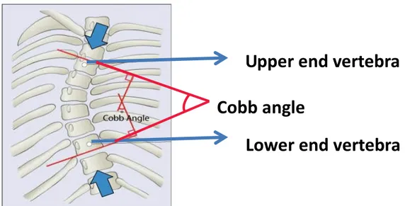
The term scoliosis (skō̍'ӏē-ō'sȋs), “Ʃκoλίωσηˮ, first used by the Hippocrates (A.D. 460– 370) and Galen (A.D.131-201), is a Greek word which means “crookedness”. It is a three-dimensional (3D) spinal deformity with a Cobb angle of 10ο or more in the coronal plane measured on a standing radiograph. The Cobb angle is the anglulation used to measure the degree of curvature of the spine (Figure 4), it is the angle formed between a line drawn parallel to the superior endplate of the upper vertebra of the scoliotic curve and that drawn parallel to the inferior endplate of the lower vertebra of the scoliotic curve. The deformity of
Figure 4: The Cobb angle used for measurement of the degree of curvature of the spine (adapted from https://www.coreconcepts.com.sg/article/cobb-angle-and-scoliosis).
2.2 Classification
There are many types of scoliosis where the cause is well-known, such as congenital, neuromuscular, degenerative, and syndromic while Idiopathic scoliosis is the type whose etiology remains most elusive. Idiopathic scoliosis comprises approximately 80% among all types of scoliosis [59]. Idiopathic scoliosis can be further classified according to the pathological type into structural or functional, according to the magnitude of the curve where the severity increases with increasing the Cobb angle, according to the anatomical site of the curve into cervical, thoracic, thoracolumbar or combinations if there is more than one angle of curvature. Idiopathic scoliosis is often classified according to the age of onset into infantile (0-3) years, juvenile (4-9) years, adolescent (10-15) years which is the most common type, and an adult type which occurs above the age of 18 [58].
Upper end vertebra
Lower end vertebra
Cobb angle
2.3 Epidemiology
The overall prevalence of IS ranges somewhat widely, with estimates varying from 0.47-5.2% [60]. There are substantial differences among the studies with regard to prevalence of IS, potentially due to different definitions of scoliosis, different age groups, different study designs and methods of diagnosis [61-67]. The most important factors which affect the prevalence of IS are ethnicity, age and gender. The prevalence of IS from 17 countries has been summarized in a meta-analysis study which included 36 studies [68]. The reported IS prevalence while different in detail, overlaps substantially in different regions: in Australia and the Middle East it was 1.9%; in Asia it ranged from 0.4 to 2.5%; in North America it was from 0.4 to 3.9%; in Europe (Spain) it was reported to be from 0.7% to 7.5%. This study reported a general prevalence of curves ≥ 10ο to be 1.34%, curves ≥ 20ο to be 0.22% and severe curves to be 0.07%. Generally prevalence decreases in areas near the equator and increases towards the north, a latitude cline that has also been observed for other common disorders including some autoimmune diseases [58, 69]. Age is another factor, which affects the prevalence. It was reported that the prevalence of IS is 3.12% in the age 16-17 years, 1% in the age 11-12 years and 0.12% in the age 6-7 years [64]. Another German study reported a prevalence of 6.5% in the group of age 11-13 years and 11.1% in the group of age 14-17 years [61]. It is well known that the prevalence of IS is greater in females than males, although the female to male ratio also varies widely among different studies from 1.5 to 11 due to the previously mentioned differences among those studies [70, 71]. A study which included a very large cohort of IS patients, recruited children from 10 years old and followed
were 1.4% and severe curves were 0.33% [72]. The female: male ratios were 2.7, 4.5, 8.1 and 8.4 for curves ≥ 10ο,≥ 20ο, ≥ 40ο and very severe respectively [72]. Patients who are more at risk of developing severe cases are the youngest, shortest, premenarchal and the higher the initial Cobb angle [73].
Figure 5: Prevalence of AIS worldwide. The prevalence of AIS (Adolescent Idiopathic scoliosis) obtained from a meta-analysis of 36 school scoliosis screening studies performed in 17 countries (adapted from [58]).
2.4 Diagnosis
2.4.1 Screening
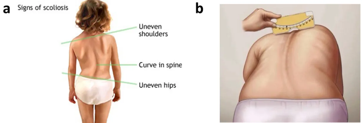
Screening of scoliosis in schools is done with the purpose to detect the condition at an early stage, which enables early intervention. Clinical signs of scoliosis include asymmetry of the trunk, shoulder and scapular prominence and uncentered head over the pelvis (Figure 6). The most common screening test for IS is the Adam’s forward bending test where the patient bends forward and examined from behind, if one side is higher than the other it is a sign of scoliosis while if the back is completely straight so the patient is normal (Figure 6). An apparatus called scoliometer is used to measure the angle of the trunk deformity which when is 7ο or more is referred to a physician for further examination (Figure 6). In some countries of Europe and East Africa another test is used called Moiré topography in addition to the forward bending test with or without a scoliometer [58]. There is an ongoing debate for the effectiveness of screening for IS. Not all health professionals agree on applying a screening program because there is a high number of false positives and negatives [74]. Adbor et al suggested that screening can reduce the rates of treatment and is therefore cost effective [75]. But many other researchers disagree because of the low sensitivity of the test and the non- predictable natural history [75]. However, the American Academy of Orthopedic Surgeons, the Scoliosis Research Society, the American Academy of Pediatrics and the Pediatric Orthopedic Society of North America decided that females should be screened twice at the age of 10 and 12 years, while males should be screened only once at the age of 13 or 14 years[76].
Figure 6: a. Signs of Scoliosis b. Adam’s forward bending test using a scoliometer for measuring the trunk deformity (http://umm.edu/health/medical/reports/articles/scoliosis), (https://www.pinterest.com/ottawascoliosis/school-screening-for-scoliosis-what-we-dont-have-i/).
2.4.2 History
The evaluating physician starts by investigating the patient’s history specially any family history, the age of onset and the history of progression of the disorder. Associated neurologic and respiratory symptoms as pain are investigated. Further pulmonary examination is done in case of history of failure to thrive in childhood and difficulty in breathing. Previous surgical and medical history is important to rule out other syndromes.
2.4.3 Physical examination and imaging
At the beginning and the end of the pubertal spurt, the skeletal maturity should be assessed. This help to predict the growth potential remaining and subsequently the potential for curve progression. The most reliable method to measure skeletal maturity is the Risser sign [77]. The process of ossification of the iliac epiphysis is divided into six stages up to its
indicated by Risser (4-5). Height also should be measured and recorded. The physician performs a standing posterior-anterior and lateral radiograph of the spine including the hip joints. The radiography is done posterior-anterior to reduce the amount of radiation to important organs in the upper torso [78]. These radiographs are viewed such that the heart is on the left side, this view is exactly like during the clinical assessment where the patient is examined from behind during Adam’s forward bending test. The curve magnitude is measured using the Cobb angle method. Other malformations in the ribs or vertebrae could also be detected. The curves are denoted by the direction of its convex side and the site of its apex. Other imaging methods like Computed Tomography Scan (CTS) and MRI are not necessarily done for IS patients except in some cases with pain, severe curvature, rapid progression of the deformity or suspected tumors, and in cases who present abnormal neurological examination as well [78]. In addition, patients who require surgical treatment undergo MRI scan to rule out any intraspinal abnormality.
2.5 Management
Management of IS patients generally ranges from observation, physiotherapy, bracing and treatment. Each patient is assessed individually and a treatment plan is tailored for each patient according to his case. Prognostic factors are very important to help the physician decide the best treatment plan for his patient. The patient’s prognosis depends mainly on the age of onset, Cobb angle and the Risser grade [79]. Factors which contribute positively to disease progression include family history, defects in connective tissue (skin and joints), flattening of kyphosis in the thorax [80], angle of deformity of trunk exceeding 10ο and growth spurt [81].
2.5.1 Physiotherapy
IS patients undergo physiotherapy as part of a conservative therapy [82-84]. Physiotherapy can either be in the form of exercises or physical treatment like electro stimulation of the paravertebral muscles [82-84]. Whether physiotherapy is effective for IS is under debate [84-86]. Several publications reported a positive effect of exercises on reducing the risk of disease progression [87-89] and reducing the Cobb angle [90-93]. On the other hand, many reviews report that those studies do not offer sufficient scientific evidence of efficacy in the reduction of scoliosis progression [84-86]. Of note, certain physiotherapy can be useful to support brace treatment and to prepare IS patients for surgical operations[82, 83].
2.5.2 Bracing
Bracing is recommended for IS patients with the aim to reduce further curve progression, alleviate back pain, prevent respiratory dysfunction and ameliorate esthetics [82]. Bracing is indicated for skeletally immature patients (Risser<3) with Cobb angle 30ο -40ο. Bracing is also indicated for curves around 25ο-30ο that showed a progression of 5ο or more. There are many types of braces which can be classified according to the material into rigid and soft, according to the time it is being worn into full time, part time and night time, or according to its design based on the site of curvature into cervical, thoracic, lumbar and sacral [94]. Examples of rigid TLSO (thoraco-lumbo-sacral orthosis) braces, which are designed for thoracic, low thoracic, thoracolumbar and lumbar scoliosis are Cheneau brace, Boston, Gensingen, Lapadula, lyonese, Sibilla, Sforzesco and Progressive action shortBrace
brace. The Charleston and the Providence braces are examples of night time braces [95]. In most studies, the evidence for the effectiveness of bracing for IS patients is of low quality [95]. It is hard to compare studies which addressed the effectiveness of brace treatment because of the inconsistency of the methods of evaluation and the presence of many different additional factors that could have affected their results [95].
2.5.3 Surgical treatment
Surgical treatment is indicated for severe cases when the curve magnitude is ≥ 45ο who are equal or less than Risser 2 and with curves ≥ 50 who are equal or greater than Risser 3. The aim of surgeries is to stop the curve progression and improve the balance and alignment of the spine. During surgeries spine fusion is performed by instrumentation and bone grafting. Fusion techniques have started early in the 1960s when Harington introduced the hook and rod constructs [96]. Later Luque introduced wires for segmental fixation [97]. Nowadays, there is third generation segmental fixation using pedicle screws [78]. In all these techniques bony anchors which include the hooks, wires and pedicle screws are placed to the vertebrae and connecting them to rod constructs [78]. Fusion surgeries can be done anteriorly, posteriorly or both. Preoperative planning should consider many factors including the stage of skeletal maturity, curve flexibility, type and magnitude of curvature and spinal balance [78].
2.6 Etiopathogenesis and different hypotheses
2.6.1 Neuromuscular
A postural equilibrium dysfunction has been suggested to play a causative role in IS [98]. Consequently, defective visual and vestibular functions have been investigated by many studies to understand their role in IS [99-103].Visual, vestibular and proprioceptive neural pathways interconnect together in certain areas of the brainstem. Lesions in these areas were found to be associated with scoliosis [104] and induction of scoliosis has been done by damaging these areas in animal studies [105]. Altered function of cerebral cortical and sub cortical has been suggested to contribute to pathogenesis of IS [101, 106, 107]. A significant reduction in the length of the spinal cord relative to the vertebral column in severe IS cases has been documented by MRI [108]. This lead to and supported the theory of disproportional neuro-osseous growth in the etiology of IS [109-112]. Differences in the volume of different regions of the brain between IS and matched controls has been reported [113]. Ten regions were shown to be significantly larger in IS patients, and twelve regions were found to be significantly smaller in IS patients. These anatomical differences of different regions of the brain have been suggested to contribute to neurological defects detected in IS patients [114]. Defects in the paraspinal muscles have also been encountered among scoliotic patients [115-120]. Several studies suggested the involvement of the paraspinal muscles in the etiology and progress of IS [121-125]. Many authors showed that there is an increase in the myoelectric response near the apex of the curve on the convex side [125-129]. The interpretation of these
motor drive of the spinal cord produced by a change in the sensory input or by central mechanism which affect the balance of forces in this area and lead to IS [98].
2.6.2 Biomechanics
Scoliotic curve progression is defined by spinal biomechanical changes [130-132]. The association between vertebral growth and IS has been extensively studied and there is extensive debate in the literature about it [133-136]. Many studies reported that IS patients are significantly taller than matched healthy controls [137-140]. It has been suggested that this deviation of growth pattern is responsible for IS because of the higher tendency of taller spines under loading to buckle out of the sagittal plane [132, 141, 142]. However, this theory has not been proved [98]. Biomechanical studies have used finite element models which are patient-specific to study the effect of biomechanical factors on the progression of IS curve. Gravity, combined with anterior spinal overgrowth were shown as aggravating factors for thoraco-lumbar scoliosis [113]. Curve progression was more enhanced by reduction of the mechanical stiffness of the disc in addition to the aforementioned factors. These disc changes could be secondarily to a primarily existing deformity and hence affecting the curve severity rather than being a primarily cause [143]. The soft tissues responsible for sustaining the stability of the spine could also play a role in the etiopathogenesis of IS. Of note, 82% of IS patients were reported to have high level of distortion of the ligamentum flavum elastic fibers when compared with controls. Moreover, 23% of IS patients showed dysfunctional metabolism of fibrillin which suggests that the elastic fiber system might play a role in the pathogenesis of IS[144].
2.6.3 Biochemistry
All the biochemical factors that have been reported in the literature and suggested to contribute to the pathogenesis of IS, are reviewed in the review in the Annex “Biochemistry of Idiopathic Scoliosis from discovery to diagnostic biomarkers”: Current scientific literature reveals potential biomarkers of the onset and progression of IS, which include hormones, systemic factors, hematological factors, and bone metabolism factors. Hormones that include insulin like growth factor-1, melatonin, estrogen, ghrelin, and leptin, have been implicated as potential diagnostic biomarkers in IS [145-149]. Systemic factors, such as osteopontin and Gi proteins; proteins involved in bone metabolism, such as matrilin-1, cartilage oligomeric matrix protein (COMP), osteocalcin; and the hematological protein calmodulin, are the other potential candidates for diagnosing IS [150-154]. These diagnostic biomarkers do not all act in isolation but are interconnected through signaling pathways. There is crosstalk between melatonin and estrogen; estrogen and growth hormones; or between melatonin and calmodulin [155, 156]. Similarly, increased levels of osteopontin regulate Gi protein-coupled receptor (GiPCR) dysfunction [157]. Other studies implicate single nucleotide polymorphisms (SNPs) in calmodulin, melatonin receptors, or matrilin-1, as potential risk factors of increasing the spinal curvature [158-160]. While theories, such as the double neuro-osseous theory implicate increased sensitivity of the hypothalamus to leptin in the development of IS, the downregulation of leptin has also been indicated in the etiopathogenesis of IS [149, 161]. Although data on the potential candidates for diagnosis appears promising, it is acknowledged that replication studies with more cases should generate a sufficient statistical power required to confirm the diagnostic
2.6.4 Genetics
A genetic component of IS is well documented. Clinical studies have noted the familial incidence of scoliosis since the 1930s, where the inheritance of scoliosis in five generations was reported by Garland et al [162]. 97% of IS patients have positive familial history [113]. Segregation studies report an increased risk of IS among first degree relatives of affected individuals, and that the risk decreases as the degree of relation to the affected individual becomes more distant [163, 164]. Moreover, twin studies have shown that the rate of concordance of IS in monozygotic twins is almost double that of the dizygotic twins [165]. It has also been reported that the rate of curve progression is very similar in twins [165]. The mode of inheritance of IS is still unclear [166] with an apparent high level of heterogeneity between different families [167-169]. The genetic nature of the disease is presumed therefore to be complex. The incomplete penetrance and variable expressivity of IS suggest complex genetics with multiple genes and environmental factors involved in the disease [170, 171]. It has been reported that there is high level of inconsistency between the results of different genetic studies in IS [172]. A recent study conducted on a huge number of twins in the Swedish twin registry estimated that the genetic component of IS susceptibility is 38%, while the environmental component is 62% [17].
2.7 Biological endophenotypes associated with IS
The inconsistency between the results of different studies in IS may be at least partly due to the heterogeneity among IS patients both at the clinical level and the genetic level. The patients differ in their age of onset, type of curvature, degree of severity, rate of progression,
common genetic background. Studies from our lab have classified IS patients into three biological endophenotypes named FG1, FG2, and FG3 (functional groups 1, 2 and 3) [151, 173]. This classification was based on a common pathological mechanism in which differential impairment of Gi-coupled receptor (G inhibitory protein-coupled receptor) signalling pathway resulting from selective inhibition of one or more of the isoforms of Giα subunit (Gi1- Gi2- Gi3). These endophenotypes represent a unique heritable trait conserved
among affected family members contrasting with the usual phenotypic heterogeneity, within the same family, in terms of curve type and severity. Moreover, some unaffected relatives of the patients can carry the same endophenotype conferring a high risk to those individuals[174]. The use of these endophenotypes allows patient stratification that has been correlated to specific molecular expression profiles and different prognosis [175]. In a complex genetic disease such as IS, those endophenotypes have potential utility to identify risk genes specific to each of these endophenotypes.
3. Genetic studies in Idiopathic Scoliosis
During the last three decades, there has been huge number of studies trying to uncover genetic factors in IS. This task is difficult due to the complexity of the disease, as evidenced by the high level of inconsistency among different genomic studies. These studies are either family based and conducted with linkage analysis, or population (case/control) based and conducted with association studies. The general goal of these studies was either to identify specific loci in the genome or certain genes, which could be related to the disease or trying to explain the mode of inheritance of this disorder.
3.1 Family based studies and suggested modes of inheritance
3.1.1 Genome wide linkage studies
Extensive effort was made to identify specific loci in the genome, which might be linked to IS by genome-wide studies as well as to understand its mode of inheritance. Several regions in the genome have been suggested to be linked to IS through family-based studies. Those with significant statistics are listed in Table III. However, there is a general lack of replication of specific loci by different groups. As an exception to this, two loci were replicated by two different groups; chromosome 9q31-q34 which was introduced by Miller at al [176] and replicated by Ocaka et al [169], and chromosome 17q25 which was first identified by Ocaka et al [169] and confirmed by Clough et al [177]. Gao et al [178], reported linkage of the chromosomal region 8q12 and suggested CDH7 as a candidate IS gene. Mutations in this gene lead to CHARGE syndrome in which a large percentage of patients have scoliosis. However, these results could not be replicated by another group [179]. The mode of inheritance for IS is inconsistent among the different studies. Of note, 65% of the studies reported AD to be the mode of inheritance for IS [180]. X-linked dominant mode of inheritance had also been suggested [166, 181]. Raggio et al suggested both AD and AR modes of inheritance in their study [182]. Recently a study suggested AD, AR and multifactorial modes of inheritance for IS [183].
Table III: Genomic regions that have been linked to Idiopathic Scoliosis (modified from[172]).
Locus
Suggested mode
of inheritance
Statistics
Reference
17p11 AD Zmax=3.2 Salehi et al.[184]
19p13.3 AD LOD=4.48 Chan et al.[185]
Xq22.3-q27.2 XLD LOD=2.23 Justice et al.[181]
9q31.2-q34.2 AD Zmax=3.64 Ocaka et al.[169]
3q12.1 5q13.3 AD Zmax=3.00 Zmax=3.01 Edery et al.[168] 12p AR AD HLOD=3.2 HLOD=3.7 Raggio et al.[182] 6q15-q21 10q23-q25.3
_
P<2x10⁻¹⁰ P<2x10⁻⁷ Marosy et al.[186]AD: autosomal dominant, XLD: X-linked dominant, AR: autosomal recessive
3.1.2 Candidate gene linkage studies
Based on the biological roles of some genes, several have been tested by linkage analysis and/or by transmission disequilibrium. A study demonstrated that an intragenic microsatellite polymorphism in the 3’ region of the matrilin 1 gene (MATN1) was linked to IS[187]. Other genes tested by studies which fall in this category did not show linkage with
IS; fibrillin (FBN1), elastin (ELN), collagen I A1, collagen IA2, and collagen II A1 (COL1A1, COL1A2, COL2A1), Aggrecan (ACAN) and melatonin receptor 1A (MTNR1A) [188-192].
3.2 Population based studies
3.2.1 Genome wide association studies
Since 2011, a number of GWAS of IS have been published. The most important findings are listed in Table IV. A GWAS study in a Japanese cohort reported an association between a locus at 10q 24.31 and IS [42]. They reported common SNPs in the vicinity of the LBX1 gene, of which the most significant was rs11190870. Several replication studies subsequently confirmed this association in both Asian and Caucasian populations [193-197]. A recent study showed that a risk allele of this SNP is associated with higher level of transcription of the promoter. Moreover in zebra fish high expression of LBX1 leads to the deformation of normal body curvature [198]. Another candidate gene, GPR126 (G-protein coupled receptor 126), has been reported by two studies to be associated with IS. The first study involved three populations; Japanese, Han Chinese and European [199]. The second study involved a Chinese population [200]. It has also been shown that the knockdown of this gene in zebra fish lead to delayed ossification of the spine [199]. The intergenic variant rs12946942 has been reported by Miyake et al [201]. This variant lies on chromosome 17 near the two genes SOX9 and KCNJ2, mutations in which are associated with campomelic dysplasia and Andersen Tawil syndrome, for which scoliosis is an aspect of the phenotype. BNC2 (basonuclin-2) and PAX1 (paired box1) are another two interesting genes suggested by two GWAS [202, 203]. However, they both need to be confirmed by additional studies.
body curvature in zebra fish [202]. PAX1 encodes a transcription factor which is involved in the development of the spine [203].
Table IV: The most important GWAS results for Idiopathic Scoliosis (modified from [204]).
SNP
Gene name
Statistics
References
rs11190870 LBX1(downstream) p=1.2x10⁻¹⁹ Takahashi et al.[42], Fan et al. [193] Gao et al.[194], Jiang et al.[195],
Liang et al.[196], Londono et al.[197], Grauers et al. [205] rs657507 GPR126 (intron) p=1.6x10⁻⁴ Kou et al. [199], Xu at al.[200],
Grauers et al.[205] rs12946942 Intergenic p= 4x10⁻⁸ Miyake et al.[201], Grauers et
al.[205] rs3904778 BNC2 p=2.5x10⁻¹³ Ogura et al.[202] rs6137473 PAX1 p=2.2x10⁻¹⁰ Sharma et al.[203]
SNP: single nucleotide polymorphism
3.2.2
Candidate genes association studies
During the last two decades, a large number of associations of candidate genes have been explored. Gorman et al [172] have comprehensively reviewed those studies. They reported that the earlier candidate gene studies (from 1992 to 2006) were mostly family based linkage studies, while most recent candidate gene studies (after 2006) were case-control association studies, due to the low power of family based methods in detecting common variants in complex diseases. Genes, which have been reported for their positive associations with either IS susceptibility and/ or severity are summarized in Table V.
Table V: Genes which have been reported to be associated with Idiopathic Scoliosis (modified from [172]).
Gene name
SNPs
Statistics
References
MATN1 rs1149048 p=0.0034 Chen et al. [160]
MMP3 rs3025058 p=0.01 Aulisa et al. [206] IL6 rs1800895 p=0.014 (G/C) p<0.001 (G/G) Aulisa et al. [206] VDR rs1544410 p=0.0054 Suh et al.[207] MTNR1B rs4753426 p=0.045 Qiu et al. [159] TPH1 rs10488682 p=0.002 (allele)
p=0.001 (genotype) Wang et al. [208]
ESR1 rs9340799 p=0.002
p=0.01
Inoue et al [209] Wu et al. [210]
ESR2 rs1256120 p=0.037 (susceptibility)
p= 0.005 (severity) Zhang et al. [211]
GPER rs3808351 rs10269151 rs4266553 p=0.004 p=0.048 p=0.028 Peng et al. [212]
IGF1 rs5742612 p=0.042 Yeung et al.[213]
IL-17RC rs708567 p=0.023 (genotype)
p=0.028 (allele) Zhou et al.[214]
DOT1L rs12459350 p=0.001 Mao et al.[215]
C17orf67 rs4794665 p=0.006 Mao et al. [215]
3.3 Sequencing studies
Because sequencing technologies were very expensive in the past and only very recently have become more affordable, few studies have attempted to detect rare causal variants in IS and this field of research is still in its infancy. Sequencing using either whole exome or targeted gene panels has identified a few genes, which might contribute to the occurrence and/or severity of scoliosis. Buchan et al documented an enrichment of rare potentially protein-altering variants in the FBN1 and FBN2 genes in severe IS cases versus controls, by performing WES (whole exome sequencing) in 91 severe cases and 337 controls, followed by targeted sequencing of these two genes in a larger cohort [217]. HSPG2, a gene coding for an extracellular matrix protein, was identified by Baschal et al [218]. In their study they reported several rare potentially damaging variants by performing WES for three patients in a multigenerational family followed by sequencing this gene in an independent cohort of 100 IS patients. POC5, which encodes for a centriolar protein, was identified by Patten at al. In this study they performed exome sequencing for three patients in a multigenerational family as a follow up study to another LA study to the same family and suggested linkage to two loci 3q12.1 and 5q13.3. Three rare variants in POC5 were reported and were shown to cause spinal deformity in zebrafish [219]. Haller et al analyzed exome sequence data of 391 severe IS cases and 843 controls. In a genome-wide gene burden test no individual gene achieved statistical significance, therefore they further collapsed genes according to gene ontology pathways and observed excess variation among genes implicated in the extracellular matrix, particularly collagen genes [220]. More recently in a study where
WES was performed for a multiplex family, a variant in the gene AKAP2 was found to be co-segregating with the phenotype [221].
3.4 Genome wide expression profiling studies
Genome wide expression profiling methods are those, which detect all the transcriptionally active genes and the relative amounts of their RNAs in the tested cells or tissues. While sequencing a genome shows the alterations that might lead to an effect, expression profiling shows the actual transcriptional state of the cell or tissue in a given state. DNA microarrays were usually used in these analyses, wherein the extracted RNAs from biological samples after processing, are allowed to hybridize to their probes fixed on a solid surface, and hence genes which are expressed can be determined [222]. The probes can include subsets of biological candidate genes, or ultimately all known protein-coding or otherwise transcribed genes. This method allows for the detection of genes whose expression changes with a disease state, and that might therefore play a role in the pathophysiology of the disease. Few studies have performed this approach in IS. Using genome wide expression profiling, Fendri et al found 145 genes differentially expressed in primary osteoblasts from vertebrae of IS patients compared to healthy controls. The most significant genes were Homeobox genes (HOXB8, HOXB7, HOXA13, HOXA10), ZIC2, FAM101A, COMP and PITX1.Clustering analysis showed that these genes belong to biological pathways important for the development of bones specifically of skeletal elements, differentiation and the structural integrity of vertebrae [223]. Another gene expression study reported the differential expression profiles of genes of the bone marrow mesenchymal stem cells and related
pathways in IS patients [224]. For expression studies in general, DNA microarrays have mostly been replaced by high throughput sequencing technologies (RNA-Seq).
3.5 Summary
In sum, the estimated heritability of IS is 38%, whereas common known variants explain less than 2% of the genetic component of the disease. On the other hand, few sequencing studies have been performed which have identified a few potential candidate genes, so this field of research is still in its infancy. Hence, according to the current state of knowledge, the genetic architecture of IS is very incompletely understood, and there is a room for much additional discovery work. New genes may identify new pathways or clarify already identified pathways in terms of modulating them to improve treatment of patients at various stages of disease progression.
4. Objectives
The main objective of our thesis is to gain more knowledge and deeper insight in the genetic and biochemical basis of IS. To identify the genetic determinants of IS we employed multiple genetic approaches.
The first approach is exome sequencing and targeted sequencing aiming to detect genes enriched with rare variants in IS.
The second approach is a GWAS approach where the goal of our analysis is to test whether the LBX1 gene locus is associated with French-Canadian cohort. LBX1 locus is so far the most solidly confirmed locus for its association with IS in different populations, but it has never been tested in French-Canadian population.
The third approach is a candidate gene approach. We chose to study the gene CHI3L1, encoding for the secreted factor YKL-40, which has been identified in a previous study in our lab (Gorman et al, manuscript submitted), in which a genome wide expression profiling method has shown that CHI3L1 is significantly over expressed in a subgroup of IS patients (biological endophenotype FG1), compared to the two other groups (FG2 and FG3) which are more prone to develop a severe scoliosis. We attempted to elucidate the biochemical correlates of circulating YKL-40 levels and by combining it with the analysis of CHI3L1 gene variants, with the risk of spinal deformity progression in the context of IS.
![Figure 1: Demonstrating different types of common variation in the Genome. Table I: Types of common variation in the Genome (modified from[1])](https://thumb-eu.123doks.com/thumbv2/123doknet/2040908.4734/23.918.175.712.210.638/figure-demonstrating-different-variation-genome-variation-genome-modified.webp)
![Table II: Estimated heritability for some complex diseases, modified from [19].](https://thumb-eu.123doks.com/thumbv2/123doknet/2040908.4734/26.918.118.805.156.611/table-ii-estimated-heritability-complex-diseases-modified.webp)
![Figure 2:Different approaches to studying disease-associated variants (adapted from [21])](https://thumb-eu.123doks.com/thumbv2/123doknet/2040908.4734/27.918.122.804.220.611/figure-different-approaches-studying-disease-associated-variants-adapted.webp)
![Figure 5: Prevalence of AIS worldwide. The prevalence of AIS (Adolescent Idiopathic scoliosis) obtained from a meta-analysis of 36 school scoliosis screening studies performed in 17 countries (adapted from [58])](https://thumb-eu.123doks.com/thumbv2/123doknet/2040908.4734/36.918.133.801.291.776/prevalence-worldwide-prevalence-adolescent-idiopathic-scoliosis-performed-countries.webp)
![Table III: Genomic regions that have been linked to Idiopathic Scoliosis (modified from[172])](https://thumb-eu.123doks.com/thumbv2/123doknet/2040908.4734/48.918.131.786.193.723/table-iii-genomic-regions-linked-idiopathic-scoliosis-modified.webp)
![Table IV: The most important GWAS results for Idiopathic Scoliosis (modified from [204])](https://thumb-eu.123doks.com/thumbv2/123doknet/2040908.4734/50.918.122.804.249.609/table-iv-important-gwas-results-idiopathic-scoliosis-modified.webp)
