Science Arts & Métiers (SAM)
is an open access repository that collects the work of Arts et Métiers Institute of
Technology researchers and makes it freely available over the web where possible.
This is an author-deposited version published in: https://sam.ensam.eu
Handle ID: .http://hdl.handle.net/10985/17458
To cite this version :
Rachele ALLENA - The discriminant role of mechanics during cell migration - Journal of Cellular
Immunotherapy - Vol. 4, n°1, p.30-34 - 2018
Any correspondence concerning this service should be sent to the repository
Administrator : [email protected]
The discriminant role of mechanics during cell migration
Rachele Allena
LBM – Institut de Biomécanique Humaine Georges Charpak, ENSAM Paris, France
A R T I C L E I N F O Keywords: Cell migration Actin polymerization Confinement Durotaxis A B S T R A C T
Cell migration is a fundamental process involved in many mechanobiological phenomena such immune re-sponse, bone remodelling and tumorogenesis. During the last decades several numerical works have been pro-posed in the literature in order to unveil its main biological, chemical and mechanical principles. Here, I will show how a computational approach purely based on mechanics is able to reproduce cell migration in different configurations including migration under confinement, in presence of durotaxis and on flat substrates. A series of models will be presented each of which is based on three main ingredients: i) the active strains of the cell reproducing the cyclic protrusion-contraction movement of the cell (i.e. the polymerization and depolymer-ization processes), ii) the adhesion forces exerted by the cell on the surrounding and ii) the intra-synchrondepolymer-ization between the active strains and the adhesion forces. I will show how mechanics play a critical role in determining the efficiency of the cell in terms of displacement, speed and forces.
1. Introduction
Cell migration is a key phenomenon taking place during many biological events such as embryogenesis, bone remodelling, immune response or tumorogenesis. As such, it involves several cell phenotypes going from osteoblasts, osteoclasts, dendritic cells and mesenchymal stem cells. Even though cells differ in morphology and function, their translocation from one site to another occurs in a very similar way. In fact, most of the cells crawl in a cyclic manner like a worm (i.e. amoeboid migration) in a process which includes three main steps [1]. First, the cell protrudes its frontal edge forming one or several pseu-dopodia, which are large extensions of the cellular membrane. Such large deformations are triggered by the polymerization of the actin fi-laments [2] and are essential for migration since they determine the direction and the speed of movement. There exist different types of pseudopodia according to their size, shape and structure. One may distinguish between lobopodia (i.e. short or finger-like pseudopodia), lamellipodia (i.e. broad and flat extensions) and filipodia (i.e. rod-like and filamentous extensions). While the time and position of formation of a pseudopod may be controlled by several external cues such as chemoattractants (chemotaxis) [3], temperature gradients (thermo-taxis) or electrical signals [4], the pseudopod grows in space and time in an independent manner [5–7]. Second, the cell adheres to the sur-rounding environment whether it is a flat substrate or a fibres network by developing the focal adhesions (FA), which correspond to anchoring points along the cell cortex. Finally, the cell contracts its rear end
through the depolymerisation of the actin filaments [8,9].
Cell migration is sensitive to both the biochemical and mechanical properties of its environment. In fact, during movement, the cell ex-plores and probes the stiffness of the extracellular matrix (ECM) through the FA, which seem to be responsible of the cellular mechan-osensibility [10–13] as well as the acto-myosin complexes that act as global sensors of rigidity [14]. In doing so, the cell shows the ability to create and maintain an asymmetric distribution of internal subdomains such as the cytoskeleton with different morphologies and mechanical properties. The transition from a symmetric and isotropic configuration (i.e. radial distribution of the actin filaments around the nucleus) to an asymmetric and anisotropic configuration (i.e. orientation of the actin filaments in the direction of migration) is called cell polarity and is highly influenced by the rigidity of the cell environment through a process known as durotaxis [15–17]. Such phenomenon consists in triggering the orientation of the actin filaments along the ECM stiffness gradient or along stress fields engendered by the neighbours cells [18,19].
Additionally, cell migration often occurs in confined environments [20]. In such cases, the surrounding ECM may vary in terms of het-erogeneity, fibres density and morphology. Many experimental works [21–23] have proved that the ECM pores dimensions, the degree of ECM alignment as well as the ECM stiffness are all critical parameters which determine whether the cell movement is enhanced or inhibited. Therefore, the cell needs to continuously adapt its shape and be able to squeeze through sub-cellular or sub-nuclear pores. The former only
https://doi.org/10.1016/j.jocit.2018.09.007 E-mail address:[email protected].
involves the deformation of the cytoplasm, whereas the latter requires the nucleus to undergo large deformations in order for the cell to be-come invasive, as it is the case during tumorogenesis for instance.
Many numerical models have been proposed in the literature to simulate cell migration on flat substrates [24–30] or in presence of durotaxis [16,31–36] and confinement [37–42].
In this paper I will present an overview of the numerical results obtained through the development of computational models, which are all based on three main ingredients:
1) the active (or biological) strains of the cell corresponding to the actin polymerization and depolymerisation processes;
2) the adhesion forces developed by the cell to grab the substrate or the surroundings;
3) the intra-synchronization between the active strain and the adhe-sion forces.
In Section2, Iwill provide the essential analytical tools of the model and in Section3the main outcomes are detailed for cell migration in different configurations including migration over flat substrates (Sec. 3.1), in the presence of ‘obstacles’ and durotaxis (Sec.3.2) and under confinement (Sec.3.3).
2. The model
2.1. Active strains
As mentioned in Sec.1, the cell protrusion-contraction movement is triggered by the polymerization and the depolymerization of the actin filaments inside the cytoplasm. Such mechanisms are intrinsic to the cell and regulated by specific molecular and/or chemical signals, which are not taken into account here. They consist in adding and removing, respectively monomers at one end to the polymer chain of the actin filament. This leads to an elongation (seeFig. 1) or to a shortening of the filament and can be considered as active strains. As such, they can be defined through an active deformation tensor Fawhich reads
=
F ( )t im im (1)
where imis a vector determining the direction of the active strain,
indicates the tensorial product and α(t) is a function of time defined as =
t sin t
T
( ) 0 2 (2)
with α0a scalar, t the time and T the period of the active strain. Ac-cording to this approach, when the sinus function increases, the pro-trusion occurs whereas when the sinus function decreases, contraction
takes place. If the cell only underwent the active strains, it would pulse on place without moving forward. In order to be able to migrate, it needs to develop a minimal amount of adhesion forces.
2.2. Adhesion forces and inter-synchronization
The cell is able to generate viscous adhesion forces through the FA at the frontal (hf) and at the rear (hr) edges. From an analytical point of
view, these forces can be described as follows = f µh h u t adh f, f Fa (3) = + f µh h u t adh r, r Fa (4)
where µ is the friction coefficient and u is the displacement of the cell centre of inertia. hrand hrare two spatial characteristic functions
de-fining the region within which the frontal and the rear adhesion forces are applied, respectively. Finally, and are two characteristic function which links the adhesion forces to the active strains. The former leads the frontal adhesion force to be applied during the contraction phase, whereas the latter allows the rear adhesion force to be developed during the protrusion phase. This approach enables to reproduce the cyclic behaviour of the cell.
2.3. Mechanics and constitutive behaviour
Let be the cell density, a the acceleration, σ the Cauchy stress, F the deformation tensor and J its determinant, then the global equili-brium equation reads
= + +
Div J F( T) a f f
adh ext (5)
Where Div is the divergence operator, fadhis the sum of fadh,fand fadh,rand fextis a term including all those forces coming from external
elements such as the wall of a microchannel or an additional viscous force associated to a slippering or soft region. We consider here the inertial effects since it has been observed that they may play a sig-nificant role, especially during the protrusion phase [43].
Overall, the cell shows a viscoelastic behaviour, which can be de-scribed by a generalized Maxwell model with springs and dashpots in parallel. The former represent the elastic solid components such as the membrane, the actin filaments or the nuclear lamina, whereas the latter represent the fluid components like the cytosol and the nucleoplasm. Then, the Cauchy stress tensor = +s f and the deformation gra-dientsF,Fsand Ffcoincide (the subscripts s and f stand for solid and
fluid, respectively). Additionally, Fsbeing the result of the contribution
of each solid domain, it also includes the active strain tensor which is taken into account through the decomposition of the deformation tensor approach [44,45]. Since the cell may undergo large rotations and deformations during migration, a full non-linear tensorial approach is required.
3. Results
The analytical framework presented in Sec.2has been used to de-velop a series of finite element simulations able to simulate single cell migration in different configurations such as on flat substrates (Sec. 3.1.), in presence of obstacles or durotaxis (Sec.3.2) and under con-finement (Sec.3.3).
The cell has an initial circular shape with diameter equal to 10 μm. It has been represented as a homogenous material in the case of mi-gration on flat substrates and in presence of obstacles and durotaxis (Young modulus equal to 1000 Pa and Poisson ratio equal to 0.3) [25,35]. However, in the case of confined migration, both the cyto-plasm (constituted by the membrane and the cytosol) and the nucleus (constituted by the lamina and the nucleoplasm) have been taken into Fig. 1. A relaxed cell with the actin filaments radially distributed. When
polymerization occurs, monomers are added to one end of the polymeric chain leading to an elongation of ΔL of the filament and therefore a deformation of the membrane which is moved from its initial position (dashed black line) to its final position (black line).
account as well as their specific mechanical properties. Therefore, we have set the Young moduli for the membrane, the cytosol, the lamina and the nucleoplasm equal to 100 Pa, 10 Pa, 3000 Pa and 25 Pa re-spectively, whereas the Poisson ratio is equal to 0.3 for the membrane and the lamina and to 0.4 for the cytosol and the nucleoplasm [37,38]. 3.1. Migration on flat substrates
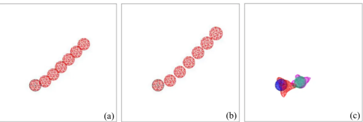
InFig. 2a and b the progression of a cell over a flat substrate in presence of an external source placed at the upper right corner of the substrate is presented at different steps from 0s to 900s. More specifi-cally,Fig. 2a shows the migration of the cell when the frontal and the rear adhesion surfaces are spatially symmetric, whereas inFig. 2b a spatial asymmetry is introduced and the rear adhesion surface is highly reduced in order to enhance the migration as experimentally observed [46–48]. The difference between the two simulations can be noticed in terms of total covered distance (100 μm (Fig. 2a) versus 130 μm (Fig. 2b)) and of migration speed and more particularly the speed during the protrusion phase (5.7 μm/mn (Fig. 2a) versus 8.2 μm/mn (Fig. 2b)). Additionally, for the asymmetric case a higher adhesion force is found at the front, which allows the cell to stick on the substrate and pull its back forward [25].
Fig. 2c presents the migration of a cell with multiple pseudopodia. In fact, in Ref. [24] two modes of motility are explored by which the cell is able to develop several false feet simultaneously (temporal sen-sing model [49]) or one at the time (spatial sensing model [50]) ac-cording to the external random source.
3.2. ‘Obstacles’ and durotaxis
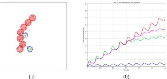
Very often cell migration takes place in heterogeneous environ-ments. As presented in Ref. [24], the cell moves over flat substrates where two sticking regions are introduced, which are supposed to in-hibit the efficiency of the cell (i.e. decrease of the migration speed) and in presence of an external source placed at the upper right corner of the substrate. Then, the cell can adopt two different strategies in order to avoid such ‘obstacles’: the ‘run-and-tumble’ [51] and the ‘look-and-run’ strategy [25]. In the former case, the cell is equipped with a velocity sensor, which allows detecting the decrease in speed as soon as the cell approaches the sticking region. In the latter case, the cell is equipped with a distance sensor, which enables to measure a priori the distance between the centre of the ‘obstacle’ and the cell centre of inertia and to program the path to take in order to perfectly avoid the ‘obstacles’ and reach the external source (Fig. 3a). In the case of the ‘look-and-run’ strategy the variation of the direction of migration is much smoother than in the case of the ‘run-and-tumble’ strategy since the cell is able ‘to see’ the ‘obstacle’ in the distance and adjust its path in advance. Ad-ditionally, for the ‘run-and-tumble’ case the cell slowly migrates across
the ‘obstacle’ and does not completely avoid it.
Substrate heterogeneity can also be provided by a difference in ri-gidity. In this specific case, migration occurs in presence of durotaxis, which orients the actin filaments and their polymerization along stiff-ness gradient of the ECM. More specifically, it has been observed that cells migrate more efficiently over stiff substrates than soft ones [52]. In Ref. [35] such a phenomenon is explored and several simulations are proposed in order to analyse the cell response to different substrate rigidities. InFig. 3b the total covered distance of the cell is reported for migration over homogeneous soft (blue line) and stiff substrate (red line) with external source at 0° and over heterogeneous substrates (i.e. gradient from stiff to soft) with an external source at 0° (green line) and at 45° (purple line). One can notice that the cell does not move over the soft substrate, which also coincides with the absence of polarity of the actin filaments (i.e. isotropic morphology and actin filaments radially distributed around the nucleus). However, the cell is much more effi-cient over stiff regions allowing the actin filaments to orient in the direction of the attractant signal (i.e. anisotropic morphology). Never-theless, as soon as the cell approaches the softer regions, it starts to pulse on place and a plateau is observed for the displacement (green and purple lines).
3.3. Migration under confinement
Migration under confinement is highly influenced by the ECM morphology. In fact, the cell must squeeze its body according to the entanglement and the size of the pores within the fibers network. In Refs. [37,38], numerical simulations are presented to reproduce cell migration across micro-channels of different size, from sub-cellular to sub-nuclear. For these simulations, the cell is constituted by both the cytoplasm and the nucleus and has a diameter of 16 μm, whereas the nucleus has a diameter of 8 μm. InFig. 4a and b, successive steps of migration through a sub-cellular (12 μm) and a sub-nuclear (7 μm) micro-channel are shown. Such results have allowed to extract the mechanical conditions determining the cell behaviour. For the cell to be invasive (i.e. cell able to enter the micro-channel but stuck in the middle),i) the adhesion force between the cell and the substrate must be higher than the contact force between the cell and the walls of the channel and ii) the cell protrusion length inside the micro-channel must be larger than half the micro-micro-channel width. These two conditions need to be satisfied for the cell to be permeative (i.e. cell able to reach the opposite side of the micro-channel) and additionally the contact force between the cell and the micro-channel must be higher around the nucleus since it is the stiffest cellular component.
In Ref. [38] it has been shown that for highly sub-nuclear micro-channels (4 μm), the cell is completely blocked at the entrance. The only way for the cell to be able to invade the micro-channel and migrate across it is to ablate the lamina. From a numerical point of view, this Fig. 2. Cell migration over flat substrates in presence of an external source place at the upper right corner of the substrate. The cell is shown at different steps of the
simulation from 0s to 900s. (a) Spatial symmetry between the frontal and the rear adhesion surfaces, (b) spatial asymmetry between the frontal and the rear adhesion surfaces and (c) migration with multiple pseudopodia.
consists in decreasing its Young modulus as presented inFig. 4c. As it can be observed, the cell becomes permeative and reaches the opposite side of the micro-channel and both the cytoplasm and the nucleus are completely squeezed. This outcome is very interesting as a similar be-haviour has been experimentally observed in migrating mammalian cells where a transient opening of the laminais caused by nuclear de-formation and is rapidly repaired [53]. Such a phenomenon may have important effects on normal and pathological immune responses and confirm that survival of leukocytes strongly depends on efficient nu-clear lamina and DNA repair machineries.
4. Discussion
In the present paper I have proposed an overview of recent nu-merical results on single cell migration in different contexts such as durotaxis and confinement, which may be observed during several biological phenomena. The computational approach used for the dif-ferent models has allowed identifying the mechanical conditions en-hancing or inhibiting cell migration in terms of efficiency (i.e. dis-placement and speed) and of developed forces. From a quantitative point of view, the numerical results are consistent with the experi-mental data that may be found in the literature. This confirms that cell mechanics constitutes an essential factor in cell migration together with molecular, genetic and chemical frameworks. The robustness of the models have been tested and discussed in previous papers, and further improvements may be envisaged in order to be able to reproduce spe-cific experimental set ups.
References
[1] Flaherty B, McGarry JP, McHugh PE. Mathematical models of cell motility. Cell Biochem Biophys 2007;49:14–28.
[2] Borisy GG, Svitkina TM. Acting machinery: pushing the envelope. Curr Opin Cell Biol 2000;12:104–12.
[3] Hoeller O, Kay RR. Chemotaxis in the absence of PIP3 gradients. Curr Biol CB 2007;17:813–7.
[4] Bahat A, Eisenbach M. Sperm thermotaxis. Mol Cell Endocrinol 2006;252:115–9. [5] Andrew N, Insall RH. Chemotaxis in shallow gradients is mediated independently of
PtdIns 3-kinase by biased choices between random protrusions. Nat Cell Biol 2007;9:193–200.
[6] Bosgraaf L, Van Haastert PJM. Navigation of chemotactic cells by parallel signaling to pseudopod persistence and orientation. PLoS One 2009;4. e6842.
[7] Karsenti E. Self-organization in cell biology: a brief history. Nat Rev Mol Cell Biol 2008;9:255–62.
[8] Jay PY, Pham PA, Wong SA, Elson EL. A mechanical function of myosin II in cell motility. J Cell Sci 1995;108:387–93.
[9] Merkel R, Simson R, Simson DA, Hohenadl M, Boulbitch A, Wallraff E, et al. A micromechanic study of cell polarity and plasma membrane cell body coupling in Dictyostelium. Biophys J 2000;79:707–19.
[10] Balaban NQ, Schwarz US, Riveline D, Goichberg P, Tzur G, Sabanay I, et al. Force and focal adhesion assembly: a close relationship studied using elastic micro-patterned substrates. Nat Cell Biol 2001;3:466–72.
[11] Tan JL, Tien J, Pirone DM, Gray DS, Bhadriraju K, Chen CS. Cells lying on a bed of microneedles: an approach to isolate mechanical force. Proc Natl Acad Sci U S A 2003;100:1484–9.
[12] Nicolas A, Geiger B, Safran SA. Cell mechanosensitivity controls the anisotropy of focal adhesions. Proc Natl Acad Sci U S A 2004;101:12520–5.
[13] Shemesh T, Geiger B, Bershadsky AD, Kozlov MM. Focal adhesions as mechan-osensors: a physical mechanism. Proc Natl Acad Sci U S A 2005;102:12383–8. [14] Kobayashi T, Sokabe M. Sensing substrate rigidity by mechanosensitive ion
chan-nels with stress fibers and focal adhesions. Curr Opin Cell Biol 2010;22:669–76. [15] Saez A, Ghibaudo M, Buguin A, Silberzan P, Ladoux B. Rigidity-driven growth and Fig. 3. (a) Cell migration over a flat substrate in presence of two sticking regions. Different time steps are represented from 0s to 900s. (b) Migration in presence of
durotaxis and more specifically: homogeneous soft substrate (blue line), homogeneous stiff substrate (red line), heterogeneous substrate with external source at 0° (green line) and heterogeneous substrate with external source at 45° (purple line).
Fig. 4. Cell migration through micro-channels with sub-cellular (12 μm) (a) and
sub-nuclear width (7 μm) (b). (c) Migration through a highly sub-nuclear micro-channel (2 μm) when the nuclear lamina has been ablated (blue = cytoplasm; red = nucleus).
migration of epithelial cells on microstructured anisotropic substrates. Proc Natl Acad Sci Unit States Am 2007;104:8281–6.
[16] Trichet L, Digabel JL, Hawkins RJ, Vedula SRK, Gupta M, Ribrault C, et al. Evidence of a large-scale mechanosensing mechanism for cellular adaptation to substrate stiffness. Proc Natl Acad Sci Unit States Am 2012;109:6933–8.
[17] Raab M, Swift J, Dingal PCDP, Shah P, Shin J-W, Discher DE. Crawling from soft to stiff matrix polarizes the cytoskeleton and phosphoregulates myosin-II heavy chain. J Cell Biol 2012;199:669–83.
[18] Zemel A, Safran SA. Active self-polarization of contractile cells in asymmetrically shaped domains. Phys Rev E - Stat Nonlinear Soft Matter Phys 2007;76:021905. [19] De R, Zemel A, Safran SA. Do cells sense stress or strain? Measurement of cellular
orientation can provide a clue. Biophys J 2008;94(5):L29–31.
[20] Friedl P, Wolf K. Plasticity of cell migration: a multiscale tuning model. J Cell Biol 2010;188:11–9.
[21] Erler JT, Weaver VM. Three-dimensional context regulation of metastasis. Clin Exp Metastasis 2009;26:35–49.
[22] Wolf K, Alexander S, Schacht V, Coussens LM, von Andrian UH, van Rheenen J, et al. Collagen-based cell migration models in vitro and in vivo. Semin Cell Dev Biol 2009;20:931–41.
[23] Egeblad M, Rasch MG, Weaver VM. Dynamic interplay between the collagen scaf-fold and tumor evolution. Curr Opin Cell Biol 2010;22:697–706.
[24] Allena R. Cell migration with multiple pseudopodia: temporal and spatial sensing models. Bull Math Biol 2013;75:288–316.
[25] Allena R, Aubry D. “Run-and-tumble” or “look-and-run”? A mechanical model to explore the behavior of a migrating amoeboid cell. J Theor Biol 2012;306:15–31. [26] Bottino D, Mogilner A, Roberts T, Stewart M, Oster G. How nematode sperm crawl.
J Cell Sci 2002;115:367–84.
[27] Mogilner A, Verzi DW. A simple 1-D physical model for the crawling nematode sperm cell. J Stat Phys 2003;110:1169–89.
[28] Rubinstein B, Jacobson K, Mogilner A. Multiscale two-dimensional modeling of a motile simple-shaped cell. Multiscale Model Simul 2005;3:413–39.
[29] Sakamoto Y, Prudhomme S, Zaman MH. Viscoelastic gel-strip model for the simu-lation of migrating cells. Ann Biomed Eng 2011;39:2735–49.
[30] Taber LA, Shi Y, Yang L, Bayly PV. A poroelastic model for cell crawling including mechanical coupling between cytoskeletal contraction and actin polymerization. J Mech Mater Struct 2011;6:569–89.
[31] Moreo P, García-Aznar JM, Doblaré M. Bone ingrowth on the surface of endosseous implants. Part 2: theoretical and numerical analysis. J Theor Biol 2009;260:13–26. [32] Dokukina IV, Gracheva ME. A model of fibroblast motility on substrates with
dif-ferent rigidities. Biophys J 2010;98:2794–803.
[33] Harland B, Walcott S, Sun SX. Adhesion dynamics and durotaxis in migrating cells. Phys Biol 2011;8:015011.
[34] Allena R, Scianna M, Preziosi L. A Cellular Potts Model of single cell migration in presence of durotaxis. Math Biosci 2016;275:57–70.
[35] Aubry D, Gupta M, Ladoux B, Allena R. Mechanical link between durotaxis, cell
polarity and anisotropy during cell migration. Phys Biol 2015;12:026008. [36] Stefanoni F, Ventre M, Mollica F, Netti PA. A numerical model for durotaxis. J
Theor Biol 2011;280:150–8.
[37] Allena R. Mechanical modelling of confined cell migration across constricted-curved micro-channels. Mol Cell Biomech MCB 2014;11:185–208.
[38] Aubry D, Thiam H, Piel M, Allena R. A computational mechanics approach to assess the link between cell morphology and forces during confined migration. Biomechanics Model Mechanobiol 2015;14:143–57.
[39] Hawkins RJ, Voituriez R. Mechanisms of cell motion in confined geometries. Math Model Nat Phenom 2010;5:84–105.
[40] Hawkins RJ, Piel M, Faure-Andre G, Lennon-Dumenil AM, Joanny JF, Prost J, et al. Pushing off the walls: a mechanism of cell motility in confinement. Phys Rev Lett 2009;102:058103.
[41] Scianna M, Preziosi L. Modeling the influence of nucleus elasticity on cell invasion in fiber networks and microchannels. J Theor Biol 2013;317:394–406.
[42] Tozluoğlu M, Tournier AL, Jenkins RP, Hooper S, Bates PA, Sahai E. Matrix geo-metry determines optimal cancer cell migration strategy and modulates response to interventions. Nat Cell Biol 2013;15:751–62.
[43] Gracheva ME, Othmer HG. A continuum model of motility in ameboid cells. Bull Math Biol 2004;66:167–93.
[44] Allena R, Mouronval A-S, Aubry D. Simulation of multiple morphogenetic move-ments in the Drosophila embryo by a single 3D finite element model. J Mech Behav Biomed Mater 2010;3:313–23.
[45] Lubarda V. Constitutive theories based on the multiplicative decomposition of de-formation gradient: thermoelasticity, elastoplasticity, and biomechanics. Appl Mech Rev 2004;57:95–109.
[46] Lämmermann T, Sixt M. Mechanical modes of “amoeboid” cell migration. Curr Opin Cell Biol 2009;21:636–44.
[47] Lauffenburger DA, Horwitz AF. Cell migration: a physically integrated molecular process. Cell 1996;84:359–69.
[48] Zaman MH, Kamm RD, Matsudaira P, Lauffenburger DA. Computational model for cell migration in three-dimensional matrices. Biophys J 2005;89:1389–97. [49] Gerish G, Malchow D, Hess B. Cell communication and cyclic-amp regulation during
aggregation of the slime mold, dictyostelium discoideum. Biochem Sens Funct 1974;25:279–98.
[50] Zigmond SH, Levitsky HI, Kreel BJ. Cell polarity: an examination of its behavioral expression and its consequences for polymorphonuclear leukocyte chemotaxis. J Cell Biol 1981;89:585–92.
[51] Erban R, Othmer HG. Taxis equations for amoeboid cells. J Math Biol 2007;54:847–85.
[52] Lo CM, Wang HB, Dembo M, Wang YL. Cell movement is guided by the rigidity of the substrate. Biophys J 2000;79:144–52.
[53] Raab M, Gentili M, Belly H de, Thiam HR, Vargas P, Jimenez AJ, et al. ESCRT III repairs nuclear envelope ruptures during cell migration to limit DNA damage and cell death. Science 2016;352(6283):359–62.