Université de Montréal
Quantitative functional neuroimaging of cerebral
physiology in healthy aging
par
Claudine Gauthier
Département de Physiologie Faculté de Médecine
Thèse présentée à la Faculté de Médecine en vue de l’obtention du grade de Doctorat
en Sciences neurologiques
Août 2012
© Claudine Gauthier, 2012
Université de Montréal
Faculté des études supérieures et postdoctorales
Cette thèse intitulée :
Quantitative functional neuroimaging of cerebral physiology in healthy aging
Présentée par : Claudine Gauthier
a été évaluée par un jury composé des personnes suivantes :
Paul Cisek, président-rapporteur Richard D. Hoge, directeur de recherche
Sylvie Belleville, membre du jury Beau Ances, examinateur externe
Florin Amzica, représentant du doyen de la FES
Résumé
Les études d’imagerie par résonance magnétique fonctionnelle (IRMf) ont pour prémisse générale l’idée que le signal BOLD peut être utilisé comme un
succédané direct de l’activation neurale. Les études portant sur le vieillissement cognitif souvent comparent directement l’amplitude et l’étendue du signal BOLD entre des groupes de personnes jeunes et âgés. Ces études comportent donc un a
priori additionnel selon lequel la relation entre l’activité neurale et la réponse
hémodynamique à laquelle cette activité donne lieu restent inchangée par le
vieillissement. Cependant, le signal BOLD provient d’une combinaison ambiguë de changements de métabolisme oxydatif, de flux et de volume sanguin. De plus, certaines études ont démontré que plusieurs des facteurs influençant les propriétés du signal BOLD subissent des changements lors du vieillissement. L’acquisition d’information physiologiquement spécifique comme le flux sanguin cérébral et le métabolisme oxydatif permettrait de mieux comprendre les
changements qui sous-tendent le contraste BOLD, ainsi que les altérations physiologiques et cognitives propres au vieillissement. Le travail présenté ici démontre l’application de nouvelles techniques permettant de mesurer le métabolisme oxydatif au repos, ainsi que pendant l’exécution d’une tâche. Ces techniques représentent des extensions de méthodes d’IRMf calibrée existantes. La première méthode présentée est une généralisation des modèles existants pour l’estimation du métabolisme oxydatif évoqué par une tâche, permettant de prendre en compte tant des changements arbitraires en flux sanguin que des changements en concentrations sanguine d’O2. Des améliorations en terme de robustesse et de précisions sont démontrées dans la matière grise et le cortex visuel lorsque cette méthode est combinée à une manipulation respiratoire incluant une composante d’hypercapnie et d’hyperoxie. Le seconde technique présentée ici est une
extension de la première et utilise une combinaison de manipulations respiratoires incluant l’hypercapnie, l’hyperoxie et l’administration simultanée des deux afin d’obtenir des valeurs expérimentales de la fraction d’extraction d’oxygène et du métabolisme oxydatif au repos. Dans la deuxième partie de cette thèse, les changements vasculaires et métaboliques liés à l’âge sont explorés dans un
groupe de jeunes et aînés, grâce au cadre conceptuel de l’IRMf calibrée, combiné à une manipulation respiratoire d’hypercapnie et une tâche modifiée de Stroop. Des changements de flux sanguin au repos, de réactivité vasculaire au CO2 et de paramètre de calibration M ont été identifiés chez les aînés. Les biais affectant les mesures de signal BOLD obtenues chez les participants âgés découlant de ces changements physiologiques sont de plus discutés. Finalement, la relation entre ces changements cérébraux et la performance dans la tâche de Stroop, la santé vasculaire centrale et la condition cardiovasculaire est explorée. Les résultats présentés ici sont en accord avec l’hypothèse selon laquelle une meilleure condition cardiovasculaire est associée à une meilleure fonction vasculaire centrale, contribuant ainsi à l’amélioration de la santé vasculaire cérébrale et cognitive.
mots-clés:
IRMf calibré, métabolisme oxydatif, vieillissement, santé vasculaire, flux sanguin cérébral, condition cardiovasculaire, cognition
Abstract
Functional MRI (fMRI) studies using the BOLD signal are done under the general assumption that the BOLD signal can be used as a direct index of neuronal activation. Studies of cognitive aging often compare BOLD signal amplitude and extent directly between younger and older groups, with the additional assumption that the relationship between neuronal activity and the hemodynamic response is unchanged across the lifespan. However, BOLD signal arises from an ambiguous mixture of changes in oxidative metabolism, blood flow and blood volume.
Furthermore, previous studies have shown that several BOLD signal components may be changed during aging. More physiologically-specific information on blood flow and oxidative metabolism would allow a better understanding of these signal changes and of the physiological and cognitive changes seen with aging. The work presented here demonstrates techniques to estimate oxidative metabolism at rest and during performance of a task. These techniques are extensions of previous calibrated fMRI methods and the first method presented is based on a
generalization of previous models to take into account both arbitrary changes in blood flow and blood O2 content. The improved robustness and accuracy of this method, when used with a combined hypercapnia and hyperoxia breathing manipulation, is demonstrated in visual cortex and grey matter. The second technique presented builds on the generalization of the model and uses a
combination of breathing manipulations including hypercapnia, hyperoxia and both simultaneously, to obtain experimentally-determined values of resting oxygen extraction fraction and oxidative metabolism. In the second part of this thesis, age-related vascular and metabolic changes are explored in a group of younger and older adults using a calibrated fMRI framework with a hypercapnia breathing manipulation and a modified Stroop task. Changes in baseline blood flow, vascular reactivity to the CO2 challenge and calibration parameter M were identified in the older participants. Potential biases in BOLD signal measurements in older adults arising from these physiological changes are discussed. Finally, the relationship between these cerebral changes and performance on the modified Stroop task, central vascular health and cardiovascular fitness are explored. The results of this
thesis support the hypothesis that greater cardiovascular fitness is associated with improvements in central vascular function, contributing in turn to improved brain vascular health and cognition.
Keywords: calibrated fMRI, oxidative metabolism, aging, vascular health, cerebral blood flow, cardiovascular fitness, cognition.
Table of contents
List of tables
!
16
List of figures
!
17
Acknowledgments
!
20
Abbreviations and acronyms
!
22
Preface
!
25
1.Introduction!28
1.1. Vascular and metabolic changes with aging!28 1.1.1. Vascular aging in the brain!31
1.1.2. Mitochondrial theory of aging!32 1.2. Aging and physical fitness!34
1.3. Aging and cognition!37
1.3.1. Mechanisms leading to changes in activation patterns!40 1.3.2. Cognitive theories!41
1.4. The BOLD contrast mechanism!44
1.4.1. Underlying neuronal activity!45 1.4.2. Oxidative metabolism!47
1.4.3. Neurovascular coupling!49
1.4.4. Blood flow response!53 1.4.5. Blood volume!56
1.4.6. Limitations of the BOLD signal!57
1.4.6.1.Confounds in aging studies!57
1.5. Calibrated fMRI techniques!59
1.5.1. Hypercapnia calibration method!59 1.5.2. Hyperoxia calibration method!63 1.5.3. R2' method!65
1.6. Baseline CMRO2!65
1.7. Quantitative studies of brain aging!70
2.A generalized procedure for calibrated MRI incorporating
hyperoxia and hypercapnia
!
77
2.1. Preface!77 2.2. Abstract !78 2.3. Introduction!79 2.3.1. Theory!81 2.4. Methods!84 2.4.1. Image acquisition!84 2.4.2. Manipulations!85 2.4.2.1.Visual stimulus!85 2.4.2.2.Gas manipulations!85 2.4.3. Data analysis!86 2.4.3.1.Visual!88 2.4.3.2.Grey matter! 882.4.3.3.Analysis of sensitivity to errors in CBF!88
2.5. Results!89 2.6. Discussion!98
2.6.1. Estimation of M parameter!98
2.6.1.1.ROI analysis!99
2.6.1.2.Susceptibility-weighted venograms!99 2.6.1.3.M mapping!100
2.6.1.4.Spatial heterogeneity of M values!100 2.6.1.5.Robustness of M estimates!101
2.6.2. CMRO2!103
2.6.3. Limitations of the study!103
2.6.3.1.M value confounds!104
2.7. Conclusion!106
2.8. Acknowledgements!106 2.9. Appendix!107
2.9.1. Davis model with hypercapnia manipulation!108 2.9.2. Chiarelli model with hyperoxia manipulation!108
2.9.3. Generalized calibration model (GCM) with hyperoxia and hypercapnia!109
2.9.4. Stability of the models during hypercapnic manipulations!110 2.9.5. Stability of the models during hyperoxia manipulations!111
3.Magnetic resonance imaging of resting OEF and CMRO2 using a
generalized calibration model for hypercapnia and hyperoxia
!
113
3.1. Preface!113 3.2. Abstract !114 3.2.1. Keywords!115 3.3. Introduction!115 3.3.1. Theory!1173.3.1.1.Determination of resting BOLD signal M and OEF!118 3.3.1.2.Determination of resting CMRO2!120
3.4. Methods!121
3.4.1. Image acquisition!121
3.4.2. Gas manipulations!122 3.4.3. Data analysis!123 3.5. Results!128 3.5.1. Gas manipulations!128 3.5.2. ROI analyses!129 3.5.2.1.MRI responses!129
3.5.2.2.Group average M vs. OEF0 curves!129
3.5.2.3.Individual grey matter ROI values of OEF0, M, and CMRO2!130
3.5.3. Parametric maps!132
3.5.3.1.Resting BOLD signal M!132
3.5.3.2.Resting oxygen extraction fraction OEF0!132 3.5.3.3.Resting oxygen metabolism CMRO2!134 3.5.3.4.Group average maps!134
3.5.3.5.Sensitivity to model parameters, possible CMRO2 changes during hypercapnia!134 3.6. Discussion!136 3.6.1. Accuracy!136 3.6.2. Stability!143 3.6.3. Similar methods!144 3.6.4. Applications!144 3.7. Conclusion!146 3.8. Acknowledgements!146
4.Age-dependence of hemodynamic response characteristics in
human fMRI
!
148
4.1. Preface!148 4.2. Abstract !149 4.3. Introduction!150 4.4. Methods!153 104.4.1. Participants!153 4.4.2. MR Image acquisition!155 4.4.3. Hypercapnic manipulation!156 4.4.4. Stroop task!156 4.4.5. Data analysis!158 4.4.6. Regions-of-interest definition!161 4.4.7. Statistical analysis!162 4.5. Results!164
4.5.1. Modified Stroop task behavioral results!164 4.5.2. Modified Stroop activation maps!165
4.5.3. Stroop evoked responses!169 4.5.4. Respiratory manipulation!170 4.5.5. Vascular parameters!171 4.5.6. Calibrated fMRI estimates!175 4.5.7. Temporal SNR!176
4.6. Discussion!179
4.6.1. BOLD measurement of the cognitive task!179
4.6.2. Blood flow measurements during the cognitive task!181 4.6.3. Hypercapnic responses!181
4.6.4. Calibrated fMRI measurements!182 4.6.5. Potential confounds!183 4.6.6. Limitations!186 4.6.7. Future directions!187 4.7. Conclusion!188 4.8. Acknowledgments!188 11
5.Relationship between cardiovascular health, cerebral
physiology and cognition in healthy aging
!
189
5.1. Preface!189 5.2. Abstract !190 5.3. Introduction!191
5.3.1. Effects of cardiorespiratory fitness!192 5.4. Methods!193
5.4.1. Participants!193
5.4.2. Maximal continuous graded exercise test !193 5.4.3. Modified Stroop task!194
5.4.4. MR Image acquisition!194 5.4.4.1.Brain exam! 194 5.4.4.2.Aortic exam! 195 5.4.5. Tonometry!195 5.4.6. Hypercapnic manipulation!195 5.4.7. Data analysis!196 5.4.7.1.Brain data!196 5.4.7.2.Aortic data!196 5.4.8. Statistical analysis!196 5.5. Results!197
5.5.1. Central vascular health and fitness!197 5.5.2. Cerebral measures!198
5.5.3. Correlations between parameters!201 5.6. Discussion!202
5.6.1. Differences between age groups!202 5.6.2. Correlations within older participants!203 5.7. Conclusion!205
5.8. Acknowledgments!206
6.General Discussion!207
6.1. Calibrated fMRI method comparisons!208 6.2. Accuracy of measurement!209
6.3. Baseline metabolism!211 6.3.1. QUO2 technique!212 6.3.2. Future improvements!214 6.4. Calibrated fMRI of aging!215
6.4.1. Calibrated fMRI of aging literature!216 6.4.2. Future improvements!217
6.5. Vascular changes with aging!219 6.5.1. Future improvements!221 6.6. Future studies!222
Conclusion
!
224
G.Appendix - Elimination of visually evoked BOLD responses
during carbogen inhalation: implications for calibrated MRI!225
G.1. Abstract !225 G.2. Introduction!226 G.3. Methods!229 G.3.1. Gas manipulations!232 G.3.2. Data analysis!233 G.4. Results!235 G.4.1. Additional tests!239
G.4.2. Subjective discomfort rating!241 G.5. Discussion!241
G.5.1. Arterial spin-labeling!242
G.5.2. Susceptibility-weighted imaging!243 G.5.3. Implications for calibrated MRI!243
G.5.3.1.Pitfalls and future directions!246
G.6. Conclusion!248
G.7. Acknowledgments!249 G.8. Appendix!249
H.Appendix - Absolute quantification of resting oxygen
metabolism and metabolic reactivity during functional
activation using QUO2 MRI!253
H.1. Abstract !253 H.2. Introduction!254 H.3. Methods!256 H.3.1. Image acquisition!256 H.3.2. Gas manipulations!257 H.3.3. Visual stimulus!258 H.3.4. Stroop task!258 H.3.5. Data analysis!259 H.3.6. Visual ROI!262
H.3.7. Stroop ROIs definition!262 H.4. Results!265
H.4.1. Gas manipulations!265
H.4.2. M, OEF0 and resting CMRO2!266 H.4.3. Task-evoked CMRO2!268
H.4.4. Vascular and metabolic profile!269 H.5. Discussion!270
H.5.1. Accuracy of estimates!271
H.5.1.1.Visual!271 H.5.1.2.Stroop!273
H.5.2. Implementation biases and confounds!276 H.6. Conclusion!280 H.7. Acknowledgments!280
Bibliography
!
281
15List of tables
Chapter 2 A generalized procedure for calibrated MRI incorporating hyperoxia and hypercapnia
Table 1 End-tidal values for each breathing manipulation 89
Table 2 Calibrated fMRI results 98
Chapter 3 Magnetic resonance imaging of resting OEF and CMRO2
using a generalized calibration model for hypercapnia and hyperoxia
Table 1 128
Table 2 131
Chapter 4 Age-dependence of hemodynamic response characteristics in human fMRI
Table 1 Neuropsychological battery results 164
Table 2 ROI quantification results 178
Chapter 5 Relationship between cardiovascular health, cerebral physiology and cognition in healthy aging
Table 1 Partial correlations between health indices in the older subjects 201 Appendix A Elimination of visually evoked BOLD responses during
carbogen inhalation: implications for calibrated MRI
Table 1 Gas mixture compositions 231
Table 2 Literature M values at 3 Tesla 245
Appendix B Absolute quantification of resting oxygen metabolism and metabolic reactivity during functional activation using QUO2 MRI
Table 1 Respiratory parameters 265
Table 2 Vascular and metabolic profile 270
List of figures
Chapter 1 Introduction
Figure 1 Near-infrared multiphoton LSM 30
Figure 2 Mitochondrial energy production 33
Figure 3 Exercise and cognition 37
Figure 4 Reliable patterns of brain activity across studies of aging 39
Figure 5 A conceptual model of the scaffolding theory of aging and cognition (STAC)
44
Figure 6 Electrical underpinnings of the BOLD signal 46
Figure 7 Neurovascular unit 50
Figure 8 Neurovascular coupling response 52
Figure 9 Tag delivery in ASL during normocapnia and hypercapnia 55
Figure 10 Example case of BOLD comparisons across lifespan 58
Figure 11 BOLD attenuation from metabolism 60
Figure 12 Deoxyhemoglobin dilution model 61
Figure 13 QUIXOTIC 68
Figure 14 Calibrated fMRI of visual activation in aging 71
Figure 15 Calibrated fMRI of Stroop activation in aging 72
Figure 16 The association between age and regional CBF as well as cortical thickness
73
Figure 17 Whole brain hemodynamic age-related changes 74
Figure 18 Decade-by-decade alterations in prefrontal CVR and CBF 75
Chapter 2 A generalized procedure for calibrated MRI incorporating hyperoxia and hypercapnia
Figure 1 Group average percent CBF and BOLD for each breathing manipulation
90
Figure 2 Individual and average M values over the visual and grey-matter ROI’s for each breathing manipulation
91
Figure 3 Individual M maps for each breathing manipulation 93
Figure 4 Spatial heterogeneity of M maps 94 Figure 5 Susceptibility-weighted venograms during each breathing
manipulation"
95
Figure 6 Effect of CBF errors on M estimates from different models and breathing manipulations
96
Figure 7 Individual and average CMRO2 estimates over the visual ROI 97
Chapter 3 Magnetic resonance imaging of resting OEF and CMRO2
using a generalized calibration model for hypercapnia and hyperoxia Figure 1 130 Figure 2 131 Figure 3 133 Figure 4 134 Figure 5 135 Figure 6 136
Chapter 4 Age-dependence of hemodynamic response characteristics in human fMRI
Figure 1 Modified Stroop task paradigm 158
Figure 2 Modified Stroop behavioral response 165
Figure 3 Modified Stroop Z-score maps 167
Figure 4 Modified Stroop task ROIs 168
Figure 5 Percent BOLD and CBF responses to the modified Stroop task 170
Figure 6 BOLD hypercapnia time courses 172
Figure 7 CBF hypercapnia time courses 173
Figure 8 Baseline CBF and CVR 174
Figure 9 M and CMRO2 values 175
Figure 10 Temporal SNR of CBF and BOLD measurements 177
Chapter 5 Relationship between cardiovascular health, cerebral physiology and cognition in healthy aging
Figure 1 Central vascular health and cardiorespiratory fitness 197
Figure 2 Stroop task performance 198
Figure 3 Cerebral hemodynamic properties 200
Appendix A Elimination of visually evoked BOLD responses during carbogen inhalation: implications for calibrated MRI
Figure 1 Visual cortex regions of interest for each subject 234
Figure 2 Percent BOLD response maps from example subject 236
Figure 3 BOLD Time course for carbogen inhalation and carbogen + visual stimulation
237
Figure 4 Individual BOLD percent changes in visual ROI 238
Figure 5 Group-average BOLD response amplitudes 239
Figure 6 Susceptibility-weighted venogram 240
Figure 7 BOLD and ASL time courses for visual stimulation during carbogen-10 breathing
240
Figure 8 Maps of percent BOLD increase during carbogen-10 breathing 246
Appendix B Absolute quantification of resting oxygen metabolism and metabolic reactivity during functional activation using QUO2 MRI
Figure 1 ROI for Stroop task 264
Figure 2 Percent CBF and BOLD changes during breathing manipulations 266
Figure 3 Intersection plots for all ROIs 267
Figure 4 M, OEF0 and resting CMRO2 values over all ROIs 268
Figure 5 Task-evoked percent CBF and BOLD changes 269
Figure 6 Task-evoked percent CMRO2 change 269
Acknowledgments
First and foremost, I want to thank my supervisor, Rick, for his mentorship, his unwavering support throughout the six years I spent in his lab and all the confidence he placed in me. Being a PI’s first PhD student is something of a special position: both parties come with the enthusiasm and idealism of ignorance and learn together both the harsh reality of their obligations and the enjoyment of their new scientific freedom. I come away with much gratitude for all the freedom, knowledge and extra attention I received. I am looking forward to working again together in the future as collaborators.
En seconde part dans cette liste de remerciement doit venir l’amie, la collègue, la coloc de bureau, la personne qui a partagé tous les hauts et les bas de ce grand défi que fût ce doctorat: Cécile. Sans son support, son travail acharné, sa générosité, son imagination, ses talents de programmation, son écoute et sans son amitié, je n’aurais pas accompli la moitié de ce que j’ai fait. Elle mérite ce doctorat autant que moi et je la remercie du fond du coeur pour l’opportunité fantastique d’avoir pu travailler avec elle. J’aimerais aussi remercier les membres passés et actuels de mon labo. Merci à Paul-Olivier pour son aide sur mon projet et sa personnalité adorable, merci à mes petits frères Brice et Sébastien pour leur enthousiasme et leur écoute, merci à Felipe pour son support affectueux et à Isabelle pour avoir adouci le départ de Cécile. Finalement merci à Louis, Tarik, Lorenzo et Élodie pour leur aide toujours souriante et merci à Anne-Marie pour avoir pourfendu avec vaillance et joie de vivre tous les défis organisationnels de ma soumission et ma défense. J’ai eu la chance de travailler sur un projet fascinant qui m’aura permis de connaître des personnes généreuses et sympathiques dans d’autres labos. Merci à Louis et aux membres de son équipe d’avoir contribué avec tant d’enthousiasme à mon projet. Un merci particulier à Laurence dont la générosité constante et tranquille a grandement contribué au succès de cette épopée, à Saïd pour avoir de son entrain sans faille rendu un test d’effort agréable à tous mes participants, et merci à Mélanie pour les prouesses organisationnelles dont elle a fait preuve. Merci à Frédéric et à son équipe pour leur aide pour tous les aspects d’imagerie optique. Un merci particulier à Michelle et Nicolas pour leur aide constante et toujours enthousiaste. Un grand merci aussi aux gens de l’INSERM de m’avoir accueillie et aidée. Merci à Frédérique et Muriel surtout, qui ont contribué une diligence et un enthousiasme sans faille à mon projet. Merci à Pierre, qui est à la fois un collègue, un ami et une source d’inspiration dans sa ferveur scientifique inépuisable. Un immense merci aux employés de l’UNF qui m’ont fourni un support constant. Merci à
Carollyn et André pour tous les sacrifices de lunch et toute l’aide qu’ils m’ont apportée dans mon doctorat. Merci à Francine d’avoir toléré pendant toutes ces années mon manque d’organisation et d’avoir de si bonne grâce remédié à mes oublis.
On ne peut survivre avec succès les années de doctorat sans le support des amis. Donc un immense merci à mes amies-soeurs: Christine, Hélène, Sigrid et Olivia. Qu’aurais-je fait sans les heures incalculables de conversations, les rires et tout ce que nous avons partagé? Elles sont une partie de moi et je les remercie du fond du coeur d’être les filles fantastiques qu’elles sont. Merci à leurs conjoints et bébés, Vincent, Sebastian, Yannick et Jon qui sont aussi devenus des amis chers. Merci à Clarisse et Jean pour m’avoir adoptée quand j’étais seule aux conférences et pour m’avoir ensuite donné une amitié précieuse. Merci aussi à Jenny, Gab, Catherine, ‘la gang’ et Charlotte, pour une petite baignade rafraîchissante chez les gens qui vivent dans le vrai monde et en font une place qui en vaut la peine. Thanks to Dimo and Henrik for their conference-started friendship and warm welcome on my post-doc tour. Merci à Anne-Julie pour les matinées d’écriture et l’amitié renouvelée. Merci aux nouveaux amis qui ont ensoleillé ma dernière année: Josh, Dom, Eric, Desirée, Gonzalo, Anita et Mark. Merci à Tarek pour toutes les discussions uniques qui ont permis l’envol de mon imagination. Merci à Patrick pour son support et pour avoir stimulé ma fibre lyrique. Et finalement merci à Marie-Claude, Aaron, Zoë et James pour ces visites de la dernière année qui m’ont tant aidée à garder les pieds sur terre et à profiter de la vie.
Je tiens à dire deux mercis vraiment très spéciaux pour moi: merci à Nicolas et Julien qui ont été pendant cette aventure une source de support vraiment précieux. Merci à Nicolas qui m’a donné le support dont j’avais besoin pour entreprendre la grande traversée. Nos discussions m’ont fait grandir et son amitié a fait de moi une personne mille fois plus forte et belle. Merci à sa famille aussi, qui m’a adoptée comme une des leur et dont le support m’a touchée énormément. Merci à Julien qui a été un grand-frère scientifique
incomparable, mais surtout un ami sincère et constant. Merci pour son humour qui fait scintiller la vie et pour son écoute dans tous les moments difficiles.
Merci à ma famille, qui a cru plus que tous à ma capacité à me dépasser, peu importe le but que je m’étais fixé. Merci à ma mère, mon père, Laurent, Mathilde, Aline et Diane pour leur fierté, leur confiance et leur patience. And finally, I want to thank Chris who allowed me by his constant support and affection to get to the end of this last demanding year with all the grace and happiness possible. I am looking forward to the next step of this
adventure together.
Abbreviations and acronyms
AD Alzheimer’s disease
AI Augmentation index
AMPA 2-amino-3-(5-methyl-3-oxo-1,2-oxazol-4-yl)propanoic acid ASL/PASL/
pCASL Arterial spin labeling/Pulsed ASL/Pseudo-continuous ASL
ATP Adenosine triphosphate
BDNF Brain-derived neurotrophic factor BOLD Blood oxygen level-dependent CaO2 Arterial content of O2
CBF Cerebral blood flow
CBV Cerebral blood volume
CMRO2 Cerebral metabolic rate of O2 CPMG Carr Purcell Meiboom Gill
CRP C-reactive protein
CRUNCH Compensation-Related Utilization of Neural Circuits CvO2 Venous content of O2
CVR Cerebrovascular reactivity ETO2/ETCO2 End-tidal O2/CO2 concentration
FDG Fluoro-D-glucose
FLAIR Fluid attenuated inversion recovery fMRI Functional magnetic resonance imaging FWHM Full width at half maximum
GABA gamma-Aminobutyric acid
GCM Generalized calibration model
GESSE Gradient echo sampling of the spin echo
GLM General linear model
GM Grey matter
GRAPPA Generalized autocalibrating partially parallel acquisition HAROLD Hemispheric asymmetry reduction in older adults Hb/dHb Hemoglobin/Deoxyhemoglobin
HO/HC Hyperoxia/Hypercapnia
HRF Hemodynamic response function
LFP Local field potentials
M Maximal possible BOLD signal change
MPRAGE Magnetization prepared rapid gradient echo
MRI Magnetic resonance imaging
mtDNA Mitochondrial DNA
MUA Multi-unit activity
n Flow-metabolic coupling constant NADH Nicotinamide adenine dinucleotide NMDA N-Methyl-D-aspartic acid
NO Nitric oxide
OEF Oxygen extraction fraction PaO2 Arterial partial pressure of O2 PASA Posterior anterior shift in aging PET Positron emission tomography
PGE2 Prostaglandin E2
PWV Pulse wave velocity
qBOLD Quantitative BOLD
QUIPSS II Quantitative imaging of perfusion using a single subtraction QUIXOTIC Quantitative imaging of extraction of oxygen and tissue
QUO2 Quantitative O2
ROI Region-of-interest
ROS Reactive oxygen species
RT Reaction time
SaO2 Arterial O2 saturation
SNR Signal-to-noise ratio
STAC Scaffolding theory of aging and cognition SvO2 Venous O2 saturation
T Tesla
T1 Longitudinal relaxation time T2/T2* Transverse relaxation time
TE Echo time
TR Repetition time
TRUST T2 relaxation under spin tagging
TSE Turbo spin echo
tSNR Temporal signal-to-noise ratio
VASO Vascular space occupancy
VERVE Venous refocussing for volume estimation
VO2max Maximal volume of O2 consumed during exercise
Preface
Longevity is a vascular question. A man is only as old as his arteries.
" " " " " " " " " -- Dr William Osler Aging is associated with a variety of cognitive and behavioral changes. In order to understand the cognitive changes associated with normal and pathological aging, we need to investigate the anatomical and physiological correlates of these
changes. While cortical atrophy and white matter lesions are known to occur at the later stages in life even in healthy older adults, functional changes that go beyond the straight-forward effects of the loss of grey-matter can also be observed. However, while the study of the anatomical changes associated with aging is becoming well established, the investigation of dynamic changes in the brain still remains a technical challenge. The technique of choice to study brain function non-invasively is functional magnetic resonance imaging (fMRI) using the blood oxygen level-dependent (BOLD) contrast. This technique’s popularity can be attributed to the fact that it is fairly simple to implement on any modern MRI scanner and is sensitive to the local activity of brain regions at the millimeter scale. Thanks to these characteristics, it can yield robust results with adequate spatial and temporal resolution that allow inferences to be made about local brain function.
The effect of aging on BOLD signal changes evoked by a variety of tasks has been the subject of intense research in recent years. However, while BOLD contrast is a useful measure of local brain activity, the information that can be derived from it is intrinsically ambiguous in nature. This is because the BOLD signal arises from an ambiguous combination of changes in oxidative metabolism, blood flow and blood volume. Furthermore, the BOLD signal is measured as a relative change from an unknown baseline. These characteristics call for caution when comparing BOLD signal changes between groups, especially when either the baseline or the
dynamic range of any of its component is suspected to differ between them. Aging is known to be associated with vascular and metabolic changes that can potentially influence all components of the BOLD signal. Therefore, while BOLD studies of aging can be helpful in identifying potential loci of task-related differences between
young and old, more physiologically-specific information is needed to understand the underlying differences in neuronal usage.
The work presented in this thesis was motivated by the need to both characterize the vascular and metabolic changes associated with aging and provide a more physiologically-specific context in which to discuss the functional changes we observe in BOLD studies of cognitive aging. The body of this thesis will be comprised of four manuscripts. Two of these will present the technical and modeling development work done in the course of my project. These techniques were developed in parallel with the aging study forming Chapter 4 of this thesis and therefore represent separate novel findings. The first manuscript, entitled “A
generalized procedure for calibrated MRI incorporating hyperoxia and hypercapnia” presents a new generalized model based on the hypercapnia and hyperoxia
calibration models that can be used with breathing manipulations causing any arbitrary change in cerebral blood flow and arterial oxygen content. This model will be used to perform calibrated fMRI in the aging manuscripts. The second
manuscript is entitled “Magnetic resonance imaging of resting OEF and CMRO2 using a generalized calibration model for hypercapnia and hyperoxia” and demonstrates the use of the generalized model in combination with at least two breathing manipulations to estimate baseline oxygen extraction fraction and oxidative metabolism. The third and fourth manuscripts will focus on quantitative studies of aging. In the third manuscript entitled “Age-dependence of hemodynamic response characteristics in human fMRI”, the changes in hemodynamic
components with aging will be explored. This manuscript explores the potential confounds associated with hemodynamic imaging in aging. The fourth manuscript entitled “Relationship between cardiovascular health, cerebral physiology and cognition in healthy aging” will discuss changes in vascular physiology in the brain and at the level of the aorta, and their link to hemodynamic signal changes will be discussed. Finally, two additional manuscripts will be presented as appendices to this thesis, since they provide additional support for the studies shown here. The first appendix will comprise a manuscript entitled “Elimination of visually evoked BOLD responses during carbogen inhalation: implications for calibrated MRI”, which presents a direct measurement through a breathing manipulation of the
calibration parameter we seek to determine through modeling in standard calibrated fMRI experiments. The second appendix is a manuscript entitled “Absolute quantification of resting oxygen metabolism and metabolic reactivity during functional activation using QUO2 MRI”. This manuscripts demonstrates the comprehensive set of measurements that can be obtained using a combination of all the techniques presented in Chapters 2 and 3. While these were not all applied in our studies of aging, future studies will include them to provide an improved quantification of BOLD signal sub-components.
1. Introduction
Neurological components of aging can be detected by decreased accuracy and speed of cognitive and sensory responses, as well as structural and functional changes. These include atrophy and decreased cerebral perfusion and
metabolism. While cortical thinning and decreases in white matter integrity can be found throughout the brain during old age, frontal and parietal cortex and the hippocampus, may be more vulnerable to the effects of age (Salat et al. 2004; Raz et al. 2010; Chen et al. 2011; Salat et al. 2012). The rate and severity of cortical thinning and white matter lesions is furthermore highly variable between individuals (Raz et al. 2010; Salat et al. 2012). The underlying causes of these anatomical changes are still unclear, but it may be that some of these changes have a vascular origin or contribution.
1.1. Vascular and metabolic changes with aging
Vascular changes may be an inevitable fact of aging, but the severity and rapidity of the progression of these changes show great heterogeneity within the population of older adults. The arterial network has two main functions: to deliver blood to all tissues in the body and dampen heart pulsations so that when blood reaches the capillaries, flow is continuous rather than pulsatile. As blood flow reaches the high resistance small diameter vessels (arterioles and capillaries) after going through the more elastic larger vessels, the remainder of the pressure wave is reflected back up the arterial tree so that flow in these small vessels becomes continuous. In young individuals, the arterial system can perform both of its functions very well and flow is continuous at the level of arterioles in all organs, except those with higher baseline blood flow such as the kidneys and the brain (O'Rourke et al. 2007).
The dampening of pulsatile flow is achieved by laminae of the protein elastin in blood vessel walls. This protein acts like rubber to allow vessel walls to be
distended during the systolic phase of the cardiac cycle and return to their original shape during the diastolic phase (O'Rourke et al. 2007). This protein is the most inert in the body and has a half-life of decades. However, like all material, elastin
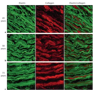
undergoes fatigue. Over an entire lifetime of being stretched at every heart beat, the laminae fracture and become less efficient. Laminae become thinner and since elastin is not replenished readily, the space created between laminae is filled with collagen (Figure 1). Collagen filling with age is thought to prevent overly large deformations of blood vessel walls. However, because collagen is not elastic, the decreased elastin to collagen ratio results in stiffer blood vessels with a larger diameter (Fritze et al. 2012). As this process unfolds, the vessels become partially distended and undergo little change in diameter to absorb the heart pulse and the added pressure of the reflection wave. As vessels harden, there is more and more mechanical stress on the already weakened elastin laminae and these processes of thinning and collagen filling accelerate. Larger vessels become more hardened and ever smaller arteries are affected by this phenomenon. Furthermore, lifestyle choices may also have a strong impact on this hardening mechanism through mitochondrial oxidative stress (Davenport et al. 2012; Zhou et al. 2012) and plaque deposition which further harden vessels (O'Rourke et al. 2007). High calorie intake and low exercise may lead to increased production of reactive oxygen species (ROS), which may contribute to elastin protein damage. ROS are highly reactive molecules since they have an unpaired valence shell electron. To fill their valence shell, ROS steal electrons from larger molecules such as DNA, proteins and lipids, thereby degrading them (Wallace 2005). Furthermore, ROS may cause decreased nitric oxide (NO) bioavailability, thereby reducing overall vascular tone and reserve (Pialoux et al. 2009). The mitochondrial theory of aging, which includes these phenomena, will be further discussed in the next section.
Fritze et al. J Vasc Res 2012;49:77–86
82
be significantly related to segregated MMP-2 (F(2,12) = 0.211; p = 0.810), MMP-9 (F(2,12) = 1.811; p = 0.205), TIMP-1 (F(2,12) = 0.505; p = 0.616) or TIMP-2 (F(2,12) = 0.187; p = 0.832).
Desmosine Content Is Not Decreasing with Age The results of the radioimmunoassays are summa-rized in figure 5 g. Differences in the desmosine content for all three groups proved not significant (F(2,15) = 3.103; p = 0.075).
Marked Decrease in the Elastogenic Potential Revealed by Gene Expression Analyses
Quantitative analysis of tropoelastin mRNA levels re-vealed that TE-mRNA was detectable in all three age groups ( fig. 6 ).
We observed a strong variation of tropoelastin levels within each age group. Even in patients younger than 60 years, some individuals expressed very low amounts (5%) of tropoelastin. However, gene expression analyses also suggested that, with increasing age, tropoelastin
expres-sion decreases (group I: 0.28 8 0.25; group II: 0.14 8 0.08; group III: 0.07 8 0.06), as displayed in figure 6 (left panel). We found a significant effect of age on tropoelas-tin mRNA levels (F(2,58) = 10.266; p ! 0.001). A post hoc Fisher LSD test revealed that tropoelastin mRNA levels did not differ significantly between group I and group II (t(32) = 1,066; p = 0.301). This result is probably influ-enced by a high variance of tropoelastin mRNA levels in young patients ( fig. 6 ) as well as the lower number of in-vestigated younger patients due to the fact of substantial-ly lower probability of CVD’s. Significant differences of tropoelastin levels were observed between group II and group III (t(45) = 3.933; p ! 0.001).
Next, a logistic regression model was employed to de-termine the impact of age, gender and DM on tropoelas-tin expression levels. This model was specified with the equation: log 10 (TE-mRNA) = –0.249 –0.019 ! age –0.029 ! gender –0.022 ! DM and revealed a highly significant overall correlation (F(3,57) = 6.09; p = 0.001). Subsequent t tests demonstrated that age was a significant predictor for TE-mRNA (t(57) = –4.035; p ! 0.001),
49 years 58 years 73 years
Elastin Collagen Elastin/collagen
a
b
c
Fig. 4. Near-infrared multiphoton LSM. Elastin autofluorescence signals (green) and SHG-signals of collagen (red) are de-picted for aortic cross sections (circumfer-ential view) of 49- ( a ), 58- ( b ) and 73- ( c ) year-old patients. Images are 120 ! 120 ! m. Co lo r v er si o n av ai la b le o n lin e
Figure 1. Near-infrared multiphoton LSM.
Elastin autofluorescence signals (green) and SHG signals of collagen (red) are depicted for aortic cross sections (circumferential view) of 49- (a),58- (b) and 73- (c) year-old patients. This figure illustrates changes in elastin and collagen ratio and structure with age. This figure and its caption were reproduced from (Fritze et al. 2012).
Arterial hypertension is both the cause and effect of even greater vessel wall stiffening. As vessels harden, the pulse pressure wave from the heart increases in velocity. This leads to an increased amplitude and velocity of the reflection wave, as blood flow with higher pressure reaches high resistance vessels and is reflected back. This positive feedback loop accelerates, leading to increasing blood pressure and ever more damage to progressively smaller blood vessels (O'Rourke et al. 2007; Redheuil et al. 2010). Arterial hypertension is therefore an indication that damage has already started to occur. Though decreases in arterial elasticity are general and affect the whole body, the aorta may be the most representative of the state of other arteries. This is because the aorta may be the first to be affected since much of the pulsatile flow absorption and therefore the mechanical damage to elastin laminae occurs at this level (Laurent et al. 2006; Redheuil et al. 2010). The relationship between aortic hardening and measures of cerebral health will be discussed in Chapter 5 of this thesis.
Inflammation may be an important contributing factor to vascular stiffening. Arterial hypertension has been found to be associated with the presence of inflammatory markers such as interleukin-6, tumor necrosis factor-alpha and C-reactive protein (CRP) (Amar et al. 2005; Laurent et al. 2007). Though the mechanisms leading to this association are not well known, molecules released as a consequence of endothelial dysfunction are thought to lead to this chronic elevation of inflammatory markers (Devaraj et al. 2011). Furthermore, CRP level was found to be predictive of pulse pressure and treatment to lower CRP levels has been shown to have a positive effect on pulse pressure (Amar et al. 2005; Devaraj et al. 2011).
1.1.1. Vascular aging in the brain
Arterial hypertension has long been known as a risk factor for stroke and is now recognized as playing a role in cognitive decline (Robbins et al. 2005; Waldstein et al. 2008; Elias et al. 2009; Brown et al. 2011; Gorelick et al. 2011). The brain may be particularly vulnerable to the effects of arterial hardening and it is thought that there is a link between arterial stiffening and cognitive decline (Waldstein et al. 2008; Elias et al. 2009; Brown et al. 2011). Several mechanisms have been
proposed to contribute to this relationship. First, the brain is one of the only organs without protection against the pulsatile nature of blood flow. With age, cerebral arteries suffer from the loss of elastin (O'Rourke et al. 2007) and a decrease in microvascular density (Sonntag et al. 1997). The increased pulsatility associated with less elastic arteries may lead to distal cerebral lesions and impaired distal microcirculation (O'Rourke et al. 2007). Secondly, the severity of white matter hyperintensities is increased with hypertension and these have been shown to be associated with cognitive decline and dementia (Sierra et al. 2004; Au et al. 2006). Finally, impaired endothelial function leading to circulation of cellular debris (Wang et al. 2007; Wang et al. 2009) has also been proposed to lead to microvascular lesions (O'Rourke et al. 2007) and hence cognitive deficits. This may also be reflected in the fact that elevated CRP levels, indicative of endothelial dysfunction, are an independent risk factor for age-associated cognitive decline (Yaffe et al. 2003).
1.1.2. Mitochondrial theory of aging
While aging cannot be reduced to a single phenomenon, one of the most enduring theories of the cellular mechanisms of aging relates accumulating damage in the cellular environment to progressive breakdown of membrane integrity and of the electron transport chain of the mitochondria. Mitochondrial damage leads to an increased formation of reactive oxygen species (ROS). The progression of
mitochondrial damage in normal aging, leading eventually to disease, is presumed to progress as described in the remainder of this section and summarized in Figure 2. Though mitochondrial aging occurs throughout the body, it is here discussed in the context of the brain.
The electron transport chain, which is used for oxidative phosphorylation and
production of extra ATP molecules from glycolysis end-products, is the main source of ROS. These are created at all times even in the best of cases, but their
production can be increased by several factors including a high-calorie diet and low exercise (Wallace 2005; Pialoux et al. 2009). Because electrons from fat and
carbohydrate oxidation are produced even when there is no ADP to be
phosphorylated (because, for example, of lack of exercise), excess protons from metabolism of fats and carbohydrates cause hyperpolarization of the mitochondrial membrane. Excess electrons fill all binding sites on electron carriers within the membrane and these electrons can then be added to O2 to form ROS. These ROS diffuse and degrade DNA, proteins and lipids. Because ROS are mainly created in mitochondria and are very reactive, these organelles are the first and worst
affected (Wallace 2005).
Since mitochondria are continually produced and destroyed in post-mitotic cells, mutations in mitochondrial DNA (mtDNA) can affect the function of cells even when they have stopped replicating. Throughout a lifetime, ROS will create somatic mutations in mtDNA and affect the functioning of proteins encoded by this mtDNA. Complexes I, III and IV of the electron transport chain (ETC) are especially prone to the effects of ROS-mediated damage, since they reside within mitochondria and some of their components are encoded by mtDNA (Aleardi et al. 2005; Coskun et al. 2011). Mitochondria become more and more inefficient as electron transport
becomes progressively uncoupled from proton pumping due to damage to these protein complexes (Wallace 2005). As the process to produce ATP loses efficiency, more and more protons are required to make ATP.
the NADHþ Hþand reduces the lipid-soluble electron
carrier coenzyme Q10(CoQ). CoQ or ubiquinone
incor-porates two electrons successively, the first electron gener-ating ubisemiquinone and the second ubiquinol. Electrons from other dehydrogenases are also transferred to CoQ. This includes complex II, succinate dehydrogenase, which collects electrons from the TCA cycle intermediate, succi-nate. From CoQ, the electrons are transferred to complex
III (b – c1complex), then to cytochrome c, then to complex
IV (cytochrome c oxidase, COX), and finally to O2to
gen-erate H2O. As the electrons traverse complexes I, III, and
IV, the energy released is used to pump protons out across the mitochondrial inner membrane to create the
electro-chemical gradient: DP¼ DC þ DmHþ(Fig. 3). The
accu-mulated power generated by 1000 mitochondria within
each of 100 trillion cells or 1017mitochondria provides
the energy that powers our lives.
The mitochondrial capacitors provide energy to drive a wide range of biological processes, but to extend mito-chondrial utility to fueling reactions and performing work, it needs to convert the capacitor’s potential energy
complex V, bound to the mitochondrial inner membrane and pointing into the matrix. Mitochondrial ATP is then exchanged by cytosolic ADP by the adenine nucleotide translocators (ANTs). Therefore, the proton-pumping complexes I, III, and IV of the ETC are coupled to ATP synthesis through the proton gradient. The efficiency by which complexes I, III, and IV pump protons out of the mitochondrion and by which the ATP synthase converts the proton gradient into ATP is known as the coupling effi-ciency. Because the calorie is a unit of heat, the more calo-ries that are burned by the cell, the more heat that is generated, whether the energy goes through the high-energy intermediate ATP or not. As it turns out, people dif-fer significantly in their coupling efficiencies. Therefore, those that are more tightly coupled generate the maximum ATP per calorie burned and the minimum heat, whereas individuals that are less tightly coupled have to burn more calories for the same amount of ATP and thus gener-ate more internal heat. This provides one of the fundamen-tal connections between our genes and our environment.
The biophysics of the linkage among calorie oxidation,
Figure 3. Mitochondrial energy production and its reaction to the pathophysiology of disease. Five features of mitochondrial metab-olism are central to the pathophysiology of the common age-related diseases: (1) energy production by OXPHOS; (2) regulation of cellular oxidation – reduction (redox) state; (3) ROS generation as a by-product of OXPHOS; (4) buffering of the cytosolic and mito-chondrial Ca2þlevels; and (5) regulation of apoptosis through activation of the mtPTP. ADP or ATP, adenosine di- or triphosphate, ANT, adenine nucleotide translocator; cytc, cytochrome c; GPx, glutathione peroxidase-1; LDH, lactate dehydrogenase; MnSOD, manganese superoxide dismutase or SOD2; NADH, reduced nicotinamide adenine dinucleotide; TCA, tricarboxylic acid cycle; VDAC, voltage-dependent anion channel; I, II, III, IV, and V, OXPHOS complexes I – V. Complex I is composed of 45 polypeptides, seven (ND1, 2, 3, 4L, 4, 5, 6) encoded by the mammalian mtDNA; complex II consists of four nDNA-encoded polypeptides; complex III consists of 11 polypeptides, one (cytb) encoded by the mtDNA; complex IV is composed of 13 polypeptides, three (COI, II, III) encoded by the mtDNA; and complex V is composed of!15 polypeptides, two (ATP6, 8) encoded by the mtDNA. (Modified from Wallace 2005, 2007; Ruiz-Pesini et al. 2007.)
WALLACE 6
Cold Spring Harbor Laboratory Press
on February 10, 2012 - Published by
symposium.cshlp.org
Downloaded from
Figure 2. Mitochondrial energy production
This figure and its caption were reproduced from (Wallace 2011).
Eventually, mitochondria become so inefficient that energy production falls below the so-called bioenergetic threshold. At this point, the cell reduces its functions to adapt to its own inefficient energy production and enters a steady-state that enables only a basic operational level of functioning. This is the stage where
damage and progression to disease start to occur. For example, the symptoms that form the hallmark of Alzheimer’s disease (AD) (including amyloid-β production and deposition) develop at this point (Wallace 2005; Bonda et al. 2010; Coskun et al. 2011).
As oxidative stress damages the cell, a variety of protective mechanisms are put in place. One of these is the cleavage of the amyloid precursor protein into amyloid-β1-42 by presenilin 1 and 2. amyloid-β1-42 has been found to have antioxidant properties and is produced as a defensive mechanism against the effects of
excess ROS (Kontush et al. 2001; Bonda et al. 2010; Coskun et al. 2011; Bartley et 33
al. 2012). However, as it accumulates, amyloid-β becomes toxic and leads to a decrease in the activity of complexes I, III and IV (cytochome c oxidase) (Canevari et al. 1999; Aleardi et al. 2005), the inhibition of Mn superoxide dismutase (Aleardi et al. 2005; Anantharaman et al. 2006) and perturbation in the functioning of the mitochondrial permeability transition pore (Bartley et al. 2012). These lead to decreased O2 consumption and ATP synthesis and to an increase in ROS
production (Aleardi et al. 2005). Leakage through the permeability transition pore leads to the last stages of cell life as it triggers the induction of apoptosis cascades (Wallace 2005; Coskun et al. 2011; Bartley et al. 2012). This leads to the loss of neurons and brain atrophy. Though markers of these mechanisms are present in normal aging brains, these effects are more pronounced in the case of diseases such as AD. It may be, however, that these effects contribute to the atrophy and cognitive decline observed even in healthy aging (Wallace 2005), especially in individuals that adopt a less healthy lifestyle. However, the specific effects of these mechanisms on cognitive processes is largely unknown. Atrophy and vascular changes are known to occur at different rates in different brain regions (Salat et al. 2004; Chen et al. 2011) and the link between mitochondrial health and regional deficits still remains to be determined.
1.2. Aging and physical fitness
The effects of aging often result in a significantly poorer quality of life and much research effort has been dedicated to alleviating the effects of aging. Lifestyle has a very substantial impact on health and quality of life. A healthy diet and regular exercise are, by far, the best recognized means to aging gracefully (Wallace 2005). However, the exact impact of regular exercise on cerebrovascular and cognitive aging remains in part unknown.
Studies on the effects of exercise can take two forms. In the first, participants with a history of regular exercise and sedentary participants are compared (Rogers et al. 1990; Yaffe et al. 2001; Churchill et al. 2002; Podewils et al. 2005; Etnier et al. 2006; Kramer et al. 2006; Larson et al. 2006; Brown et al. 2010; Brown et al. 2011). While these studies have the advantage of taking into account the long-term
effects of exercise, they are subject to various confounds because the people who 34
exercise regularly may also be more likely to have a healthy lifestyle in other respects (Churchill et al. 2002; Kramer et al. 2006). People that engage in regular exercise may also be more likely to have a healthy diet, refrain from smoking and excessive drinking for example. The other type of study follows individuals with a similar background through a regular exercise training intervention and, typically, some other form of activity such as cognitive training in a control group of matched participants (Colcombe et al. 2006; Voss et al. 2010; Erickson et al. 2011; Prakash et al. 2011; Hopkins et al. 2012). While this type of study avoids the potential confounds of unmatched lifestyle choices, it does not address the question of long-term effects of regular exercise. However, with the combined information of these two types of studies, a cohesive picture of the effects of physical fitness on
vascular and cognitive health is starting to emerge.
Retrospective population studies of questionnaire-based self-reported physical activity and cross-sectional studies including a cardiorespiratory fitness test have suggested that regular exercise may be partly protective for dementia (Podewils et al. 2005; Larson et al. 2006) and cognitive decline (Rogers et al. 1990; Yaffe et al. 2001; Churchill et al. 2002; Etnier et al. 2006; Kramer et al. 2006; Brown et al. 2010; Brown et al. 2011). Typically, recent studies quantify VO2max, to estimate cardiorespiratory fitness by measuring the amount of O2 used for metabolism during maximal effort.
Despite the potential confounds of cross-sectional studies, a lifetime of regular exercise seems to have a beneficial effect on cognitive health. The effects of a physical training regimen on cognitive health are, however, more subtle (Churchill et al. 2002; Kramer et al. 2006) and may depend on many factors including the amount of exercise done on the day of the experiment and genetic polymorphisms (Hopkins et al. 2012). A meta-analysis of fitness training studies showed that fitness training results in improved cognitive scores, especially for tasks requiring executive control processes (Colcombe et al. 2003). Older adults’s performance on executive function tasks may be improved by an aerobic physical training regimen. Furthermore, this type of intervention may also lead to patterns of functional
connectivity more similar to those seen in younger adults (Voss et al. 2010;
Prakash et al. 2011). Furthermore, grey matter atrophy associated with aging may 35
be reduced by a similar physical training program (Colcombe et al. 2006; Erickson et al. 2011).
Cardiorespiratory fitness also has a positive effect on vascular health. Physically fit older women were found to have lower blood pressures and higher
cerebrovascular reserve (Brown et al. 2010). Results in animals have also shown that regular exercise leads to increased cerebrovascular reserve, possibly through angiogenesis (Swain et al. 2003). The positive effects of regular exercise on cognition and cerebral blood flow (CBF) in aging may be due in part to increased antioxidant activity leading to increased NO bioavailability and improvements in vascular tone (Pialoux et al. 2009; Davenport et al. 2012) (Figure 3). In a study of post-menopausal women, Pialoux and colleagues (Pialoux et al. 2009) found an inverse relationship between VO2max and oxidative stress markers, but a positive relationship between antioxidant activity and VO2max, indicating better
mitochondrial function in more active older women. A similar relationship was seen between cerebrovascular conductance and oxidative stress markers, indicating that part of these effects may be due to changes in the regulation of vascular tone. Animal studies suggest that exercise may result in increased serotonin and brain-derived neurotrophic factor (BDNF) levels in the brain, which may in turn lead to neurogenesis and cognitive improvement (Churchill et al. 2002; Vaynman et al. 2004; Hopkins et al. 2010; Spoelgen et al. 2011; Davenport et al. 2012). Though difficult to establish non-invasively, some of the same mechanisms may be involved in cognitive improvements following regular exercise regimen that have been detected in adult human populations (Hopkins et al. 2012). IGF-1, VEGF and BDNF, by their role in initiating both the angiogenesis and neurogenesis cascades, are thought to be the main effectors in regulating the effects of regular exercise on cerebrovascular function(Cotman et al. 2007; Davenport et al. 2012) (Figure 3).
Figure 3
ACCEPTED
Copyright © 2012 by the American College of Sports Medicine. Unauthorized reproduction of this article is prohibited.
Figure 3. Exercise and cognition
Proposed vascular mechanisms in the association between exercise training and increased cognitive plasticity. This figure and its caption were reproduced from (Davenport et al. 2012). Though the link between vascular health, fitness and cognition is becoming more established, some of the difficulties in pinning down the relationship between
vascular health and cognition may arise from the fact that the exact impact of aging on cognition is still a much debated subject.
1.3. Aging and cognition
Aging is associated with changes in cognitive processing. While decreases in processing speed have been observed across the majority of cognitive tasks studied in older adults, there is some debate as to their specificity and source. One of the most obvious confounds to take into account is that all sensory systems degrade with age. Within the visual system, for example, aging leads to hardening and increased density of the crystalline lens, a decrease in resting pupil diameter, increased opacity of the lens and retinal cell loss. All these factors combine to greatly reduce the light that enters the eye as we age. By 60 years of age, it is estimated that two thirds of retinal luminance has been lost (Cabeza et al. 2005). Because less information reaches the brain, this means that older brains must process information with a lower starting signal-to-noise ratio (SNR). This is true of other sensory systems as well, and must be kept in mind when assessing the effects of aging in cognitive tasks that include a significant sensory component.
Some of the decreased performance, both in terms of errors and reaction time, may therefore be attributed to the effects of decreased sensory system
performance (Cabeza et al. 2005). However, certain tasks seem to be specifically impacted by age over and beyond these general sensory slowing effects.
Furthermore, though decreased hemodynamic responses are expected in a system showing the effects of wear, it is the presence of specific and reproducible patterns of increased functional signals in specific types of task which has most intrigued the cognitive neuroscience community and will be a focus of the following discussion.
The functions thought to be most affected by the physiological changes associated with age fall under the category of ‘executive functions’. This term is used to
describe a range of functions such as planning, working memory, inhibition, problem-solving and monitoring. These functions generally involve a network of structures that includes some frontal component (Petrides 2005). This, combined with the ubiquitous observation of an increased frontal component in functional imaging studies of cognitive aging and the known plasticity of the brain even in later life, has led to the formulation of several theories of neuronal processing changes in healthy cognitive aging.
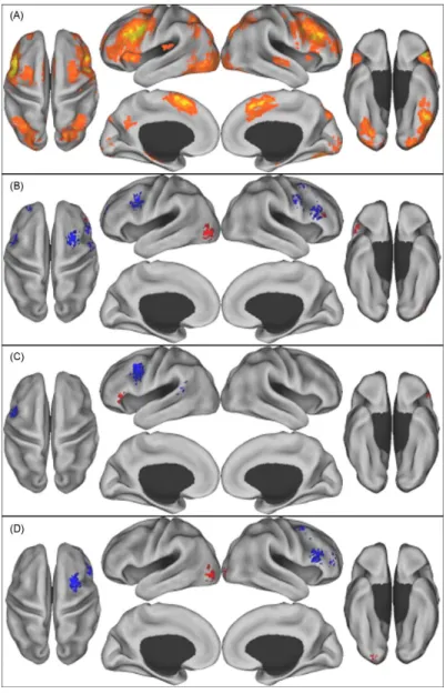
Recently, Spreng and colleagues published a meta-analysis of age-related differences reported across many cognitive domains in BOLD and positron
emission tomography (PET) studies (Spreng et al. 2010). This study shows that it is possible to generalize across cognitive domains to find reliable age-related differences in activation foci (Figure 4). Most of these foci fell within the task positive network, which is known to be activated across many tasks and is one of the most reliable networks found in functional connectivity analyses (Fox et al. 2005; Toro et al. 2008). This study compared maps of various cognitive tasks acquired in high-performing and more poorly performing older adults. While left frontal areas showed consistently higher signals in older adults when age groups were matched for performance, frontal areas on the right hemisphere showed higher signal changes when older adults’ performance was poorer than their younger counterparts’. When looking more specifically at studies of executive function, a bilateral increase in functional signal was seen over frontal areas, likely
because performance was not used to distinguish groups of older adults. There may therefore be a selective advantage to greater involvement of the left prefrontal areas, and older adults showing enhanced left frontal recruitment may be better able to maintain their performance at the level achieved by young subjects.
Increased right prefrontal signal changes on the other hand, may reflect degrading of specialized neural constructs and therefore recruitment of brain areas not
specific for the task.
not contribute to this cluster), consistent with the TPN. Old adults who performed more poorly also activated right RLPFC and the left thalamus (Fig. 1D andTable 3).
3.5. Domain specific results
Perception, memory encoding, memory retrieval and executive function independently demonstrated a pattern of activity consis-tent with the TPN, including reliable clusters in lateral prefrontal
regions, aIfO, SMA, and vOC (Figs. 2–5). These findings are consistent with a previous large-scale meta-analysis of domain specific cognition (Cabeza and Nyberg, 2000). Notable domain specific clusters were also apparent and the results are discussed in turn. 3.6. Perception
Perceptual studies, most of which were in the visual modality, showed extensive visual cortical activation, as would be expected Fig. 1. Reliable patterns of brain activity across studies. A: Activation likelihood clusters across all studies and age groups. B: Age differences from all studies. C: Age differences from those studies where old and young adults had equivalent performance. D: Age differences from those studies where old adults had poorer performance relative to young adults. Red = young adults > old adults, Blue = old adults > young adults. Activation likelihood clusters (FDR p < .01) are shown on an inflated surface map in Caret (Van
Essen, 2005). Some clusters may not appear contiguous due to mapping clusters on the surface maps; for example, this can occur when neighboring gyri, but not the
intermediary sulcus, were included in a statistically reliable cluster in the original image volume.
R.N. Spreng et al. / Neuroscience and Biobehavioral Reviews 34 (2010) 1178–1194 1184
Figure 4. Reliable patterns of brain activity across studies of aging
A: Activation likelihood clusters across all studies and age groups. B: Age differences from all studies. C: Age differences from those studies where old and young adults had equivalent performance. D: Age differences from those studies where old adults had poorer performance relative to young adults. Red = young adults > old adults, Blue = old adults > young adults. Figure reproduced from (Spreng et al. 2010).
The theories addressing the hemodynamic signal changes observed across cognitive studies seek to address two aspects: the localization of signal changes (with particular attention being given to signal increases) and the underlying cause of these changes. While both aspects inform each other to some extent, the most difficult to address is no doubt the underlying cause of these changes. Two main ideas are proposed to explain these phenomena. One is the idea of compensation and the other is the idea of dedifferentiation.
1.3.1. Mechanisms leading to changes in activation patterns
Compensation hypotheses express the idea that as the brain ages, there is an accumulation of damage that will eventually lead to deficits, and that deficits can be alleviated at least partially by recruiting additional neural resources. These can be in the same area, so that a given area normally used for a given task is now more active than previously, or by recruiting additional brain regions that are not normally involved in the task in younger individuals (Cabeza 2002; Reuter-Lorez et al. 2008; Reuter-Lorenz et al. 2010). This type of theory implies that additional recruitment will provide a processing advantage that is usually observed as lower error rates or faster reaction times.
Dedifferentiation hypotheses state that as we age, the specialization of brain regions acquired during development breaks down, so that tasks which in early adulthood gave rise to very localized hemodynamic signal increases, now give rise to more loosely localized networks (Baltes et al. 1997; Park et al. 2009; Cappell et al. 2010; Reuter-Lorenz et al. 2010). Thus, aging leads to similar or the same functional responses to different tasks. In other words, there is an increased
correlation amongst the activation patterns and observed performance on a variety of tasks.
Evidence for dedifferentiation was identified in a study by Baltes and colleagues (Baltes et al. 1997), showing that as participants age the intercorrelation between different cognitive scores, perceptual speed and sensory information all increase. While visual and auditory performance can explain a large proportion of cognitive score variability during aging, the authors privilege a general dedifferentiation process to explain their results. According to this view, the degradation of sensory