Possible Role of MKP-1 in Glioblastoma cells
radioprotecton following CXCL12 actvaton
Matthias Dedobbeleer
1, Nicolas Goffart
1, Estelle Willems
1, Emmanuel Di Valentin
2, Bernard Rogister
1,3,4 1 Nervous System Diseases and Treatments, GIGA-Neurosciences Research Center, University of Liège, Liège, Belgium2GIGA-Viral Vector Plateform, University of Liège, Liège, Belgium. 3Department of Neurology, CHU and University of Liège, Liège, Belgium. 4GIGA – Development, Stem Cells and Regenerative Medicine, University of Liège, Liège, Belgium.
In patients with glioblastoma multiform (GBM), recurrence is the rule despite continuous advances in surgery, radio- and chemotherapy. Within these
most frequent primary brain tumors, glioblastoma stem cells or initiating cells (GIC) have recently been described and were shown to be involved in
these recurrences. Our lab recently demonstrated that GIC, once injected into the striatum of immunodeficient nude mice, exhibit a tropism for the
subventricular zones (SVZ), one of the adult neurogenic niches bringing them an appropriate molecular and cellular environment to growth. After
irradiation of these mice, we still discovered cells inside the SVZ. We then questionned the role of the CXCL12/CXCR4 pathway in radioprotection
phenotype. After demonstrating that CXCL12 could play a radioprotective role, we wanted to know by which mechanism it happens. Knowing that
MKP1, the major regulator of the MAP kinase pathway, shown a higher phosphorylation profile after CXCL12 stimulation, and that this protein is
involve in many cancers and that its role in glioblamstoma remain unclear, we wanted to know could have a radioprotective role link or not to the
CXCL12/CXCR4 signalling pathway.
Contact :
mdedobbeleer@
ulg.ac.be
FRIA
B
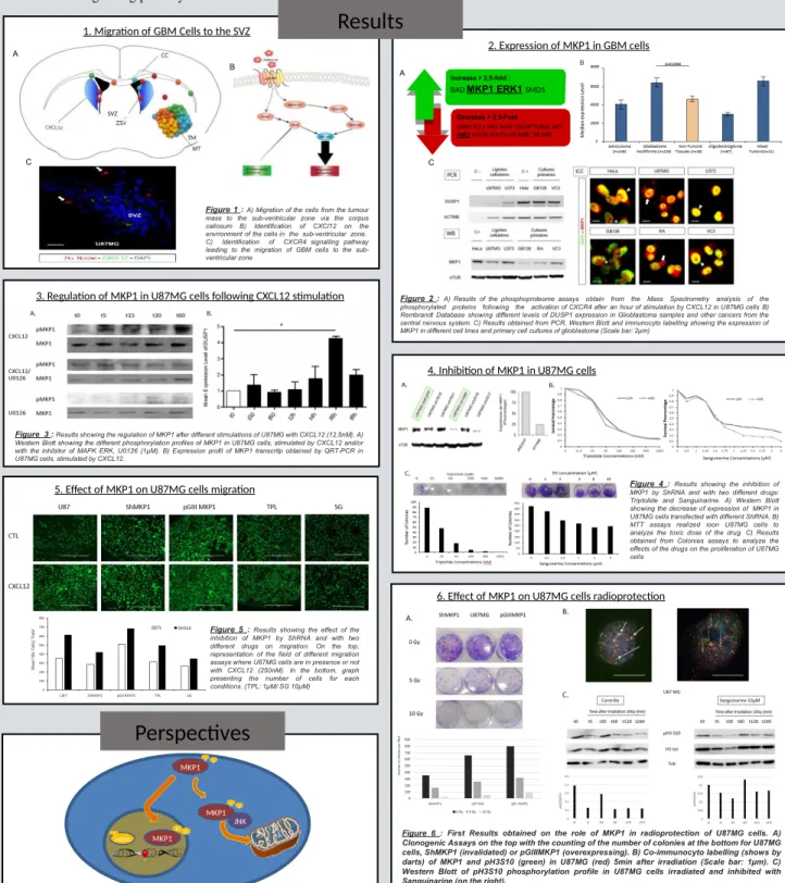
Figure 1 : A) Migration of the cells from the tumour mass to the sub-ventricular zone via the corpus callosum B) Identification of CXCl12 on the environment of the cells in the sub-ventricular zone. C) Identification of CXCR4 signalling pathway leading to the migration of GBM cells to the sub-ventricular zone A
Introduction
Results
SVZ TM Decrease > 2.5-FoldGRB2 PLCγ IKKγ AurA CD2AP TUBα1 AKT
JNK1 mTOR STAT3 LC8 RAB7 Rb HRC Increase > 2,5-fold :
BAD
MKP1 ERK1
SMD5 AFigure 2 : A) Results of the phosphoproteome assays obtain from the Mass Spectrometry analysis of the phosphorylated proteins following the activation of CXCR4 after an hour of stimulation by CXCL12 in U87MG cells B) Rembrandt Database showing different levels of DUSP1 expression in Glioblastoma samples and other cancers from the central nervous system. C) Results obtained from PCR, Western Blott and immunocyto labelling showing the expression of MKP1 in different cell lines and primary cell cultures of glioblastoma (Scale bar: 2µm)
C
Figure 3 : Results showing the regulation of MKP1 after different stimulations of U87MG with CXCL12 (12,5nM). A) Western Blott showing the different phosphorylation profiles of MKP1 in U87MG cells, stimulated by CXCL12 and/or with the inhibitor of MAPK ERK, U0126 (1µM). B) Expression profil of MKP1 transcritp obtained by QRT-PCR in U87MG cells, stimulated by CXCL12.
C
Figure 4 : Results showing the inhibition of MKP1 by ShRNA and with two different drugs: Triptolide and Sanguinarine. A) Western Blott showing the decrease of expression of MKP1 in U87MG cells transfected with different ShRNA. B) MTT assays realized ioon U87MG cells to analyze the toxic dose of the drug. C) Results obtained from Colonies assays to analyze the effects of the drugs on the proliferation of U87MG cells
Figure 5 : Results showing the effect of the inhibition of MKP1 by ShRNA and with two different drugs on migration. On the top, representation of the field of different migration assays where U87MG cells are in presence or not with CXCL12 (250nM). In the bottom, graph presenting the number of cells for each conditions. (TPL: 1µM/ SG 10µM) Nu m ber o f co lo n ie s pe r fle d
Figure 6 : First Results obtained on the role of MKP1 in radioprotection of U87MG cells. A) Clonogenic Assays on the top with the counting of the number of colonies at the bottom for U87MG cells, ShMKP1 (invalidated) or pGIIIMKP1 (overexpressing). B) Co-immunocyto labelling (shows by darts) of MKP1 and pH3S10 (green) in U87MG (red) 5min after irradiation (Scale bar: 1µm). C) Western Blott of pH3S10 phosphorylation profile in U87MG cells irradiated and inhibited with Sanguinarine (on the right).
Perspectives
Futher investigations with 2 different hypothesis of radioprotection mechanisms
will be study with the JNK-docking pathway and the Histone H3-H2AX pathway,
in order to clarify the radioprotective role of MKP1 in glioblastoma.
H2AX P MKP1 P P JNK MKP1P P MKP1 P P H 3