Université de Montréal
IL-23 Receptor and IL-12 Receptor Expression is Restricted to Distinct Cell Types in the IL-23R-GFP Reporter Mouse
par Lisa Bellemare
Département de Microbiologie et Immunologie Faculté de Médecine
Mémoire présenté à la Faculté des études supérieures En vue de l’obtention du grade de Maîtrise ès Sciences
En Microbiologie et Immunologie
August 2012
Université de Montréal
Mémoire intitulé:
IL-23 Receptor and IL-12 Receptor Expression is Restricted to Distinct Cell Types in the IL-23R-GFP Reporter Mouse
Présenté par: Lisa Bellemare
Évalué par le jury suivant:
Dr. Iannis Adamopoulos Dr. Martin Guimond
Dr. Sylvie Lesage
Abstract
Inflammatory bowel diseases (IBD) are characterised by uncontrolled immune responses in the gut. Genome-wide association studies (GWAS) have identified a protective polymorphism for IBD in the IL23R gene. IL23R codes for the IL-23r protein, one of the two subunits of IL-23R. IL-23R belongs to the IL-12R family, which contains many heterodimeric receptors. For example, both IL-12R and IL-23R share the IL-12Rβ1 subunit. Nevertheless, IL-12R and IL-23R are associated with different immune processes (Th1 vs. Th17).
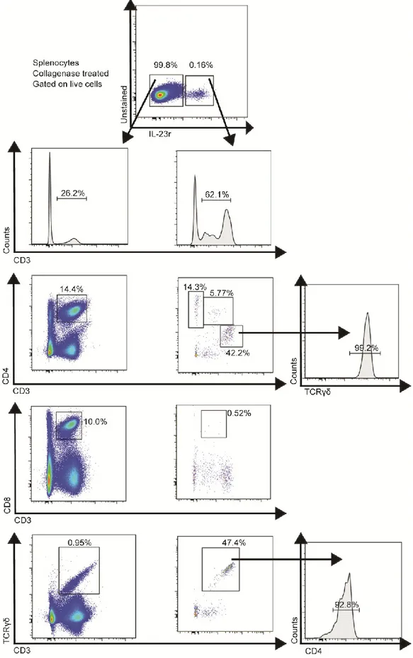
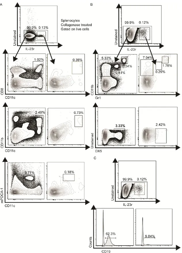
This thesis characterizes the cellular patterns of expression of both IL-23R and IL-12R, to further elucidate their roles in inflammation. We established that IL-23R and IL-12R were never co-expressed together, even though they share the IL-12Rβ2 subunit. Analysis of murine splenocytes revealed that IL-23R is expressed by some TCRγδ T-cells, a few B-cells, CD4+ T-cells and several Lti-like cells. IL-12R protein was found in a few B-cells.
The analysis of IL-23R and IL-12R expression in different organs revealed that the lamina propria of the small intestine was the organ containing the largest proportion of IL-23r+ cells. IL-12R+ cells were found in constant numbers throughout the organs.
Finally, in vitro cultures showed that IL-23R and IL-12R had crossed reaction to IL-12 and IL-23. Study of IL-23R in IBD should always be accompanied by IL-12R analysis, because both receptors could have complementary roles.
Key words: Inflammatory bowel diseases, IL-23, IL-12, IL-23R, IL-12R, small intestine lamina propria, Lti-like cells, IL-23R-GFP reporter mouse
Résumé
Les maladies inflammatoires de l'intestin (MII) sont caractérisées par des réponses immunitaires incontrôlées dans l'intestin. Des études génétiques ont associé un polymorphisme dans le gène de l'IL23R à la résistance aux MII. IL23R code pour la protéine de l’IL-23r, une sous-unité du récepteur à l’IL-23 (IL-23R). Ce récepteur appartient à la famille de l’IL-12R, contenant plusieurs récepteurs hétérodimériques. D’ailleurs, IL-12R et IL-23R partagent la sous-unité IL12Rb1. Néanmoins, ces deux récepteurs favorisent des réponses immunitaires distinctes (Th1 vs Th17).
Ce mémoire caractérise les dynamiques d’expression cellulaires de l’IL-23R et l’IL-12R, afin d’élucider leurs rôles dans l’inflammation. Nous avons établi qu’IL-23R et IL-12R ne sont jamais co-exprimés, malgré qu’ils partagent la sous-unité IL-12Rβ1. Parmi les cellules de rates de souris, la protéine IL-23r est trouvée dans certaines cellules T TCRγδ ou T CD4+, quelques cellules B et des cellules Lti-like. La protéine IL-12Rβ2 est exprimée par quelques cellules B.
L’analyse de l’expression de l’IL-23R et l’IL-12R dans différents organes révéla que la plus grande proportion de cellules exprimant l’IL-23R se retrouve dans la lamina propria de l'intestin grêle, alors que les cellules exprimant l’IL-12Rβ2 ont été retrouvées en proportion équivalente dans tous les organes lymphoïdes. Ces observations appuient les études génétiques suggérant un rôle prédominant de l’IL23R dans les intestins.
Finalement, des cultures in vitro suggèrent que l’IL-23R ou l’IL-12R avaient des réactions croisées à l’IL-12 ou l’IL-23. L’étude de l’IL-23R dans les MII devrait donc être complémentée par l’étude de l’IL-12R, car les deux récepteurs pourraient avoir des rôles complémentaires.
Mots clés: maladies inflammatoires de l'intestin, IL-23, IL-12, IL-23R, IL-12R, lamina propria de l’intestin grêle, Lti-like cells, souris IL-23R-GFP
Table of contents
Abstract ... i
Résumé ... ii
Table of contents ... iii
List of Figures ... v
Abbreviations used in this thesis ... vii
Acknowledgments ... viii
Introduction ... 9
1. Inflammatory Bowel Diseases ... 9
Crohn’s Disease... 9
Ulcerative Colitis... 11
IBD epidemiology ... 12
Causes... 13
2. IL-23R and IL-12R biology ... 22
IL-12R/IL-23R/IL-35R/IL-27R family of receptors ... 22
IL-12/IL-23/IL-35/IL-27/p40 homodimer families of cytokines ... 24
IL-23R biology ... 27
Signals leading to the upregulation of the receptor ... 27
Signals at the receptor levels ... 28
Signaling cascade ... 28
IL-12R biology ... 30
IL-23R and IL-12R functions ... 32
3. Mouse models ... 37
Roles and importance of IL-23R and IL-12R in mouse models other than colitis 37 Colitis models ... 40
Importance of IL-23R in different colitis models ... 43
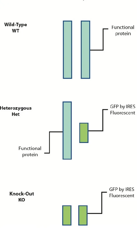
IL-23R-GFP reporter mouse ... 46
Research question and hypothesis ... 50
Methods ... 52
1. Lymphocytes extraction from murine organs ... 52
1.1 Spleen, lymph nodes, thymus, mesenteric lymph nodes ... 53
1.3 Lamina propria of the small intestine ... 54
1.4 Lungs ... 55
1.5 Cell counting ... 56
1.6 Flow cytometry ... 56
2. In vitro culture ... 61
2.1 CD4+T-cells and TCRγδ+T-cells ... 61
2.2 B-cells (IL-12Rβ2+ and IL-12Rβ2-) ... 61
2.3 Lti-like cells... 62
2.4 NK cells ... 62
3. Measurements of supernatants ... 63
3.1 Ig ELISA ... 63
3.2 Cytokine measurements by Flow Cytomix ... 64
4. Statistics ... 65
Results ... 66
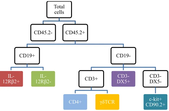
Objective 1: Identify the cell populations expressing IL-23R and IL-12R using the IL-23r-GFP reporter mouse and a monoclonal antibody, respectively ... 66
Objective 2: Characterize the cell populations expressing IL-23R and IL-12R in different organs, especially the gut ... 83
Objective 3: Understanding the specificity of the cytokine responses of different cell types following stimulation with IL-12 or IL-23 ... 104
1. Lti-like cells... 104 2. T-cells ... 109 3. B-cells... 112 4. NK cells ... 115 Discussion ... 118 Bibliography ... 130 Appendix 1 ... cxxxv Appendix 2 ... cxxxix Appendix 3 ... cxli
List of Figures
Figure 1: Pathogenesis of IBD ... 14
Figure 2: Receptors of IL-12R family ... 23
Figure 3 : Cytokine structure ... 25
Figure 4: IL-23R induction and signaling cascade... 29
Figure 5: IL-12R induction and signaling cascade... 31
Figure 6: IL-23r-GFP reporter mouse ... 49
Figure 7 : Sorting Strategy for cell culture ... 60
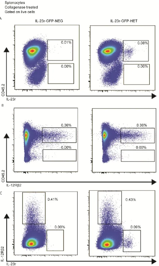
Figure 8: IL-23r and IL-12Rβ2 are of hematopoietic origin, are expressed in naïve mice and are not co-expressed ... 68
Figure 9: IL-23r is expressed by different T-cell receptor bearing T-cells ... 69
Figure 10 : IL-23R is rarely found in B-cells, DC, NK and monocytes ... 70
Figure 11: IL-23r is found in increased proportion in RagKO mice despite a lack of T-cells and B-T-cells, while IL-12Rβ2 is found in very low proportion ... 73
Figure 12: DC, monocytes and NK are not the major cell types expressing IL-23R in RagKO animals ... 74
Figure 13: A Lin(-) c-kit+ cell population express IL-23R and can be found both in WT and RagKO mice ... 76
Figure 14 : Lti-like cells are dependent upon collagenase treatment ... 78
Figure 15: IL-12Rβ2 is expressed by B-cells in naïve mice ... 80
Figure 16: CD43+B-cells are enriched in the IL-12Rβ2+ B-cells population ... 81
Figure 17: Compilations of IL-23R expressing cells in the spleen ... 82
Figure 18: IL-23R and IL-12Rβ2 expressing cells are found in low numbers in the thymus ... 84
Figure 19: IL-23R and IL-12Rβ2 expressing cells are found in low numbers in the bone marrow and are not co-expressed on the same cells ... 87
Figure 20: Lti-like are rare in bone marrow and are rare expressors of Il-23R, while a strong proportion of IL-23R+ cells are B-cells ... 88
Figure 21: Peripheral lymph nodes contain slightly higher percentages of IL-23R positive cells than the spleen ... 90
Figure 23: Lungs contain a population of IL-23r positive cells which are maily CD3+ and some Lti-like cells ... 94 Figure 24: Percentage of IL-23r+ in the lung decreases in RagKO mice ... 95 Figure 25: A third of IL-23R+ cells in the lamina propria are T-cells ... 97 Figure 26: Lamina propria of the small intestine contains high numbers of IL-23R
positive cells and those cells are in vast majority Lti-like cells ... 98 Figure 27: IL-23r+ cells are found in very high numbers in the lamina propria of
RagKO mice ... 100 Figure 28 : Distribution of IL-23r+ cells in the small intestine lamina propria ... 101 Figure 29 Compilation of IL-23R expression in different organs: the small intestine LP
is the organ with the highest proportion of IL-23r+ cells of all the organs analysed. ... 102 Figure 30: Lti-like cells can produce high amounts of IL-22 following IL-23 in vitro
stimulation ... 106 Figure 31:Lti-like cells from the lamina propria respond to IL-23 and IL-2 ... 108 Figure 32: CD4+ and γδ T-cells cytokine secretion following in vitro stimulation ... 111 Figure 34: Ig production by IL-12Rβ2+ B-cells ... 114 Figure 35: NK proliferation is impaired when stimulated with IL-12 ... 116 Figure 36: Presence of IL-12 and IL-12Rβ2 is required for INFγ production by NK
Abbreviations used in this thesis
BM Bone marrow
CD Crohn’s disease
CIA Collagen induced arthritis
DC Dendritic cells
EAE Experimental autoimmune encephalomyelitis
GFP Green-fluorescent protein
IBD Inflammatory bowel diseases
IL-12R Interleukin-12 receptor
IL-23R Interleukin-23 receptor
IL-27R Interleukin-27 receptor
IL-35R Interleukin-35 receptor
lDC Lymphoid dendritic cells
LN Lymph nodes
mDC Myeloid dendritic cells
MS Multiple sclerosis
NK Natural killer cells
pDC Plasmacytoid dendritic cells
Acknowledgments
I want to thank everyone who helped throughout this thesis. Members of my lab, past and present: Fanny, Mary-Cruz, Véronique, Erin, Adam and Genevieve. I also want to thank my supervisor, Dr. Sylvie Lesage, who gave me the best project for me and guided me throughout this whole process.
I want to thank my family, my parents, my sister and my brother, who are always interested by whatever I do. I love you all.
1. Inflammatory Bowel Diseases
Inflammatory bowel diseases (IBD) are a family of illnesses including Crohn’s disease (CD), ulcerative colitis (UC) and indeterminate colitis, among others[1]. This introduction will focus mainly on Crohn’s disease and ulcerative colitis, as they are the two most prevalent and most studied in this family of diseases1. Even though both diseases share certain hallmark characteristics, there are striking differences between their etiology, genetic predisposition and the clinical presentation of patients.[1]
Crohn’s Disease
The prevalence of autoimmune and inflammatory diseases such as Multiple sclerosis (MS) and systemic lupus erythematosus (SLE) is often higher in women than in men, suggesting a role of sex hormones in the pathogenesis of the disease. However, CD is diagnosed in equal number between men and women[2], which could indicate that factors other than sex hormones are determinants in this disease.
The most common symptom of CD is chronic diarrhea, often accompanied by weight loss and abdominal pain. Some CD patients also suffer from symptoms that can manifest outside the GI tract, including pain in joints, bones and muscles[2]. However, those symptoms are not helpful in leading to a direct diagnosis of CD.
Indeed, to diagnose this disease, a variety of tests are required. Ileocolonoscopies, which involves the observation of both the colon and the ileum, as well as biopsies from both are essential to rule out other diseases, including UC[2]. Diagnosis of CD is usually made through the exclusion of known infectious origins and other gastrointestinal diseases, after which CD is confirmed through pathological tests, such as histological analysis of biopsy samples.
1 Around 10% of people suffering from IBD suffer from indeterminate colitis, a disease with hallmarks
of both CD and UC1. Danese, S. and C. Fiocchi, Ulcerative Colitis. New England Journal of Medicine, 2011. 365(18): p. 1713-1725..
Pathology
Inflammation during CD can be found throughout the GI tract, from the mouth to the anus. However, it is not continuous, and areas of unaffected tissue are found interspersed between diseased sections of tissue. The inflammation is transmural, meaning all layers of gut epithelium extending into the lamina propria may be invaded with lymphocytes. Inflammation can also result in granulomas: macrophages and T-cells forming structures where tissues are necrotizing[3].
Treatment
As is the case for many other relapse-remitting disease chronic diseases, treatment of CD consists of induction of remission and maintenance of remission[4]. There are no definitive treatments available right now. Disease remission is first induced using various agents, such as corticosteroids, antibiotics, aminosalicylates and other immunosuppressive drugs. New biological agents, for example anti-TNF-α monoclonal antibodies, are more and more frequently utilized.
However, those immunosuppressive agents cannot be used on a long-term basis for the management of CD, especially in the case of corticosteroids and antibiotics. For instance, continued treatment with corticosteroids is associated with bone demineralization and increased susceptibility to infections. Various studies analyzed the effect of 5-aminosalicylates with anti-TNFα and other drug combinations, but opinions are divergent regarding the best course of treatment and how to maintain remission in CD patients, since no drug combinations are completely devoid of side-effects nor do they effectively prevent relapses[4].
Ulcerative Colitis
Ulcerative colitis(UC) is more common than Crohn’s disease2[1, 5]. Very often, it is also milder than CD and is easier to treat. However, it still severely impedes the quality of life of patients affected by it.
Like CD, UC is a relapsing-remitting disease, where symptomatic and asymptomatic periods alternate, with no specific patterns of timing[5]. The most frequent symptom of UC is bloody diarrhea[6]. Depending on the severity of the disease, it can be accompanied by weight loss, fever, abdominal pain and other symptoms[6].
Because there are no standard hallmarks of the disease, diagnosis relies on a combination of colon biopsy and histological findings, while medical history and stool analysis may be used to rule out known infections causes and other intestinal diseases[6]. Histological findings, such as continuous inflammation contained in the colon and restricted to the first layer of the intestine lining will confirm the UC diagnosis. Currently, one of the main research goals in this field is to identify disease markers that could facilitate UC diagnosis, including genetic markers.
Pathology
Even though CD and UC have many similar characteristics and symptoms, there are also some very important differences amongst the two: UC is restricted to the colon, inflammation is restricted to the mucosal layer and it is usually continuous[1]. Colonoscopy is therefore essential in order to establish a correct diagnosis, as treatment and consequences of UC can be very different from those with CD. For example, as one of the main consequences of UC is colon cancer, and patients who suffer from UC for a decade or more must be followed for possible cancerous lesions in the colon [1].
2 Reported by Danese et al., incidence of UC is 1.2 to 20.3 cases per 100,000 people, while CD
Treatment
Similar to CD, there are conceptually two steps in the treatment of UC: induction of remission and maintenance of remission.
As most patients are diagnosed during an episode of inflammation, treatment starts by making an attempt to decrease gut inflammation. 5-aminosalicylates, orally and/or rectally, is effective in around 50% of patients in inducing clinical remission[1]. If it does not work, glucocorticoid therapy is given, and as a last resort anti-TNFα monoclonal antibodies are administered. In the second treatment phase, the goal is to maintain the patient in an asymptomatic phase. Various immunosuppressive therapies can be used (but not usually glucocorticoids, for the same reasons mentioned in CD treatment, where the side effects of long-term treatments with glucocorticoids overcome its possible benefits). Many studies have looked at the potential use of infliximab in a long-term treatment option for medium-to-severe disease; these studies showed positive results for many of the patients participating[7]. However, there could be possible risks for adverse events, such as cancer and/or serious infections[7]. The final UC treatment, which leads to complete remission but also brings various serious life-impediments is a colectomy, or the complete removal of the colon[1].
IBD epidemiology
In Western countries, the incidence of inflammatory and autoimmune diseases, such as multiple sclerosis and more relevant to this thesis, CD, has been increasing over the last seventy years. For example, incidence of CD has more than tripled [8] since the 1950s. Danese and Fiocchi characterized IBD as “disorders of modern society”[1]. An interesting study from Molodecki et al. reviewed 260 articles from different countries measuring incidence and prevalence of IBD through time and different regions of the world [5]. This analysis concluded that 75% of CD studies and 60% of UC studies showed statistically significant increases of incidence of their respective diseases studied. Perhaps more importantly, no CD studies showed a decrease in the incidence of the disease.
Causes
One of the main problems in the identification of the causes of IBD is that upon their first medical visit, patients already exhibit advanced clinical symptoms of the disease. [9] Early IBD patients are difficult to find. However, it is widely accepted that IBD, in general, are the result of an anormal immune response against the microbiota in the gut, leading to an uncontrolled immune response and inflammation[9]. Nevertheless, there is no consensus on the causative factors of IBD. This is still a “chicken or the egg” dilemma, where some scientists believe that defects in mucosal immune responses in the presence of normal flora in the gut leads to IBD, while others posit that defects in resident gut microflora lead to abnormal immune responses[10].
Evidently, IBD are multifactorial diseases, where there is an interplay between the genetic susceptibility of the host, bacterial infections, inadequate innate and adaptive immune responses and weakness of the mucosal barrier in the gut which may contribute to the pathology of IBD[11]. See Figure 1 for a better understanding of IBD factors.
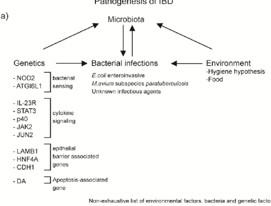
Figure 1: Pathogenesis of IBD
a) There are four factors interacting together that can lead to IBD: genetics, environment, bacterial infections and the microbiota of the individual. Polymorphisms in certain genes can increase susceptibility to some infections, but also influence types of bacteria colonizing the gut. The composition of the microbiota can increase or decrease susceptibility to specific infections, but bacterial infections can modify the gut microbiota. Finally, different environmental factors, including food and cleanliness, can change the frequence of bacterial infections and the composition of the gut microbiota. b) The order in which the different factors can lead to IBD is not unanimous in the scientific community. In the first hypothesis, the gut microbiota in an individual is normal, but abnormal immune responses, triggered by genetic susceptibility or bacterial infection, disturb the normal immune responses, leading to IBD. The second hypothesis proposes that abnormal gut flora is the first factor leading to abnormal immune responses, which increase in genetically susceptible individuals, leading to IBD.
Hygiene hypothesis
One of the reasons frequently evoked to explain why those autoimmune diseases are more prevalent in industrialized nations is due to the decrease of infectious agents encountered during childhood in those specific environments. Some scientists even suggest that the lower number of infections encountered throughout childhood is the single most important factor[8]. This explanation is also known as the hygiene hypothesis.
For example, the frequency of intestinal infections in children has massively decreased in Western countries when compared to developing countries. These intestinal infections are crucial in shaping the intestinal flora. Parasitic infections can also stimulate Toll-like Receptors (TLR), leading to the production of various pro and anti-inflammatory cytokines, which can help in intestinal repairs and control of bacterial infections, both elements which can be defective in IBD[12].
Bacterial infections
Studies of blood and gut samples from IBD patients sometimes incriminate one or another bacterium, such as adherent enteroinvasive Escherichia coli and Mycobacterium avium subspecies paratuberculosis [3]. For example, biopsy samples from CD patients showed a subspecie of E. coli that adhere to intestinal tissue in CD patients than in healthy controls. However, transfer of intestinal bacteria into colitis susceptible monkeys was not sufficient to induce IBD, demonstrating the importance of other factors to induce IBD[13]. Another example of bacteria suspected to play a role in IBD is M. avium subspecies paratuberculosis, which is known to cause Johne’s Disease in cattle, a disease highly similar to CD. Some studies reported identification of M. avium subspecies paratuberculosis in some CD patients, but this bacterium is not systematically found in patients samples analyzed[13].
Microbiota
Even if we humans like to think that we are “clean”, we are in fact colonized by hundreds of billions of bacteria. The gut contains the largest amount of those bacteria,
followed by the skin. Humans have an extremely diversified intestinal flora. One of the first indications that bacteria have a role to play in IBD was demonstrated by the efficacy of antibiotics in inducing remission and decreasing symptoms in IBD patients[3].
Furthermore, many mouse models of IBD are resistant to methods of inducing colitis when they are highly treated with antibiotics or deprived of a normal gut flora[9]. However, in other settings, mice deprived of gut microbiota can be more susceptible to colitis, demonstrating a protective role of gut bacteria against colitis induction. Also, some IBD mouse models, such as the Helicobacter hepaticus transfer model, rely on the addition of a specific bacterium to induce gut inflammation[14]. Finally, many genes linked to IBD in humans have roles to play in innate immune responses, and especially in bacterial recognition[3]3.
Genetics
To better understand and identify genes that could be involved in IBD, various genetics studies have been done through the years. More than 30 susceptibility loci have been associated with IBD[15]. A more recent analysis using even more patients and healthy controls increased the number of CD risk loci to 71[16]. CD and UC both share similarities in their pathogenesis yet are different in other ways, as reflected by the genetic polymorphisms linked with the diseases; some are associated with CD only, some are linked with UC only and others are linked with both diseases. [17]. For example, genetic polymorphisms present in genes of the IL-23R pathway, such as STAT3, p40 and IL23R are linked to both UC and CD[1]. However, the first genetic polymorphism linked with increased susceptibility to IBD was NOD2, and is only associated with CD[17]. Polymorphisms in ATG16L1, a gene associated with both autophagy and NOD2 signaling, are also only associated with CD. Polymorphisms in DAP, death associated protein, are often linked with UC[18]. Other genes possibly involved in UC pathogenesis include epithelial-barrier associated genes, such as HNF4A, CDH1 and LAMB1[18]. However, these UC-only genes will not be further
discussed in this study, as they are not directly linked to immunology, even if they may be crucial in understanding the full scope of pathogenesis of the disease.
One of the tools used to understand the genetics of IBD is the genome wide association study (GWAS), where genomes from patients are compared to genomes from healthy controls. Using genetic markers, there is an attempt to identify allelic patterns, where rare alleles are more frequently associated with one phenotype[17]. Through GWAS, many genetic loci were identified, revealing candidate genes involved in the pathogenesis of IBD. Only a few of these genes will be discussed herein.
NOD2
The first gene to be associated with CD was NOD2(encoded by CARD15)[19]; this gene is only associated with CD and not UC. The NOD2 protein can sense muramyl dipeptide, a subunit of peptidoglycan found in bacteria. So far, three different polymorphisms of NOD2 have been identified, all of which are found in the LRR coding domain which detects the presence of MDP when expressed in monocytes[17]. This leads to a reduced detection of the PAMPs in bacteria and a subsequent decreased activation of the NF-κB pathway. However, NOD2 genetic polymorphism are not enough to induce CD on its own; only 15% of CD patients have one or two polymorphic alleles (SNP identified through GWAS) of NOD2[20], so most CD patients (85%) have a disease independent of NOD2 mutations.
The mechanism by which NOD2 could increase disease risk is poorly understood. One hypothesis is that NOD2 signalling can regulate and decrease TLR2 signaling; without NOD2, TLR2 signaling induces an increased amount of IL-12 and leads to dysregulated immune functions[20]. However, there is increasing evidence that IL-12 is not the most important cytokine involved in the pathogenesis of IBD. In mouse models of colitis, NOD2 deficiency is insufficient to cause colitis by itself; another stimulus is required to induce gut inflammation.
ATG16L1
ATG16L1 is important in autophagy, the biological process by which the cell “recycles” its components. Autophagy is central for antigen preparation, protein degradation and other important processes of immune responses[21]. In a North American-wide study, a polymorphic allele of ATG16L1 was the third most frequent polymorphism associated with CD, after IL-23R and NOD2[21]. Other studies, including one in Germany, replicated these findings[22]. Interestingly enough, ATG16L1 is highly expressed in the epithelium layer of the intestine, as well as in APC and T-cells, and is important for responses against Mycobacterium infections, sometimes suspected in the pathogenesis of CD[11].
A link between ATG16L1 and NOD2 signaling has recently been revealed; NOD2 signaling is essential for the formation of the autophagosome following bacterial stimulation[23]. Without autophagosome formation in DCs, antigen presentation is impaired and the immune response against the bacterium is impaired. This could explain how polymorphisms in both ATG16L1 and NOD2 could work together to create susceptibility to IBD.
Polymorphisms in genes involved in bacterial handling and recognition, such as NOD2 and ATG16L1, increase susceptibility to IBD. Those genes are mostly expressed in epithelial cells and DC. Bacterial signaling through NOD2 and ATG16L1 is one of the first steps in the induction of the immune response. Those immune responses, when out of control, can lead to chronic inflammation and IBD. The signaling through NOD2 and ATG16L1 also induces a plethora of inflammatory mediators, but also the induction of the adaptive immune response. An example of an important inflammatory cytokine is IL-23. It is known that DCs are an abundant source of IL-23 and IL-12; could the signaling induced by those cytokines be important in the pathogenesis of IBD? Genetic studies point towards an involvement of IL-23R and its signaling cascade.
IL-23R
The polymorphism identified in IL-23R was the second IBD-associated polymorphism found in humans[24]. After a GWAS study comparing approximately 600 healthy controls with 600 patients having ileal CD, a polymorphism was found in the intracellular domain of the receptor where an arginine at position 381 is modified for a glutamine (R381Q polymorphism). This polymorphism is protective against colitis. In addition to the R381Q polymorphism, nine other polymorphisms were identified in the IL-23R region, including in the intergenic region between IL-23R and IL-12Rβ2 (both genes are next to each other on chromosome 6), but no polymorphism were identified in IL-12Rβ2. Other studies also found involvement of IL-23R with psoriasis and ankylsoing spondylitis[25].
Following the identification of this polymorphism, multiple studies have tried to identify the biological impact of the amino acid substitution. One of them took CD4+CD45RO+ T-cells from patients with either WT or R381Q coding alleles and activated them with αCD3 and αCD28 antibodies[25]. Measurements of INFγ, IL-17 and IL-22 revealed that the cells coming from individuals with the R381Q polymorphism secreted less cytokines than cells coming from individuals with the wild-type allele. R381Q CD8+ T-cells stimulated in the same conditions also produced less cytokines. IL-23 induced STAT3 phosphorylation was also decreased in R381Q cells. The R381Q polymorphism induces a change in the amino acid sequence of the IL-23r4 subunit. This amino acid substitution is between the transmembrane domain of the receptor and the possible JAK2 recruitment site[26]. This could lead to either impairment of recruitment of the receptor to the cell surface or problems in the downstream signaling cascade. Failure or decrease of JAK2 recruitment could lead to decreased activation of STAT3 (and other factors), as well as decreased cytokine production. This group’s findings were also consistent with other studies where IL-23 signaling on R381Q T-cells led to less phosphorylation of STAT3, but also STAT1. Another study demonstrated that the number of circulating Th17 T-cells was not different between the two alleles, but the responses and pathogenicity following IL-23 stimulation was decreased in patients bearing the protective allele[27]. Together, these
4 IL-23R is the receptor composed of two subunits, IL-23r and IL-12rβ2. Further explanation on the
findings indicate that IL-23R signaling is essential in the pathogenesis of both UC and CD. Furthermore, less signaling through the receptor following cytokine binding could lead to decreased inflammatory signalling and therefore protects against colitis.
Among other genetic polymorphisms identified in IL-23R which differ between healthy controls and IBD patients, one of them, r10889677, was found to be in the 3’ untranslated region (3’UTR) of the receptor. This polymorphism altered the sequence in the UTR of the IL-23r mRNA which led to a different mRNA[28]. People bearing this variant produced more IL-23R protein. One mechanism proposed was that miRNA Let-7 could not bind and regulate the translation of the IL-23R mRNA, leading to increased signaling and immune responses, including feedback regulation. Patients with a difference in the UTR region are more susceptible to colitis, as more signaling through IL-23R could induce more inflammation.
As IL-23R variants seem to have very strong implications with IBD, other genetic studies looked carefully at possible genes involved in the IL-23R signaling cascade and their possible implication in increasing or decreasing susceptibility to IBD. Susceptibility alleles were found in IL12B(p40, which forms IL-23 when paired with p19 or IL-12 when paired with p35), STAT3, IL12RB1, JAK2 and JUN2[29, 30]. Those genes are essential in the activity5 and signaling cascade of IL-23R[17].
Since the identification of different polymorphisms in IL-23R, many investigators have tried to create new therapies to control IBD by modulating IL-23 and IL-23R responses. One study used ustekinumab, a α-p40 monoclonal antibody against IL-12 and IL-23. This randomized trial did not lead to dramatic effects when compared to placebo. It did have a beneficial effect for some patients, but overall, did not lead to drastic improvements[31], which could indicate the possible need for therapies targeted only to IL-23R and not both IL-12R and IL-23R.
So, even though some genetic variants are found in CD and not UC and vice-versa, IL-23R is common to both diseases. Understanding the roles of IL-23R in one or both
diseases is crucial for diagnostics, prevention, and treatment of IBD, which are all currently strongly lacking in the field of IBD.
2. IL-23R and IL-12R biology
IL-12R/IL-23R/IL-35R/IL-27R family of receptors
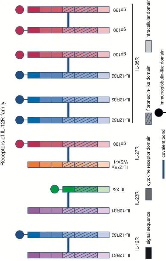
The IL-12R family, which includes IL-12R, IL-23R, IL-35R and IL-27R, are all heterodimeric receptors : they are made of two different subunits linked together with covalent bonds[32, 33](Figure 2: Receptors of 12R family). 12Rβ1 and IL-12Rβ2(which form together IL-12R) both have homology to gp130[34], one of the subunits of IL-27R. IL-23r is highly similar to IL-12Rβ1 and IL-12Rβ2: all three subunits contain in their extracellular domain a signal sequence, two cytokine receptor domains, but only IL-23r and IL-12Rβ2 possesses an N-terminal Ig-like domain[35]. However, IL-23r, unlike IL-12Rβ2 and gp130, does not contain three fibronectin-like extracellular domains[35]. In humans and mice, IL-23r and IL-12Rβ2 are 150kbp apart on chromosome 1 or 6, respectively.
It is sometimes hard to differentiate between IL-23 receptor, composed of two different subunits, and the IL-23r subunit itself. The ambiguity between IL-23R and its IL-23r subunit will be resolved in this thesis in the following way: IL-23r with a small r, will denote the subunit, while IL-23R with the capital R, will denote the functional IL-23R protein, made of the IL-23r and IL-12Rβ1 subunits.
IL-27R is composed of gp130 and WSX-1(also known as IL-27Ra6)[36]. gp130 also possesses an N-terminal Ig-like domain7. IL-35R can act through three different receptors: heterodimers made of gp130 and IL-12Rβ2, homodimers made of gp130 or homodimers made of IL-12Rβ2[37]. Each receptor is comprised of subunits highly similar to one another, however, they bind to different cytokines and are expressed in different cell types, leading to very different in vivo roles. One must be also cautious when analyzing mice knock-out data; for example, IL-12Rβ2KO mice lack responsiveness to IL-12, but could also show decreased Treg functions through the lack or the decreased number of IL-35R.
6 http://www.informatics.jax.org/marker/MGI:1355318 7 Idem
Figure 2: Receptors of IL-12R family
The IL-12R family are all heterodimeric receptors (IL-35R can be composed of IL-12Rβ2 or gp130 homodimers). All the subunits contain a signal sequence, a fibronectin-like domain and an intracellular domain. All receptors contain at least one subunit with an immunoglobulin like domain. The two subunits are linked together through a covalent bond. IL-23r is the only receptor subunit without a cytokine receptor domain, which probably explains why IL-23 is known to mostly bind to the IL-12Rβ1 subunit.
IL-12/IL-23/IL-35/IL-27/p40 homodimer families of cytokines
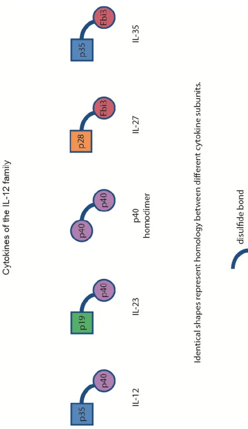
IL-12 family cytokines are heteromeric complexes, where two different subunits linked with a disulfide bond form a functional cytokine[33]. IL-12 is made up of a p35 chain and a p40 chain. IL-27 is made up of p28 and EBI3, which are homologuous to p35 and p40, respectively[33]. EBI3 can also bind to p35, forming the IL-35 cytokine. IL-23 is composed of p40 and p19, where p19 is also homologuous to p35.
The result is therefore four highly similar cytokines. However, even though they share many similarities they have drastically different roles. IL-35 is essential for the suppressive activity of Tregs in mice. IL-12 induces INFγ, inhibits IL-17 and is characteristic of Th1 response. IL-23 leads to IL-17 cytokine production and Th17 responses and IL-27 can induce Th1, but strongly inhibits the development of Th17[33]. Addition of IL-35 in T-cell proliferation assays will strongly decrease the proliferation of T-cells. This inhibition of proliferation is similar to the effect of the addition of Tregs in the same assay[38]. IL-35 is secreted by Tregs and can act on other T-cells to restrain their proliferation[39].
The last and often overlooked member of this cytokine family is the p40 homodimer. This homodimer can bind to IL-12Rβ1 with both high and low affinity[40]. When injected in vivo following LPS injection, p40 homodimer decreases levels of INFγ in the serum[41]. This homodimer can be either an agonist or an antagonist[42] of IL-12 secretion, and thus regulates IL-12 signaling. Because of the strong links and shared subunits between the cytokines, one must be careful when interpreting data from murine cytokine knock-outs8.
To understand the role of each cytokine, one must understand their source and the stimuli leading to 12, 23, 27 and 35 secretion. DCs are a source of IL-12, IL-23 and IL-27 following stimulation through different TLR signaling cascades[33], while IL-35 is mostly secreted by Tregs[39]. The quantities of each cytokine found during an immune reaction depend on the ligands recognized by the DC.
Figure 3 : Cytokine structure
Ebi3 and p40 are homologuous to each other. Both are thought to be constantly produced in various cell types, especially DC and monocytes. Upon signaling through PAMPs or other signals, p35, p19 and p28 are believed to be produced. The different subunits then bind to p40 or Ebi3 through a covalent bond and are secreted as an heterodimer by the various cells. The p40 homodimer secretion and biology is not well understood.
For example, signaling through TLR2 will induce strong secretion of p40 and p19, but no secretion of p35[33]. As is frequently seen in inflammatory responses, NF-κB is known to bind to the p40 promoter[43] and probably regulates the secretion of IL-23 and IL-12. The strongest p40 promoter activity by NF-κB was recorded in the intestine of mice and is therefore of interest when studying the biology and role of IL-23R in the gut. Ebi3 and p40, the α chain of the cytokines, are secreted in high numbers, while the β chain (p19, p35) is the limiting factor of cytokine secretion and depends on the stimulation of both APCs and Tregs[39].
Soon after the identification of p19, mice ubiquitously expressing p19 subunit were created[44]. Those animals suffered from impaired growth, infertility and most importantly, systemic inflammation. In naïve WT mice, p40 mRNA is produced at high levels in many different cells without any inflammation. Levels of p40 mRNA production did not increase in the p19 ubiquitous mice, but high levels of IL-23 were found, as well as systemic inflammation. This indicates that p40 is always secreted, but needs production of p19 to induce its effects. However, it could also mean p19 could have biological effects independent of p40.
IL-23R biology
Signals leading to the upregulation of the receptor
Naïve CD4+ T-cells do not express IL-23R [35]. When naïve CD4+ T-cells were transfected with a hyperactivated STAT3 and cultured with IL-6, TGFβ and αIL-4 antibody, IL-23R mRNA levels increased seven fold compared to cells not transfected, demonstrating that STAT3 is important in the upregulation of IL-23R[45]. One of the downstream signals of IL-23R, IL-17, increased dramatically when compared to controls, showing that mRNA was translated into proteins and affected cell signaling. Under the same conditions, using STAT3 KO naïve T-cells, IL-23R mRNA slightly decreased, but other downstream cytokines such as IL-22 and IL-17 decreased tremendously, demonstrating a relationship between STAT3, 17 and IL-22. Another group also showed with the same STAT3 CD4+ T-cell-deficient mouse model that STAT3 was required for the upregulation of IL-23R mRNA following IL-6, TGFβ or IL-21 exposure[46]. Together, these studies indicate an important role of STAT3 in IL-23R upregulation.
APCs can secrete cytokines which induce naïve T-cells to differentiate into Th17; these cytokines include TGFβ, IL-6 and IL-21. Th17 cytokines can also induce IL-23R at the surface of naïve cells. Using RT-qPCR analysis, one group demonstrated that IL-6 (in combination with αCD3/αCD28 stimulation) could lead to up-regulation of IL-23R[46]. IL-21 could also upregulate IL-23R. However, in presence of TGFβ+IL-6 and/or IL-21, IL-23R mRNA did not increase as much. According to the authors of this article, and in contrast to previously published papers, TGFβ actually had an inhibitory effect on the induction of IL-23R mRNA.
Interestingly, even though both IL-23R and NOD2 have been identified as important for the pathogenesis of IBD in genetic studies colitis induction by C. rodentium or S. thyphimurium have been shown to induce colitis in mice, Nod1 and Nod2 are apparently not important for the upregulation of either IL-23 or IL-23R[47]. The role of ATG16L1 in bacterial colitis is important, but its relationship to IL-23R is
unknown. Very little is known about IL-23R induction in other cell types; other data suggest that those cells may not need to upregulate IL-23R because they constitutively express it, even in a steady state.
Signals at the receptor levels
When IL-23R was first characterized in humans, it was found that IL-23 could bind to both subunits IL-23R and IL-12Rβ1 [35]. In the absence of IL-23, IL-12Rβ1 and IL-23R are not bound together; the binding of the cytokine brings the two subunits in close proximity and leads to the dimerization of the receptor. Following dimerization, signaling can take place through IL-23R and the IL-23 cascade is therefore activated[48].
Signaling cascade
The signaling cascade of IL-23R following IL-23 binding is highly similar to IL-12R, which uses JAK2, TYK2, STAT1, STAT3, STAT4, AND STAT5[35]. Signaling through IL-23R mainly induces the phosphorylation of STAT3, but also weakly phosphorylates STAT4 (and STAT1/5)[45] in Th17 cells. Tyrosine Kinase 2 ((TYK2) and JAK2 are bound to the IL-12Rβ1 subunit, which will phosphorylate STAT3 upon IL-23 binding. This stimulation induces IL-17 secretion in naïve T-cells; in the absence of Tyk2, IL-17 levels are decreased, indicating an important role for Tyk2 in inducing IL-17 production following IL-23 stimulation[49].
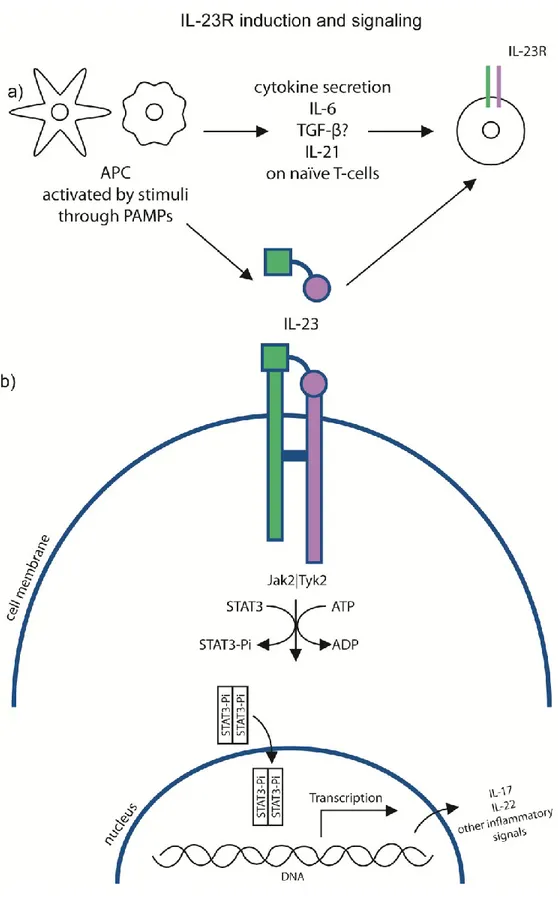
Figure 4: IL-23R induction and signaling cascade
a) APCs are one of the main source of cytokines leading to the upregulation of IL-23R. APC are also a good source of IL-23. The stimuli activating APC will determine the nature of the cytokines.
b) Following 23 binding to 23R, a covalent bond will be formed between the two subunits of IL-23R and the downstream signaling cascade will be activated. Kinases will phosphorylate STAT3, which will dimerize. Once the dimerized-STAT3 reaches the nucleus, it will activate the transcription of various inflammatory signals, including IL-17 and IL-22.
IL-12R biology
2.1.1 Signals leading to the upregulation of the receptor
IL-18, another very important Th1 cytokine, just like IL-12[50], is known to be important in the upregulation of IL-12Rβ2 on the surface of developing Th1 cells, which assembles with IL-12Rβ1 and produces the functional IL-12R[34, 51]. IL-27, another member of the IL-12 family, is also believed to be an early Th1 cytokine which would lead to the upregulation of IL-12Rβ2. Following this upregulation, IL-12 could then signal through IL-12R and stabilize the Th1 phenotype[32]. Signals that could lead to IL-12R upregulation in cell types other than T-cells are not very well-known.
Signals at the receptor levels
Even though IL-12R is made up of IL-12Rβ1 and IL-12Rβ2 in both mice and humans, the affinity of IL-12 for its receptor is different. In humans, IL-12 binds with low and high affinity to both subunits; in mice, IL-12 seems to bind very strongly with IL-12Rβ1 but only possesses low affinity for IL-12Rβ2[52]. However, in mouse studies, it was showed that without IL-12Rβ2, STAT4 phosphorylation was greatly reduced and INFγ production (a hallmark of IL-12 activity) was inhibited in response to IL-12[52]. So, even though IL-12 only weakly binds to IL-12Rβ2, this subunit is essential for signaling.
Signaling cascade
Upon binding of IL-12 to IL-12R, the Jak2/Tyk2 kinases associated with the receptor phosphorylate the receptor's tyrosine residues [53]. IL-12 binding to IL-12R leads to the activation (dimerization and phosphorylation) of STAT4, but also of STAT1,3,5[34, 45]. The activated STATs can than translocate into the nucleus and activate a plethora of genes, including IFNγ. One of the hallmarks of IL-12 signaling is INFγ secretion; in the absence of Tyk2, INFγ secretion after IL-12 stimulation is greatly reduced[49] (Figure 5).
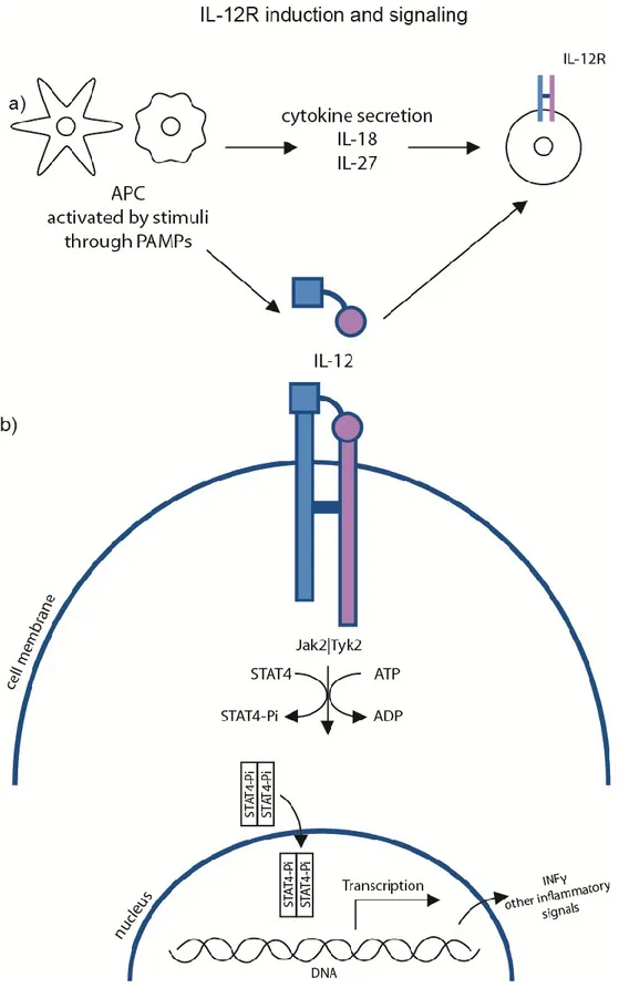
Figure 5: IL-12R induction and signaling cascade
a) APCs are one of the main source of cytokines leading to the upregulation of IL-12R. APC are also a good source of IL-12. The stimuli activating APC will determine the nature of the cytokines.
b) Following IL-12 binding to IL-12R, the downstream signaling cascade will be activated. Kinases will phosphorylate STAT4, which will dimerize. Once the dimerized-STAT4 reaches the nucleus, it will activate the transcription of various inflammatory signals, including INFγ.
IL-23R and IL-12R functions
Th17 T-cells
Th17 T-cells are CD4+ T-cells that secrete IL-17 [54]. Th17 cells may play an important role in defending against extracellular pathogens[54]. Other experiments demonstrated that Th17 cells were highly inflammatory and very important in animal models of inflammatory diseases, including colitis and EAE [54]. IL-23 is required for the maintenance and pathogenicity of Th17[54]. Th17 can also produce IL-22 and other inflammatory cytokines.
Two different types of Th17 have recently been described: the classical Th17, which are differentiated from naïve T-cells using IL-6 and TGF-β, and "alternative" Th17 cells which were induced using IL-6, IL-23 and IL-1β[55]. T-cells can differentiate into Th17 in the absence of IL-23/12 [56], but can also form Th17 pathogenic cells in the absence of TGF-β[57]. Th17 cells differentiated in the presence of IL-23 had a more pathogenic phenotype than the classical ones; this could be one way in which IL-23 is important to induce inflammation, through production of pathogenic Th17 cells. Another mechanism by which IL-23 seems to affect T-cells function is through the inhibition of IL-10; without IL-10, Th17 may lack this feedback inhibition loop which could hinder their immune responses [55]. IL-23 can also induce GM-CSF production from Th17 T-cells; this cytokine has potent effects on APCs, leading to even more IL-23 production and increasing the inflammatory environment [58] and the potency of pathogenic Th17 cells.
γδ T-cells
γδ cells are a rare subtype of cells, representing less that 5% of total T-cells[59]. However, they are enriched in epithelial tissues, such as the skin and are found in high numbers in the gut. Unlike αβ T-cells, they do not need APC to be activated; their T-cell receptors recognize small bacterial patterns and not specific antigens [59]. γδ T-cells found in epithelial tissues are mature and can either be important in homeostatic processes or in the defence against pathogens.
Using the IL-23R-GFP reporter mouse, it was previously demonstrated that γδT-cells express IL-23R at steady state[60]. IL-23R+ were CCR6+ and INFγ-, while IL-23R- γδT-cells were CCR6+ and INFγ+[61]. γδT-cells stimulated with IL-23 induced IL-17, IL-22 and IL-21; this cytokine production was dependent on IL-23R.
Th1 T-cells.
Th1 T-cells are essential for clearance of intracellular pathogens[62]. They are characterized by their high secretion of INFγ. The most important Th1-inducing cytokines are derived from APCs; they are IL-12 and IL-18[50]. DCs and macrophages, upon activation, will secrete various cytokines; if they are primed to secrete IL-12 and IL-18, it will lead to the up-regulation of IL-12R and differentiation of naive T-cells into Th1 cells.
Up-regulated Th1 T-cell responses have been associated with many diseases, including multiple sclerosis, rheumatoid arthritis, lupus and type I diabetes [62]. On the other hand, down-regulation of Th1 responses leading to a decrease in IL-12 can lead to increased malignancy. It has also been shown that increasing the concentration of IL-12 in tumors can decrease the immunoregulatory environment of the tumor and increase the immune response against the malignancy[63].
a)Th17/Th1 dichotomy
Generally speaking, cytokines leading to the differentiation of a certain Th subtype will also inhibit the differentiation of another subtype. For example, Th17-inducing cytokines will inhibit the formation of Th1[50].
However, even if Th1 cells are associated with INFγ production and Th17 cells with IL-17 production, it was identified that in situations of autoimmunity, for example, Th17 cells actually produced INFγ[51]. In a study by Lee et al., OT-II naïve CD4+ T-cells in culture with irradiated splenic cells p40KO, OVAp9, αINFγ,
9 OVA, short for ovalbumin, is the specific peptide recognized by OT-II (CD4+ T-cells).In this
4(both antibodies), IL-6 and TGF-β for seven days gave rise to Th17 cells. About half of the Th17 differentiated cells produced IL-17, but few produced INFγ or INFγ and IL-17. Those Th17 differentiated cells were then transferred into new culture conditions. Upon stimulation with IL-12 (and OVAp, irradiated p40KO splenic cells, and αIL-4/αINFγ), more than 60% of the cells started producing INFγ. However, when stimulated with IL-23, 50% of the cells continued the production of only IL-17. This experiment shows that even if Th17 do not express (or express very low levels of) IL-12Rβ2, they can still respond to IL-12 and produce INFγ. In the same study, the authors also showed that TGFβ and not IL-23 was required in vitro to maintain IL-17 production by Th17 cells. These experiments cast a shadow on the specific response to IL-12 and IL-23; nevertheless, all of these results were obtained in vitro and merits verification in an in vivo model. Transcriptome analysis of Th17 cells cultured with IL-12 revealed a Th1-skewed gene expression, which shows a plasticity in Th17 cells depending on the cytokine environment, but most importantly a responsiveness to IL-12 even when expression of IL-IL-12Rβ2 is very low. It could be that this responsiveness to IL-12 in vitro is an artefact of the in vitro conditions[64]. IL-23 is important in driving inflammatory responses in the gut; this could include the production of INFγ by Th17 following IL-23 stimulation[64].
Other cell types
The importance of IL-23R in certain T-cell subtypes is reasonably well understood, but IL-23R expression is not restricted to T-cells. Experiments measuring IL-23R expression in other cell types are rare. One of the reasons is due the lack of appropriate antibodies to study 23R expression. Very often, in order to measure IL-23R presence, cells are stimulated with IL-23 and cytokines such as IL-22 and IL-17 are measured to evaluate IL-23 responsiveness. It is interesting how some articles describe certain cell populations as IL-23 responsive and IL-23R expressing[65], as measured by IL-23R mRNA in certain cells populations, but others respond to IL-23 even in the presence of only very small quantities of IL-23R mRNA. This could indicate that IL-23 may signal through a receptor other than IL-23R; it could also mean that only small quantities of mRNA are required for protein production and signaling.
a) NK and NKT-cells
In a recent paper, DX5+TCRβ+ cells, known as NKT-cells, were found to secrete high amounts IL-17 when stimulated with αCD3 and IL-23. Those cells also expressed IL-23R mRNA before stimulation. NK cells, on the other hand, did not respond to the same stimulation and did not express IL-23R mRNA [66]. Those NKT-cells, when stimulated with either IL-23 or αCD3, still produced IL-17. The authors also isolated NKT-cells from an 23RKO mouse and stimulated them with 23. IL-23 stimulation IL-IL-23RKO NKTcells did not induce IL-17 production. Further characterization revealed that NK1.1- NKT cells were responsible for producing the most IL-17 following IL-23/αCD3 stimulation. In this article by Rachitskaya et al, there is a suggestion that early IL-17 production following splenocyte stimulation with αCD3 and IL-23 is made by NKT-cells and that this mechanism is independent of IL-6, which would be a mechanism different from the one inducing IL-23R in T-cells.
It is generally believed that NK cells can directly respond to IL-12[67]. It is also known that IL-12 stimulation of NK cells in the inflammatory phase of immune responses induces INFγ[34]. IL-12 is usually associated with activation and proliferation of NK cells. IL-12 stimulated NK cells are also strong producers of INFγ.
b) B-cells
No expression of either IL-12R or IL-23R by B-cells in mice has been reported in the literature. Interestingly, it is known that IL-12Rβ2 acts as a tumor suppressor gene in human chronic B cell malignancies[68, 69], which led the authors of that paper to believe that IL-12/IL-12R signaling could be important in the regulation of B cells. Another study from the same group showed that some B cells in human tonsils expressed IL-12Rβ2 mRNA[70]. In the long-term study of IL-12Rβ2KO mice10[69], it was observed that B220+ B cells were activated and plasma-cell hyperplasia (B-cell malignancies) were high, possibly indicating a role of IL-12Rβ2 in controlling B cell proliferation and malignancy.
c) Dendritic cells, monocytes and macrophages
DCs, monocytes and macrophages are mostly recognized as a source of IL-23 and IL-12, but not as responding to IL-12 and IL-23, which could indicate that they do not express IL-23R and IL-12R[33]. However, there are many reports of INFγ secretion from these cells, including following stimulation with IL-12 alone[71], which would indicate the presence of a receptor to IL-12 on those cells. Others report the requirement of IL-18 and IL-12 through a STAT4 dependent mechanism to induce INFγ secretion[72]. Macrophages are specifically believed to respond to IL-23 and therefore express IL-23R, but data is conflicting about this conclusion[67].
d) Lti-like
Lymphoid tissue inducer cells (Lti) are required during embryonic development to create secondary lymphoid organs, such as lymph nodes [73]. Lti-like cells are cells similar to Lti that continue to be found in both mice and human in adulthood [73]. They can produce IL-22 and IL-17, are dependent on the RORγT transcription factor, can be found in germ-free mice, and can respond to IL-23[73]. They were found to be important in the αCD40 colitis models as a source of INFγ[74]. At the mRNA levels, IL-23R was expressed in high amounts [35]. The definition of Lti-like cells that can respond to IL-23 varies from one research group to another. They usually are Lin-, Thy1 (CD90) high, Sca-1+, and RORγT[64]. Another name frequently used is innate lymphocytes. Lti-like cells were found to increase in numbers during gut inflammation.
IL-22 production by innate lymphocytes is crucial for gut homeostasis, especially for epithelial cell proliferation, mucus production and overall maintenance of the epithelial barrier in the gut[64].
3. Mouse models
Before the discovery of IL-23 and its receptor, all biological phenotypes of p40 were associated with IL-12. Many experiments had to be re-examined and results were re-analyzed. Very often, roles attributed to IL-12 and IL-12R were actually caused by IL-23 and IL-23R.
Roles and importance of IL-23R and IL-12R in mouse models other
than colitis
IL-23R and IL-12R have been studied in animal models of autoimmune diseases, bacterial and fungal infections and malignancies. In this section, a brief overview of the involvement of those receptors in the disease pathogenesis will be given in order to emphasize the importance of studying the biology of 12R and IL-23R.
EAE
Experimental autoimmune encephalitis (EAE) is an animal model of MS, where the animals are immunized with MOG (myelin oligodendrocyte glycoprotein), a peptide of myelin which is often the target of autoreactive T-cells in MS[75]. IL-12 was considered the main orchestrator of brain inflammation through generation of Th1-producing INFγ. However, Cua et al. showed that EAE was totally inhibited in the p19KO and p40KO animal models but not in the p35KO models[76, 77], showing that IL-23 is a crucial cytokine in the development of EAE, while IL-12 is dispensable11. The local IL-23 production in the brain was required for induction of brain inflammation.
γδT cells are IL-23R+ and are known to accumulate in great numbers in the CNS of mice affected by EAE [61]. γδT cells can control and decrease the activity of Foxp3+ Tregs and inhibit the conversion of naїve CD4+ T cells into Tregs [61], which could be one of the ways IL-23R can mediate brain inflammation.
11 In that study by Cua et al., p35KO actually had a worse clinical state after two weeks than the WT
Other articles showed that IL-23RKO animals, similar to p19KO, were resistant to EAE induction, demonstrating the crucial role of the receptor in the induction of EAE [60, 78]. IL-12 is known to be important in the generation of Th1 pathogenic T cells; however, induction of EAE in IL-12Rβ2KO mice resulted in a more severe disease than in controls[79]. p19 mRNA was also higher in IL-12Rβ2KO mice, which may indicate a role for IL-12R in the control of IL-23R mediated inflammation. As one review clearly indicated, IL-23 is crucial for the development of encephalitogenic Th17 and IL-17 production, but also through the modulation of an array of inflammatory cytokines, including GM-CSF and IL-22 and through its actions on a variety of cells not limited to autoreactive T-cells[80].
Collagen induced arthritis
Collagen induced arthritis (CIA) is a murine model for rheumatoid arthritis. Genetically susceptible mice are immunized twice subcutaneously at the tip of the tail with collagen and develop a specific immune response against their joints. Using p35KO and p19KO mice, it was demonstrated that mice lacking IL-23 were completely protected from CIA, while IL-12-deficient mice actually had a worse disease than WT mice[81]. Mice lacking both IL-12 and IL-23 (p40KO) were also protected from CIA. The levels of IL-17 and TNF were slightly higher in the p35 knock-out strain compared to the WT animals, which could explain the slightly worse outcome of those animals. In this model, IL-23 seems to be the pathogenic cytokine, probably through the Th17 and IL-17 pathway[82].
IL-23-induced arthritis
The single hydrodynamic injection of IL-23 minicircle DNA resulted in joint-destructive arthritis [83], clearly demonstrating the possible pathogenic effects of increased IL-23.
Lupus nephritis
C57BL/6–lpr/lpr are susceptible to lupus erythemathosus and are therefore the animal model of human lupus, a disease characterised by systematic inflammation caused by autoreative T-cells and autoantibodies. When bred with IL-23RKO, the progeny were susceptible to lupus had lower cytokines, lower auto-antibodies levels and no nephritis, showing the importance of IL-23R in the pathogenesis of lupus in mice[84].
Infectious diseases
a) Experimental Cerebral Malaria
The role of IL-12/IL-12R in the development of malaria is still not well understood. In a mouse model of experiment cerebral malaria (ECM), it was shown that IL-12Rβ2KO mice did not suffer from ECM and survived much longer than the controls, but IL-12Rβ2KO mice had an unrestrained parasite proliferation and died of anemia[85]. When p40KO, p35KO, p19KO and IL-12Rβ1 animals were infected, they suffered from strong ECM and died at day 5. When this study was published, the role of IL-12Rβ2 in forming IL-35R was unknown; this could be an indication of the importance of IL-35R in the pathogenesis of ECM.
b) Klebsiella pneumoniae
In a K. pneumoniae mouse lung infection model, IL-23 was essential for the production of IL-17 by T cells. Without IL-17, the bacterial burden was much higher. However, using p19KO, p35KO and p40KO mice, they showed that mortality following bacterial infection was the same in p19KO, p35KO and p40KO mice, indicating an important role for IL-12 as well, even if IL-12 was not required for IL-17 production[86].
c) Listeria monocytogenes
Listeria monocytogenes is often used to study immune responses against intracellular bacteria. In a study by Riol-Blanco et al., Riol-Blanco et al. examined the importance of IL-23R during the infection of this pathogen[87]. IL-23R was important
in the early stage of the bacterial infection in order to induce the production of IL-17 by γδT-cells and decrease the liver bacterial burden. However, IL-23R was not required for the immune cells to be recruited to the abdomen following the infection. Also, in the absence of IL-23R, the decrease in IL-17 was correlated with an increase in INFγ, showing a switch from Th17 to Th1 response.
Colitis models
There are currently no colitis mouse models that exhibit all characteristics of IBD seen in humans[9]. However, many mouse models demonstrate that the genes identified in genetic studies in humans were as important to the development of the disease in mice, including IL-23R. Mouse models are relevant to human diseases because many immune responses are homologous between the two mammals and very often rely on the same signaling pathways[9].
However, there are many limitations of mouse models when interpreting IBD in humans[9]:
Murine inflammation is usually found in the colon; most patients suffering from CD have inflammation in the ileum.
Inflammation in the mouse is chronic, while in humans most suffer from a relapse-remitting disease.
Types of mouse colitis models
Depending on what aspects on intestinal inflammation need to be studied, one must use a specific rodent model. There are different approaches for inducing intestinal inflammation[9]:
1. Chemically-induced colitis (such as DSS, Dextran sulfate sodium and TNBS, 2,4,6-trinitrobenzene sulfonic acid)
2. Mixed techniques (such as αCD40 injections in RagKO mice) 3. Genetic modifications (such as IL-10KO)
5. Transfer of cells into a genetically susceptible mouse (such as T-cells transferred into RagKO mice)
a) DSS
DSS is awhite chemical that can easily be dissolved in water12. When added to the mice drinking water at concentrations between 1%-5% it induces an acute colitis characterized by colon shortening, neutrophil recruitment, weight loss, and ulcers in the mucosa of the intestine[88]. This model of colitis induction is useful when studying the importance of innate immune responses in the development of colitis in both SCID[88] and RagKO mice[65], which are deprived of T and B-cells and therefore of adaptive immune responses. However, DSS-induced colitis can also be used in immunocompetent mice, such as mice with T and B-cells.
b) TNBS
The TNBS model is another chemically induced model of colitis; however, unlike DSS, it depends on T-cells to induce intestinal damages[88]. Briefly, TNBS is mixed with ethanol and is given through the rectum. This model uses ethanol and TNBS to break the mucosal barrier in the intestine and induce gut inflammation[89].
c) αCD40 antibodies
Injection of an agonistic CD40 monoclonal antibody in a RagKO mouse induces systemic inflammation and rapid weight loss[90]. Lesions and pathology are observed in the liver and the colon, while the spleen increases in size. Thus, this model is not simply a colitis model; however the intestinal pathologies induced by this model are particularly useful in understanding inflammation in an innate immune model.
To understand the pathogenesis of the disease, monoclonal antibodies against different cytokines, including IL-12 and TNFα, were injected in vivo to understand the role of each cytokine in the pathogenesisof the disease. Amongst the ones tested, anti-p40, a monoclonal antibody against IL-12 and IL-23, was the most effective.
INFγ and-TNF-α inhibited the strong weight loss (wasting disease), but did not have an effect on colitis or cytokine serum levels[90]. The anti-p40 monoclonal antibody treatment indicated a possible role of IL-12 and IL-23 in the inflammation developed after αCD40 injection. In the next section, induction of inflammation with αCD40 in p19KO, p35KO and p40KO mice will be further addressed.
d) IL-10KO
A longitudinal study of IL-10KO mice revealed that many of them spontaneously developed colitis[91]. Inflammation started at three weeks of age, mostly in the colon, but after three months 100% of IL-10KO suffered from intestinal inflammation. Treatment with anti-INFγ decreased the disease severity but did not entirely inhibit it, even if analysis showed a skewed Th1 immune response in the gut of those animals. These findings have been replicated, but demonstrated that IL-23 and not IL-12 was crucial in the development of colitis in IL-10KO animals[92].
e) T-cell transfer
Another interesting model of colitis13 is the transfer of CD45RBhighCD4+ T cells into a SCID or RagKO mouse. Upon this transfer, the mice suffer from a wasting disease, massive weight loss and strong intestinal inflammation[93].
f) Helicobacter hepaticus
H. hepaticus is an intestinal bacteria used to study to role of the microbiota and infections in the development of colitis. When given orally in 129SvEvRagKO, it induces inflammation of the gut, which is then a T-cell-independent gut inflammation[94]. However, if CD45RBhighCD4+ T cells are transferred into the 129SvEvRagKO mouse and H. Hepaticus given orally the gut inflammation is much more severe, thereby showing a T-cell-dependent gut inflammation[94]. In both cases, transfer of Tregs can inhibit the gut inflammation. Transfer of H. Hepaticus in IL-10KO mice also induces colitis[95], but infection with H. Hepaticus in WT animals