ION CHANNELS, RECEPTORS AND TRANSPORTERS
CFTR and defective endocytosis: new insights
in the renal phenotype of cystic fibrosis
François Jouret&Olivier Devuyst
Received: 30 August 2008 / Accepted: 23 September 2008 / Published online: 7 October 2008
# Springer-Verlag 2008
Abstract Inactivation of the chloride channel cystic fibro-sis transmembrane conductance regulator (CFTR) causes cystic fibrosis (CF). Although CFTR is expressed in the kidney, no overwhelming renal phenotype is associated with CF. Recent studies have shown that the level of CFTR mRNA in mouse kidney approaches that found in lung. CFTR is particularly abundant in the apical area of proximal tubule cells, where it co-distributes with the Cl−/ H+ exchanger ClC-5 and Rab5a in endosomes. The biological relevance of CFTR in proximal tubule endocy-tosis has been tested in CF mouse models and CF patients. Mice lacking CFTR show a defective receptor-mediated endocytosis, as evidenced by impaired uptake of 125I-β2
-microglobulin, a decreased expression of the cubilin receptor in the kidney, and a significant excretion of cubilin and its low-molecular-weight ligands into the urine. Low-molecular-weight proteinuria (and particularly transferrinuria) is similarly detected in CF patients in comparison with normal controls or patients with chronic lung inflammation. These studies suggest that the functional loss of CFTR impairs the handling of low-molecular-weight proteins by the kidney, supporting a role of CFTR in receptor-mediated endocytosis in proximal tubule cells. The selective proteinuria should be integrated in the pathophysiology of multi-systemic compli-cations increasingly observed in CF patients.
Keywords Proximal tubule . Kidney . Chloride channel . Cystic fibrosis transmembrane conductance regulator . Protein metabolism
Cystic fibrosis: pathology and genetics
Cystic fibrosis (CF, OMIM #219700) is the most common lethal autosomal recessive disease in Caucasians, affecting as many as one in 2,500 live births. Cystic fibrosis is a multisystemic disease that essentially results from the obstruction of exocrine glands by an excessive mucus production. The accumulation of thick mucus in the human airways causes obstructive lung disease promoting chronic bacterial infection and inflammation, the hallmark of CF. A similar mucus accumulation in pancreatic ducts, bile ducts, and the intestine may lead to pancreatic insufficiency, liver damage, and meconium ileus (intestinal obstruction), respectively, in some patients with CF [73]. In addition, the absence or obstruction of the vas deferens causes male infertility in most CF patients, and altered cervical mucus production reduces female fertility [55]. The airway manifestations represent the main cause of morbidity and mortality in CF patients. Nowadays, improvement in medical care often preserves life expectancy beyond the third decade [73].
The fact that patients with CF have a typically high concentration of NaCl in the sweat, coupled with an increased lumen-negative transepithelial voltage, pointed early on to a defective chloride transport and salt homeostasis as the cause of organ damage in CF [73,79]. In 1989, Collins, Riordan and colleagues demonstrated that CF was due to loss-of-function mutations in theCFTR gene that encodes a Cl− channel named CFTR—for cystic fibrosis transmembrane conductance regulator [48, 71]. The CFTR gene, also named as ABCC7, is located on chromosome 7q31.2 and spans approximately 290 kb of genomic DNA (27 exons) encoding a 1,480-amino acid protein [20]. Over 1,000 CF-associated mutations have been reported thus far in CFTR (CF Genetic Analysis Consortium, http://www.genet.sickkids.on.ca/cftr/) and
DOI 10.1007/s00424-008-0594-2
F. Jouret
:
O. Devuyst (*) Division of Nephrology,Université catholique de Louvain Medical School, Avenue Hippocrate, 10,
1200 Brussels, Belgium
have been classified into five groups according to their structural or functional consequences on Cl− conduction [73]. The in-frame deletion of three bases encoding a phenylalanine residue at position 508 (ΔF508) represents the most common mutation in CF population, affecting the correct processing and maturation of CFTR [5]. About 5% to 10% ofCFTR mutations are due to premature truncation or non-sense alleles and are associated with the most severe CF phenotypes [74]. Most otherCFTR defects are unique to a particular family or to only a handful of cases across the world. The type of CFTR mutations seems directly linked to the pancreatic phenotype, whereas the high variability in pulmonary complications among siblings carrying identical mutations strongly supports the influence of the environment and modifier genes in the disease severity [74]. Moreover, patients with the CF phenotype have been reported with no or only one mutation in the CFTR gene [29]. These cases exemplify the broad clinical spectrum ofCFTR-linked disease and support the existence of additional genes involved in the CF phenotype.
Structure and function of CFTR
The CFTR protein is a member of the adenosine triphosphate (ATP)-binding cassette (ABC) superfamily of integral mem-brane transporters [25]. The ABC transporters function as mediators of unidirectional organic solute transport and include multidrug resistance proteins, such as MDR and P-glycoprotein, and a number of prokaryotic and eukaryotic small nutrient and molecule transporters [38]. CFTR is organized symmetrically in two transmembrane domains (TMD1 and TMD2) and two nucleotide binding domains (NBD1 and NBD2), separated by a large, polar, regulatory (R) domain unique within the ABC family [71]. Each membrane-spanning domain contains sixα helices, portions of which form the Cl− pore. CFTR is regulated by cyclic adenosine monophosphate (cAMP)-dependent phosphoryla-tion of the R domain via protein kinase A (PKA), followed by ATP-dependent gating events initiated by ATP binding to the cytoplasmic nucleotide-binding domains (NBD1 and NBD2) and resulting in transepithelial Cl−transport [25,79]. The N and C termini of CFTR are both intracellularly oriented. The C terminus harbors a conserved type I PDZ domain-binding motif, which interacts with several PDZ-domain proteins including the Na+/H+exchange regulatory factors (NHE-RF1 and NHE-RF2) and the actin-binding protein ezrin [31]. The dynamic regulation of CFTR binding to such scaffolding proteins may determine its dimeric organization into macromolecular functional units containing regulatory partners and other channels. Further-more, the phosphorylation of the R domain is also regulated by NHERF1, NHERF2, and ezrin [32]. The insertion of
CFTR channels in the plasma membrane also involves a complex of proteins including the PDZ-domain proteins NHERF1 and CAL as well as the SNARE SYN6 and the Rho GTPase TC10 [6]. Consequently, molecular switches regulate CFTR-mediated Cl− secretion by modulating both its channel activity and its intracellular trafficking [52]. On the other hand, PKA is involved in the exocytic insertion and endocytic retrieval of CFTR from the plasma membrane and distinct Rab GTPases, which are small catalysts responsible for cargo selection, vesicle motility, and endosome docking and fusion to the appropriate membranes, participate in the intracellular transport and the plasma membrane localization of CFTR [32]. CFTR interacts functionally with other channels, including the outwardly rectifying Cl− channels and the Na+channel ENaC, and it participates in exocytosis and the formation of macromolecular complexes at the plasma membrane, in close contact with receptors, signaling proteins, and the cytoskeleton [32]. Therefore, the role of CFTR extends well beyond Cl− permeability, as supported by its unique structure and its subcellular distribution in epithelial cells.
Segmental and subcellular distribution of CFTR in the kidney
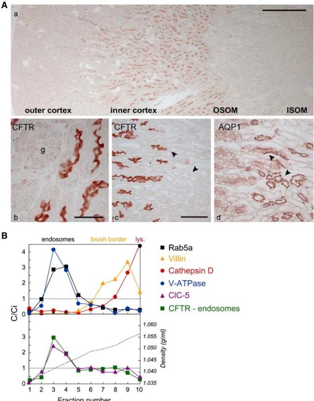
CFTR is located in the apical membrane of numerous secretory epithelia, including airways, colonic crypts, pancreatic and sweat ducts, and male genital tract [79]. However, several studies have demonstrated the expression of CFTR in the developing and mature mammalian kidney [12, 15,43, 58]. CFTR mRNA is detected in all nephron segments of the rat and human kidney, but is particularly abundant in the cortex and outer medulla [58]. By immunostaining, CFTR was detected at the apical surface of both proximal and distal tubules of rat kidney but not in the outer medullary collecting ducts [12]. Recent studies in mouse kidney [43] revealed that CFTR is mainly expressed in the apical area of proximal tubular (PT) cells (pars recta, S3 segment; Fig. 1a), with a subcellular distribution compatible with endosomes as shown by co-distribution with ClC-5 and vacuolar H+-ATPase (V-ATPase) in Rab5a-enriched fractions (Fig. 1b). In the human kidney, CFTR protein expression was detected in the PT, in addition to the thin limbs of Henle’s loop, distal tubules, and collecting ducts [12,15,58]. CFTR is also expressed in the branching ureteric bud during early nephrogenesis [15]. Of note, a functional truncated isoform (TNR-CFTR) made of the TMD1, NBD1 and R domains has also been detected in rat and human kidney with a distinct ontogeny pattern and a minor plasma membrane expression [15,39,58].
Besides its location in the plasma membrane, CFTR is located in intracellular organelles along the endocytic and
A
B
a
b c d
Fig. 1 Localization of CFTR in mouse kidney. A Distribution of CFTR in mouse kidney. (a) CFTR is detected preferentially at the junction between the inner cortex and the outer stripe of the outer medulla (OSOM). (b) In the cortex, CFTR is located in the apical area of proximal tubules. (c, d) The segmental co-localization of CFTR and AQP1 indicates that CFTR is particularly abundant in the apical area of the distal S3 segments of the proximal tubule, just before the transition with the descending thin limb (arrowheads). No specific staining is observed inCftr−/−kidney (not shown). Bars 500μm in a; 50 μm in b; and 100μm in c and d. g glomerulus. B Subcellular distribution of
CFTR in mouse kidney: Percoll gradient analyses. Percoll gradients of total mouseCftr+/+kidney resolve a low-density peak (fractions 2–4) including the early endosomal maker Rab5a, an intermediate density peak (fractions 7–9) including the brush border component villin, and a bottom peak enriched in lysosomes (cathepsin D). Distributions after centrifugation are presented by comparison with the initial concentration (C/Ci values >1 reflect organelle enrichment, and values <1 reflect organelle depletion). Typical densities are indicated by a broken line in the lower panel. CFTR co-distributes with ClC-5 and the vacuolar H-ATPase (E1 subunit) in the endosomal fractions. Modified from [43]
secretory pathways, in which it might act as a pH regulator by importing Cl−in parallel to H+accumulation [3]. Mutant epithelial cells derived from CF patients exhibit no cAMP-dependent regulation of endocytosis and exocytosis, unless transfected with cDNA encoding wild-type CFTR [4]. Incubation of freshly isolated nasal polyps from CF patients harboring theΔF508 mutation with 3-(2,4-dinitro-anilino)-3′-amino-N-methyldipropylamine (DAMP), used as a semi-quantitative marker of vesicular acidification, showed that DAMP accumulation was significantly lowered in specific biosynthetic compartments, i.e. trans-Golgi and pre-lyso-somal organelles [1]. Moreover, the monitoring of mem-brane potential in a light microsomal fraction from CF and non-CF epithelial cells showed that acidification is limited in CF cells by a high ΔΨ resulting from insufficient Cl− counterion conductance. In turn, defective acidification may induce lysosomal enzyme deficiencies and abnormal trafficking and processing of newly synthesized polypep-tides in cells lacking CFTR [3]. However, the role of CFTR in regulating organelle pH remains controversial, with hyper- rather than hypo-acidification suggested to occur in CF respiratory epithelial cells. Indeed, cell ratiometric imaging with luminally exposed pH-sensitive green-fluo-rescent protein have demonstrated that CFTR decreases the pH of endosomal organelles because of a loss of CFTR inhibitory effects on Na+ transport and a defect in cyclic guanosine 3,5 monophosphate signaling cascade [69]. In addition, recycling of transferrin receptor is impaired in CFTR mutant lung epithelial cells, with possible functional consequences at both the plasma membrane and within endosomal compartments [70].
The renal phenotype of cystic fibrosis
Despite the expression of CFTR in the mammalian kidney [12, 15], no overwhelming renal phenotype is associated with CF. Patients with CF are prone to develop episodes of hyponatraemic, hypochloraemic dehydration with metabolic alkalosis, resembling Bartter syndrome [47, 81]. A reduced renal NaCl excretion and decreased capacity to dilute and concentrate urine has also been reported [59]. However, some of these modifications could result from a primary defect in kidney function or simply reflect changes in the extracellular fluid volume caused by excessive losses of NaCl in sweat and feces. Microscopic nephrocalcinosis has been detected in an autopsy series of CF patients ranging in age from birth to 36 years [46], and the incidence of kidney stones in CF patients may also be increased [27]. However, the relative contribution of lithogenic factors, such as hypocitraturia, hyper-oxaluria and -uricosuria, or impaired hydration remains elusive.
In CF mouse models, renal Na clearance studies showed no significant difference between controls and Cftrtm2Cam mice harboring the ΔF508 mutation under
basal conditions or after acute extracellular volume expansion [49]. In addition,Cftrtm2CamΔF508 mice were equally able to reduce Na+excretion under chronic dietary salt restriction but displayed an increased amiloride sensitivity, compatible with a functional interaction bet-ween CFTR and the Na+channel ENaC in collecting duct principal cells [50, 51]. Recently, Lu et al. used CFTR-deficient mice to demonstrate that CFTR regulates the ATP sensitivity of the K+ channel renal outer medullary potassium channel (ROMK) in the thick ascending limb (TAL) of the loop of Henle, which could explain why CF patients are prone to develop the pseudo-Bartter features of hypokalemic metabolic alkalosis [54].
Interestingly, CF patients show an enhanced renal clearance of many drugs including aminoglycosides [75], which pointed to a putative defect in receptor-mediated endocytosis in proximal tubule cells [78]. Indeed, by analogy to other intracellular Cl− transporters such as the Cl−/H+ exchanger, ClC-5, CFTR may play a role in membrane recycling and/or vesicular pH regulation in kidney cells [17]. However, until recently, the possible role of CFTR in regulating endocytosis in kidney PT cells had not been substantiated in mouse models or CF patients.
Receptor-mediated endocytosis in the proximal tubule A significant amount of albumin and low-molecular-weight (LMW) plasma proteins is continuously filtered through the glomerular basement membrane, to be reabsorbed by PT cells [2]. As an example, albumin concentration in the ultrafiltrate is in the range of 20 to 30 mg/l, which corresponds to a daily filtered load of albumin of 3.5 to 5.5 g, of which less than 1% is excreted in the final urine [26]. By definition, LMW proteins are characterized by a molecular mass lower than that of albumin (∼69 kDa). They include hormones (parathor-mone (PTH), insulin, growth hor(parathor-mone), carrier or storage proteins (retinol-, vitamin D- and folate-binding pro-teins), enzymes (cytochrome C, lysozyme), cell-surface antigen components (β2-microglobulin), immunoglobulin
light chains, and other proteins (cystatin C, Clara cell CC16 protein, and α1-microglobulin). Most of these
filtered LMW proteins are reabsorbed and metabolized by PT cells, and the human urine is virtually devoid of plasma proteins under physiological conditions (Fig. 2). Such massive uptake of proteins accounts for as much as 80% of the total metabolic clearance of small proteins and peptides and plays a key role in hormone and vitamin homeostasis [7].
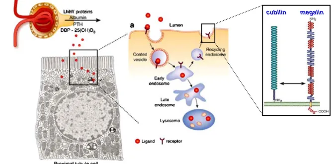
The uptake of LMW proteins by PT cells essentially involves receptor-mediated endocytosis, while fluid-phase capture can be considered as quantitatively negligible [9]. During receptor-mediated endocytosis, filtered proteins are concentrated at the apical cell surface, and their concentra-tion in the endocytic invaginaconcentra-tion exceeds that in the extracellular space several fold. Clathrin-mediated endocyto-sis represents the predominant pathway for protein uptake across the apical membrane of PT cells, with an endocytic pathway consisting of five main interrelated compartments: (1) microvilli and clathrin-coated pits, (2) early endosomes, (3) dense apical tubules responsible for apical recycling, (4) late endosomes, and (5) lysosomes [10]. The process requires two multiligand receptors, megalin and cubilin, that are abundantly expressed at the brush border of PT cells [7]. Ligand binding and interactions between both receptors induce their internalization into coated vesicles and their subsequent delivery to endosomes and lysosomes for ligand processing and receptor degradation or recycling (Fig. 2). Receptor-mediated endocytosis of albumin depends on the integrity of the actin cytoskeleton and the microtubules [26], whereas progression along the endocytic apparatus requires a sustained vesicular acidification from early to late endo-somes and finally to lysoendo-somes [22,80] (Fig.3). Indeed, the drop in pH in the successive endocytic compartments triggers receptor–ligand dissociation and modulates vesicle trafficking, endosomal fusion events, and coat formation [40]. In PT cells, the endosomal acidification is driven by the
electrogenic vacuolar H+-ATPase (Fig.4), whose inhibition by pharmacological agents like bafilomycin A-1 or toxic agents like the heavy metal cadmium severely impairs the uptake of albumin and LMW proteins in vitro and in vivo
cubilin megalin
Fig. 2 Reabsorption of low-molecular-weight proteins by proximal tubule cells. Albumin and low-molecular-weight (LMW) proteins (including PTH, vitamin-D binding protein (DBP), and 25(OH) vitamin D) that are naturally filtered by the glomerulus into the primary urine are endocytosed by PT cells via the megalin–cubilin receptor pathway. Following internalization in coated vesicles, the
receptor–ligand complexes progress along the endocytic pathway. The endosomes undergo a progressive acidification that results in the dissociation of the receptor–ligand complexes, with megalin and cubilin (inset) being recycled in the apical membrane, whereas the ligand is directed to lysosomes for degradation. Modified from [2,18]
vesicular pH
6.8
6.0
5.4
6.4
5.0
7.4
Fig. 3 Vesicular acidification and receptor-mediated endocytosis. The endocytic pathway in proximal tubule cells involves coated pits and coated vesicles, followed by early endosomes that form recycling endosomes or mature to late endosomes and lysosomes. There is a progressive, ATP-dependent acidification of the endosomes (pH 5 to 6) and lysosomes (pH 4.6–5.0) that is necessary for dissociation of the ligand–receptor complex, recycling of receptors to the apical membrane, and progression of ligands into lysosomes. The approximate pH values of the different compartments are indicated. Modified from [17]
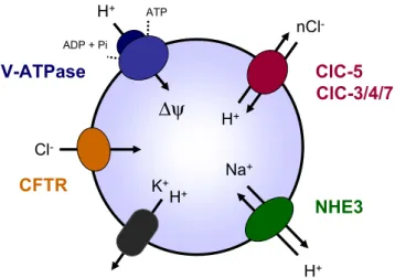
[37,84,86]. The translocation of H+from the cytoplasm into the endosomes generates a transmembrane electrical poten-tial (ΔΨ) resulting in a rapid inhibition of V-ATPase activity. Thus, in order to limit the formation of an endosomal-positive membrane potential, either anions have to concur-rently enter vesicles or cations have to leave (Fig.4). In most cases, acidification of intracellular vesicles seems dependent on a parallel Cl− conductance that provides the electrical shunt necessary to neutralize the H+electrical gradient [42]. Furthermore, the intravesicular Cl−concentration itself could directly affect the V-ATPase activity [60] as well as the vesicle recycling independently of its effect on pH [22].
The paradigm of Dent’s disease
Recent investigations on the pathophysiology of Dent’s disease and the Cl−/H+ exchanger, ClC-5, have provided exciting information on the role of chloride transporters along the endocytic pathway [42]. Dent’s disease (OMIM #300009) is a rare X-linked renal tubulopathy characterized by LMW proteinuria associated with hypercalciuria, which may provoke nephrolithiasis, nephrocalcinosis, and renal failure [18, 77]. Low-molecular-weight proteinuria is the most consistent manifestation of Dent’s disease, detected in all affected males and obligate carrier females. In contrast, there is considerable inter- and intra-familial variability in the other manifestations of the disease, including progression to renal failure [77]. Dent’s disease is caused by mutations in the CLCN5 gene that encodes ClC-5, an electrogenic Cl−/H+
exchanger [53,66,76]. ClC-5 belongs to the CLC family of
Cl channels/transporters that have been discovered and characterized by Jentsch and colleagues ([41] for review). ClC-5 consists of 746 amino acids and forms diamond-shaped homodimers composed of two repeated halves that span the membrane in opposite orientation [19]. Each subunit has its own pore responsible for the selective coupling of the Cl− flux to H+ counter-transport [66, 76]. In vitro studies have demonstrated that natural mutations in ClC-5 lead to a loss of function [53]. Furthermore, genetic inactivation of the Clcn5 gene in mouse mimics the severe PT dysfunction observed in Dent’s disease [67,85].
The complex phenotype of Dent’s disease is probably explained by the expression of ClC-5 in multiple nephron segments, including the PT, the TAL, and the α-type intercalated cells [16, 18, 33]. Particularly, ClC-5 co-distributes with the V-ATPase in early endosomes along the endocytic apparatus in PT cells [16,33], where it is thought to ensure the Cl− permeability in parallel with H+ transport (Fig. 4), as suggested by a decreased ATP-dependent fluorescence quenching of low-density vesicles isolated from ClC-5-deficient mice and loaded in vitro by acridine orange [34]. Further measurements of endosomal pH and Cl− concentration in PT cells cultured from ClC-5-deficient mice provided additional evidence for ClC-5 involvement in acidification of early endosomes but not of late endosomes and Golgi [36]. Other members of the CLC family of vesicular channels/transporters have been shown to play a role in vesicular acidification, including ClC-3 and ClC-7 [28,36]. Furthermore, recent studies have demonstrated that ClC-5 inactivation induces a generalized trafficking defect in PT cells, with loss of megalin and cubilin at the brush border and impaired lysosome biogenesis, which also contributes to defective endocytosis and urinary loss of LMW ligands and lysosomal enzymes [8,61].
Receptor-mediated endocytosis in CFTR-deficient mouse kidney
As mentioned above, the distribution of CFTR in the apical endosomes of mouse PT cells [43] pointed to its possible involvement in renal endocytosis. This hypothesis was recently substantiated by using CF mouse models to characterize the role of CFTR in the kidney. Plasma and urine analyses revealed that baseline renal function was normal in Cftr−/− (Cftrtm1Cam) mice. However, the urinary excretion of the LMW Clara Cell protein (CC16, 16 kD) was significantly increased inCftr−/−mice in comparison to controls, reflecting a defect in PT cell apical endocytosis. This was supported by the demonstration of a significant decrease in the renal uptake of radiolabelled 125I-β2
-microglobulin in Cftr−/− mice and a lower renal uptake of aminoglycosides in comparison to wild-type littermates [43].
V-ATPase
ClC-5
NHE3
Cl -K+ H+ Na+ nCl -H+ H ATP ADP + PiCFTR
H+ClC-3/4/7
∆ψ
Fig. 4 Ion transport processes involved in endosomal acidification. The endosomal acidification is achieved by ATP-driven transport of cytosolic H+ through the V-ATPase. The positive electrical gradient (ΔΨ) is dissipated by cation (H+, K+, Na+) leakage, as well as by parallel Cl− permeability through ClC-5 and most likely other anion transporters. Recent studies have demonstrated that vesicular CLC isoforms (ClC-3 to ClC-7) act as voltage-dependent Cl−/H+exchangers. ClC-6, restricted to the nervous system, is not depicted. Adapted from [22]
The endocytic uptake of aminoglycosides and LMW proteins, like CC16 andβ2-microglobulin, is mediated by
the multiligand receptors, megalin and cubilin [7]. In contrast to megalin, which is a member of the low-density lipoprotein receptor family, cubilin, also known as the intestinal intrinsic factor (IF)-B12 receptor, is a highly conserved membrane glycoprotein with little structural homology to known endocytic receptors and is character-ized by the absence of a transmembrane domain [2]. High-affinity binding of purified megalin to cubilin N-terminal region has been shown in vitro, suggesting that megalin participates in the endocytosis and intracellular trafficking of cubilin [57]. The apical sorting of cubilin and its participation in receptor-mediated endocytosis critically depend on its reciprocal interaction with the transmembrane protein amnionless (AMN) identified as a key factor for mouse gastrulation [11, 44]. Cubilin contributes ligand-binding regions of the receptor complex, whereas AMN ensures the membrane anchorage, biosynthetic processing, and recycling of the complexes at the plasma membrane [24]. Interestingly, there was a selective decrease of cubilin expression in the straight (S3) segment of the PT ofCftr−/− mice mirrored by an increased urinary excretion of cubilin ligands such as transferrin and CC16 [43]. Further inves-tigations demonstrated that the lack of CFTR in the kidney was not associated with changes in the biosynthesis of cubilin but, rather, with a significant increase in the excretion of cubilin in the urine of Cftr−/− mice. No significant changes in kidney and urine abundance of megalin were observed. Taken together, these data suggest that the lack of CFTR in renal PT cells induces instability of cubilin at the brush border, leading to its accelerated shedding into urine. Although the lack of suitable reagents prevented to investigate the specific role of AMN in the process, it is tempting to hypothesize that such cubilin instability could be due to the improper processing or trafficking of one of its partners in relation with the lack of CFTR in that nephron segment [43].
Renal phenotype in mice and humans harboring theΔF508 mutation
TheΔF508 mutation is the most common mutation in the CF population, with almost 90% of Caucasian CF patients having at least oneΔF508 allele [73]. This mutation affects the processing and maturation of CFTR to its fully glycosylated form [5], with retention of ΔF508-CFTR in the endoplasmic reticulum (ER) by molecular chaperones [20] and subsequent degradation via the ubiquitin-protea-some pathway [87]. However, the ΔF508-CFTR can essentially function as a cAMP-regulated Cl− channel both in the ER and at the plasma membrane under distinct
permissive conditions [23,64]. The processing of ΔF508-CFTR has been shown to revert to that of wild-type ΔF508-CFTR as the incubation temperature is reduced [13]. Airway and gallbladder cells from Cftrtm1Eur mice homozygote for the ΔF508 mutation show increased cAMP-induced Cl−
con-ductance when cultured at 27°C [23].
The Cftrtm1Eur mouse model expressing the ΔF508 mutant CFTR was generated by double homologous recombination [21, 82]. Six-week-old mice homozygous for the ΔF508 mutation (CftrΔF/ΔF) have abnormalities typical of CF such as growth retardation, focal hypertrophy of goblet cells in the intestinal crypts, and higher basal nasal potential difference with reduced response to for-skolin in trachea and intestine. However, the CftrΔF/ΔF tissues show a residual Cl−conductance, suggesting that the mutantΔF508-CFTR is partially processed and reaches the plasma membrane. This may explain the minor phenotype of theCftrΔF/ΔFversusCftr−/−mice (e.g., absence of lethal intestinal obstruction). Although such rescue phenomenon has not been clearly demonstrated thus far in man, residual Cl− transport activity has also been observed in rectal biopsies ofΔF/ΔF patients with milder CF phenotype [83]. Moreover, the expression ofΔF508-CFTR in man is tissue-specific, suggesting that the variable severity of CF in different organs may reflect heterogeneity of residual expression [45,65].
In mouse CftrΔF/ΔF kidney, the mRNA abundance of ΔF508-CFTR was ∼2-fold reduced in comparison to controls [43]. The mutant ΔF508-CFTR showed defective glycosylation, with a large individual variability in its residual expression in the apical area of PT cells. Consequently, the renal uptake of radiolabelled 125I-β2
-microglobulin was either unchanged or decreased (∼30%) in comparison to wild-type mice. In addition, the electrolyte and water handling has previously been demonstrated as largely preserved in the PT ofCftrtm2camΔF508 mice [49]. Taken together, these data suggest that, in mouse kidney, the ΔF508-CFTR is variably processed into its mature form, reaching the plasma membrane and ensuring correct function in someCftrΔF/ΔFmice.
Clinical and biological investigations of a large cohort of CF patients, all harboring at least one ΔF508 mutation, showed a mild but significant albuminuria and LMW proteinuria versus healthy controls. The specificity of the LMW proteinuria in CF was supported by the lack of significant changes in patients with chronic lung inflam-mation due to active asthma [43]. These findings may impact on the long-term renal function since LMW proteinuria can trigger tubulo-interstitial injury progressively leading to renal failure [2]. Moreover, the increased urinary loss of the cubilin-ligand, transferrin, could participate in the iron deficiency and lower circulating transferrin levels that are commonly reported in CF patients [62,68].
Conclusions and perspectives
The spectrum of CF, which was previously considered as a respiratory and digestive disease associated with a rapidly fatal outcome, has broadened considerably over the last decade. Recent studies performed in CF mouse models and patients with CF have demonstrated that the functional loss of CFTR is associated with a moderate but significant defect in LMW protein handling by the kidney. These data support a role of CFTR in cubilin-mediated endocytic pathway in PT cells. Data obtained in mice and patients harboring the ΔF508 mutation also give insights into the tissue-specific and species-dependent processing of wild-type and mutant CFTR [63]. Further investigations in different CF mouse models [30] and in other animal models that may be more relevant for the disease [72] should help to clarify the role of CFTR in other specialized kidney functions, such as Ca2+ handling and NaCl homeostasis [17]. One should also consider that subtle abnormalities in kidney development, function, and/or morphology may appear in CF patients at a later stage—as longer survival may be associated with a potential for developing multi-organ complications [73].
Finally, in addition to the functional loss of CFTR, specific kidney diseases may involve an abnormal activity or expression of CFTR. For example, several lines of evidence have shown that CFTR is upregulated and activated in the cells lining the cysts, playing a role in cyst fluid accumulation in autosomal dominant polycystic kidney disease, the most common inherited nephropathy [14, 35, 56, 65, 88]. Investigation of these situations may also prove useful to gain knowledge in the role of CFTR during nephrogenesis and in the mature kidney.
Acknowledgments We thank R. Beauwens, A. Bernard, E. Boulpaep, J-J. Cassiman, P.J. Courtoy, E.I. Christensen, H.R. De Jonge, W.B. Guggino, T. Leal, P. Lebecque, B.J. Scholte, P. Steels, and S. Terryn for helpful discussions and support. The authors studies are supported by the Belgian agencies FNRS and FRSM, the Foundation Alphonse et Jean Forton, Concerted Research Actions, Inter-University Attraction Poles, the Association Belge de Lutte contre la Mucoviscidose, a grant from Amgen, and the EuReGene (FP6) and EUNEFRON (FP7) projects of the European Community.
References
1. Barasch J, Kiss B, Prince A et al (1991) Defective acidification of intracellular organelles in cystic fibrosis. Nature 352:70–73 2. Birn H, Christensen EI (2006) Renal albumin absorption in
physiology and pathology. Kidney Int 69:440–449
3. Bradbury NA (1999) Intracellular CFTR: localization and func-tion. Physiol Rev 79:S175–S191
4. Bradbury NA, Jilling T, Berta G et al (1992) Regulation of plasma membrane recycling by CFTR. Science 256:530–532
5. Cheng SH, Gregory RJ, Marshall J et al (1990) Defective intracellular transport and processing of CFTR is the molecular basis of most cystic fibrosis. Cell 63:827–834
6. Cheng J, Wang H, Guggino WB (2005) Regulation of cystic fibrosis transmembrane regulator trafficking and protein expres-sion by a Rho family small GTPase TC10. J Biol Chem 280:3731–3739
7. Christensen EI, Birn H (2002) Megalin and cubilin: multifunc-tional endocytic receptors. Nat Rev Mol Cell Biol 3:256–266 8. Christensen EI, Devuyst O, Dom G et al (2003) Loss of chloride
channel ClC-5 impairs endocytosis by defective trafficking of megalin and cubilin in kidney proximal tubules. Proc Natl Acad Sci USA 100:8472–8477
9. Christensen EI, Maunsbach AB (1979) Effects of dextran on lysosomal ultrastructure and protein digestion in renal proximal tubule. Kidney Int 16:301–311
10. Conner SD, Schmid SL (2003) Regulated portals of entry into the cell. Nature 422:37–44
11. Coudroy G, Gburek J, Kozyraki R et al (2005) Contribution of cubilin and amnionless to processing and membrane targeting of cubilin–amnionless complex. J Am Soc Nephrol 16:2330–2337 12. Crawford I, Maloney PC, Zeitlin PL et al (1991)
Immunocyto-chemical localization of the cystic fibrosis gene product CFTR. Proc Natl Acad Sci USA 88:9262–9266
13. Denning GM, Anderson MP, Amara JF et al (1992) Processing of mutant cystic fibrosis transmembrane conductance regulator is temperature-sensitive. Nature 358:761–764
14. Devuyst O, Beauwens R (1998) Ion transport and cystogenesis: the paradigm of autosomal dominant polycystic kidney disease. Adv Nephrol Necker Hosp 28:439–478
15. Devuyst O, Burrow CR, Schwiebert EM et al (1996) Develop-mental regulation of CFTR expression during human nephro-genesis. Am J Physiol 271:F723–F735
16. Devuyst O, Christie PT, Courtoy PJ et al (1999) Intra-renal and subcellular distribution of the human chloride channel, CLC-5, reveals a pathophysiological basis for Dent’s disease. Hum Mol Genet 8:247–257
17. Devuyst O, Guggino WB (2002) Chloride channels in the kidney: lessons learned from knockout animals. Am J Physiol Renal Physiol 283:F1176–F1191
18. Devuyst O, Pirson Y (2007) Genetics of hypercalciuric stone forming diseases. Kidney Int 72:1065–1072
19. Dutzler R, Campbell EB, Cadene M, Chait BT, MacKinnon R (2002) X-ray structure of a ClC chloride channel at 3.0 A reveals the molecular basis of anion selectivity. Nature 415:287–294 20. Egan ME, Glockner-Pagel J, Ambrose C et al (2002)
Calcium-pump inhibitors induce functional surface expression of ΔF508-CFTR protein in cystic fibrosis epithelial cells. Nat Med 8:485–492
21. Ellsworth RE, Jamison DC, Touchman JW et al (2000) Compara-tive genomic sequence analysis of the human and mouse cystic fibrosis transmembrane conductance regulator genes. Proc Natl Acad Sci USA 97:1172–1177
22. Faundez V, Hartzell HC (2004) Intracellular chloride channels: determinants of function in the endosomal pathway. Sci STKE 233:re8 23. French PJ, van Doorninck JH, Peters RH et al (1996) F508 mutation in mouse cystic fibrosis transmembrane conductance regulator results in a temperature-sensitive processing defect in vivo. J Clin Invest 98:1304–1312
24. Fyfe JC, Madsen M, Højrup P et al (2003) The functional cobalamin (vitamin B12)-intrinsic factor receptor is a novel complex of cubilin and amnionless. Blood 103:1573–1579 25. Gadsby DC, Vergani P, Csanady L (2006) The ABC protein
turned chloride channel whose failure causes cystic fibrosis. Nature 440:477–483
26. Gekle M (2005) Renal tubule albumin transport. Annu Rev Physiol 67:573–594
27. Gibney EM, Goldfarb DS (2003) The association of nephroli-thiasis with cystic fibrosis. Am J Kidney Dis 42:1–11
28. Graves AR, Curran PK, Smith CL, Mindell JA (2008) The Cl−/H+ antiporter ClC-7 is the primary chloride permeation pathway in lysosomes. Nature 453:788–792
29. Groman JD, Meyer ME, Wilmott RW et al (2002) Variant cystic fibrosis phenotypes in the absence of CFTR mutations. N Engl J Med 347:401–407
30. Grubb BR, Boucher RC (1999) Pathophysiology of gene-targeted mouse models for cystic fibrosis. Physiol Rev 79: S193–S214
31. Guggino WB (2004) The cystic fibrosis transmembrane regulator forms macromolecular complexes with PDZ domain scaffold proteins. Proc Am Thorac Soc 1:28–32
32. Guggino WB, Stanton BA (2006) New insights into cystic fibrosis: molecular switches that regulate CFTR. Nat Rev Mol Cell Biol 7:426–436
33. Günther W, Luchow A, Cluzeaud F et al (1998) ClC-5, the chloride channel mutated in Dent’s disease, colocalizes with the proton pump in endocytotically active kidney cells. Proc Natl Acad Sci USA 95:8075–8080
34. Günther W, Piwon N, Jentsch TJ (2003) The ClC-5 chloride channel knock-out mouse—an animal model for Dent’s disease. Pflugers Arch 445:456–462
35. Hanaoka K, Devuyst O, Schwiebert EM et al (1996) A role for CFTR in human autosomal dominant polycystic kidney disease. Am J Physiol 270:C389–C399
36. Hara-Chikuma M, Wang Y, Guggino SE et al (2005) Impaired acidification in early endosomes of ClC-5 deficient proximal tubule. Biochem Biophys Res Commun 329:941–946
37. Herak-Kramberger CM, Brown D, Sabolic I (1998) Cadmium inhibits vacuolar H+-ATPase and endocytosis in rat kidney cortex. Kidney Int 53:1713–1726
38. Higgins CF (1992) ABC transporters: from microorganisms to man. Annu Rev Cell Biol 8:67–113
39. Huber S, Braun G, Burger-Kentischer A et al (1998) CFTR mRNA and its truncated splice variant (TRN-CFTR) are differ-entially expressed during collecting duct ontogeny. FEBS Lett 423:362–366
40. Hurtado-Lorenzo A, Skinner M, El Annan J et al (2006) V-ATPase interacts with ARNO and Arf6 in early endosomes and regulates the protein degradative pathway. Nat Cell Biol 8:124–136
41. Jentsch TJ (2007) Chloride and the endosomal-lysosomal path-way: emerging roles of CLC chloride transporters. J Physiol 578:633–640
42. Jentsch TJ, Maritzen T, Zdebik AA (2005) Chloride channel diseases resulting from impaired transepithelial transport or vesicular function. J Clin Invest 115:2039–2046
43. Jouret F, Bernard A, Hermans C et al (2007) Cystic fibrosis is associated with a defect in apical receptor-mediated endocytosis in mouse and human kidney. J Am Soc Nephrol 18:707–718 44. Kalantry S, Manning S, Haub O et al (2001) The amnionless gene,
essential for mouse gastrulation, encodes a visceral-endoderm-specific protein with an extracellular cysteine-rich domain. Nat Genet 27:412–416
45. Kalin N, Claass A, Sommer M et al (1999)ΔF508 CFTR protein expression in tissues from patients with cystic fibrosis. J Clin Invest 103:1379–1389
46. Katz SM, Krueger LJ, Falkner B (1988) Microscopic nephrocalci-nosis in cystic fibrosis. N Engl J Med 319:263–266
47. Kennedy JD, Dinwiddie R, Daman-Willems C, Dillon MJ, Matthew DJ (1990) Pseudo-Bartter’s syndrome in cystic fibrosis. Arch Dis Child 65:786–787
48. Kerem B, Rommens JM, Buchanan JA et al (1989) Identification of the cystic fibrosis gene: genetic analysis. Science 245:1073–1080 49. Kibble JD, Balloch KJ, Neal AM et al (2001) Renal proximal
tubule function is preserved in Cftrtm2cam ΔF508 cystic fibrosis mice. J Physiol 532:449–457
50. Kibble JD, Neal AM, Colledge WH et al (2000) Evidence for cystic fibrosis transmembrane conductance regulator-dependent sodium reabsorption in kidney, using Cftrtm2cam mice. J Physiol 526:27–34
51. Letz B, Korbmacher C (1997) cAMP stimulates CFTR-like Cl− channels and inhibits amiloride-sensitive Na+channels in mouse CCD cells. Am J Physiol 272:C657–C666
52. Li C, Naren AP (2005) Macromolecular complexes of cystic fibrosis transmembrane conductance regulator and its interacting partners. Pharmacol Ther 108:208–223
53. Lloyd SE, Pearce SH, Fisher SE et al (1996) A common molecular basis for three inherited kidney stone diseases. Nature 379:445–449
54. Lu M, Leng Q, Egan ME et al (2006) CFTR is required for PKA-regulated ATP sensitivity of Kir1.1 potassium channels in mouse kidney. J Clin Invest 116:797–807
55. Lyon A, Bilton D (2002) Fertility issues in cystic fibrosis. Paediatr Respir Rev 3:236–240
56. Magenheimer BS, St John PL, Isom KS et al (2006) Early embryonic renal tubules of wild-type and polycystic kidney disease kidneys respond to cAMP stimulation with cystic fibrosis transmembrane conductance regulator/Na(+),K(+),2Cl(−) Co-transporter-dependent cystic dilation. J Am Soc Nephrol 17:3424–3437
57. Moestrup SK, Kozyraki R, Kristiansen M et al (1998) The intrinsic factor-vitamin B12 receptor and target of teratogenic antibodies is a megalin-binding peripheral membrane protein with homology to developmental proteins. J Biol Chem 273:5235–5242
58. Morales MM, Carroll TP, Morita T et al (1996) Both the wild type and a functional isoform of CFTR are expressed in kidney. Am J Physiol 270:F1038–F1048
59. Morales MM, Falkenstein D, Lopes AG (2000) The cystic fibrosis transmembrane regulator (CFTR) in the kidney. An Acad Bras Cienc 72:399–406
60. Moriyama Y, Nelson N (1987) The purified ATPase from chromaffin granule membranes is an anion-dependent proton pump. J Biol Chem 262:9175–9180
61. Nielsen R, Courtoy PJ, Jacobsen C et al (2007) Endocytosis provides a major alternative pathway for lysosomal biogenesis in kidney proximal tubular cells. Proc Natl Acad Sci USA 104:5407–5412
62. O’Connor TM, McGrath DS, Short C et al (2002) Subclinical anemia of chronic disease in adult patients with cystic fibrosis. J Cyst Fibros 1:31–34
63. Ostedgaard LS, Rogers CS, Dong Q et al (2007) Processing and function of CFTR-DeltaF508 are species-dependent. Proc Natl Acad Sci USA 104:15370–15375
64. Pasyk EA, Foskett JK (1995) Mutant (delta F508) cystic fibrosis transmembrane conductance regulator Cl− channel is functional when retained in endoplasmic reticulum of mammalian cells. J Biol Chem 270:12347–12350
65. Persu A, Devuyst O, Lannoy N et al (2000) CF gene and cystic fibrosis transmembrane conductance regulator expression in autosomal dominant polycystic kidney disease. J Am Soc Nephrol 11:2285–2296
66. Picollo A, Pusch M (2005) Chloride/proton antiporter activity of mammalian CLC proteins ClC-4 and ClC-5. Nature 436:420–423 67. Piwon N, Günther W, Schwake M et al (2000) ClC-5 Cl–channel disruption impairs endocytosis in a mouse model for Dent’s disease. Nature 408:369–373
68. Pond MN, Morton AM, Conway SP (1996) Functional iron deficiency in adults with cystic fibrosis. Respir Med 90:409– 413
69. Poschet JF, Fazio JA, Timmins GS et al (2006) Endosomal hyperacidification in cystic fibrosis is due to defective nitric oxide-cylic GMP signalling cascade. EMBO Rep 7:553–559
tion of cellubrevin endocytic compartments and defective endo-somal recycling in cystic fibrosis respiratory epithelial cells. J Biol Chem 277:13959–13965
71. Riordan JR, Rommens JM, Kerem B et al (1989) Identification of the cystic fibrosis gene: cloning and characterization of comple-mentary DNA. Science 245:1066–1073
72. Rogers CS, Hao Y, Rokhlina T et al (2008) Production of CFTR-null and CFTR-DeltaF508 heterozygous pigs by adeno-associated virus-mediated gene targeting and somatic cell nuclear transfer. J Clin Invest 118:1571–1577
73. Rowe SM, Miller S, Sorscher EJ (2005) Cystic fibrosis. N Engl J Med 352:1992–2001
74. Rowntree RK, Harris A (2003) The phenotypic consequences of CFTR mutations. Ann Hum Genet 67:471–485
75. Samaniego-Picota MD, Whelton A (1996) Aminoglycoside-induced nephrotoxicity in cystic fibrosis: a case presentation and review of the literature. Am J Ther 3:248–257
76. Scheel O, Zdebik AA, Lourdel S et al (2005) Voltage-dependent electrogenic chloride/proton exchange by endosomal CLC pro-teins. Nature 436:424–427
77. Scheinman SJ (1998) X-linked hypercalciuric nephrolithiasis: clinical syndromes and chloride channel mutations. Kidney Int 53:3–17 78. Schmitz C, Hilpert J, Jacobsen C et al (2002) Megalin deficiency
offers protection from renal aminoglycoside accumulation. J Biol Chem 277:618–622
79. Sheppard DN, Welsh MJ (1999) Structure and function of the CFTR chloride channel. Physiol Rev 79:S23–S45
dependent acidification in endocytic vesicles from kidney prox-imal tubule. Measurement of pH in individual endocytic vesicles in a cell-free system. Biophys J 59:1208–1217
81. Sojo A, Rodriguez-Soriano J, Vitoria JC, Vazquez C, Ariceta G, Villate A (1994) Chloride deficiency as a presentation or complication of cystic fibrosis. Eur J Pediatr 153:825–828 82. van Doorninck JH, French PJ, Verbeek E et al (1995) A mouse
model for the cystic fibrosisΔF508 mutation. EMBO J 14:4403– 4411
83. Veeze HJ, Halley DJ, Bijman J et al (1994) Determinants of mild clinical symptoms in cystic fibrosis patients. Residual chloride secretion measured in rectal biopsies in relation to the genotype. J Clin Invest 93:461–466
84. Wagner CA, Finberg KE, Breton S, Marshansky V, Brown D, Geibel JP (2004) Renal vacuolar H+-ATPase. Physiol Rev 84:1263–1314 85. Wang SS, Devuyst O, Courtoy PJ et al (2000) Mice lacking renal
chloride channel, CLC-5, are a model for Dent’s disease, a nephrolithiasis disorder associated with defective receptor-medi-ated endocytosis. Hum Mol Genet 9:2937–2945
86. Wang Y, Cai H, Cebotaru L et al (2005) ClC-5: role in endocytosis in the proximal tubule. Am J Physiol Renal Physiol 289:F850–F862
87. Ward CL, Omura S, Kopito RR (1995) Degradation of CFTR by the ubiquitin–proteasome pathway. Cell 83:121–127
88. Yang B, Sonawane ND, Zhao D et al (2008) Small-molecule CFTR inhibitors slow cyst growth in polycystic kidney disease. J Am Soc Nephrol 19:1300–1310