A1/• 3Ô.3
Université de Montréal
The kallikrein-kinin system in relation to retinal vessel tone in the streptozotocin-diabetic rat model
Par
Ashraf Khanjari Dehnavi
Département de Sciences biomédicales Faculté de Médecine
Mémoire présenté à la Faculté des études supérieures en vue de l’obtention du grade de
Maître ès sciences (M.Sc.) en Sciences biomédicales
Décembre, 2002
Université
de Montréal
Direction des bibliothèques
AVIS
L’auteur a autorisé l’Université de Montréal à reproduite et diffuser, en totalité ou en partie, par quelque moyen que ce soit et sur quelque support que ce soit, et exclusivement à des fins non lucratives d’enseignement et de recherche, des copies de ce mémoire ou de cette thèse.
L’auteur et les coauteurs le cas échéant conservent la propriété du droit d’auteur et des droits moraux qui protègent ce document. Ni la thèse ou le mémoire, ni des extraits substantiels de ce document, ne doivent être imprimés ou autrement reproduits sans l’autorisation de l’auteur.
Afin de se conformer à la Loi canadienne sur la protection des renseignements personnels, quelques formulaires secondaires, coordonnées ou signatures intégrées au texte ont pu être enlevés de ce document. Bien que cela ait pu affecter la pagination, il n’y a aucun contenu manquant.
NOTICE
The author of this thesis or dissertation has granted a nonexclusive license allowing Université de Montréal to reproduce and publish the document, in part or in whole, and in any format, solely for noncommercial educational and research purposes.
The author and co-authors if applicable retain copyright ownership and moral rights in this document. Neither the whole thesis or dissertation, nor substantial extracts from it, may be printed or otherwise reproduced without the author’s permission.
In compliance with the Canadian Privacy Act some supporting forms, contact information or signatures may have been removed from the document. While this may affect the document page count, it does not represent any loss of content from the document.
Ce mémoire intitulé:
The kallikrein-kinin system in relation to retinal vessel tone in the streptozotocin-diabetic rat model
Présenté par: Asliraf Khanjari Dehnavi
A été évalué par un jury composé des personnes suivantes
(Pedro D’Orléans-Juste) Président-rapporteur (Haroutioun M. Hassessian) Directeur de recherche (Adel Giaid) Membre du jury
III
SOMMAIRE
Environ 3% de la population mondiale dont 1.5 millions de canadiens souffrent du diabète. La morbidité associée au diabète de type I ou II (diabète insulino-dépendant ou non-insulino-dépendant), résulte de complications sérieuses, telles que; microangiopathie, néphropathie, neuropathie et rétinopathie. La prevalence de tous les types de rétinopathie dans la population diabétique augmente avec la durée du diabète et l’âge du patient.
Des données soutenant la participation du système kallikreine-kinine dans la pathogenèse du diabète a été obtenue à partir de patients diabétiques et de modèles animaux de diabète par plusieurs groupes. Les kinines sont des agents vasoactifs efficaces dans la microcirculation, où elles produisant les effets par des récepteurs sur le muscle lisse des microvaissaux ou des cellules endothéliales suite à une réaction inflammatoire ou un dommage tissulaire.
Des données de la littérature ont démontré que le système kallikreine-kinine est exprimé au niveau des yeux de lapins et de porcs. Aussi, à l’aide d’analyses RT-PCR et Southern blot, l’expression et la localisation des differentes composantes de ce système tels que la kallikreine tissulaire, le kininogene de faible poids moléculaire et les récepteusrs B1 et récepteurs B2 à forte densité ont été identifiées dans la rétine humaine. En outre, l’hybridation in situ a identifié la localisation cellulaire des ARN messagers des composantes de ce système dans les tissus oculaires et dans les
cellules endothéliales des vaisseaux sanguins oculaires humains. Cependant, il n’y a aucune étude sur la fonction physiologique du système kallikreine-kinine et son rôle potentiel dans le développement de la rétinopathie diabétique au niveau de l’oeil. Le but de cette étude était d’étudier le rôle des kinines dans la régulation du tonus vasculaire rétinien, avec trois objectifs spécifiques:
.i) déterminer la nature des récepteurs aux kinines impliqués en utilisant des agonistes et antagonistes selectifs,
ii) investïguer les voies de signalisation impliquées dans l’effet vasoactif des kinines dans la rétine, et
iii) étudier l’effet de la bradykinine (BK) et de la des-Arg9-BK sur le tonus des vaisseaux rétiniens chez le rat aduit Wistar diabétique suite à l’injection de
streptozotocine (SIZ).
Les résultats de cette étude montrent que:
j) la BK dilate les vaisseaux rétiniens chez les rats contrôles et diabétique, et l’effet vasodilatateur de la BK passe par la stimulation des récepteurs B2,
ii) la des-Arg9-BK ne dilate pas les vaisseaux rétiniens chez le rat contrôle,
iii) la des-Arg9-BK dilate les vaisseaux rétiniens chez les rats diabétiques et l’effet vasodilatateur de la des-Arg9-BK passe par la stimulation des récepteurs B1, iv) l’effet vasodilatateur des kinins implique une G-proteins (G/G0),
y) la libération de l’oxyde nitrique et l’augmentation du calcium intracellulaire par
l’influx à partir du milieu extracellulaire ne sont pas impliqués dans les effets de la BK et la des-Arg9-BK. Mais,
V
vi) l’augmentation de Ca2 intracellulaire à partir des réserves intracellulaires sensible et insensible à l’1P3 et la libération de prostaglandine ‘2 (prostacycline;
PGI2), métabolite de la voie de la cyclooxygénase, sont impliquées dans les effets de la BK et la des-Arg9-BK.
Nos résultats fournissent pour la première fois des évidences fonctionnelles directes que le système kallikreine-kinine joue un rôle crucial dans la régulation du tonus vasculaire rétinien et que ce système peut avoir un rôle principal durant les premières étapes de développement de la rétinopathie diabétique.
SUMMARY
About 3% of the world population including 1.5 million Canadians suffer from diabetes. The morbidity associated with long-standing diabetes of either type I or II (insulin-dependent or non-insulin-dependent diabetes mellitus respectively) results from a number of serious complications, namely microangiopathy, nephropathy, neuropathy and retinopathy. The prevalence of retinopathy in the diabetic population increases with the duration of diabetes and is directly correlated with patient age.
The true cause of diabetic microvascular disease is unknown. There is increasing evidence, from diabetic animal models and diabetic patients, which supports the involvement of tissue kallikrein in diabetes mellitus. Kinins are potent vasoactive agents in the microcirculation, producing effects through receptors on microvessel smooth muscle or endothelial celis following inllammatory insuit or tissue damage.
Previous studies have demonstrated that kallikrein-like enzymatic activity exists in tissue homogenates of rabbit and swine eyes. Furthermore, it has been shown by molecular studies that mRNAs for key components of the kallikrein-kinin system, including tissue kallikrein and low molecular weight kininogen, are present in human ocular tissues. In addition, high levels of kinin B1 and B2 receptors have been detected in human retina. Also, in situ hybridization lias identified mRNAs for the components of the kallikrein-kinin system in ocular tissues and in endothelial ceils of human ocular blood vessels. However, there lias been no study on the physiologic
VII
function of the kallikrein-kinin system in ocular tissues and its potentiai foies ifl the deveiopment of diabetic retinopathy.
The aim of the current study was to investigate the effect of kinins on retinai vesse! tone, with three specific objectives:
j) to establish which receptors mediate the vascuiar response to kinins using se!ective agonists and antagonists,
ii) to determine which signa! transduction pathways mediate the vascu!ar effects of kinins in the retina of heaithy aduit maie Wistar rats, and
iii) to study the effect of Bradykinin (BK) and des-Arg9-BK on retinai vesse! toile in the aduit streptozotocin (STZ)-diabetic rat.
The present study shows that:
i) BK dilates retinal vessels in control and STZ-diabetic rats,
ii) the vasodi!ator effect of BK is mediated by stimulation of B2 receptors, iii) des-Arg9-BK is without effect on retina! vessel tone in control rats, but
des-Arg9-BK dilates retinal vessels in STZ-diabetic rats,
iv) the vasodi!ator effect of des-Arg9-BK is mediated by stimulation of B1 recep tors,
y) downstream of B1 or B2 receptors, the vasodilator effect of kinins is transduced
by G/G0-proteins, and
vi) the release of nitric oxide (NO) or the influx of extrace!!u!ar Ca2 are not invo! ved in the vasodilation evoked by either BK or des-Arg9-BK. In contrast, libera
tion of Ca2 from both intracellular, 1P3-sensitive and non-1P3-sensitive, pools are involved as well as the release of prostaglandin 12 (prostacyclin; PGI2).
This study is the first to provide direct ftnctional evidence that the kallikrein-kinin system may play a role in the retinal circulation, and may be a factor early in the pathological development of diabetic retinopathy.
IX
CONTENTS
SOMMAIRE.III-V SUMMARY.VI-VIII CONTENTS IX-XII TABLES XIII FIGURES XIV-XVI ABBREVIATIONS XVII-XVIII ACKNOWLEDGMENTS XIX CHAPTER I 1. INTRODUCTION 1 1.1. DIABETES MELLifUS 1-21.1.1. TYPE I DIABETES MELL1TUS 2
1.1.2. TYPE II DIABETES MELLITUS 3
1.1.3. PATHOLOGY 0F DIABETES 3-4
1.2. THE RETINA AND ifS BLOOD VESSELS 4-7
1.3. OCULAR DIABETIC COMPLICATIONS 8
1.3.1. CLASSIFICATION AND INCIDENCE 0F DIABETIC
RETINOPATHY 8
1.3.2. NONPROLIFERATWE DIABETIC RETINOPATHY 9
1.3.3. PROLIFERATIVE DIABETIC RETINOPATHY 9
1.3.4. VASODILATION IN DIABETIC RETINOPATHY 10
1.3.5. RETLNOPATHY IN STZ-DIABETIC RAT MODEL 10-11
1.4.1. KININ BIOSYNTHESIS.11-13
1.4.2. KININ METABOLISM .14
1.4.3. BIOLOGICAL EFFECTS 15
1.4.4. KININ RECEPTORS 15-19
1.5. VASCULAR SIGNAL TRANSDUCTION PATHWAYS 20-21
1.5.1. KININ-STIMULATED RELEASE 0f NifRIC OXIDE 21-22
1.5.2. KJNIN-STIMULATED INCREASE 0F INTRACELLULAR
CALCIUM 23-24
1.5.3. KININ-STIMULATED RELEASE 0F PROSTACYCLIN 25-26
1.6. THE KALLIKREIN-KININ SYSTEM IN RELATION TO THE EYE 27
2. HYPOTHESIS 27
3.AIM 28
4. SPECIFIC OBJECTIVES 28
CHAPTER Il
1. MATERIALS AND METHODS 29
1.1. BIOLOGICAL MODEL 29 1.2. EXPERIMENTAL PROTOCOL 30-31 1.3. CHEMICAL AGENTS 31 1.4. STATISTICAL ANALYSIS 32 CHAPTER UI 1. RESULTS 33 1.1.CONTROL RAT 34
1.1.1. EfFECT 0F BRADYKININ ON RETINAL VESSEL TONE 34
XI
1.1.3. B2 RECEPTOR ANTAGONISM .36
1.1.4. B1 RECEPTOR ANTAGONISM 37
1.2. INTRACELLULAR AND MEMBRANE PATHWAYS WHICH MEDIATE
THE KININ EVOKED VASODILATION IN CONTROL RATS 38
1.2.1. G0/G-PROTEINS 3$-39
1.2.2. NITRIC OXIDE SYNTHASE INHIBITION 40
1.2.3. CALCIUM CHANNEL BLOCKERS 41
1.2.4. 1P3-SENSITWE INTRACELLULAR CALCIUM STORES 42-43
1.2.5. NON-1P3-SENSITWE INTRACELLULAR CALCIUM STORES 44
1.2.6. COX-2 INHIBITORS 45-46
1.2.7. PROSTACYCLIN SYNTHASE INHIBITORS 47
1.3. STREPTOZOTOCIN-DIABETIC RAT 48
1.3.1. EFFECT 0f des-Arg9-BRADYKININ ON RETINAL VESSEL
TONE 49-50
1.3.2. EFFECT 0F BRADYKININ ON RETINAL VESSEL TONE 51
1.3.3. RESPONSE TO des-Arg9-BRADYKININ FOLLOWING
B1 RECEPTOR ANTAGONISM 52
1.3.4. RESPONSE TO BRADYKININ FOLLOWING B2 RECEPTOR
ANTAGONISM 53
1.4. INTRACELLULAR AND MEMBRANE PATHWAYS WHICH MEDIATE
THE KININ EVOKED VASODILATION IN STZ-DIABETIC RATS 54
1.4.1. RESPONSE TO des-Arg9-BRADYKININ FOLLOWING
INHIBITION 0F G0/G1-PROTEINS 54-55
1.4.2. RESPONSE TO des-Arg9-BRADYKININ FOLLOWING NITRIC
OXIDE SYNTHASE INHIBITION 56
CHANNEL BLOCKADE .57
1.4.4. RESPONSE TO des-Arg9-BRADYKININ FOLLOWING ANTAGONISM
0F 1P3-SENSffWE INTRACELLULAR CALCIUM STORES 58-59
1.4.5. RESPONSE TO des-Arg9-BRADYKININ FOLLOWING ANTAGONISM
0F NON-1P3-SENSITWE INTRACELLULAR CALCIUM STORES 60
1.4.6. RESPONSE TO des-Arg9-BRADYKTNIN FOLLOWING COX-2
INHIBITION 61
1.4.7. RESPONSE TO des-Arg9-BRADYKININ FOLLOWING PROSTACYCLIN
SYNTHASE INHIB1TION 62-63
CHAPTER IV
DISCUSSION 64-70
CONCLUSION 7 1-72
XIII
TABLE
LIST
TABLE I CATEGORIES 0F DIABETES MELLifUS 2
TABLE II COMPARISON 0F TYPE I VS TYPE II DIABETES MELLFFUS 2
TABLE III PRIMARY STRUCTURE 0F MAMMALIAN KININS 12
TABLE IV BIOCHEMICAL FEATURES AND PHARMACOLOGY 0f KININ
LIST 0F FIGURES
FIGURE 1 TUE RETINAL CIRCULATION IN RELATION TO TUE
CHOROID AND THE SCLERA 6
FIGURE 2 THE RETINA AND ifS BLOOD VESSELS 7
FIGURE 3 KININ FORMATION AND INACTWATION 13
FIGURE 4 PRINCIPAL KININASES: THEIR CLEAVING SifES FOR
BRADYK1NIN AND KALLIDIN 14
FIGURE 5 AN EXAMPLE 0F A B1 RECEPTOR AMINO ACID
SEQUENCE 16
FIGURE 6 TUE PATHWAY FOR G-PROTEIN DEPENDENT SIGNAL
TRANSDUCTION 20
FIGURE 7 NifRIC OXIDE AND VASCULAR SMOOTH MUSCLE
CELLS 22
FIGURE $ TUE PHOSPHATIDYLINOSifOL PATHWAY 24
FIGURE 9a PROSTACYCLIN AND VASCULAR SMOOTH MUSCLE
CELLS 25
FIGURE 9b ARACHIDONIC ACID AND ifS METABOLITES 26
FIGURE 10 DOSE-RESPONSE CURVE FOR THE EFFECT 0F U-46619
ON RETINAL VESSEL DIAMETER 33
FIGURE 11 EFFECT 0F BK ON THE CONTROL RAT RETINAL
VESSEL DIAMETER 34
FIGURE 12 EFFECT 0F des-Arg9-BK ON THE CONTROL RAT RETINAL
xv
FIGURE 13 B2 KININ RECEPTORS IN BK INDUCED CONTROL
RAT RETII’1AL VASODILATION 36
FIGURE 14 B1 KININ RECEPTORS IN BK INDUCED CONTROL
RAT RETINAL VASODILATION 37
FIGURE 15 G0/G-PROTE1NS IN BK INDUCED CONTROL RAT
RETINAL VASODILATION 39
FIGURE 16 NITRIC OXIDE IN BK INDUCED CONTROL RAT
RETINAL VASODILATION 40
FIGURE 17 EXTRACELLULAR CALCIUM INFLUX IN BK INDUCED
CONTROL RAT RETINAL VASODILATION 41
FIGURE 1$ ETHANOL (vehicle for BHQ) IN BK INDUCED CONTROL
RAT RETINAL VASODILATION 43
FIGURE 19 1P3-SENSff IVE INTRACELLULAR CALCIUM STORES IN
BK INDUCED CONTROL RAT RETINAL VASODILATION 43
FIGURE 20 NON-1P3-SENSrnVE INTRACELLULAR CALCIUM STORES
IN BK INDUCED CONTROL RAT RETINAL VASODILATION 44
FIGURE 21 DMSO (vehicle for L-745.337) IN BK INDUCED CONTROL
RAT RETINAL VASODILATION 46
FIGURE 22 THE COX-2 PATHWAY IN BK INDUCED CONTROL RAT
RETINAL VASODILATION 46
FIGURE 23 PROSTACYCLIN IN BK INDUCED CONTROL RAT RETINAL
VASODILATION 47
FIGURE 24 EFFECT 0F des-Arg9-BK ON STZ-DIABETIC RAT RETINAL
VESSEL DIAMETER 50
FIGURE 25 EFFECT 0F BK ON STZ-DIABETIC RAT RETINAL
FIGURE 26 B1 KININ RECEPTORS IN des-Arg9-BK INDUCED
STZ-DIABETIC RAT RETINAL VASODILATION 52
FIGURE 27 B2 KININ RECEPTORS N BK 1NDUCED STZ-DIABETIC
RAT RETINAL VASODILATION 53
FIGURE 28 G0/G1-PROTEINS IN des-Arg9-BK INDUCED STZ-DIABETIC
RAT RETINAL VASODILATION 55
FIGURE 29 NITRTC OXIDE IN des-Arg9-BK INDUCED STZ-DIABETIC
RAT RETINAL VASODILATION 56
FIGURE 30 EXTRACELLULAR CALCIUM INFLUX IN des-Arg9-BK
[NDUCED STZ-DIABETIC RAT RETINAL VASODILATION 57
FIGURE 31 ETHANOL (vehicle for BHQ) IN des-Arg9-BK INDUCED
STZ-DIABETIC RAT RETINAL VASODILATION 59
FIGURE 32 1P3-SENSITWE INTRACELLULAR CALCIUM STORES IN
des-Arg9-BK INDUCED STZ-DIABETIC RAT RETINAL
VASODILATION 59
FIGURE 33 NON-1P3-SENSffWE INTRACELLULAR CALCIUM STORES IN
des-Arg9-BK 1NDUCED STZ-DIABETIC RAT RETINAL
VASODILATION 60
FIGURE 34 THE COX-2 PATHWAY IN des-Arg9-BK INDUCED
STZ-DIABETIC RAT RETINAL VASODILATION 61
FIGURE 35 PROSTACYCLIN IN des-Arg9-BK INDUCED STZ-DIABETIC
RAT RETINAL VASODILATION 62
FIGURE 36 SUMMARY 0F THE EffECTS 0F KININS ON RETINAL
XVII
LIST 0F ABBREVIATIONS
AA Arachidonic Acid
AC Adenylate Cyclase
ACE Angiotensin-I-Converting Enzyme
ATP Adenosine Triphosphate
BHQ 2,5-Di-tert-butylhydroquinone
BK Bradyldnin
[Ca2J Intracellular Calcium
cADP-ribose Cyclic ADP-ribose
cAMP Cyclic Adenosine Monophosphate
cGMP Cyclic Guanosine Monophosphate
cPLA2 Phospholipase A2
COX-2 Cyclooxygenase-2
des-Arg9-BK des-Arg9-Bradykinin
des-Arg10-Hoe 140 des-Arg9-D-Arg-[Hyp3,Thi5,D-Tic7,Oic8J -Bradykinin
DG Diacylglycerol
eNOS endothelial Nitric Oxide Synthetase
GC Guanylyl Cyclase
GdC13 Gadolinium Chloride
GP G-Protein
HMWK High Molecular Weight Kininogen
Hoe-140 D-Arg-[Hyp3, Thi5,D-Tic7, Oic]-BK IDDM Insulin Dependent Diabetes Mellitus
IL-1 [3 Interleukin-1
[3
iNOS Inducible Nitric Oxide Synthetase
1P3 Inositol Triphosphate
KD Kallidin
LMWK LowMolecular Weight Kininogen
L-NAME No-Nitro-L-Arginine Methyl Ester MAP kinase Mitogen Activated Protein Kinase
Mins Minutes
NIDDM Non-Insulin Dependent Diabetes Mellitus
NO Nitric Oxide
02 Oxygen
PC-PLC Phosphatidylcholine specific Phospholipase C PDGF Platelet Derïved Growth Factor
PGI2 Prostacyclin
PIP2 Phosphatidylinositol 4,5-diphosphate
PKC Protein Kinase C
PLA2 Phospholipase A2
PLC Phospholipase C
PLD Phospholipase D
R Membrane Receptor
RT-PCR Reverse Transcription-Polymerase Chain Reaction
RyR Ryanodine Receptor
sGC Soluble Guanylate Cyclase
STZ Streptozotocin
TNFΠTumor Necrosis Factor-a
TPC Trans-2-phenylcyclopropylamine
U-46619 9,11 -dideoxy-9a ,11 a—methanoepoxyprostaglandin F. VEGf Vascular Endothelial Growth factor
XIX
ACKNOWLEDGMENTS
It is with ail my heart that I thank my parents, sisters, and brothers who have stood by me through ail the highs and lows, flot just in my scoiastic endeavours, but in life as weil. I also give a well deserved thank you to my director, Dr. Haroutioun M.
CHAPTER I
1. INTRODUCTION
1.1. DIABETES MELLITUS
Diabetes mellitus is a chronic disorder of carbohydrate, fat and protein metabolism. About 3% of the world population, including 1.5 million Canadians, suffer from diabetes, making it one of the most common noncommunicable diseases ‘.
This disease can be categorised into two common variants (Type I and Type II) which differ in their pattern of inheritance, responses to insulin, origins, and less commonly in specific genetic defects of 13-cell function (Table I) 2
& (Table II) ‘.
Type I diabetes, also called IDDM, and previously referred to as juvenile onset diabetes, accounts for about 10 % of ail cases of primary diabetes. This is mainly due to a defect in insulin secretion. Most patients (80%-90%) have type II diabetes, also termed NIDDM and previously referred to as aduit onset diabetes. Its onset follows a defect in the response to insulin due to either a non responsive insulin receptor or to a defect in the transduction machinary .
It should be stressed that while the two catagories of diabetes have different pathologic mechanisms, the long-term complications produced in blood vessels, kidneys, nerves, and eyes are the same. Such complications are the major causes of morbidity and finally death from diabetes’.
2
TABLE I. CATEGORIES 0f DIABETES MELLITUS Primary Diabetes
-Type I (insulin-dependent diabetes mellitus, IDDM) -Type II (non-insulin-dependent diabetes mellitus, NIDDM)
-Genetic defects of f3-cell function (including maturity-onset diabetes of the young [MODY])
Secondary Diabetes
-Infections (e.g.,congenital rubella)
-Endocrinopathies (e.g., pituitary tumours)
-Drugs (e.g.,corticosteroids)
-Other genetic disorder (e.g., Down’s syndrome)
-Gestational diabetes mellitus
Modified from the report of the Executive Committee on the Diagnosis and Classification of Diabetes Mellitus: Diabetic Care 20: 1183-1197, 19972
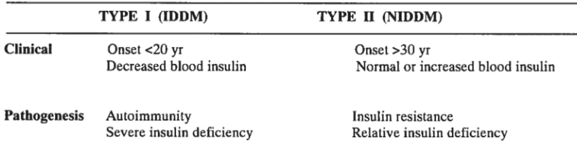
TABLE II. COMPARISON 0f TYPE I VS TYPE II DIABETES MELLITUS
TYPE I (IDDM) TYPE II (NIDDM)
Clinical Onset <20 yr Onset >30 yr
Decreased blood insulin Normal or increased blood insulin
Pathogenesis Autoimmunity Insulin resistance
Severe insulin deficiency Relative insulin deficiency
Modified from pathologic basis of disease. W.B. Saunders Company. Philadelphia. 9 13-929, 1999’.
1.1.1. TYPE I DIABETES MELLITUS
Ibis form of diabetes resuits from a severe drop in the level of circulating insulin due to a reduction in 3-ceil mass. Three interrelated mechanisms are responsible for islet ceil destruction: genetic susceptibility, autoimmunity, and an environmental insuit ‘.
1.1.2. TYPE II DIABETES MELLITUS
Pathogenesis of type II diabetes mellitus remains enigmatic. Genetic factors are even more important in type II than in type I diabetes. The two metabolic defects that characterize type II diabetes are: a derangement in 3-ce11 secretion of insulin and a decreased response of peripheral tissues to insulin (insulin resistance)1•
1.1.3. PATHOLOGY 0F DIABETES
The morbidity associated with long-standing diabetes of both types resuits from a number of serious complications, namely microangiopathy, nephropathy, neuropathy and retinopathy .
There is extreme variability among patients with regards to the time of onset of these complications, their severity, and the particular organ(s) that are involved. In those whom are able to control well their diabetes, the onset may be delayed.
The cause of diabetic microvascular disease has not been established‘.
The overproduction of cytokines including IL-113 and TNFŒ following an autoimmune response leads to the destruction of pancreatic 13-celis and consequently the induction of diabetes mellitus type I “.
Furthermore, most of the available experimental and clinical evidence suggests that the complications are a consequence of the metabolic derangement, in particular the hyperglycemia “s. The oxidative stress following hyperglycemia, may activate NF-iB which induces kinin B1 receptors. The latter is generally under-expressed in normal conditions but it is highly inducible by
4
inflammatory mediators.
In addition, there is evidence to Support the invoivement of the kallikrein-kinin system in diabetes meilitus 6• Both the overproduction of
cytokines and hyperglycemia stimulate the kallikrein-kinin system which is known to promote arteriolar vasodilation ‘.
1.2. THE RETINA AND ITS BLOOD VESSELS
The retina is a thin, multilayered sheet of fleurai tissue that unes the inner aspect of
the posterior two-thirds of the wall of the ocuiar globe. It extends aimost as far anteriorly as the ciiiary body, ending at that point in a ragged edge termed the ora serrata. The outer surface of the sensory retina is apposed to the retinal pigment epithelium and thus associated with Bruch’s membrane, the choroid, and the sciera (Fig. 1)8,9• The inner surface of the retina is in contact with the vitreous.
The retina receives its biood supply from two sources: the choriocapillaris immediateiy outside of Bruch’s membrane, which supplies the outer third of the retina; and branches of the centrai retinai artery, which suppiy the inner two-thirds (Fig. 2). The capillaries of the retina vary in diameter from about 3 to 6 tm and they are surrounded by a thick basal lamina, pericytes, and endothelial ceils. The retinal biood vessels have a non-fenestrated endotheiium as do cerebral blood vessels. Such an endotheiiai anatomy aiiows oniy extremeiy smali molecules such as 02 and C02 to pass into or out of the retina. It forms part of the biood-retinai barrier and regulates the selective exchange of material and celis between the retina and the biood . In
contrast, in other tissues, the clefts between the capillary endothelial celis are wide open, therefore almost ail dissolved substances of the plasma can pass from the blood into the tissues .
The blood-retinal barrier in its entirety consists of two anatomic characteristics: 1) tight junctions between the endothelial ceils of the retinal vessels, 2) tight junctions between the retinal pigment epithelial celis .
The blood retinal barrier serves to maintain the proper ionic balance required for retinal function The retina has the highest demand for 02 per gram tissue in the body. A marked drop in the supply of 02 to the retina can resuit in iscliemia and the pathological manifestations of disease such as diabetic retinopathy .
6 Bruch’ s membrane Retinal Pigment Epithelium Choroid Retinal Ora serrata Vitreous Sciera Retina (neural layer) Sciera Choroid Retina Sensory ratina
FIE 1. The retinal circuiation in relation to the choroid and the sciera. See text for
CphUiaImcartery andvein Larger vess&s on ratinai surface Ganglion -ccli layer Inner ntiIear -layer Endothera ces
Centrai rett& vessels
Ï
Basal lamina
Chornwl
{
FIGURE 2. The retina and its blood vessels (the branches from the choriocapillaries and central retinal attery). In right hand, the structure of the retinal capillaries, from outside: basal lamina, pericytes, and endothelial cells.
$
1.3. OCULAR DIABETIC COMPLICATIONS
1.3.1. CLASSIFICATION AND INCIDENCE 0F DIABETIC
RETINOPATHY
Visual impairment leading to total blindness is among the most feared consequences
of long-standing diabetes This disease is currently the fourth leading cause of
acquired blindness in Western countries “s. is estimated that if a patient is
diagnosed as a diabetic at age 30, there is a 10% chance he wiÏl have some degree of
diabetic retinopathy by age 37 and up to 90% chance by age 55 ‘.
The ocular
involvement may take the form of cataract formation, glaucoma or retinopathy .
The
prevalence of ail types of retinopathy in the diabetic population increases with the
duration of diabetes and patient age .
With advances in therapy, the life span of diabetic patients bas improved, but the prevaience of secondary retinal disease has greatly increased. Approximateiy 60% of diabetic patients develop retinopathy 15 to
20 years after their original diagnosis. furthermore, about 2% of the diabetic
population have visual impairment, severe enough to be considered legafly blind,
that is attributable to retinopathy o There are two forms of diabetic retinopathy
1.3.2. NONPROLIFERATIVE DIABETIC RETINOPATHY
Early in the course of diabetic retinopathy, certain physiologic abnormalities are seen. These include impaired autoregulation of the retinal vasculature, alterations in retinal blood flow, and breakdown of the blood-retinal barrier .
Retinal microvascular changes that occur in nonproliferative retinopathy are limited to the confines of the retina and do not extend beyond the retinal limiting membrane .
Characteristic findings include dilation of veins, microaneurysms, intraretinal hemorrliages, diffuse edema, and focal exudates .
The nonperfusion following impaired autoregulation of the retinal vasculature produces hypoxia which can later lead to proliferative retinopathy3.10•
1.3.3. PROLIFERATIVE DIABETIC RETINOPATHY
The proliferative stage of diabetic retinopathy is subdivided into a proliferative phase (neovascularization) and a contraction (cicatricial) phase 3.b0•
The neovascularization occurs in response to severe ischemia and hypoxia of the retina 3,10
The new capillaries contain both endothelial celis and pericytes but are incompletely formed and poorly supportedo.
10
1.3.4. VASODILATION IN DIABETIC RETINOPATHY
Changes in vascular flow and caliber begin very early after injury and develop at varying rates, depending on the severity of the injury ‘.
In the retinal circulation, pericytes play a contractile function. Thus, pericyte degeneration in early stages of diabetic retinopathy as a consequence of diabetes leads to vasodilation Furthermore, retinal vessel damage due to diabetes stimulates the overproduction of cytokines derived from lymphocytes and macrophages, and activation of Nf-KB or the release of vasodilator agents which promote arteriolar vasodilation The vasodilation is associated with retinal ischemia. It is perhaps an attempt by the retinal circulation to releave the ischemia through vasodilation leading to increased blood flow.
1.3.5. RETINOPATHY IN STZ-DIABETIC RAT MODEL
Ocular complications following diabetes have been reported in a variety of laboratory species 1214rn
In animal models as in human, the deveiopment of diabetic retinopathy depends on the duration and severity of hyperglycemia. The rat as a model of diabetic retinopathy offers advantages over other large animais in terms of cost and housing requirements and relatively rapid onset of significant anatomic criteria such as capiiiary cell loss. Although, because of the short life span of rats ail changes that occur in the human retina due to diabetes do not appear in the rat, but the early stages of diabetic retinopathy such as blood-retinal barrier breakdown and
degenerative changes in pericytes have been demonstrated in the diabetic rat and
have been partly ascribed to VEGF 13,15
The vasodilator agents which mediate early
vasodilation have flot been established
1.4. THE KALLIKREIN-MNIN SYSTEM
The kallikrein-kinin system includes the kinins, their precursors (HMWK, LMWK, and 1-kininogen), and their relative enzymes including plasma and tissue kallikreins,
T-kininogenase, kininase I, kininase 16-23•
1.4.1. MNIN BIOSYNTHESIS
Kinins are potent (subnanomolar Kd), short-lived (30 seconds or less) mediators. They belong to a small family of structurally related 9-11 amino acid peptides
including BK, kallidin (KD; Lys-BK), T-kinin (IYe-Ser-BK; exclusively in rats), and
12
Bradykinin (BK) Arg-Pro-Pro-Gly-Phe-Ser-Pro-Phe-Arg-OH
Ka1lidn Lys-Arg-Pro-Pro-Gly-Phe-Ser-Pro-Phe-Arg-OH
T-Kinin Ile-Ser-Arg-Pro-Pro-GIy-Phe-Ser-Pro-Phe-Arg-OH
des-Arg9.BK Arg-Pro-Pro-Gly-Phe-Ser-Pro-Phe-OH
Modified from Brain kallikrein-kinin system: from receptors to neuronal pathways and physiological functions. Handbook of chemical neuroanatomy. 16: peptide receptors, part I. 241-300 16
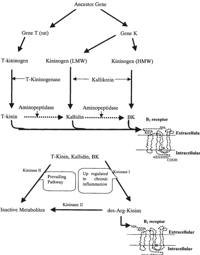
BK and KD are generated following the proteolytic cleavage of their respective
precursors, HMWK and LMWK by plasma and tissue kallikreins 22.23
(Fig. 3). A single gene codes for plasma kaÏlikrein, whereas tissue kallikrein is a member of a
multigene family that shows different patterns of tissue-specific gene expression 22
T-kinin was identified exclusively in the rat 24.25
and could be generated from T
kininogen under the enzymatic action of T-kininogenase26•
Kinins stimulate B1 or B2
receptors on the celi surface to produce their biological effects 21,22
TABLE III. PRIMARY STRUCTURE 0F MAMMALIAN KININS
Ancestor Gene Gene T (rat)
/
z
Kininogen (LMW) Gene K B1 receptor NH2 :2::. Extracellular Intracellular COOHFIGURE 3. Kinin formation and inactivation. Ail the components of the kallikrein-kinin system
are shown in this diagram. The B1 receptor is flot readily expressed undet normal physiotogical condition, it can be upregulated during chronic pathological states (e.g. inflammation, hyperthermia, diabetes and epilepsy)16
See text for details.
T-kininogen Kininogen (HMW) T-Kininogenase Kallikrein
J
AminopeptidaseI
AminopeptidaseT—kinin ...+....
*
BK B2receptor T-Kinin, Kallidin, BK Kininase Il ‘OOH Kininase II14
1.4.2. MNIN METABOLISM
Metabolic degradation is an important mechanism by which kinin action is
terminated. Kinins are broken down by a group of amino- , carboxy- and
endopeptidases found in blood, tissues and biological fluids 1622,27,28
(fig. 4). In
plasma and arterial walls, kinins are metabolized by carboxy-terminal degradation 29•
Kininase I (carboxypeptidase N of plasma or carboxypeptidase M of ceil
membranes) removes the C-terminal Arg and generates the des-Arg-kinin
homologues. Kininase II (also named angiotensin-I-converting enzyme, ACE), a dipeptidyl carboxypeptidase, liberates the C-terminal Phe8-Arg9 and Ser6-Pro7 in a
sequential order 16•
Kininase I metabolites are also kininase II substrates 27•
Kininase I
(carboxypeptidase N ,and carboxypeptidase M) Kallidin
Lys-Arg’-Pro2-Pro3-G1y4-Phe5-Ser6-Pro7-Phe8-Arg9
(ACE) Kininase II
ENZYME TISSUE
Kininase I Carboxypeptidase N Serum30
Carboxypeptidase M ccli membrane3’
Kininase II Angiotensin oenverting enzyme (ACE) Membrane,Serum, Endothelium, Brain,
Kidney, Lung, cerebrospinai fluid 32,33
fIGURE 4. Principal kininases: Theit cleaving sites for bradykinin and kallidin, the tissues where high activites are found 16
1.4.3. BIOLOGICAL EFFECTS
The biological effects of kinins include contraction or relaxation of smooth muscle, effects on epithelial ion transport, actions on endothelial celis and promotion of arteriolar vasodilation. Kinins participate in the acute inflammatory response of microvasculature and aid in tissue repair The control of celi function and mitogenesis are generally mediated via stimulation of B2 receptors
1.4.4. MNIN RECEPTORS
Kinins interact with their ceil surface G-protein coupled receptors (Fig. 5) to produce a variety of biological effects, such as vasodilation, stimulation of celi proliferation, production of pain and inllammatory responses 22
16
FIGURE 5. The dog B1 receptof amino acid sequence. Each circle represents an amino acid residue with the single letter code inset. The transmembrane domains are designated by Roman numerals. Clear circles indicate identical residues between dog and human B1 receptors. Asterisks designate potential phosphorylation sites. Modified from molecular cloning and pharmacological charactetization of the canin Bi and B2 bradykinin receptors. Bio!. Chem. 382: 123-129 17
There are at least two types of kinin receptors. They are designated B1 and B2
receptors Although, the existence of a B3 receptor was suggested in the
pulmonary tissue of guinea pig, but later the same group showed that it is invalid
By in situ hybridization the B1 receptor gene was localised to human chromosome 14
q 32.1-q32.2 38,
chromosome 14 in hand q32 41,42•
The sequence of kinin receptors lias seven
hydrophobic segments which form transmembrane helices.
B1 receptors were described for the first time in the isolated rabbit aorta They have
now been documented in isolated preparations taken from the cardiovascular, urinary as well as from intestinal systems, and in cultured ceils of vascular, endothelial,
mesangial, tracheal, bone and fibroblast origin 3536,37,43•
Although the B1 receptor is
constitutively present and functional in the canine cardiovascular system 13,
in the vas deferens and stomach of the mouse ,
this receptor is generally under-expressed in normal conditions but it is highly inducible by infiammatory mediators such as
bacterial lipopolysaccliaride, interleukins, overproduction of cytokines and growth
factors 20,46
B2 receptors have been identified for the first time in rabbit isolated jugular veins
using pharmacological in vitro assays-‘.
B2 receptors mediate the majority of in vivo
effects of kinins, including bronchoconstriction, hypotension, acute inflammatory reactions, pain and hyperalgesia 36.47,
Both B1 as well as B2 receptors have been
shown to mediate vasodilation
A comparison of B1 and B2 receptors according to size and interspecies homology is
presented in Table IV 16,
The amino acid sequence of the human B1 receptor (353 amino acid protein) is 36% identical to the amino acid sequence of the human B2
18
receptors is $0.6 and B1 receptors is 70.6. The natural agonists for B2 receptors are BK and KD, and for B1 receptors the natural agonists are des-Arg’°-KD and des
Arg9-BK. Highly potent and selective peptide as well as non-peptide agonists and
antagonists for B2 receptors and only peptide selective agonists and antagonists for B1 receptors are availabi e. No nonpeptide B1 receptor antagonists have been
described .
The peptide antagonist D-Arg- [Hyp3,Thi5,D-Tic7,Oic8,des-Arg9]
-BK([desArg’°]-Hoel4O lias been introduced as a selective B1 receptor antagonist‘
TABLE IV. BIOCHEMICAL FEATURES AND PHARMACOLOGY 0F MNIN RECEPTORS”
Receptor B2 B,
Family Rhodopsin superfamily of
G-protein-coupled receptors ‘ Rhodopsin superfamily of G-protein-coupled receptors Number of amino acids (molecular wt.) rat 366 (41.0 to 41.7 kDa) 50 human 364 (41.0 to 41.5kDa) M mouse 366 (41.5 kDa) 52 rabbit 367 (41.5 kDa) rat 337 (38.4 kDa) 16 human 353 (40.4 kDa) mouse 334 (38kDa) -rabbit 352 (39.5 kDa) 6 Homology (%)
Between human and rat Between human and mouse Between human and rabbit Between rabbit and rat Between rabbit and mouse Between mouse and rat
80.6 81.7 81.7 79.8 82.3 90.7 70.6 72.9 76.4 69.7 74.2 $8.4 Signal transduction mechanism
Phospholipase A,, C and D, cAMP, cGMP, ion channels
Phospholipase A and C Order of potency of natural agonists BK»des-Arg’-BK KD»des-Arg10-KD des-Arg’-BK»BK des-Arg10-KD»KD Peptide-selective agonist Ph’P(CH2NH)Arg9-BK 57 Sar[D-Phe’]des-Arg9-BK58 Non-peptide-selective FR190997 agonist Peptide-selective Hoe-140 . antagonist FR 173657 FR 167344 LF 16.0335 Bradyzide
-Hoe-140 is D-Arg-[Hyp, Thi5,D-Tic7, Oic’]-BK
.
- FR190997 is
($-[2,6-dichloro3-[N-[(E)-4(N-methylcarbamoyl)cinnamidoacetyl]-N-methyl-aminolbenzyloxy] -2-methyl-4-(2-pyridylmethoxy)quinoline)6°
-FR167344 is
(3-bromo-8-[2,6-dichloro-3-[N-[(E0-4-(n,n-dimethylcarbamoyl)cinnamidoacetylJ-N-methylamino]benzyloxy]-2-methylimidazo[1 ,2-aJpyridine hydrochloride)6’
-FR173657 is dichlorobenzyloxy]-2-methylquinoline)6 - LF1 6.0335 is (1 -[[3-[(2,4-dimethylquinolin-8-yl)oxymethylJ-2,4-dichloro-phenyljsulphonylj-2(S)-[[4-[(aminoiminomethyl)phenylcarbonyl]pjperazjn-1-ylÏcarbonyllpyrroljdine)62 -Bradyzide is ((2S)-1-[4-(4-benzhydrylthiosemicarbazido)-3-nitrobenzenesulfonyl]pyrrolidine-2-carboxylic acid[2-(2-dimethyl-aminoethyl)methylamino]ethyl]amide)63 Non-peptide-selective antagonist flot available [Leu’]des-Arg9-BK Lys [Leu’]des-Arg9-BK AcLysJD-Na17,I1e’Jdes-Arg -BK59 flot available
20
1.5. VASCULAR SIGNAL TRANSDUCTION PATHWAYS
The signal initiated by the binding of kinins to their receptors on the ceil surface is
transduced by G-proteins to intracellular pathways (fig. 6). In general, kinin
receptors activate Gaq/1 1 and Gai to stimulate intracellular pathways 16,64•
Second messengers
Kinins
. .
Cytoplasm
FIGURE 6. The patbway for G-protein dependent signal transduction. KR, kinin receptors; G, G protein incluing its three subunites (a, 13 and y). See text for details.
Kinins stimulate a variety of intracellular pathways depending on cellular types.
Activation of intracellular pathways by kinins resuits in the accumulation of cAMP
or cGMP, activation of phospholipases A2, C or D. These second messengers will then produce the release of prostaglandins or NO, the opening of ion channels, and
Celi membrane .
+
release of inositol phosphates or DG from membrane inositol phospholipids. The
resuit is mobilization of [Ca2]1 and activation of several isoforms of PKC 22.66.67
Moreover, it is also suggested that the activation of protein tyrosine kinases and
phosphatases as well as a MAP kinase are involved in kinin-mediated signal
transduction6g-70
1.5.1. MNIN-STIMULATED RELEASE 0F NITRIC OXIDE
Kinins could stimulate L-arginine tlirough the kinin receptors on the surface of
endothelial ceils to release NO 22,7i
Nitric oxide is a lipophulic compound, therefore
it does not require a ceil surface receptor to mediate its action on vascular smooth
muscle. Indeed, NO produced by the endothelium probably moves by diffusion into
the contiguous vascular smooth muscle. The NO derived from L-arginine activates GC in smooth muscle celis leading to accumulation of cGMP which mediates
vascular relaxation 72
22
Endothelial Ccli
Vascular Smooth Muscle CeIl
Kinins
FIGURE 7. Nitric oxide and vascular smooth muscle ceils. Production of endothelium-derived nitric oxide from L-arginine. R, kinin receptors ; NO, nitric oxide; OC, guanylyl cyclase; cGMP, cyclic guanosine monophosphate; GTP, guanosine triphosphate72
Nitric oxide (NO)
1.5.2. MNIN-STIMULATED INCREASE 0f INTRACELLULAR CALCIUM
Kinin-stimulated increase of [Ca2] can be achieved via extracellular influx or
through release of Ca2 from intraceilular stores There are five possible routes for
Ca2 entry into endotheliai ceils including receptor-operated, voltage-gated Ca2
channels, Ca2 leak channels, stretch-activated Ca2 channels, and Na/Ca2
exchange Kinins increase Ca2 influx into endothelial ceils by opening ion
channels .
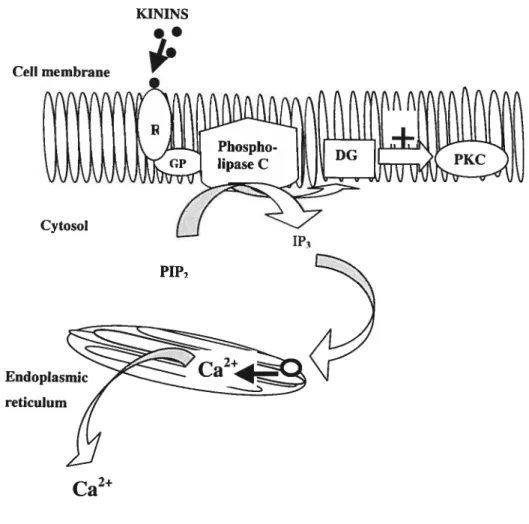
Furthermore, mediated by the 1P3 pathway, endothelial ceil Ca2 can be increased by kinins (Fig. 8). Following stimulation of kinin receptors, PLC acts upon
PIP2 to generate DG and 1P3. Subsequentiy, 1P3 mobilizes intraceliular Ca2 from internai stores to induce its effects Whiie a DG-activated PKC allows plasma membrane influx of extracellular Ca2
24
Celi membrane
MNINS
FIGURE 8. The phosphatidylinositol pathway. R, kinin receptors; GP, G-protein; PIP2,
phosphatidyl-inositol 4,5-diphosphate; 1P3, inositol triphosphate; DG, diacylglycerol; PKC, protein kinase C
PIP%
‘P3
reticulum
1.5.3. MNIN-STIMULATED RELEASE 0F PROSTACYCLIN
Kinins could stimulate phospholipids through kinin receptors on endothelial celis to produce AA, a 20 carbon, unsaturated, fatty acid formed from phospholipids in the celi membrane by PLA2 . Subsequently, AA is metabolized by cyclo-oxygenases
including COX1, a constitutively expressed isoform that is involved in a range of physiological functions, and the COX2 isoform which is inducible and expressed by extracellular stimuli such as pro-inftamatory cytokines in different celis. Further along the pathway, endoperoxides are metabolized by prostacyclin synthetase to produce PGI2 (Fig. 9a & 9b)73. The synthesis of PGI2 occurs predominantly in the endothelium and causes an increase of AC activity leading to the formation of vasodilator cAMP from ATP
Blood R
JJ.
Endothelial AA celi cox PGI2FIGURE 9a. Prostacyclin (PG12) and vascular smooth muscle ceils. Kinins cause the release of
metabolites of Arachidonic acid (AA), PGI2, through activation of its specific endothelial receptor. AC, adenylate cyclase; cAMP, cyclic adenosin monophosphate: ATP, adenosine triphosphate; KR, kinin receptors;
cox,
cyclo-oxygenases26 Phospholipids Phospholipase-A2 PROSTAGLANDINS PGE2, PGD2, PGF2 HYDROPEROXY AND HYDROXY FATFY ACIDS
Arachidonic acid Cyclo-oxygenase Endoperoxides isomerase Prostacyclin synthetase lipoxygenase PROSTACYCLIN (PGI2) Vasodilation THROMBOXANE-A2
FIGURE 9b. Arachidonic acid and its metabolites‘.
1.6. THE KALLIKREIN-MNIN SYSTEM IN RELATION TO THE EYE
Previous studies have demonstrated that the kallikrein-like enzymatic activities exist
in tissue homogenates of rabbit and swine eyes Later, another group, usÏng RT PCR, Southern blot analyses, and in sitit hybridization histochemistry, identified the
expression and localization of components of the kallikrein-kinin system in human
retina, choroid, cillary body, optic nerve, and endothelial cells of ocular blood
vessels 76
b date, there has flot been a study on the physiological function of the
kallikrein-kinin system in ocular tissues and its potential roles in the development of
the diabetic retinopathy.
2. HYPOTHESIS
Kinins are present in the retina, and kinins are known to be strong vasodilators. I
hereby propose the hypothesis that B2 receptors mediate vasodilation in the healthy
rat, and that early in diabetes kinin B1 receptors are induced and potentiate the
28
3. MM
To investigate the capacity for and mechanism of vasodilation produced by kinins in the retinal circulation of diabetic rats and their healthy control.
4. SPECIFIC OBJECTIVES
My specific objectives are to:
1) investigate the effect of BK and des-Arg9-BK on retinal vesse! tone in the healthy and STZ diabetic rat.
2) establish the receptors which mediate the vascular effects of kinins in the healthy and diabetic retina.
3) detemiine the signal transduction pathways which mediate the vascular effects of
CHAPTER II
1. MATERIALS & METHOBS
1.1. BIOLOGICAL MODEL
Male Wistar rats (150-250g); age (37-52 days), were purchased from Charles River (Saint Constant, Québec) and used as the control group. They were kept in our animal facilities at 22 °C, 12 h light/dark cycle and fead with Rodent Laboratory Chow # 5001 (Purina company, Canada) aU libituni.
For the diabetic group, Wistar rats were fasted overnight and made diabetic
following intraperitoneal injection of a single dose of STZ (65 mg/kg) dissolved in sodium citrate (pH 4.5). STZ is known to cause DNA alkylation and subsequent DNA strand breaks in pancreatic islet celis. This induces an insulitis and produces a
mode! of insulin-dependent diabetes mellitus Only rats with blood glucose 20 mM were considered diabetic. Also, the same rats showed polyuria after thay
became hyperglycemic. To measure the blood glucose, the rat’s tait was washed with
warm water and dried. Then, it was pricked and its blood glucose measured using test strips (Roche company, Québec).
30
1.2. EXPERIMENTAL PROTOCOL
The blood glucose of rats was evaluated just before the rats were decapitated to
remove their eyes. The eyes were enucleated and placed immediately in ice-cold
Krebs buffer (pH 7.4) of the following composition in mM: 120 NaCl, 4.5 KC1, 2.5 CaC12, 1.0 MgSO4, 27 NaHCO3, 1.0 KH2PO4, and 10 glucose. A circular incision was made in the posterior segment of the eye near the ora serrata to remove the
anterior segment and vitreous body with minimal handiing of the retina. The remaining eyecup was fixed with pins to a wax base containing 270 .il of Krebs buffer (pH 7.35-7.45) and maintained at 37°C. The preparations were allowed to stabilize for 15 mins. The outer vessel diameter was recorded with a video camera
and responses were quantified by a digital image analyser (Scion image).
A first measure was made and considered as baseline. Afterwards, U-46619 was used to
apply a tone to retinal vessels before measuring the effect of kinins. A concentration
response curve (Fig. 10) was made, and 1 tM U-46619 was considered optimal for
the rest of the experiments. In a first set of experiments, the vessels were pretreated
with U-46619 (1iM), then cumulative concentration-response curves to BK and des
Arg9-BK in the control and diabetic groups were made. The agonists or antagonists were used and vascular diameter was recorded before and after topical application of each concentration of agent, at which time a stable response was achieved. The
responses are expressed as percent change in the outer diameter of the vessel compared to U-46619 constricted vessels. It should be noted that in the present
have a larger overail diameter than corresponding arteries. Therefore, we chose the
vessels which appeard to have larger diameter.
1.3. CHEMICAL AGENTS
For this study, we used the following products: BK, a peptide-selective agonist for the B2 receptor 57;
des-Arg9-BK, a peptide-selective agonist for the B1 receptor
Hoe-140, a peptide-selective antagonist for the B2 receptor 49;
and des-Arg’°-Hoe 140, a peptide-selective antagonist for the B1 receptor were purchased from
Peninsula laboratories (California). GdC13, a channel blocker ; STZ, a DNA
aikylating agent 75;
U-46619, a stable thromboxane A2 mimetic and a vasoconstrictor
agent °
; L-NAME, irreversible inhibitor of eNOS and a reversible inhibitor of
iNOS81 ; L-745.337, a specific COX-2 inhibitor 82;
TPC, a PGI2 synthase inhibitor 83
and BHQ, specifically mobilizes Ca2 from 1P3-sensitive Ca2 stores by inhibiting
microsomal and sarcoplasmic reticulum Ca2-ATPase activity 84
were purchased from Sigma. cADP-ribose which functions as a physiological regulator of Ca2
release and which mobilizes Ca2 from internai pools that are distinct from 1P3—
sensitive poois 85
and NF023, a selective and direct G-protein antagonist for a
subunits of the G0/G group $6
32
1.4. STATISTICAL ANALYSIS
Each set of experiments were conducted on 3-8 rats, where n represents the number of rats which were used. Ml resuits are expressed as mean ± SEM. Resuits are analysed using Student’s t-test for paired samples and a one-way analysis of variance (ANOVA) followed by the Dunnett test was used for multiple comparisons with control group. A probability of
p<O.O5
was considered statistically significant.CHAPTER III
1. RESULTS
The resuits of this study are divided into two parts. The first part has been conducted in the healthy rat and the second part in the STZ-diabetic rat. Throughout the
experiments, U-46619 was used to give vessels tone and thus allow vasodilation to
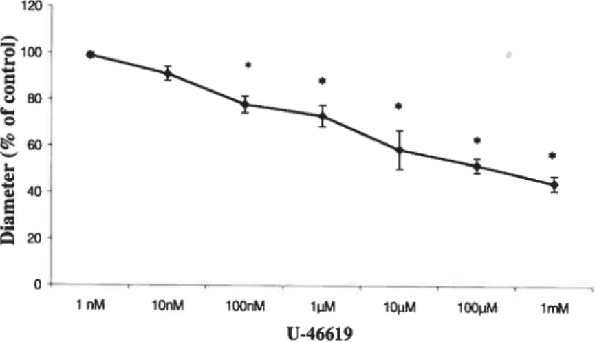
be studied. In an initial set of experiments, a dose-response cuwe was constructed for
U-46619 to set the optimal concentration to use in the experiments. U-46619 was
applied serially to retinal preparations with concentrations ranging from 1 nM to 1 mM (Fig. 10). U-46619 induced a dose-dependent vasoconstriction of rat retinal vessels. The vasoconstrictor effect was significant (j<O.O5, n=4) at concentrations
100 nM. The optimal concentration of U-46619 was decided to be 1 tM which
induces vascular tone similar to phsiological levels.
120
20
0
1 nM lOnM lOOnM liiM lOpM 100M lmM
U-46619
FIGURE 10. Dose-response curve for the effect of 46619 on retinal vessel diameter.
U-46619, was applied at a serial concentration from 1 nM to lmM for 10 mïns to apply tone to the retinal vessels. Resuits are expressed as mean ± SEM of data obtained from 4 separate retinas (4
34
1.1. CONTROL RAT
The following experiments were done in control healthy rat retinas, with the aim to investigate the effect of BK on retinal vesse! diameter
1.1.1. EFFECT 0F BRADYMNIN ON RETINAL VESSEL TONE
After a baseline measure was taken, and U-46619 (1iM) was applied, retinas were treated with serial concentrations of BK ranging from 100 pM to 10 nM. Treatment duration for each concentration was 15 mins (Fig. 11). The resuits show that BK induces a dose-dependent relaxation of retinal vessels following the vasoconstrictor effect of U-46619. The vasodilator effect of BK is significant (p<0.05, n=13) starting at a concentration of 100 pM. * 160 * 140 * 120 100
E
E.
_
1 iM lOOpM mM l0nM U-46619 BKFIGURE 11. Effect of BK on the control rat retinal vessel diameter. A baseline measure was obtained. Then, U-46619 (1IM) was used and the second measure was obtained, after, BK was applied at a serial concentration from 100 pM to 10 nM for 15 mins and the diameter of the vessels was measured each time.
(*p<O.OS.
n=13) compared to the vasocostrictor effect of U-46619.1.1.2. EFFECT 0F des-Arg9-BRADYMNIN ON RETINAL VESSEL TONE
b determine the effect of des-Arg9-BK on control rat retinal vesse! diameter, U-466 19 was used to constrict retinal vessels after which serial concentrations of des Arg9-BK were applied over concentrations ranging from 10 pM to 10 nM, with a treatment duration of 15 mins (Fig. 12). Resuits show that des-Arg9-BK does flot affect (p>.0.05, n=7) retina! vessel diameter following the vasoconstrictor effect of U-466 19. Even at a concentration 100 times higlier than what is needed of BK to induce retinal vasodi!ation (des-Arg9-BK (10 nM) vs BK (100 pM)).
120
3 100 $0
1flliflli
1 jiM lOpM lOOpM mM lOnMU-46619 des-Arg9-BK
FIGURE 12. Effect of des-Arg9-BK on the control rats retinal vessel diameter. A baseline measure was obtained and then U46619 was used after 10 mins the second measurement was done. after that a serial concentration of des-Arg9-BK ranging from 10 pM to 10 nM with a treatment duration of 15 mins was added. The diameter of the vessels was measured each time. The effect of des-Arg9-BK compared with the vasoconstrictor effect of U-46619 (n=7).
36
1.1.3. B2 RECEPTOR ANTAGONISM
To determine which kinin receptor mediates vasodilation evoked by BK under
healthy conditions, the following set of experiments were conducted. Retinal vessels
were vasoconstricted using U-46619 (1 liM). Afterwards, retinas were pretreated
with Hoe-140 (10 1iM), the B2 receptor specific antagonist, for 15 mins, before adding BK (10 nM) (Fig. 13). As the resuits show, Hoe-140 was without effect on retinal vesse! tone. More importantly, BK failed to di!ate the vesse!s pretreated with Hoe-140 (p<O.Ol, n=7) when compared to the vasodilator effect of BK alone (Fig.
11). This suggests that B2 kinin receptors participate in BK-induced retinal
vasodilation. 120 100 $0 60 40
o
FIGURE 13. B2 kinin receptors in BK induced control rat retinal vasodilation. A baseline
measure was obtained, then U-46619 (1cM) was used after 10 mins the second measurement was done, Hoe-140 (10 uM) was applied and after 15 mils the third measurement was obtained, 3K (10 nM) was used and final measurement was done. The effect of BK is suppressed by Hoe-140 (n=7).
1 M 10 uM lOnM
1.1.4. B1 RECEPTORANTAGONISM
b address further the question of whether or flot B1 receptors are involved in BK induced retinal vesse! dilation, the foïlowing set of experiments were performed. We used the B1 receptor specific antagonist, des-Arg’°-Hoe-140. Retinal vesse!s were vasoconstricted by U-46619 (1 tM). Then, they were pretreated with des-Arg’°-Hoe 140 (10 1iM) for 15 mins before adding BK (10 nM) (Fig. 14). The resu!ts show that des-Arg10-Hoe-140 was without effect on retina! vesse! tone. Nevertheless, in contrast to the effect of Hoe-140, des-Arg10-Hoe-140 faiÏed to inhibit the effect of BK (n=5). This suggests that BK-induced retina! vasodilation is flot mediated by the B1 kinin receptor.
E
1Œ4
U-46619 des-Arg10-Hoe-140 BK
FIGURE 14. ldnin receptors in 3K induced control rat retinal vasodilation. A baseline measure was obtained, then U-46619 (1 11M) was used after 10 mins the second measurement was done, des-Arg10-Hoe-140 (10 [KM) was applied and after 15 mins the third measurement was obtained, 3K (10 nM) was used and final measurement was donc. The effect of 3K is suppressed by des-Arg10-Hoe-140 (n=5).
38
1.2. INTRACELLULARAND MEMBRANE PATHWAYS WHICH MEDIATE THE MNIN EVOKED VASODILATION IN CONTROL RATS
In the following series of experiments, the intracellular and membrane pathways which mediate the effect of BK were investigated. b identify these signal transduction pathways we used a battery of signal transducer inhibitors.
1.2.1. G0/G1-PROTEINS
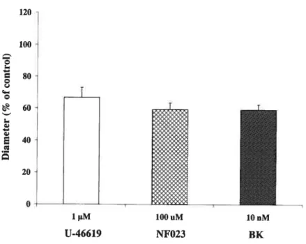
Using NF023 we investigated the participation of G0/G1-proteins in the vasodilation evoked by BK. Retinal vessels were vasoconstricted with U-46619 (1 !IM). Then, they were pretreated with NF023 (100 tM) for 25 mins before adding BK (10 nM) (fig. 15). The resuit shows that Nf023 was without effect on retinal vessel diameter, and that BK failed to dilate these vessels
(p<O.Ol,
n=4) when compared to the vasodilator effect of BK in the absence of Nf023 (Fig. 11). This demonstrates that the vasodilator effect of BK is transduced by G0/G-proteins.120 100 80 60 E 40 20 o U-46619 NF023 BK
FIGURE 15. G0/G-proteins in 13K induced control rat retinal vasodilation. A baseline measure
was obtained, then U-46619 (1M) was used after 10 mins the second measurement was done,
Nf023 (100 .tM) was applied and after 25 mins the third measurement was obtained, BK (10
nM) was used and final measurement was done. The effect of 13K is suppressed by NF023
r
1tM lOOuM lOnM
40
1.2.2. NITRIC OXIDE SYNTHASE INHIBITION
To determine whether or flot NO mediates the BK evoked vasodilation, L-NAME was used to inhibit NOS. Retinas were exposed to U-46619 to apply vascular toile.
Afterwards, the preparation was pretreated wïth L-NAME (100 tM), for 20 mins
before adding BK (1 nM) (Fig.16). The resuits obtained from these experiments show that L-NAME was without effect on retinal vessel diameter. However, BK induced a significant vasodilation (p<O.O5, n=5) in the retina. Therefore, NO is flot
involved in the vasodilator effect of BK in the retina.
FIGURE 16. Nitric oxide in BK induced control rat retinal vasodilation. A baseline measure
was obtained. Then, U-46619 (1tM) was used after 10 mins the second measurement was done.
After that , L-NAME (100 tM) was applied for 20 mins, then the third measurement was
obtained. Finally, 3K (1 nM) was used and the final measurement was donc. The vasodilator effect of 3K is flot blocked by L-NAME. (tp<O.O5, n=5).
* 140 120 o o o ‘60 E lpM 10iM mM U-46619 L-NAME BK
1.2.3. CALCIUM CHANNEL BLOCKERS
Is Ca2 influx necessary for the BK evoked vasodilation? b address this question, GdCÏ3 was used to block the Ca2 influx pathway of endothelial ceils. Retinas were exposed to U-46619 to provide a tone to vessels. Then, they were treated with GdCI3 (10 mM) for 15 mins before addition of BK (1 nM) (Fig. 17). The resuits show that GdCI3 was without effect on retinal vessel tone compared to the vasoconstrictor effect of U-46619. However, BK induced a significant vasodilation
(p<O.05,
n=5).This suggests that Ca2 influx is flot required for BK to induce vasodilation in the retina. 160 * ‘140 T 120 o c100 T 6O 20 1tM lOmM mM U-46619 GdCI3 BK
FIGURE 17. Extracellular calcium influx in BK induced control rat retinal vasodilation. A baseline measure was obtained. Then, U-46619 (1 tM) was used after 10 mins the second measurement was done. Afterwards, GdC13 (10 mM) was applied for 15 mins, then the third measurement was obtained. Finally, BK(1 nM) was used and the final measurement was done. The GdCI3 did flot block the effect of BK. (*p<O.05, n=5).
42
1.2.4. 1P3-SENSITIVE INTRACELLULAR CALCIUM STORES
To verify whether 1P3-sensitive Ca2 stores are involved in the effect of BK, we used BHQ. As BHQ was dissolved in ethanol, experiments were done to verify if the same concentration of ethanol which was used as vehicle affects the retinal vessel response to BK. Retinal vessels were vasoconstricted using U-46619 (1 pM). Afterwards, retinas were pretreated with ethanol (0.0001%) for 10 mins before adding BK (1 nM) (Fig. 18). When compared to the vasoconstrictor effect of U-466 19 the results show that ethanol is not able to affect the diameter of these vessels, and BK induces a significant vasodilation (p<0.05, n=5) in the presence of ethanol. This shows that the ethanol vehicle does not affect retinal vessel responses to BK. After these control studies, BHQ was tested on the retina (fig. 19). Following treatment with U-46619 (1 iM), retinas were pretreated with BHQ (1 iM) for 10 mins before the addition of BK (1 nM). As the resuits show, BHQ does flot change the diameter of vessels when compared to U-46619. Moreover, the vasodilator effect of BK was inhibited by BHQ (p.<O.Ol, n=6) (Fig. 11), which suggests that the release of Ca2 from 1P3-sensitive intracellular Ca2 stores is implicated in the vasodilator effect of BK.
=‘ 100 80 * T I 140 ‘ 120 40-E . 20-o 1tM 10-4% U-46619 Ethanol BK
fIGURE 18. Ethanol (vehicle for BHQ) in 3K induced control rat retinal vasodilation. A baseline measure was obtained. Then, U-46619 (1 &M) was used after 10 mins the second measurement was done. Afterwards , ethanol (1OE4%) was applied for 10 mins, then the third measurement was obtained. finatly, 3K (1 nM) was used and the final measurement was done,
(*p<o.o5,
n=5). 120 1 nM 60 40 20 o 1tM mM U-46619 BHQ BKFIGURE 19. 1P3-sensitive intracellular calcium stores in 3K induced control rat retinal vasodilation. A baseline measure was obtained. Then, U-46619 (1 M) was used after 10 mins the second measurement was donc. Afterwards, BHQ (1 tM), was applied for 10 mins, then the third measurement was obtained. Finally, 3K (1 nM) was used and the final measurement was donc (n=6).
44
1.2.5. NON-1P3-SENSITIVE INTRACELLULAR CALCIUM STORES
In the following set of experiments, cADP-ribose was used to investigate the role of 1P3-insensitive intracellular Ca2 p001 in BK-induced rat retinal vasodilation. Retinal vessels were vasoconstricted by U-46619 (1 tM). Then, the vessels were treated with cADP-ribose (10 M) for 25 mins before addition of BK (lOnM) (Fig.20). The resuits show that cADP-ribose does flot affect the diameter of the vessels compared to the vasoconstrictor effect of U-46619. Morever, the vasodilator effect of BK is blocked
(p<O.Ol,
n=5) when compared to the vasodilator effect of BK alone (Fig. 11). This suggest that 1P3-insensitive intracellular Ca2 poois are implicated in the effect of BK. 120 .100 80 60 40 E . 20 o1iM 10.rvI lOiiM
U-46619 cyclic ADP-ribose BK
FIGURE 20. Non-1P3-sensitive intracellular calcium stores in BK induced control rat retinal vasodilation. A baseline measure was obtained. Then, U-46619 (1 1iM) was used after 10 mins the second measurement was done. Afterwards , cyclic ADP-ribose (10 M), was applied for 25 mins, then the third measurement was obtained. Finally, BK(10 nM) was used and the final measurement was done (n=5).
1.2.6. COX-2 INifiBITORS
b determine whether or flot products of the cyclooxygenase pathway are involved
in the retinal vasodilator effect of BK, we used L-745.337, a specific COX-2
inhibitor. As L-745.337 is soluble in DMSO, control experiments were done to verify that a same concentration of DMSO as was used for a vehicle does flot affect retinal vesse! tone. Retina! vessels were vasoconstricted by U-46619 (1 tM). Then,
they were treated with DMSO (0.0015%) for 30 mins before addition of BK (1 nM) (Fig. 21). The resuits show that DMSO does flot affect vessel diameter, and that BK can induce vasodilation (p<0.05, n=3). In the second set of experiments, retinal
vessels were vasoconstricted using U-46619 (1 tM). Then, they were pretreated with L-745.337 (1 tM) for 30 mins before adding BK (1 nM) (Fig.22). The resuits show
that L-745.337 does flot affect the diameter of vesse!s. Moreover, BK can flot induce
retinal vesse! dilation (p<0.01, n=$) when compared to the vasodi!ator effect of BK
in the absence of L-745.337 (Fig. 11). This suggests that the cyc!ooxygenase
46 1 M 15x10-3% mM 160 140 120 100 80 60 40 .E 20 O U-46619 DMSO BK
fIGURE 21. DMSO (vehichie for L-745.337) in BK induced control rat retinal vasodilation. A baseline measure was obtained. Then, U-46619 (1 iM) was used after 10 mins the second measurement was done. Mterwards, DMSO (15x i0 %) for 30 mins was applied, then the third measurement was obtained. Finally, 3K (1 nM) was used and the final measurement was donc. The effect of BK is present. (*p<O.05, n=3).
120
Ï
.100 Q ç 80; ____ Q 60 ?40 E 20 — 1 ..M U-46619FIGURE 22. The COX-2 pathway in 3K induced control rat retinal vasodilation. A baseline measure was obtained. Then, U-46619 (111M) was used after 10 mins the second measurement was donc. Afterwards, L-745.337 for 30 mins was applied, then the third measurement was obtained. Finally, BK (1 nM) was used and the final measurement was donc (n=8).
1 nl.t
1.2.7. PROSTACYCLIN SYNTHASE INHIBITORS
To verify whether or flot PGI2 is iflvolved hi the retiflal vasodilator effect of BK we used TPC. Retinal vessels were vasoconstricted with U-46619 (1 tM). Then, they were pretreated with TPC (5 tM) for 25 mins before adding BK (1 nM) (Fig. 23).
The resuits show that TPC did flot affect the diameter of vessels. Morever, BK was flot able to dilate retinal vessels (p<O.O1, n=6) when compared to the vasodilator effect of BK in the absence of TPC (Fig. 11). This suggests that PGI2 is implicated in the vasodilator effect of BK in the retina.
z
2
FIGURE 23. Prostacyclin in BK induced control rat retinal vasodilation. A baseline measure was obtained. Then, U-46619 (1tM) was used after 10 mins the second measurement was done. Afterwards, TPC (5 tM) for 25 mins was applied, then the third measurement was obtained. Finally, BK (1 nM) was used and the final measurement was donc (n=6).
lpM 5iM mM
48
In summary, our data from the control rat group show that:
j) BK dilates retinal vessels and Hoe-140 inhibits the vasodilation induced by BK, ii) des-Arg9-BK is without effect on retinal vessel diameter,
iii) the vasodilator effect of BK is mediated by stimulation of B2 receptors, but flot B1 receptors,
iv) the signal initiated via stimulation of B2 receptors by BK is transduced by G0/G-proteins,
y) NO and extracellular Ca2 influx are flot involved in the effect of BK,
vi) the products of the cyclooxygenase pathway are involved in the retinal vasodilator effect of BK,
vii) PGI2 mediates the vasodilation, and
viii) intracellular Ca2 pools from both 1P3-sensitive and 1P3 insensitive stores are necessary for the vasodilator effect of BK.
1.3. STREPTOZOTOCIN-DIABETICRAT
In this section, we will show the kinin effects on retinal vessel diameter in STZ diabetic rats. Rats were made diabetic as described in section 11.1.1. Rats injected with STZ but which did flot develop diabetes (blood glucose ranging from 5 to 10 mM) were taken as controls to verify any potential direct cytotoxic effect of STZ on the retina. It should be stressed that those rats which became diabetic (blood glucose >20 mM), did so as early as 1 day after injection of STZ.
1.3.1. EFFECT 0F des-Arg9-BRADYMNIN ON RETINAL VESSEL TONE
To investigate what is the effect of des-Arg9-BK on SIZ-diabetic rat retinal vesse! tone, rats were put into 6 separate groups. The first group received norma! saline (contro! group), the second group were STZ-injected rats which did flot become diabetic, and the other four groups are STZ-injected diabetic rats: 1 day, 4, 7 and 21 days after the administration of STZ. After each period of treatment, the eyes have been prepared as was previously described. Retinas were exposed to U-46619 (1tM) to app!y a tone to the vessels. Ten minutes later, des-Arg9-BK was used in seria! concentrations ranging from 10 pM to 1 nM. The duration of treatment with des Arg9-BK was for 15 mins (Fig.24). In the control group which received normal saline (n=7) and in the STZ-injected non-diabetic rat group (n=5), des-Arg9-BK did not affect the diameter of vessels when compared to the vasoconstrictor effect of U 46619. Mso, in 1 day STZ-injected diabetic rats, des-Arg9-BK did not affect the diameter of the vesse!s (p>O.05, n=3) when compared to the vasoconstrictor effect of U-46619. However, in the other groups, 4 days (n=6), 7 days (n=6), and 21 days
(n=5), des-Arg9-BK changed the diameter of the vesse!s significant!y (p<0.05) and dose-dependently when compared to the vasoconstrictor effect of U-46619.
160 140 ‘120 c100 -Q