Elsevier Editorial System(tm) for Clinical Neurophysiology
Manuscript Draft Manuscript Number: CLINPH-D-18-11532R2
Title: Clinical and electrophysiological investigation of spastic muscle overactivity in patients with disorders of consciousness following severe brain injury
Article Type: Full Length Article
Section/Category: Sleep and Disorders of Consciousness
Keywords: Spasticity; Modified Ashworth Scale; H/M ratio; coma; minimally conscious state; unresponsive wakefulness syndrome
Corresponding Author: Ms. Géraldine Martens,
Corresponding Author's Institution: Coma Science Group, GIGA Research & Neurology Department, University and University Hospital of Liege, Liege, Belgium
First Author: Géraldine Martens
Order of Authors: Géraldine Martens; Thierry Deltombe; Marguerite Foidart-Dessalle; Steven Laureys; Aurore Thibaut
Cover letter : Martens G et al, CLINPH-D-18-11532R2
1
October 18, 2018
Dear Fer Mesman, ir,
We are pleased to resubmit the revised version of CLINPH-D-18-11532 “Clinical and electrophysiological investigation of spastic muscle overactivity in patients with disorders of consciousness following severe brain injury”. We are very grateful for the rapid reviewing process and addressed the concerns as outlined below.
We addressed the editorial comments regarding unnecessary files in the abstract, figure legends and tables and provided additional details on specific references. We followed the reviewer’s advice to rephrase a sentence in the introduction.
We hope to have addressed the referees’ suggestions in a timely, organized and satisfactory manner.
We report no conflict of interest and the paper is not published or under consideration elsewhere.
Thank you for your consideration, time and continued support. Respectfully,
Géraldine Martens
Cover letter : Martens G et al, CLINPH-D-18-11532R2
2 In response to the Editorial Office Comments:
1. Abstract. You state: "We removed the Highlights and Keywords from the separate abstract-file which now only includes the abstract and the authors' names, affiliations, funding sources and declaration of interest."
Please leave ONLY the Abstract in this file. The rest has no meaning.
We removed the authors’ names, affiliations, funding sources and declaration of interest to leave only the abstract in the file.
2. The Legends of Figures 1-2 are embedded in the text. Please move them to the end of the manuscript-file.
We moved the Legends of Figures 1-2 to the end of the manuscript-file. 3. Refer to Supplementary Table S2 as well.
We referred to Supplementary Table S2 in the manuscript. Please note that we updated the order of the Supplementary Tables so that Table S1 appears before Table S2 in the manuscript. We therefore also updated the Supplementary Material file.
4. Please provide volume & page numbers, or else a doi, for:
Deltombe T, Lejeune T, Gustin T. Botulinum toxin type A or selective neurotomy for treating focal spastic muscle overactivity? Ann Phys Rehabil Med. 2018;
Since this article is still in press, we provided the doi (10.1016/j.rehab.2018.07.008) in the references. The reference now reads:
Deltombe T, Lejeune T, Gustin T. Botulinum toxin type A or selective neurotomy for treating focal spastic muscle overactivity? Ann Phys Rehabil Med. 2018; available online:
10.1016/j.rehab.2018.07.008
5. Can you add the Editor names of the book "Coma and Disorders of Consciousness"? Martens G, Foidart-Dessalle M, Laureys S, Thibaut A. How Does Spasticity Affect Patients with Disorders of Consciousness? In: Coma and Disorders of Consciousness. Cham:
Springer International Publishing; 2018. p. 119-35.
We added the Editor names (i.e., Scnakers C and Laureys S) in the references. The reference now reads:
Cover letter : Martens G et al, CLINPH-D-18-11532R2
3
Martens G, Foidart-Dessalle M, Laureys S, Thibaut A. How Does Spasticity Affect Patients with Disorders of Consciousness? In: Schnakers C, Laureys S, editors. Coma and Disorders of Consciousness. Cham: Springer International Publishing; 2018. p. 119–35.
In response to the comments of Reviewer #1:
I found one inappropriate description.
In introduction: "The ratio of the maximal H-reflex amplitude . . . and reflects the percentage of excited motoneurons activated upon electrical stimulation in comparison . . . " should "The ratio . . . and reflects the percentage of excited motoneurons via the H-reflex in comparison . . . ".
We thank the reviewer for his careful reading and adapted the sentence in the manuscript as suggested. It now reads:
The ratio of the maximal H-reflex amplitude and of the maximal M response amplitude is called Hmax/Mmax ratio and reflects the percentage of excited motoneurons via the H-reflex in comparison to the direct activation of the motoneurons (Katz et al. 1992)
Abstract
Objective: The clinical and electrophysiological profile of spastic muscle overactivity (SMO) is poorly documented in patients with disorders of consciousness (DOC) following severe cortical and subcortical injury. We aim at investigating the link between the clinical observations of SMO and the electrophysiological spastic over-reactivity in patients with prolonged DOC. Methods: We prospectively enrolled adult patients with DOC at least 3 months post traumatic or non-traumatic brain injury. The spastic profile was investigated using the Modified Ashworth Scale and the Hmax/Mmax ratio. T1 MRI data and impact of medication were analyzed as well. Results: 21 patients were included (mean age: 41±11 years; time since injury: 4±5 years; 9 women; 10 traumatic etiologies). Eighteen patients presented signs of SMO and 11 had an increased ratio. Eight patients presented signs of SMO but no increased ratio. We did not find any significant correlation between the ratio and the MAS score for each limb (all ps > 0.05). The presence of medication was not significantly associated with a reduction in MAS scores or Hmax/Mmax ratios.
Conclusions: In this preliminary study, the Hmax/Mmax ratio does not seem to reflect the clinical MAS scores in patients with DOC. This supports the fact they do not only present spasticity but other forms of SMO and contracture.
Significance: Patients with DOC are still in need of optimized tools to evaluate their spastic profile and therapeutic approaches should be adapted accordingly.
1
Clinical and electrophysiological investigation of spastic muscle overactivity
in patients with disorders of consciousness following severe brain injury
Martens, G.1, Deltombe T.2, Foidart-Dessalle M.3, Laureys S.1 & Thibaut A.1
1
Coma Science Group, GIGA Research & Neurology Department, University and University Hospital of Liege, Liege, Belgium
2
Departments of Physical Medicine and Rehabilitation, CHU UCL (Université catholique de Louvain) Namur site Godinne, Belgium
3
Departments of Physical Medicine and Rehabilitation, University and University Hospital of Liege, Liege, Belgium
Corresponding author: Géraldine Martens (geraldine.martens@ulg.ac.be) GIGA-Research Coma Science Group and Neurology Department
University and University Hospital of Liège Avenue de l’Hopital, 11
Liège Belgium
Funding: The study was supported by the University and University Hospital of Liège, the Belgian National Funds for Scientific Research (FRS-FNRS), Human Brain Project (EU-H2020-fetflagship-hbp-sga1-ga720270), Luminous project (EU-H2020-fetopen-ga686764), DOCMA project (EU-H2020-MSCA–RISE–778234), Marie Sklodowska-Curie Actions (H2020-MSCA-IF-2016-ADOC-752686), the James McDonnell Foundation, Mind Science Foundation, the European Commission, the Public Utility Foundation ‘Université Européenne du Travail’, "Fondazione Europea di Ricerca Biomedica", the Bial Foundation. The funders had no role in study design, data collection and analysis, decision to publish, or preparation of the manuscript.
*Manuscript
2
Declaration of interests: None of the authors have potential conflicts of interest to be disclosed.
3 Abstract
Objective: The clinical and electrophysiological profile of spastic muscle overactivity (SMO) is poorly documented in patients with disorders of consciousness (DOC) following severe cortical and subcortical injury. We aim at investigating the link between the clinical observations of SMO and the electrophysiological spastic over-reactivity in patients with prolonged DOC.
Methods: We prospectively enrolled adult patients with DOC at least 3 months post traumatic or non-traumatic brain injury. The spastic profile was investigated using the Modified Ashworth Scale and the Hmax/Mmax ratio. T1 MRI data and impact of medication were analyzed as well.
Results: 21 patients were included (mean age: 41±11 years; time since injury: 4±5 years; 9 women; 10 traumatic etiologies). Eighteen patients presented signs of SMO and 11 had an increased ratio. Eight patients presented signs of SMO but no increased ratio. We did not find any significant correlation between the ratio and the MAS score for each limb (all ps > 0.05). The presence of medication was not significantly associated with a reduction in MAS scores or Hmax/Mmax ratios.
Conclusions: In this preliminary study, the Hmax/Mmax ratio does not seem to reflect the clinical MAS scores in patients with DOC. This supports the fact they do not only present spasticity but other forms of SMO and contracture.
Significance: Patients with DOC are still in need of optimized tools to evaluate their spastic profile and therapeutic approaches should be adapted accordingly.
Key words: Spasticity, Modified Ashworth Scale, H/M ratio, coma, minimally conscious state, unresponsive wakefulness syndrome
4 Highlights:
Severely brain-injured patients are prone to developing spastic muscle overactivity
No correlation between electrophysiological spastic component and clinical observation was found
Subcortical lesions may explain discrepancies between clinical and electrophysiological components
5 Abbreviations
DOC: Disorders of Consciousness
EMCS: Emergence from the Minimally Conscious State EMG: Electromyography
MAS: Modified Ashworth Scale MCS: Minimally Conscious State MRI: Magnetic Resonance Imaging SMO: Spastic Muscle Overactivity UMN: Upper Motor Neuron
UWS: Unresponsive Wakefulness Syndrome VS: Vegetative State
6 1. Introduction
About a third of patients who underwent a stroke or a traumatic brain injury will develop upper motor neuron (UMN) syndrome with spastic muscle overactivity (SMO) (Wissel et al. 2010, 2013; Martens et al. 2018). This syndrome can occur following any central nervous system lesion involving the corticospinal tract and parapyramidal tracts along the cortex, brainstem and spinal cord. UMN syndrome is classically described with positive (e.g., SMO) and negative signs (e.g., muscle weakness, fatigability) (Thibaut et al. 2013). SMO includes different forms of muscle hypertonia including spasticity, spastic dystonia, spastic co-contraction, associated reactions and spastic myopathy (Gracies et al. 2010a; Yelnik et al. 2010). A commonly accepted definition of spasticity is the clinical observation of an increase in velocity-dependent stretch reflexes (Gracies 2005b) (e.g., soleus muscle after stroke). Spastic dystonia is an inappropriate muscle activation at rest sensitive to passive stretch (e.g., finger flexors muscles after stroke). Spastic co-contraction is an inappropriate antagonist activation during agonist active mobilization (e.g., gastrocnemius co-contraction during tibialis anterior activation, finger flexors co-contraction during finger extensors activation). Associated reactions, also called spastic muscle overflow, is an inappropriate activation distant from the initial muscle contraction (e.g., elbow flexors activation when moving from sitting to standing). Lastly, spastic myopathy is the muscle modification following brain damage even in the absence of spasticity. Importantly, the prevalence of these different types of SMO in different muscles and etiologies has never been studied. A better determination and understanding of these different clinical manifestations in different etiologies is fundamental as the treatment should probably differ (Deltombe et al. 2018). In patients with disorders of consciousness (DOC) – encompassing unresponsive wakefulness syndrome/vegetative state (UWS/VS) (Laureys et al. 2010), minimally conscious state (MCS)
7
(Giacino et al. 2002) or emergence from the MCS (EMCS) – the proportion of patients suffering from SMO is extremely high, ranging from 57 to 89% (Martens et al. 2017). SMO may significantly alter quality of life especially if the patient is bedridden and lacks of voluntary command since these two conditions (i.e., disuse and immobilization) favor SMO to occur or increase (Gracies 2005a). Patients with DOC are often facing these two situations together, in addition to not being able to express any potential discomfort or pain (Schnakers et al. 2010; Chatelle et al. 2014). It seems therefore crucial to develop a reliable way to assess SMO to better understand and, consequently, treat it. The most widely tool used in daily practice is the Modified Ashworth Scale (MAS), a 6-level ordinal scale measuring the resistance to a passive movement. (Bohannon and Smith 1987). This bedside assessment may be sharpened by the study of the H-reflex which reflects the synaptic input elicited by synchronous electrical excitation of Ia afferent fibers and is affected by heterogeneous afferent inputs activating both mono- and oligosynaptic pathways (Burke et al. 1984). When a motor nerve is stimulated, two responses are recordable from the innervated muscle: a direct potential (called M response) corresponding to the direct activation of the efferent alpha motor fibers and a delayed reflex potential (called H response) corresponding to the activation via afferent Ia fibers of the alpha motor fibers at the spinal level. The ratio of the maximal H-reflex amplitude and of the maximal M response amplitude is called Hmax/Mmax ratio and reflects the percentage of excited motoneurons via the H-reflex in comparison to the direct activation of the motoneurons (Katz et al. 1992). The Hmax/Mmax ratio can sometimes be an indicator of the severity of spasticity due partly to the hyperexcitability of the alpha motoneuron through sensitive inputs and is increased in patients with clinical features of SMO (Angel and Hofmann 1963; Funase and Miles 1999; Huang et al. 2006). This quantitative measurement is therefore commonly used to assess the effects of surgical interventions aiming at decreasing the spasticity (Fève et al. 1997; Roujeau et al. 2003; Deltombe et al.
8
2006, 2008; Stokic and Yablon 2012). Both the MAS and the Hmax/Mmax ratio seem to be reliable and evolve in the same way in chronic stroke patients suffering from spasticity: when one decreases, the other decreases as well (Deltombe et al. 2008). The MAS score and the Hmax/Mmax ratio value for one spastic joint showed a correlation in this population (Pizzi et al. 2005; Ghotbi et al. 2006) even though this relation was found to be nonlinear in another study (Bakheit et al. 2003). While this relationship is widely studied in stroke, little is known regarding patients with DOC who usually show more extensive brain lesions. Therefore, this study aims at investigating the link between the MAS score (as a marker of SMO) and the Hmax/Mmax ratio (as an indirect marker of spasticity) with the help of neuroimaging in patients with DOC following severe brain injury. Results will provide insight in the relationship between clinical and electrophysiological spastic manifestations as well as their underlying structural pathophysiology.
9 2. Methods
2.1.Materials
This cross-sectional study was performed on clinically stable patients with DOC following severe traumatic or non-traumatic (ischemia or hemorrhage) brain injury admitted to the Institutional University Hospital for one week of diagnostic assessments including behavioral and neuroimaging examinations. Inclusion criteria were: 1) a diagnosis of UWS, MCS or eMCS according to international criteria (Giacino 2004); 2) age over 18 and 3) more than three months post injury. Exclusion criteria were: 1) documented neurological disorder prior to the injury; and 2) presence of skin or musculoskeletal lesions. The study was approved by the institutional ethical committee and written informed consents were obtained from the legal representatives. SMO was first evaluated by the same trained physiotherapist in the patient’s room using the MAS. Each patient was lying in bed to test the wrist flexors (with the fingers loose) and ankle plantar flexors. When a contracture (i.e., loss of passive range of motion by muscle shortening and joint retraction (Gracies 2005a; Kwah et al. 2012)) was identified by the physiotherapist during her testing, it was reported in addition to the MAS score for the affected joint. The patient was then brought to the hospital EMG lab to undergo an electrophysiological assessment performed by a trained physician. The maximal H response and M response were collected with increasing stimulation intensities using a Medtronic Keypoint EMG/EP/IOM Nerve Diagnostic System (Medtronic, Denmark). The surface electrodes were applied over the palmaris longus for the upper limb and over the soleus for the lower limb while the patient was lying in bed. Patient’s antispastic medication (e.g., Baclofen, Lorazepam, Clonazepam) was collected on admission to the hospital by reviewing the medical and nursing records. T1 MRI data was acquired during the same week and was analyzed by focusing attention on lesions in the pyramidal tract at the cortical level (i.e., the primary motor cortex and the supplementary motor area) and the subcortical level (i.e., basal
10
ganglia and brainstem). We then classified our sample in two subgroups according to the lesions: 1) “Both” = cortical lesions (i.e., involving the primary motor cortex and/or the supplementary motor area) and subcortical lesions (i.e., involving the basal ganglia and/or the brainstem); 2) “Subcort” = subcortical lesions only.
2.2.Statistics
Statistical analyses were performed using R 3.4.3 (R Core Team 2017). We used the Spearman’s correlation to compare the MAS scores and the Hmax/Mmax ratio for each limb. We also assessed the correlation between the MAS scores, the Hmax/Mmax ratios and the time since injury. We used the Mann-Whitney U test to investigate the difference of MAS scores and Hmax/Mmax ratios according to medication and contracture (i.e., presence vs. absence for both). Comparison between the groups presenting cortical and subcortical lesions or subcortical lesions only and the groups presenting an increased (i.e., ≥ 0.5 – (Deltombe et al. 2008)) Hmax/Mmax ratio or a normal ratio was performed using a Fisher’s exact test. Results were considered significant at the 5% level (p<0.05).
11 3. Results
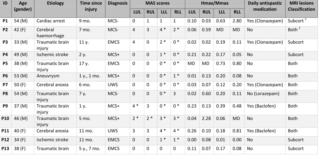
We included 21 patients (5 UWS, 12 MCS, 4 EMCS; mean age: 41±11 years; mean time since injury: 4±5 years; 9 women; 10 with traumatic etiology). Demographic data, Hmax/Mmax ratios, MAS scores and the main MRI lesions are reported in Table 1.
TABLE 1
According to the MAS, 18 patients out of 21 (86%) suffered from SMO (MAS ≥ 1 for at least one limb) and 7 (33%) presented severe SMO (MAS ≥3 for at least one limb). Presence of SMO was found in 2 patients (10%) for the upper limbs only, in 4 patients (19%) for the lower limbs only and in 12 patients (57%) for both upper and lower limbs. Regarding the Hmax/Mmax ratio, 11 patients (52%) presented a higher ratio (≥50%) for at least one limb. The Hmax/Mmax ratio was increased in 5 patients (24%) for the right lower limb, in 4 patients (19%) for the left lower limb, in 5 patients (24%) for the right upper limb and was below that threshold for the left upper limb for every patient, as presented in Figure 1.
FIGURE 1
We found no significant correlation between the MAS score and the Hmax/Mmax ratio for each limb (r= -0.23, p=0.24 [left lower]; r=0.10, p=0.67 [right lower]; r=0.25, p=0.28 [left upper]; r=0.12, p=0.63 [right upper]). Nine patients (43%) presented both SMO and increased Hmax/Mmax ratio for at least one limb. Ten patients (48%) presented a dissociation between the presence of SMO and an increase in Hmax/Mmax ratio (9 presence of SMO and Hmax/Mmax ratio not increased and 1 no SMO detected and increased Hmax/Mmax ratio).
12
Eighteen patients (86%) had contractures (5 had wrist retraction and 16 fixed equinovarus feet). A summary of the interrelations between presence of SMO; increased Hmax/Mmax ratio; contracture is presented in Figure 2. The presence of contracture was associated with higher MAS score for the upper limbs (U=16; p=0.04) but not for the lower limbs (U=22.5; p=0.15).
FIGURE 2
The time since injury did not correlate with the MAS mean scores (R= -0.06; p=0.79) or the Hmax/Mmax mean values (R= -0.27; p=0.25). Eleven patients (48%) were receiving oral medication to treat the spasticity (6 baclofen, 4 clonazepam, 1 lorazepam). This sample did not show lower MAS scores than the patients without antispastic treatment for both the upper (U=45.5; p=0.51) and lower limbs (U=42; p=0.37). Regarding the influence of medication on the Hmax/Max ratio, no significant difference between these two groups was observed either for the upper (U=60.5; p=0.45) and the lower limbs (U=34; p=0.10).
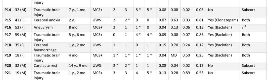
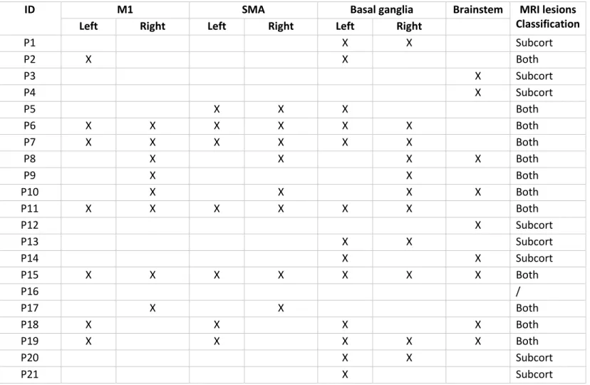
Regarding the relationship with MRI lesions, 8 patients with both increased Hmax/Mmax ratio and increased MAS score had both cortical and subcortical lesions and two patients with both conditions had subcortical lesions only. In the sample with increased MAS scores only, two had both cortical and subcortical lesions, 5 had subcortical lesions only and one had no interpretable MRI due to material artifacts. Only one patient presented with an increased Hmax/Mmax ratio only and had both cortical and subcortical lesions. The MRI lesions classification is presented in Table 2 and the localization of the main MRI lesions is presented in Supplementary Table S1.
13
When comparing the subpopulations of patients who presented both cortical and subcortical lesions (n=12) and the patients who present subcortical lesions only (n=8), there is a trend for an increased Hmax/Mmax ratio in the group of patients with both cortical and subcortical lesions (9/12 with both cortical and subcortical lesions presented an increased Hmax/Mmax ratio; while 2/8 with subcortical lesion only presented an increased Hmax/Mmax ratio – see Supplementary Table S2) even though the difference in proportions is not significant (p=0.07).
14 4. Discussion
The aim of the present study was to investigate the relationship between the MAS scores and the Hmax/Mmax ratio in a sample of severely brain-injured patients with DOC. The first is a clinical scale easily administered at the bedside while the second is a quantitative assessment investigating synaptic excitability and requires specific medical equipment. The MAS assesses the resistance to a passive movement (i.e., the stiffness) and has therefore a qualitative component which partly explains the inter-rater variability. A limitation of this scale is that it is performed at only one mobilization speed and doesn’t appreciate the velocity dependent increase in muscle tone which characterizes the spasticity; in contrast with the Tardieu Scale. The Tardieu Scale takes into account the influence of three different velocities (low, normal and fast) as well as the angle of contraction outbreak (Tardieu et al. 1954; Held and Pierrot-Deseilligny 1969; Gracies et al. 2010b). This scale might therefore be better suited to assess the SMO of patients with DOC since they are highly prone to present contractures. However, validation studies with this scale in adult populations are still needed (Boyd and Graham 1999; Mehrholz et al. 2005; Thibaut et al. 2013; Gracies et al. 2015). Even though the MAS is widely reported in the literature, another major drawback is that no distinction is made between severe SMO and ankyloses, contracture or even active resistance to mobilization and vegetative crisis, frequently suspected in DOC. Indeed, the last stage of the MAS is only defined as “affected joint is rigid in flexion or extension”. However, patients with DOC often present the association of SMO and contracture, which is confirmed by the results of this study (especially concerning the upper limbs). The cause of this co-presentation is probably the chronicity of the motor impairment in addition to their constant immobilization. Meanwhile, others neurologic conditions, such as stroke, also frequently associate SMO and motor impairment with immobilization but without severe contracture
15
such as observed in DOC (even though patients with stroke are not exempt of the risk of developing contracture, sometimes in a time span of a few weeks only after injury (Ward 2012)). The duration of immobilization is also typically more important in DOC patients. In our cohort, 66% of the patients suffered from a moderate muscle hypertonia (MAS <3) while 86% presented fixed contracture confirming the high rate of contracture even in the absence of severe SMO in patients with DOC after severe brain injury. In a previous study assessing the prevalence of SMO in 65 brain-injured patients with DOC, we have found 42% of upper limb contracture and 57% of fixed equinovarus feet. Such repeated findings support the major role of serious and extended brain lesions in the occurrence of contracture, in addition to the alterations in the patients’ state of consciousness.
Contractures may, in turn, affect the Hmax/Mmax ratio that investigates the proportion of the motoneuron pool that can be reflexively activated and is known to be pathologically increased in spastic patients (Angel and Hofmann 1963; Funase and Miles 1999; Huang et al. 2006). This increase can be explained either by an excessive facilitation of the H-reflex and/or an insufficient inhibition (Nielsen et al. 1993). As underlined above, a high proportion of patients presented contractures in our sample, which significantly affected the MAS scores, coupled sometimes with their immobilization that is likely to affect the Hmax/Mmax ratio. Indeed, prolonged immobilization impacts the presynaptic control of the Ia afferences, presumably via a decreased GABAergic inhibition, and thereby increases the H-reflex amplitude without affecting the M response (Clark et al. 2006; Lundbye-Jensen and Nielsen 2008b, 2008a). Future studies should investigate this aspect since the relation between the presence of contracture and the Hmax/Mmax ratio is poorly documented. The Hmax/Mmax ratio is used for both clinical and research purposes because it provides quantitative information in a reliable and sensitive way and enables an accurate follow-up of the patients or comparisons between groups. To the best of the authors’ knowledge, the present study is
16
the first one using this assessment to evaluate spasticity in patients with DOC following severe traumatic or non-traumatic brain injury and to correlate the results with neuroimaging findings. Both the MAS and the Hmax/Mmax ratio do not require the patient’s participation which is a considerable advantage in this non-communicating population. Our results show no significant correlation between the MAS and the ratio for the two muscular groups investigated (i.e., the wrist flexors and the ankle plantar flexors, which are commonly affected). This would mean that the Hmax/Mmax ratio does not reflect the clinical SMO as assessed by the MAS in severely brain-injured patients with DOC. Previous studies already emphasized the inter-subject variability (Levin and Hui-Chan 1993) along with the differences between the H reflex amplitudes and the clinical degree of spasticity. Such finding provides argument that SMO observed in patients with DOC does not seem to represent spasticity as defined by a velocity dependent increase in muscle tone associated to an hyperexcitability of the myotatic reflex. This supports the fact that patients with DOC do not only present spasticity but other forms of SMO and contracture. Another hypothesis is that the high MAS scores observed in a subset of patients in the present study might sometimes be due to the presence of contracture only and is therefore not reflected by an increase in the Hmax/Max ratio. When comparing these results to other populations, there seems to be a lack of consensus in the literature regarding the potential correlation between the Hmax/Mmax ratio and the MAS scores. Many authors report poor or no correlation between them in populations presenting multiple sclerosis (Morita et al. 2001) or stroke (Katz et al. 1992; Pisano et al. 2000; Bakheit et al. 2003) while several authors report a significant correlation between these two components in stroke and spinal cord injury patients (Milanov 1999; Pizzi et al. 2005; Huang et al. 2006). However, patients with DOC show more extended lesions than patients with stroke who usually suffer from cortical damage while patients with DOC often combine cortical and subcortical damage, as shown in the MRI results. In the present
17
study, a higher proportion of patients combining cortical and subcortical lesions presented an increased Hmax/Mmax ratio (73%) as compared to the patients with an increased ratio presenting subcortical lesions only (29%). This suggests that the Hmax/Mmax ratio increases when the lesions are cortical rather than when located near the brainstem. We could therefore draw the hypothesis that, in patients with DOC, the cortical lesions are less electrophysiologically expressed since they may be partly obscured by deeper structures’ lesions. Therefore, the ratio does not necessarily reflect the location of the lesions and is not systematically increased. This could explain the heterogeneity of the ratios measured in our sample. Regarding the influence of the time since injury on both MAS scores and the ratios it was non-significant which is not in line with our previous results, however, the present sample size is much smaller as compared to our previous cross-sectional study (Thibaut et al. 2015). The same applies for the presence of oral medication.
About half of the population studied was receiving drug treatment to reduce spasticity but there was no significant influence of the presence of medication on both the MAS scores and the Hmax/Mmax ratios, even though these two assessments tools are widely used to assess the clinical effects on antispastic interventions. Two hypotheses might explain this observation: 1) the medication was not effective enough in reducing the manifestations of spasticity, as previously suggested in DOC (Thibaut et al. 2015), even though the drugs used (i.e., baclofen) have previously shown clinical benefits in spastic patients (Gracies et al. 1997), or 2) the medication was effective in reducing spasticity, but due to the small sample size it may not be reflected in our statistical tests. However, the elevation of the Hmax/Max ratio may still help clinicians to extract the neuronal hyperexcitability input from the different components of SMO and contribute deciding to use antispastic medication.
The sample size is indeed a limitation of this proof of concept study and need to be considered before generalizing our results. Another limitation is that we only used the MAS to
18
assess SMO while other measurements such as the Tardieu Scale might be best suited to capture all the clinical components of spasticity. Regarding the electrophysiological assessment, we focused on the Hmax/Mmax ratio only while other measurements taking into account the mechanical component of reflex excitability, such as the T response and the Tmax/Mmax ratio, could have provided complementary data. It also appeared that our population was heterogeneous regarding time since injury, etiology and level of consciousness, which could also have impacted our findings. Finally, the chronicity of the motor alteration in these patients has some impact on the interpretation of the findings since the occurrence of contracture may hide the underlying SMO. Despite these limitations, this study is the first of its kind showing the poor correlation between the MAS scores and the Hmax/Mmax ratios, along with the absence of significant influence of either the localization of the MRI lesions and the medications used, in the specific population of patients with DOC following severe brain injury, underlying the complexity of their spastic profiles and the well-known intricate pathophysiology behind. Even though these are preliminary results, the Hmax/Mmax ratio does not appear as the prime candidate to evaluate the severity of SMO in patients with DOC since it does not seem to reflect the clinical MAS scores. These results need to be confirmed on larger samples to obtain more accurate and validated results reading the correlation between the clinical and electrophysiological outcomes as well as the structural brain lesions. Further studies with this population should also investigate the usefulness of other tools (e.g., Tardieu scale, Tmax/Mmax ratio, passive resistance torque) to assess SMO since it is a major issue affecting the general condition of patients with DOC, altering not only the motor ability but also likely preventing them to show several signs of consciousness and causing pain in this non-communicating population. Investigating the contribution of the different components of SMO (i.e., muscle hypertonia including spasticity, spastic dystonia, spastic co-contraction, associated reactions and spastic myopathy) would
19
also allow to better target the optimal therapeutic strategy. In the meantime, therapeutic approaches have to be adapted to the spastic profile of patients with DOC on an individual basis.
20 5. References
Angel RW, Hofmann WW. The H Reflex in Normal, Spastic, and Rigid Subjects. Arch Neurol. 1963;8:591.
Bakheit AM, Maynard VA, Curnow J, Hudson N, Kodapala S. The relation between Ashworth scale scores and the excitability of the alpha motor neurones in patients with post-stroke muscle spasticity. J Neurol Neurosurg Psychiatry. 2003;74:646–8.
Bohannon RW, Smith MB. Interrater reliability of a modified Ashworth Scale of muscle spasticity. Phys Ther. 1987;67:206–7.
Boyd R, Graham HK. Objective measurement of clinical findings in the use of botulinum toxin type A in the management of spasticity in children with cerebral palsy. Eur J Neurol. 1999;6:23–36. Burke D, Gandevia SC, McKeon B. Monosynaptic and oligosynaptic contributions to human ankle
jerk and H-reflex. J Neurophysiol. 1984;52:435–48.
Chatelle C, Thibaut A, Whyte J, De Val MD, Laureys S, Schnakers C. Pain issues in disorders of consciousness. Brain Inj. 2014;28:1202–8.
Clark BC, Manini TM, Bolanowski SJ, Ploutz-Snyder LL. Adaptations in human neuromuscular function following prolonged unweighting: II. Neurological properties and motor imagery efficacy. J Appl Physiol. 2006;101:264–72.
Deltombe T, Detrembleur C, Hanson P, Gustin T. Selective tibial neurotomy in the treatment of spastic equinovarus foot: a 2-year follow-up of three cases. Am J Phys Med Rehabil. 2006;85:82–8.
Deltombe T, Jamart J, Hanson P, Gustin T. Soleus H reflex and motor unit number estimation after tibial nerve block and neurotomy in patients with spastic equinus foot. Neurophysiol Clin Neurophysiol. 2008;38:227–33.
Deltombe T, Lejeune T, Gustin T. Botulinum toxin type A or selective neurotomy for treating focal spastic muscle overactivity? Ann Phys Rehabil Med. 2018; available online:
10.1016/j.rehab.2018.07.008
Fève A, Decq P, Filipetti P, Verroust J, Harf A, N’Guyen JP, et al. Physiological effects of selective tibial neurotomy on lower limb spasticity. J Neurol Neurosurg Psychiatry. 1997;63:575–8. Funase K, Miles TS. Observations on the variability of the H reflex in human soleus. Muscle Nerve.
1999;22:341–6.
Ghotbi N, Olyaei GR, Hadian MR, Ansari NN, Bagheri H. Is there any relationship between the Modified Ashworth Scale scores and alpha motoneuron excitability indicators? Electromyogr Clin Neurophysiol. 2006;46:279–84.
Giacino JT. The vegetative and minimally conscious states: consensus-based criteria for establishing diagnosis and prognosis. NeuroRehabilitation. 2004;19:293–8.
Giacino JT, Ashwal S, Childs N, Cranford R, Jennett B, Katz DI, et al. The minimally conscious state: definition and diagnostic criteria. Neurology. 2002;58:349–53.
21 2005a;31:535–51.
Gracies JM. Pathophysiology of spastic paresis II: emergence of muscle overactivity. Muscle Nerve. 2005b;31:552–71.
Gracies JM, Bayle N, Vinti M, Alkandari S, Vu P, Loche CM, et al. Five-step clinical assessment in spastic paresis. Eur J Phys Rehabil Med. 2010a;46:411–21.
Gracies JM, Brashear A, Jech R, McAllister P, Banach M, Valkovic P, et al. Safety and efficacy of abobotulinumtoxinA for hemiparesis in adults with upper limb spasticity after stroke or traumatic brain injury: A double-blind randomised controlled trial. Lancet Neurol. 2015;14:992–1001. Gracies JM, Burke K, Clegg NJ, Browne R, Rushing C, Fehlings D, et al. Reliability of the Tardieu
Scale for assessing spasticity in children with cerebral palsy. Arch Phys Med Rehabil. 2010b;91:421–8.
Held J, Pierrot-Deseilligny E. Reeducation Motrice des Affections Neurologiques. Paris. 1969; Huang C-Y, Wang C-H, Hwang I-S. Characterization of the mechanical and neural components of
spastic hypertonia with modified H reflex. J Electromyogr Kinesiol. 2006;16:384–91.
Katz RT, Rovai GP, Brait C, Rymer WZ. Objective quantification of spastic hypertonia: correlation with clinical findings. Arch Phys Med Rehabil. 1992;73:339–47.
Kwah LK, Herbert RD, Harvey LA, Diong J, Clarke JL, Martin JH, et al. Passive Mechanical
Properties of Gastrocnemius Muscles of People With Ankle Contracture After Stroke. Arch Phys Med Rehabil. 2012;93:1185–90.
Laureys S, Celesia GG, Cohadon F, Lavrijsen J, Leon-Carrion J, Sannita WG, et al. Unresponsive wakefulness syndrome: a new name for the vegetative state or apallic syndrome. BMC Med. 2010;8:68.
Levin MF, Hui-Chan C. Are H and stretch reflexes in hemiparesis reproducible and correlated with spasticity? J Neurol. 1993;240:63–71.
Lundbye-Jensen J, Nielsen JB. Central nervous adaptations following 1 wk of wrist and hand immobilization. J Appl Physiol. 2008a;105:139–51.
Lundbye-Jensen J, Nielsen JB. Immobilization induces changes in presynaptic control of group Ia afferents in healthy humans. J Physiol. 2008b;586:4121–35.
Martens G, Foidart-Dessalle M, Laureys S, Thibaut A. How Does Spasticity Affect Patients with Disorders of Consciousness? In: Schnakers C, Laureys S, editors. Coma and Disorders of Consciousness. Cham: Springer International Publishing; 2018. p. 119–35.
Martens G, Laureys S, Thibaut A. Spasticity management in disorders of consciousness. Brain Sci. 2017;7:162.
Mehrholz J, Wagner K, Meissner D, Grundmann K, Zange C, Koch R, et al. Reliability of the Modified Tardieu Scale and the Modified Ashworth Scale in adult patients with severe brain injury: a comparison study. Clin Rehabil. 2005;19:751–9.
Milanov I. Clinical and neurophysiological correlations of spasticity. Funct Neurol. 1999;14:193–201. Morita H, Crone C, Christenhuis D, Petersen NT, Nielsen JB. Modulation of presynaptic inhibition
and disynaptic reciprocal Ia inhibition during voluntary movement in spasticity. Brain. 2001;124:826–37.
22
following muscle stretch in spastic spinal cord injured patients than in healthy subjects. Exp Brain Res. 1993;97:173–6.
Pisano F, Miscio G, Del Conte C, Pianca D, Candeloro E, Colombo R. Quantitative measures of spasticity in post-stroke patients. Clin Neurophysiol. 2000;111:1015–22.
Pizzi A, Carlucci G, Falsini C, Verdesca S, Grippo A. Evaluation of upper-limb spasticity after stroke: A clinical and neurophysiologic study. Arch Phys Med Rehabil. 2005;86:410–5.
R Core Team. R: A language and environment for statistical computing. R Foundation for Statistical Computing, Vienna, Austria. 2017.
Roujeau T, Lefaucheur J-P, Slavov V, Gherardi R, Decq P. Long term course of the H reflex after selective tibial neurotomy. J Neurol Neurosurg Psychiatry. 2003;74:913–7.
Schnakers C, Chatelle C, Majerus S, Gosseries O, De Val M, Laureys S. Assessment and detection of pain in noncommunicative severely brain-injured patients. Expert Rev Neurother. 2010;10:1725– 31.
Stokic DS, Yablon SA. Effect of concentration and mode of intrathecal baclofen administration on soleus H-reflex in patients with muscle hypertonia. Clin Neurophysiol. 2012;123:2200–4. Tardieu G, Shentoub S, Delarue R. A la recherche d’une technique de mesure de la spasticité. Rev
Neurol. 1954;91:143–4.
Thibaut A, Chatelle C, Wannez S, Deltombe T, Stender J, Schnakers C, et al. Spasticity in disorders of consciousness: A behavioral study. Eur J Phys Rehabil Med. 2015;51:289–397.
Thibaut A, Chatelle C, Ziegler E, Bruno M-A, Laureys S, Gosseries O. Spasticity after stroke: physiology, assessment and treatment. Brain Inj. 2013;27:1093–105.
Ward AB. A literature review of the pathophysiology and onset of post-stroke spasticity. Eur J Neurol. 2012;19:21–7.
Wissel J, Manack A, Brainin M. Toward an epidemiology of poststroke spasticity. Neurology. 2013;80:S13-19.
Wissel J, Schelosky LD, Scott J, Christe W, Faiss JH, Mueller J. Early development of spasticity following stroke: a prospective, observational trial. J Neurol. 2010;257:1067–72.
Yelnik AP, Simon O, Parratte B, Gracies JM. How to clinically assess and treat muscle overactivity in spastic paresis. J Rehabil Med. 2010;42:801–7.
23
Figure 1: Proportions of patients presenting SMO, spasticity and contracture according to the MAS (≥1), the Hmax/Mmax ratio (≥0.5) and clinical notes, respectively. DOC= disorders of consciousness; LUL= left upper limb; RUL= right upper limb; LLL= left lower limb; RLL= right lower limb; MAS= Modified Ashworth Scale.
Figure 2: Venn diagram presenting the interrelations between patients presenting with increased Modified Ashworth Scale (≥ 1), increased Hmax/Mmax ratio (≥ 0.5 - (Deltombe et al. 2008)), contracture (presence versus absence) and none of these characteristics (=*).
24 6. Tables
Table 1: Demographic data, MAS scores, Hmax/Mmax ratios, medication, MRI lesions of the study sample. mo. =months; y. =years; MCS =minimally conscious state; EMCS =emergence from the minimally conscious state; UWS =unresponsive wakefulness syndrome; LUL =left upper limb; RUL =right upper limb; LLL =left lower limb; RLL =right lower limb; MAS =Modified Ashworth Scale; MD =Missing Data; MRI =magnetic resonance imaging; * =presence of contracture (i.e., loss in passive range of motion); 1 =presence of subcortical lesions only (i.e., basal ganglia and/or the brainstem); 2 =presence of cortical and subcortical lesions (i.e., primary motor cortex and/or the supplementary motor area and/or basal ganglia and/or the brainstem); 3 =no interpretable MRI
ID Age (gender)
Etiology Time since injury
Diagnosis MAS scores Hmax/Mmax Daily antispastic medication
MRI lesions Classification LUL RUL LLL RLL LUL RUL LLL RLL
P1 54 (M) Cardiac arrest 9 mo. MCS- 0 1 1 1 0.10 0.03 0.63 2.80 Yes (Clonazepam) Subcort 1
P2 42 (F) Cerebral haemorrhage
7 mo. MCS- 4 3 4 * 2 * 0.06 0.59 MD MD No Both 2
P3 33 (M) Traumatic brain injury
11 y. EMCS 4 0 2 * 0 * 0.02 0.02 0.19 0.11 Yes (Clonazepam) Subcort
P4 49 (M) Ischemic stroke 2 y. MCS+ 0 0 1 * 0 * 0.21 0.22 0.17 0.05 No Subcort
P5 38 (M) Traumatic brain injury
17 y. EMCS 0 0 0 * 0 * MD MD 0.73 0.80 No Both
P6 53 (M) Aneuvrysm 1 y., 1 mo. MCS+ 0 0 0 * 1 * 0.01 0.13 0.20 0.08 No Both
P7 50 (F) Cerebral anoxia 6 mo. UWS 0 0 0 * 0 * 0.03 0.07 0.12 0.20 Yes (Clonazepam) Both
P8 54 (M) Traumatic brain injury
7 y. MCS- 0 0 0 * 3 0.02 0.60 0.20 0.11 No (Lorazepam) Both
P9 37 (M) Traumatic brain injury
1 y. MCS+ 4 * 3 0 * 0 * 0.23 0.13 0.39 0.48 Yes (Baclofen) Both
P10 46 (M) Traumatic brain injury
5 mo. MCS+ 2 * 2 * 3 * 3 * 0.04 2.28 0.06 MD No Both
P11 40 (F) Cerebral anoxia 11 mo. UWS 3 3 4 * 4 * 0.26 0.10 0.18 0.81 Yes (Baclofen) Both
P12 34 (F) Ischemic stroke 11 mo. EMCS 0 0 1 * 1 * 0.00 0.08 0.01 0.00 No Subcort
25 injury
P14 32 (M) Traumatic brain injury
7 y., 1 mo. MCS+ 2 3 5 * 5 * 0.08 0.08 0.02 0.05 No Subcort
P15 41 (F) Cerebral anoxia 2 y. UWS 2 2 * 0 0 0.07 0.63 0.03 0.81 Yes (Clonazepam) Both
P16 53 (F) Aneuvrysm 8 mo. MCS+ 2 1 1 * 0 0.04 0.13 0.06 0.13 Yes (Baclofen) / 3
P17 59 (M) Traumatic brain injury
5 y., 6 mo. MCS+ 0 1 4 * 4 * 0.09 0.08 0.07 0.86 Yes (Baclofen) Both
P18 35 (F) Cerebral haemorrhage
1 y., 2 mo. UWS 1 1 0 1 0.15 0.70 0.24 0.12 Yes (Baclofen) Both
P19 18 (F) Traumatic brain injury
4 mo. MCS+ 1 * 1 * 1 * 1 * 0.04 MD 0.50 0.25 Yes (Baclofen) Both
P20 32 (M) Cardiac arrest 14 y., 9 mo. UWS 2 * 2 * 1 1 0.08 0.04 0.02 0.13 No Subcort
P21 19 (M) Traumatic brain injury
1 y., 2 mo. MCS+ 3 3 4 5 * 0.13 0.28 0.89 0.53 No Subcort
Table 2: Relationships between the clinical profile and the MRI lesions of the 21 patients included in the study. 1 = ratio ≥0.5; SMO =spastic muscle overactivity according to the Modified Ashworth Scale (score ≥1); Both =presence of cortical and subcortical lesions (i.e., primary motor cortex and/or the supplementary motor area and/or basal ganglia and/or the brainstem); Subcortical =presence of subcortical lesions only (i.e., basal ganglia and/or the brainstem); / =no interpretable MRI
Clinical profile MRI lesions SMO + high Hmax/Mmax
ratio 1
SMO only High Hmax/Mmax ratio only
None
Both 8 2 1 1
Subcortical 2 5 0 1
Figure 1
Figure 2
Table S1: Localization of the main MRI lesions for the study sample. M1 =primary motor cortex; SMA =supplementary motor area; MRI =magnetic resonance imaging; Subcort =presence of subcortical lesions only (i.e., basal ganglia and/or the brainstem); Both =presence of cortical and subcortical lesions (i.e., primary motor cortex and/or the supplementary motor area and/or basal ganglia and/or the brainstem); / =no interpretable MRI.
ID M1 SMA Basal ganglia Brainstem MRI lesions Classification Left Right Left Right Left Right
P1 X X Subcort P2 X X Both P3 X Subcort P4 X Subcort P5 X X X Both P6 X X X X X X Both P7 X X X X X X Both P8 X X X X Both P9 X X Both P10 X X X X Both P11 X X X X X X Both P12 X Subcort P13 X X Subcort P14 X X Subcort P15 X X X X X X X Both P16 / P17 X X Both P18 X X X X Both P19 X X X X X Both P20 X X Subcort P21 X Subcort
Table S2: Table used for the Fisher’s exact test of independence. Increased ratio =ratio ≥ 0.5.
Subcortical Lesions Both Cortical and Subcortical Lesions
Increased Hmax/Mmax ratio 2 9
Normal Hmax/Mmax ratio 6 3
AUTHOR CONCURRENCE FORM
CLINICAL NEUROPHYSIOLOGY
Official Organ of the International Federation of Clinical Neurophysiology (IFCN) http://www.journals.elsevier.com/clinical-neurophysiology
Prof. Ulf Ziemann
Editor-in-Chief Instructions:
1. Each submission to Clinical Neurophysiology needs a filled out Author Concurrence Form. Please fill in the Title of the manuscript below.
2. Collect signatures from all authors.
3a. Scan the form and submit it together with the paper. The submission item is Author Agreement. 3b. If this is not an option, please fax the form to the Editorial Office: +31-20-485-3881.
Title of the Manuscript: Clinical and electrophysiological investigation of spastic muscle overactivity in patients with disorders of consciousness
A signature below certifies compliance with the following statements.
Copyright Transfer: In consideration of the acceptance of the above manuscript for publication, the copyright is transferred to IFCN and administered by Elsevier. All proprietary rights other than copyright (such as patent rights) are reserved to the author(s), as well as the right to use original figures and tables in future works, provided full credit is given to the original publication. If the manuscript is work prepared by employee(s) of the United Kingdom or the United States government as part of their official duties, copyright cannot be fully transferred to the IFCN, and authors must check the appropriate box below.
● This manuscript was written in the course of employment by the United Kingdom and it is subject to Crown copyright.
● This manuscript was written in the course of employment by the United States Government and it is not subject to copyright in the United States.
Authorship Responsibility:
The submission is a truthful, original work without fabrication, fraud or plagiarism, and contains no libellous or unlawful statements.
It has not been published previously except in abstract form.
The manuscript is not under consideration for publication, nor will it be submitted for publication, elsewhere until a final decision has been made by this journal.
The undersigned certify that each author has participated sufficiently in the work to take responsibility for its truthfulness and validity, has read the complete manuscript, and concurs with its content.
Conflict of interest disclosure:
All funding sources supporting this work are acknowledged. The authors will disclose to the editor any pertinent financial interests associated with the manufacture of any drug or product described in this manuscript.
ALL AUTHORS MUST SIGN
Author Géraldine Martens Date July 19, 2018
Author Thierry Deltombe Date July 19, 2018 _____ ____________
Author Aurore Thibaut Date July 20, 2018
Author Marguerite Dessalle Date July 25,,2018
Author Steven Laureys Date July 25, 2018