O
pen
A
rchive
T
oulouse
A
rchive
O
uverte
(OATAO)
OATAO is an open access repository that collects the work of some Toulouse
researchers and makes it freely available over the web where possible.
This is
an author's
version published in:
https://oatao.univ-toulouse.fr/23117
Official URL :
https://doi.org/10.1016/j.otsr.2017.02.016
To cite this version :
Any correspondence concerning this service should be sent to the repository administrator:
tech-oatao@listes-diff.inp-toulouse.fr
Accadbled, Franck and Vial, Julie and Sales de Gauzy, Jérôme Osteochondritis dissecans
of the knee. (2018) Orthopaedics & Traumatology: Surgery & Research, 104 (1). S97-S105.
ISSN 1877-0568
OATAO
Osteochondritis
dissecans of the knee
F.
Accadbled
a,∗,
J. Vial
b,
J. Sales de Gauzy
aa Service de chirurgie orthopédique et traumatologique, hôpital des enfants, CHU de Toulouse, 330, avenue de Grande-Bretagne, 31059 Toulouse cedex 9,
France
b Service de radiologie, hôpital des enfants, CHU de Toulouse, France
Keywords: Osteochondritis dissecans Knee Adolescent Sports Arthroscopy
a b s t r a c t
Osteochondritis dissecans (OCD) of the knee is an idiopathic, focal, subchondral-bone abnormality that can cause instability or detachment of a bone fragment and overlying articular cartilage, with subsequent progression to osteoarthritis. The diagnosis is usually made during adolescence. Mechanical factors play a major role in the pathophysiology of OCD. When the radiographic diagnosis is made early in a patient with open physes, healing can often be obtained simply by restricting sports activities. The degree of lesion instability can be assessed by magnetic resonance imaging. When the lesion remains unstable and the pain persists despite a period of rest, surgery is indicated. Arthroscopic exploration is always the first step. Drilling of the lesion produces excellent outcomes if the lesion is stable. Unstable lesions require fixation and, in some cases, bone grafting. Defects must be filled, depending on their surface area. Although many surgical techniques are available, the therapeutic indications are now standardized.
1. Introduction
Osteochondritis dissecans (OCD) of the knee is an idiopathic, focal, subchondral-bone abnormality that can cause instability or detachment of a bone fragment and overlying articular cartilage, with subsequent progression to osteoarthritis. The diagnosis may be made in childhood or adulthood. Data reported at the 2005 SoF-COT symposium indicated a mean age at diagnosis of 16.5 years
[1]. The terms “open physes” and “closed physes” should be pre-ferred over the terms “juvenile” and “adult”. Despite advances in our understanding of this enigmatic condition, many issues remain unresolved, particularly regarding the pathophysiology, indications for magnetic resonance imaging (MRI), MRI signs of instability, current treatment methods, and feasibility of develop-ing a treatment algorithm.
2. Epidemiology
A recent epidemiological study failed to identify any cases in children younger than 6 years of age, and the diagnosis was made 3.3 times more often between 12 and 19 years than between 6 and 11 years[2]. The incidence was 9.5/100,000. The risk of developing OCD was 3.8 times higher in boys than in girls. Most patients are athletes[3]. The most common site is the medial femoral condyle,
∗ Corresponding author.
E-mail address:faccadbled@gmail.com(F. Accadbled).
particularly on its lateral surface. The lateral femoral condyle and patella were affected less often and the tibial plateau very rarely. 3. What is the pathophysiology?
3.1. Histology
A histological study evaluated osteochondral plugs from the center of OCD lesions[4]. A cleft was visible between the fragment and the surrounding trabecular bone. Interestingly, the fragments had not always been rated as unstable at arthroscopy despite being clearly separated from their bed. Thus, arthroscopic instability may be a delayed finding compared to the natural course of the condition. The subchondral bone was fractured and the fragment necrotic, although which of these two abnormalities occurred first was unclear. The fragment was composed of necrotic trabecular bone, viable trabecular bone, or cartilage with no bone. The deep surface of the fragment was covered with dense fibrous or carti-laginous tissue, resembling a non-union. Active bone remodeling was apparent beneath the surface of the basal side of the lesion. Thus, the necrotised fragment either undergoes re-ossification fol-lowed by incorporation or remains separated, as in a non-union, and eventually detaches.
3.2. Micro-trauma hypothesis
This hypothesis is, by far, supported by the best level of evidence. The initiating factor may be impingement on the anterior tibial
Fig. 1. Arthroscopic view of the anterior tibial spine in contact with the lateral sur-face of the medial femoral condyle during anterior cruciate ligament reconstruction.
spine. Wechter et al. found greater posterior and medial tibial slope in patients with medial condyle OCD, suggesting a role for micro-trauma from the anterior tibial spine[5]. The smaller intercondylar notch width in patients with OCD may promote impingement[6]. We found that the anterior tibial spine was larger in patients with OCD[7], in keeping with reports by Fairbank and Smillie (Fig. 1). In young baseball catchers, who frequently assume a crouched position, McElroy et al. showed that the OCD lesions were located more posteriorly on the femoral condyle[8]. A more distal loca-tion of the footprint of the posterior cruciate ligament (PCL) may add repetitive traction loading to the impingement[9]. Smillie[10]
then Cahuzac et al.[11]previously pointed out the close relation-ships between the OCD lesion and the femoral PCL footprint. In an MRI study, Laor et al. found disruption of the secondary physis, i.e., the edge of the enchondral ossification center of the condyle
[12]. This micro-trauma-induced disruption, akin to epiphysiode-sis, may result in uneven growth with the production of irregular subchondral bone that may predispose to OCD. Patients with lateral condyle OCD often have a discoid meniscus responsible for repet-itive abnormal loading. Complete discoid menisci were associated with central OCD and incomplete discoid menisci with peripheral OCD[13].
3.3. Other hypotheses
Other hypotheses have been put forward but failed to gar-ner support. As suggested by the designation “osteochondritis dissecans” used in 1888 by König, a moderate inflammatory foreign-body response has been described. A role for endocrine disturbances has been suggested. Reports of familial cases[14], particularly in identical twins, prompted genome-wide association studies, which recently identified several candidate loci in humans
[15]. Ischemia responsible for subchondral bone necrosis is often incriminated. Causes of ischemia may include trauma or arterial micro-thrombosis resulting in focal disruption of the blood supply. 3.4. In brief
OCD is clearly a multifactorial condition in which micro-trauma plays a major role. Multicenter studies and task forces such as Research in Osteo Chondritis of the Knee (ROCK) will provide fur-ther insights in the near future.
4. Clinical diagnosis
There are three main presentations:
• incidental discovery in an asymptomatic individual; • mechanical pain during sports (the most common);
• nearly continuous mechanical pain with swelling and locking of the joint.
Pain upon weight bearing is the predominant symptom in 80% of cases[1]. Patients may also report swelling, catching, or locking of the joint. In patients with an unstable or loose fragment, transient quadriceps blocks may result in buckling of the knee. A system-atic physical examination must be performed, as the pain may be unrelated to the OCD, which is then discovered incidentally.
The physical findings are often limited. Femoro-tibial align-ment in the coronal plane should be assessed. Medial condyle OCD is associated with varus and lateral condyle OCD with valgus of the knee[16]. Finger pressure on the femoral condyle at various degrees of knee flexion may elicit the patient’s usual pain. The Wil-son test consists in bending the knee at 90◦then passively moving it to 30◦ of flexion while rotating the foot medially. If the usual pain occurs during the test and resolves when the foot is rotated laterally, the test is positive. The Wilson test detects only medial condyle lesions and is interpretable only when positive. It is a help-ful follow-up tool. A joint effusion[3]or a sudden increase in pain intensity suggests an unstable lesion.
The functional impact of OCD is usually moderate. Thus, the International Knee Documentation Committee (IKDC) score is higher than in other knee disorders of adolescence[17].
5. Imaging studies 5.1. Radiographs
Radiography is the first step. An antero-posterior view, a lat-eral view, and a tunnel view with the knee flexed at 60◦should be obtained. A skyline view is required if an OCD lesion of the patella or trochlea is suspected. As OCD is bilateral in about 15% of cases, radiographs of the contralateral knee should be taken.
5.1.1. Stages of OCD
Bedouelle described a classification during an instructional course lecture delivered in 1988[18](Fig. 2). Although this clas-sification is extremely accurate, distinguishing stages Ia and Ib and stages IIa and IIb can be difficult [1,11]. Furthermore, in addition to the radiographic changes, intraoperative findings con-tribute to define the stages. Consequently, a simpler and strictly radiographic three-stage classification was suggested at the 2005 SoFCOT symposium. The stages are focal lucency, attached frag-ment, and detached fragfrag-ment, defined based on the bone trabecula abnormalities, irrespective of the condition of the overlying carti-lage and viability of the fragment. A more accurate evaluation can now be obtained by MRI[1].
5.1.2. Location and surface area
The Cahill and Berg classification separates the antero-posterior view into five segments, from medial to lateral. On the lateral view, the Harding classification distinguishes anterior (A), middle (B), and posterior (C) sites, based on the Blumensaat line and on the tangent to the posterior femoral cortex (Fig. 3). The surface area is relevant to the prognosis and follow-up and is now more easily measured due to the widespread availability of digitized imaging systems. Finally, skeletal maturity should be assessed and the patient clas-sified as having open or closed physes.
Fig. 2. Radiographic classification developed by Bedouelle. Stage Ia: defect seen as a well-defined lucent image whose contours are thickened and slightly more opaque. Stage Ib: defect containing calcifications, which are often multiple. Stage IIa: nodule seen as a denser and sometimes irregular image separated from the bed by a lucent line. Stage IIb: nodule with an intraoperatively visible, thin, short fissure. Stage III or sleigh-bell aspect: sequestered lesion that is denser, sometimes lamellar, and often demarcated from the surface of the condyle. The sequester remains anchored to the bed. Stage IV: loose body that is completely free within the joint cavity.
5.1.3. Differential diagnosis
OCD must be differentiated from simple irregularity of the pos-terior femoral condyle contours, which is a normal variant seen in patients with open physes, chiefly between 6 and 10 years of age. In contrast to OCD, there is no surrounding sclerotic rim and the abnormality attenuates over time.
5.1.4. In brief
Standard radiographs ensure the diagnosis of OCD and are useful for monitoring the healing of the lesion. However, several param-eters cannot be assessed, including the condition of the overlying cartilage, viability of the fragment, condition of the interface with the epiphyseal bone, and residual growth potential of the epiphy-seal cartilage.
5.2. When is MRI indicated and what are the signs of instability? MRI is the most informative investigation in OCD, provided spe-cific sequences are obtained. To choose the appropriate sequences, the radiologist must have clinical details on the patient. MRI is not performed routinely. Lefort grade I OCD, defined as a surface area smaller than 350 mm2with open physes, does not require first-line
imaging studies other than radiographs. Criteria for MRI are Lefort grade II or III, surface area greater than 350 mm2, or closed physes.
In intermediate cases, the decision to perform MRI is taken on a case-by-case basis.
5.2.1. Technical considerations
Depending on the location of the lesion, images are acquired in at least two planes. A 1.5- or 3-Tesla machine should be used, with an appropriate surface coil to ensure good image quality. T1-weighted sequences should be obtained with good tissue reso-lution to allow a detailed analysis of the bone signal. T2-weighted sequences with fat-signal suppression are preferable over proton density-weighted sequences, which are less specific for detecting fluid at the epiphyseal bone interface. Three-dimensional gradient echo sequences with various fat suppression techniques provide a detailed assessment of the cartilage and differentiate the epi-physeal cartilage from the surface cartilage by showing a thin low-signal line between the layers (Fig. 4). With T2* mapping, this type of sequence shows whether the fragment is composed of cartilage and/or bone[19]. To assess fragment viability, a gadolin-ium injection must be performed (post-gadolingadolin-ium T1-weighted sequence with fat saturation). The use of gadolinium in chil-dren is controversial, however. The post-gadolinium T1-weighted sequence with fat saturation visualizes a hypervascular line in con-tact with the growth plate if the physis is open.
5.2.2. MRI signs
5.2.2.1. Diagnostic information. MRI confirms the radiographic diagnosis by visualizing the fragment, which is usually hypo-intense on T1 images (Fig. 5) and heterogeneous on T2 images
(Fig. 6). MRI provides a better assessment of volume and may show
an osteochondral fragment extending beyond the normal epiphy-seal contour, a defect at the site that shed the fragment, or loose fragments within the joint cavity (Fig. 7).
In doubtful cases, MRI differentiates OCD from a simple irregular ossification. In this last case, there is no extension to the notch, and neither is there any intra-osseous edema. Other findings include a jigsaw-puzzle appearance of the subchondral bone and spicules
Fig. 3. Two other radiographic classifications. A. Cahill and Bergt. B. Harding.
Fig. 4. Magnetic resonance imaging, coronal view, gradient echo sequence with fat-signal suppression: thin line of low fat-signal between the epiphyseal cartilage and the surface cartilage.
Fig. 5. Magnetic resonance imaging, coronal view, T1-weighted sequence: low-signal fragment within the subchondral bone.
Fig. 6. Magnetic resonance imaging, sagittal view, proton density sequence with fat-signal suppression: heterogeneous low signal from the fragment within the subchondral bone on the T2 image.
Fig. 7. Magnetic resonance imaging, coronal view, proton density sequence with fat-signal suppression: fragment protruding beyond the epiphyseal contour with disappearance of the overlying surface cartilage.
Fig. 8. Irregular ossification of the lateral condyle. Differential diagnosis of osteo-chondritis dissecans.
(secondary ossification centers). The surface of the overlying carti-lage appears normal (Fig. 8).
5.2.2.2. Prognostic information.
5.2.2.2.1. Appearance of the surface cartilage. An abnormal sur-face cartilage is recognized as a factor of adverse prognostic significance [20]. Consequently, the surface cartilage must be examined in detail. It is normally thin, regular, and pale grey. The cartilage overlying an OCD lesion may be thicker and may gener-ate a lower intensity and more heterogeneous signal, indicating chondral edema. At a more advanced stage, cracks may develop, generating flaps or defects.
5.2.2.2.2. Condition of the interface with the epiphyseal bone. An assessment of the interface is crucial, as it indicates whether the course is progressing towards separation or re-integration of the fragment. Normal signal from the basal side suggests re-integration. In contrast, a high-signal rim on T2 images indicates either granulation tissue or synovial fluid.
5.2.2.2.3. Viability of the fragment. Viability can be assessed only on a post-gadolinium T1 image with fat saturation. Identical or higher signal intensity compared to the epiphyseal bone indicates
that the fragment receives a blood supply and is therefore viable. Lower signal, in contrast, indicates progression towards sequestra-tion of the fragment.
5.2.2.2.4. Residual growth potential. The distinction between closed and open physes can be refined by determining whether residual epiphyseal cartilage is present. Visibility of epiphyseal car-tilage on the gradient echo sequence with fat-signal suppression indicates considerable potential for growth.
5.2.2.3. Classification. In 1991, Dipaola et al. suggested a classifica-tion based on correlaclassifica-tions with arthroscopy findings[21]: • stage 1: thickening of articular cartilage and low signal changes; • stage 2: articular cartilage breached; low-signal rim behind
frag-ment indicating fibrous attachfrag-ment;
• stage 3: articular cartilage breached; high signal changes behind fragment indicating synovial fluid between fragment and under-lying subchondral bone;
• stage 4: loose body. 5.2.3. Instability
Instability is the key factor governing the prognosis and treat-ment decisions. De Smet et al. defined instability based on four criteria evaluated on T2 images[21]: (1) high-signal intensity line beneath the lesion, (2) cystic area beneath the lesion, (3) high-signal intensity line through the articular cartilage, and (4) focal articular defect. These criteria were established using arthroscopy findings as the gold standard, with instability defined as a break in the articular cartilage or a mobile flap detected using the palpation arthroscopic probe. Both signs had 100% sensitivity and specificity in patients with closed physes and 100% sensitivity with only 11% specificity in those with open physes[22]. In this last group, how-ever, specificity reached 100% in patients meeting the following MRI criteria: intensity of the high-signal line beneath the lesion identical to that of the adjacent joint fluid, second, outer rim of low-signal intensity (Fig. 9), and multiple breaks in the subchon-dral bone plate. Cysts indicate instability when multiple or larger than 5 mm in diameter[22](Fig. 10).
The low specificity of MRI in patients with open physes may be ascribable to the inverse relationship between the thickness of the epiphyseal cartilage and age. In young patients, cracks or mobil-ity may be absent due to the thick cartilage layer overlying the fragment, even if this last is mobile on its bed. Arthroscopic signs
Fig. 9. Magnetic resonance imaging, coronal view, T2-weighted sequence with fat-signal suppression: double line with an internal high-fat-signal line of fluid intensity rimmed by a low-signal line, indicating instability.
Fig. 10. Magnetic resonance imaging, sagittal view, T2-weighted sequence with fat-signal suppression: two cysts around the fragment.
of instability would then arise later than MRI signs. The above-mentioned histological findings support this hypothesis.
Older age predicts instability. In a study of 119 patients (68% of boys) by Siegall et al., MRI was useful only between 13 and 17 years of age[23]. Instability was extremely uncommon in patients younger than 13 years (2/71, 3%) but was consistently present in those older than 17 years (7/7, 100%)[23].
6. Natural history
OCD can heal spontaneously or worsen over time. In patients with open physes, younger age is associated with a higher like-lihood of healing with no treatment other than restricting sports activities until the pain resolved[11,24,25]. If the lesion fails to heal, the patient experiences intermittent pain, which may last for years, until the fragment becomes unstable. This event is a turning point in the course of OCD, after which osteoarthritis will develop eventually (Fig. 11).
6.1. Healing
Healing is variably defined either as full resolution of the symptoms, irrespective of the imaging study findings, or the devel-opment of signs of re-ossification. We believe patients should be monitored until the radiographs are completely normal, since progression of the lesion may resume after years of quiescence. Because this process extends over many years, patients are fre-quently lost to follow-up. OCD lesions diagnosed in adulthood are latent lesions[1,27].
6.2. Prognostic factors
Age is a key prognostic factor. Outcomes are better in patients with open physes[3,11,23,24]. The 2005 SoFCOT symposium con-firmed this fact and showed that the age threshold was 12 years for both genders pooled[1].
Another major determinant is the surface area of the lesion, with better outcomes in patients with smaller surface areas[26]. The threshold defined at the 2005 SoFCOT symposium was 240 mm2.
In a prospective study of 47 knees in 42 patients with open phy-ses who were treated non-operatively, Wall et al. found that the mean surface area in the group whose lesions healed was 209 mm2,
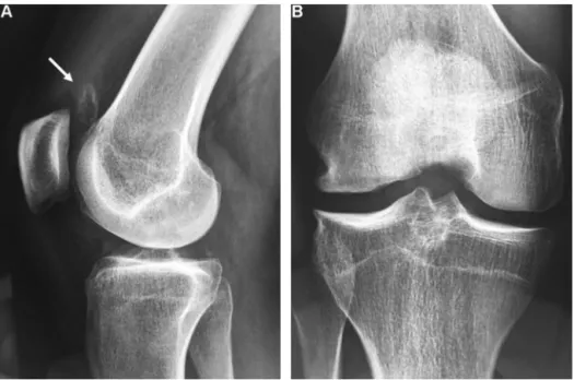
Fig. 11. Osteochondritis dissecans of the medial condyle in an 18-year-old male. A. Fragment loose in the supra-patellar bursa (arrow). B. Notch view showing osteoarthritis of the medial tibio-femoral compartment.
compared to 288 mm2 in the group with persistent lesions[27].
Visibility by MRI of a cyst measuring 1.3 mm or more in diameter predicts failure to heal[26]. The factors identified in these last two studies have been used to develop two nomograms for predicting healing in patients with open physes[26,27]. Both are reproducible and can be helpful in clinical practice.
7. What are the current treatment methods
Numerous treatment methods have been evaluated, often in small retrospective studies. Nevertheless, there is some agreement regarding the main strategies at each stage[28]. Symptomatic and stable OCD that fails to heal with non-operative treatment is man-aged by drilling. Unstable OCD requires fixation of the fragment. Finally, when the fragment has detached, the defect must be filled by an osteochondral graft. The criteria developed by Hughston et al. are used to evaluate the results by determining a clinical score and a radiographic score, each rated on a 0–4 scale[24]. Two clinical scores, Lyshölm’s and the IKDC, are used also but are not specific
[17,29].
7.1. Non-operative treatment
When the diagnosis is incidental, no treatment is needed but follow-up should be provided until radiographic healing is docu-mented. In patients with open physes who experience pain but have no evidence of instability, non-operative treatment for 3–6 months is the first-line strategy.
The importance of restricting sports activities is universally recognized. Pivoting, jumping, and repeated impacts should be avoided, as well as any activity that elicits the pain. Depending on the location of the lesion, discontinuing specific activities may be sufficient. Routinely discontinuing all sports is, in our opinion, nei-ther desirable nor feasible. When adherence to the restrictions is poor, a long-leg cast is useful, although complete immobilisation has not been proven effective.
In patients aged 8 to 14 years (mean, 11.4 years), Cahuzac et al.[11] and subsequently Sales de Gauzy et al.[24]reported that restricting sports activities was nearly always sufficient to ensure clinical and radiographic healing after a mean follow-up
of 8 months. Other studies produced less optimistic results. For instance, in girls with a mean age of 11.3 years and boys with a mean age of 12.4 years, Krause et al. found healing rates of only 33% after 6 months and 50% after 12 months[26].
Persistent pain after 6 months and/or the development of signs of instability requires surgical treatment.
7.2. Surgical treatment 7.2.1. Arthroscopic exploration
Arthroscopy is now the main method used for the surgical treatment of OCD. The initial exploration guides the therapeutic decisions.
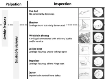
The International Cartilage Repair Society (ICRS) developed a four-grade classification based on findings upon inspection and pal-pation: grade I, stable lesions with a continuous but softened area covered by intact cartilage; grade II, lesions with partial discontinu-ity that are stable when probed; grade III, lesions with a complete discontinuity that are not yet dislocated (“dead in situ”); and grade IV, empty defects as well as defects with a dislocated fragment or a loose fragment within the bed[30]. The ROCK classification is more accurate, more reproducible, and easy to remember but fails to consider findings upon probing[31]. We suggest a modified ver-sion, in which instability is assessed by probing, irrespective of the appearance of the lesion (Fig. 12).
Arthroscopy is the reference standard for assessing instability. As indicated above, however, we advocate the use of MRI as the reference standard, given its greater sensitivity (Fig. 13).
Before starting the procedure, all the potentially required equip-ment must be available, to ensure that all situations can be managed adequately. For instance, arthroscopy may show a loose fragment requiring an internal fixation implant or osteochondral transfer set. If the necessary equipment is not available, performing the appro-priate procedure at a later date is preferable.
7.2.2. Drilling
Drilling creates new pathways in the subchondral bone through which blood vessels can penetrate the fragment. Two techniques are available, trans-articular drilling and retro-articular drilling. To
Fig. 12. Arthroscopic classification of knee OCD lesions.
Fig. 13. Osteochondritis dissecans of the medial condyle in a 13-year-old boy. Shadow sign (arrows) without instability by arthroscopy, whereas signs of instability were visible by MRI (seeFig. 8).
avoid confusion, these terms should replace the terms “retrograde” and “anterograde”.
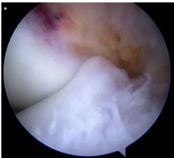
Trans-articular drilling is performed during arthroscopy. The lesion is usually identified based on its greyish or yellowish color, demarcation, and/or softer consistency upon probing, compared to the adjacent white cartilage. A Kirschner wire 1.2 to 1.5 mm in diameter is used, with a low-speed power drill. A cannula is useful for directing the pin, protecting the soft tissues, and pre-determining the depth of the drill hole (usually 20 mm). A total of five to ten drill holes are created by switching between the antero-medial and antero-lateral portals (Fig. 14). Postoperatively, patients are maintained without weight bearing by the use of crutches for 4 to 6 weeks. Sports activities can be resumed after 3 to 6 months depending on the postoperative course. The only drawback of this technique is that it violates the normal surface cartilage. It is our preferred surgical method.
Retro-articular drilling involves inserting a pin into the epiph-ysis under fluoroscopy guidance, from the outside to the inside of the knee, without entering the joint cavity. A parallel wire guide is then inserted to guide the other drillings. Risks include soft tis-sue injury, fragment mobilization, and incomplete perforation of
Fig. 14. Trans-chondral drilling to treat stable osteochondritis dissecans. Arthro-scopic view.
Fig. 15. Fixation of an unstable fragment using an autologous osteochondral plug. A. Arthroscopic view. B. Magnetic resonance imaging 6 months later.
the lesion. Retro-articular drilling is technically more challenging, requires a longer operative time, and requires radiation exposure. With trans-articular drilling to treat 40 patients aged 10 to 16 years, Rammal et al. obtained good clinical outcomes in 97.5% and good radiographic outcomes in 95% of cases[32]. A systematic lit-erature review found no evidence that either method was better than the other[29].
7.2.3. Fixation
An unstable fragment requires fixation to ensure stability. Fixation is also indicated for loose but intact fragments with macro-scopically normal surface cartilage and a layer of subchondral bone. In 1957, Smillie[10]reported a technique that used metallic nails. Since then, many fixation implants have been evaluated (staples, screws with or without a head and, more recently, bio-absorbable nails and screws)[33]. Bio-absorbable materials have gained pref-erence in North America[28]. No further procedure is needed for implant removal. A cancellous bone graft harvested from the tibial metaphysis is indicated when there is a hinged cartilage fragment with a subchondral bone defect, to provide a base that supports the fixed fragment. In this situation, the instrumental arthroscopy portal is extended into an antero-medial arthrotomy. The bed is freshened, the graft is implanted into it, and the fragment is repo-sitioned and fixed.
Fixation can also be achieved biologically using one or more osteochondral plugs collected, using dedicated tools, from the non-weight-bearing part of the intercondylar notch[34](Fig. 15). This is our preferred technique. The diameter and number of plugs vary
Palpation Inspection
eue ball
No abnormality detectable
Shadow
Cartilage intact but subtly demarcated •
-Wrinkle in the rug
Cartilage is demarcated with a fissure, buckle
and/or wrinkle
Locked door
Cartilage fissuring, unable to hinge open
Trapdoor
Cartilage fissuring, able to hinge open
Crater
with the size of the lesion. In most cases, one or two plugs mea-suring 6 or 8 mm in diameter and 15 mm in length are used. An alternative consists in hybrid fixation combining metallic screws and osteochondral plugs, as described by Lintz et al., who pointed out the unsatisfactory outcomes of screw fixation alone in patients with closed physes[35].
Tabaddor et al. reported good outcomes after fixation using bio-absorbable implants[33]. Of the 24 patients, 18 had closed physes. Arthrotomy with cancellous bone grafting was required in 5 patients. The mean number of implants inserted per patient was 2.3 (range, 1–7). Radiographs obtained after a mean of 19 months showed complete healing in 13 patients, partial healing in 9 patients, no change in 1 patient, and loose bodies without healing in 1 patient.
Fixation of unstable OCD fragments using osteochondral plugs only has also produced good outcomes[34].
7.2.4. Bone and cartilage reconstruction
Reconstruction is required when the fragment is too severely damaged to be repositioned and fixed or is detached and not found. Fortunately, these situations are rare in pediatric patients. The defect must be carefully evaluated to determine its location, sur-face area, and depth. This information is obtained preoperatively by MRI and intraoperatively using a probe or cylindrical template. Both cartilage and bone are lost, particularly after the bed is fresh-ened. Simple removal of the fragment is followed eventually by the development of osteoarthritis. The various techniques with their indications and outcomes were described in detail by Versier et al. for the French Arthroscopy Society (SFA) in 2010[36]. Micro-fractures serve to stimulate the generation of fibrocartilage, which can fill defects of up to 4 cm2. The outcomes are satisfactory in the
short term but deteriorate over time. A biological membrane can be placed over the site of micro-fracture and cancellous bone grafting to enhance chondrogenesis, a method known as autologous matrix-induced chondrogenesis (AMIC). The mesenchymatous stem cells released by the bone marrow through the micro-fractures concen-trate under the membrane, within the defect. Kusano et al. reported good outcomes of AMIC in 11 patients whose lesions had a mean surface area of 4.2 cm2[37].
Mosaic osteochondral transplantation can be used for defects of up to 4 cm2but is ideally used for defects smaller than 2 cm2.
Osteo-chondral plugs are placed in a mosaic arrangement to fill the defect. A multicenter study by the French Arthroscopy Society included 61 cases with a mean defect surface area of 1.7 cm2[38]. The outcomes
were good, with Hughston scores of 4 in 33% of cases and 3 in 40% of cases after 8 years of follow-up. This is our preferred technique. Articular congruence of the plugs and donor site morbidity are the main limitations.
In patients with larger defects, autologous chondrocyte trans-plantation can be used. With this technique, 5-year outcomes were good or very good in 91% of defects measuring 1.5 to 12 cm2[39].
An alternative consists in a massive cylindrical osteochondral auto-graft taken from the ipsilateral condyle.
7.3. Long-term outcomes
They vary with the severity of the lesion and treatment used. In 1991, Twyman et al. reported moderate-to-severe osteoarthritis in 32% of cases after a mean follow-up of 33 years[40], in contrast to the data reported in 1977 by Linden et al.[41]. It should be borne in mind that the patients in these studies were managed over half a century ago.
Fig. 16. Treatment algorithm.
8. Towards a treatment algorithm
The goal of treatment is to obtain healing before skeletal matu-rity. Nomograms have some predictive value in stable OCD lesions
[26,27]. Persistent pain reported by the patient and family after
time away from sports indicates a need for surgical treatment, especially in patients with closed or nearly closed physes. An arthroscopic evaluation is always the first step in the surgical man-agement. We suggest a treatment algorithm based on a review of the recent literature and on our own experience (Fig. 16). 9. Conclusion
OCD is clearly a multifactorial condition with a predominant role for mechanical influences acting via repetitive micro-trauma. In symptomatic cases, first-line MRI is indicated in boys older than 13 years and girls older than 11 years, as well as in all patients with persistent pain despite non-operative treatment. The MRI signs of instability have been redefined. The line beneath the frag-ment should have the same high-signal intensity as the adjacent joint fluid. A low-signal outer rim surrounding the high-signal line should be visible, as well as multiple breaks in the subchondral bone. Cysts indicate instability if they are multiple or larger than 5 mm in diameter. The treatment should be determined based on the stage of the lesion. Restricting sports activities is often sufficient to ensure healing in patients with open physes. Surgical innovations have been introduced. Fixation is required if the lesion is unstable by MRI or arthroscopy. Mosaic osteochondral transplantation is a technique of choice. Follow-up must be provided until complete radiographic healing of the lesion.
Disclosure of interest
The authors declare that they have no competing interest. References
[1]Lefort G, Moyen B, Beaufils P, de Billy B, Breda R, Cadilhac C, et al. L’ostéochondrite disséquante des condyles fémoraux. Analyse de 892 cas. Rev Chir Orthop Reparatrice Appar Mot 2006;92 [2S97-2S141].
[2]Kessler JI, Nikizad H, Shea KG, Jacobs Jr JC, Bebchuk JD, Weiss JM. The demo-graphics and epidemiology of osteochondritis dissecans of the knee in children and adolescents. Am J Sports Med 2014;42:320–6.
[3]Hefti F, Beguiristain J, Krauspe R, Moller-Madsen B, Riccio V, Tschauner C, et al. Osteochondritis dissecans: a multicenter study of the European Pediatric Orthopedic Society. J Pediatr Orthop Part B 1999;8:231–45.
[4]Uozumi H, Sugita T, Aizawa T, Takahashi A, Ohnuma M, Itoi E. Histologic findings and possible causes of osteochondritis dissecans of the knee. Am J Sports Med 2009;37:2003–8.
[5]Wechter JF, Sikka RS, Alwan M, Nelson BJ, Tompkins M. Proximal tibial mor-phology and its correlation with osteochondritis dissecans of the knee. Knee Surg Sports Traumatol Arthrosc 2015;23:3717–22.
[6]Chow RM, Guzman MS, Dao Q. Intercondylar notch width as a risk factor for medial femoral condyle osteochondritis dissecans in skeletally immature patients. J Pediatr Orthop 2016;36:640–4.
[7]Cavaignac E, Perroncel G, Thepaut M, Vial J, Accadbled F, De Gauzy JS. Relation-ship between tibial spine size and the occurrence of osteochondritis dissecans: an argument in favour of the impingement theory. Knee Surg Sports Traumatol Arthrosc 2015;25:2442–6.
[8]McElroy MJ, Riley PM, Tepolt FA, Nasreddine AY, Kocher MS. Catcher’s knee: posterior femoral condyle juvenile osteochondritis dissecans in children and adolescents. J Pediatr Orthop 2016 [PMID: 27442212].
[9]Ishikawa M, Adachi N, Yoshikawa M, Nakamae A, Nakasa T, Ikuta Y, et al. Unique anatomic feature of the posterior cruciate ligament in knees associated with osteochondritis dissecans. Orthop J Sports Med 2016;4 [2325967116648138]. [10]Smillie IS. Treatment of osteochondritis dissecans. J Bone Joint Surg Br
1957;3B:248–60.
[11]Cahuzac JP, Mansat C, Clement JL, Pasquie M, Gaubert J. The natural history of osteochondritis dissecans of the knee in children. Rev Chir Orthop Reparatrice Appar Mot 1988;74:121–4.
[12]Laor T, Zbojniewicz AM, Eismann EA, Wall EJ. Juvenile osteochondritis disse-cans: is it a growth disturbance of the secondary physis of the epiphysis? AJR Am J Roentgenol 2012;199:1121–8.
[13]Deie M, Ochi M, Sumen Y, Kawasaki K, Adachi N, Yasunaga Y, et al. Relationship between osteochondritis dissecans of the lateral femoral condyle and lateral menisci types. J Pediatr Orthop 2006;26:79–82.
[14]Gornitzky AL, Mistovich RJ, Atuahuene B, Storey EP, Ganley TJ. Osteochondritis dissecans lesions in family members: does a positive family history impact phenotypic potency? Clin Orthop Relat Res 2017;475:1573–80.
[15]Yellin JL, Trocle A, Grant SF, Hakonarson H, Shea KG, Ganley TJ. Candidate loci are revealed by an initial genome-wide association study of juvenile osteo-chondritis dissecans. J Pediatr Orthop 2017;37:e32–6.
[16]Jacobi M, Wahl P, Bouaicha S, Jakob RP, Gautier E. Association between mechan-ical axis of the leg and osteochondritis dissecans of the knee: radiographic study on 103 knees. Am J Sports Med 2010;38:1425–8.
[17]Rothermich MA, Nepple JJ, Raup VT, O’Donnell JC, Luhmann SJ. A comparative analysis of International Knee Documentation Committee Scores for common pediatric and adolescent knee injuries. J Pediatr Orthop 2016;36:274–7. [18]Bedouelle J. L’ostéochondrite disséquante des condyles fémoraux chez l’enfant
et l’adolescent. In: Cahiers d’Enseignement de SoFCOT. Paris: Expansion Scien-tifique Franc¸aise; 1988. p. 61–93.
[19]Ellermann J, Johnson CP, Wang L, Macalena JA, Nelson BJ, LaPrade RF. Insights into the epiphyseal cartilage origin and subsequent osseous manifestation of juvenile osteochondritis dissecans with a modified clinical MR imaging proto-col: a pilot study. Radiology 2017;282:798–806.
[20]De Smet AA, Ilahi OA, Graf BK. Reassessment of the MR criteria for sta-bility of osteochondritis dissecans in the knee and ankle. Skeletal Radiol 1996;25:159–63.
[21]Dipaola JD, Nelson DW, Colville MR. Characterizing osteochondral lesions by magnetic resonance imaging. Arthroscopy 1991;7:101–4.
[22]Kijowski R, Blankenbaker DG, Shinki K, Fine JP, Graf BK, De Smet AA. Juvenile versus adult osteochondritis dissecans of the knee: appropriate MR imaging criteria for instability. Radiology 2008;248:571–8.
[23] Siegall E, Faust JR, Herzog MM, Marshall KW, Willimon SC, Busch MT. Age predicts disruption of the articular surface of the femoral condyles in knee OCD: can we reduce usage of arthroscopy and MRI? J Pediatr Orthop 2016, http://dx.doi.org/10.1097/BPO.0000000000000796.
[24]Sales de Gauzy J, Mansat C, Darodes PH, Cahuzac JP. Natural course of osteo-chondritis dissecans in children. J Pediatr Orthop B 1999;8:26–8.
[25]Hughston JC, Hergenroeder PT, Courtenay BG. Osteochondritis dissecans of the femoral condyles. J Bone Joint Surg Am 1984;66:1340–8.
[26]Krause M, Hapfelmeier A, Moller M, Amling M, Bohndorf K, Meenen NM. Healing predictors of stable juvenile osteochondritis dissecans knee lesions after 6 and 12 months of nonoperative treatment. Am J Sports Med 2013;41: 2384–91.
[27]Wall EJ, Vourazeris J, Myer GD, Emery KH, Divine JG, Nick TG, et al. The healing potential of stable juvenile osteochondritis dissecans knee lesions. J Bone Joint Surg Am 2008;90:2655–64.
[28]Yellin JL, Gans I, Carey JL, Shea KG, Ganley TJ. The surgical management of osteochondritis dissecans of the knee in the skeletally immature: a survey of the Pediatric Orthopaedic Society of North America (POSNA) membership. J Pediatr Orthop 2017;37:491–9.
[29]Gunton MJ, Carey JL, Shaw CR, Murnaghan ML. Drilling juvenile osteochon-dritis dissecans: retro- or transarticular? Clin Orthop Relat Res 2013;471: 1144–51.
[30]Brittberg M, Winalski CS. Evaluation of cartilage injuries and repair. J Bone Joint Surg Am 2003;85–A:58–69.
[31]Carey JL, Wall EJ, Grimm NL, Ganley TJ, Edmonds EW, Anderson AF, et al. Novel arthroscopic classification of osteochondritis dissecans of the knee: a multi-center reliability study. Am J Sports Med 2016;44:1694–8.
[32]Rammal H, Gicquel P, Schneider L, Karger C, Clavert JM. Erratum to: juvenile osteochondritis of femoral condyles: treatment with transchondral drilling. Analysis of 40 cases. J Children Orthop 2014;8:449.
[33]Tabaddor RR, Banffy MB, Andersen JS, McFeely E, Ogunwole O, Micheli LJ, et al. Fixation of juvenile osteochondritis dissecans lesions of the knee using poly 96L/4D-lactide copolymer bioabsorbable implants. J Pediatr Orthop 2010;30:14–20.
[34]Miura K, Ishibashi Y, Tsuda E, Sato H, Toh S. Results of arthroscopic fixation of osteochondritis dissecans lesion of the knee with cylindrical autogenous osteochondral plugs. Am J Sports Med 2007;35:216–22.
[35]Lintz F, Pujol N, Pandeirada C, Boisrenoult P, Beaufils P. Hybrid fixation: evalu-ation of a novel technique in adult osteochondritis dissecans of the knee. Knee Surg Sports Traumatol Arthrosc 2011;19:568–71.
[36]Versier G, Dubrana F. Treatment of knee cartilage defect in 2010. Orthop Trau-matol Surg Res 2011;97:S140–53.
[37]Kusano T, Jakob RP, Gautier E, Magnussen RA, Hoogewoud H, Jacobi M. Treat-ment of isolated chondral and osteochondral defects in the knee by autologous matrix-induced chondrogenesis (AMIC). Knee Surg Sports Traumatol Arthrosc 2012;20:2109–15.
[38]Ollat D, Lebel B, Thaunat M, Jones D, Mainard L, Dubrana F, et al. Mosaic osteochondral transplantations in the knee joint, midterm results of the SFA multicenter study. Orthop Traumatol Surg Res 2011;97: S160–6.
[39]Peterson L, Minas T, Brittberg M, Lindahl A. Treatment of osteochondritis disse-cans of the knee with autologous chondrocyte transplantation: results at two to ten years. J Bone Joint Surg Am 2003;85–A:17–24.
[40]Twyman RS, Desai K, Aichroth PM. Osteochondritis dissecans of the knee. A long-term study. J Bone Joint Surg Br 1991;73:461–4.
[41]Linden B. Osteochondritis dissecans of the femoral condyles: a long-term follow-up study. J Bone Joint Surg Am 1977;59:769–76.