Please create your abstract using the following template and send this word document to biobarriers2014@mx.uni-saarland.de
Please do not modify the format of this template! Abstract limit is 1 page A4.
NOTE: Please save your abstract file according to the name of the presenting author as a ‘doc’ file (Author A B.doc).
Development of anti-HPV lipoplexes formulations for the treatment
of cervical cancer
Anna Lechanteur1 , Tania Furst1, Brigitte Evrard1, Patrick Roncarati2, Philippe
Delvenne2, Géraldine Piel1, Pascale Hubert2
1Laboratory of Pharmaceutical Technology and Biopharmacy - CIRM, University of Liège, Liège, Belgium 2Laboratory of Experimental Pathology, GIGA-CANCER, University of Liège, Liège, Belgium
Human Papillomaviruses (HPV) are responsible for several diseases and some of them (such as HPV16 and HPV18) can induce cervical cancer. In this case the two HPV E6 and E7 oncoproteins are essential players in order to immortalize keratinocytes by decreasing tumor suppressor genes (p53 and pRb). Nowadays, cervical cancer is known to be the third most frequent cause of death in women and treatment are associated with morbidity and high level of recurrence.
Gene therapy is a promising strategy to treat cancer. We focused on RNA interference (siRNA) to target mRNA coding for both HPV oncoproteins (E6 and E7) and also for an anti-apoptotic protein (MCL-1). This protein is over expressed in high grade lesions compared to low grade lesions and healthy exocervix. To develop a treatment, we would like to use a vector appropriate for in vivo study. SiRNA will be encapsulated in lipidic nanovectors to form lipoplexes. This association is essential to protect siRNA, to allow the diffusion into the cervical mucus and to cross the anionic cellular membrane.
The aim of this study is to develop a local treatment of cervical cancer by the use of antiE6, antiE7 and antiMCL-1 siRNA encapsulated in cationic liposomes.
First, we validated the efficiency of these antiE6 [1], antiE7 [2] and antiMCL1 [3]
siRNA with a commercially transfection agent (Oligofectamine®). Some objectives
are achieved on SiHa and CaSki cells: high transfection efficiency; extinction of E6, E7 and MCL-1 mRNA; decreased proliferation and induction of apoptosis.
In a second
step, Oligofectamine® was replaced by cationic liposomes with good
physicochemical characteristics (size, surface charge, stability…). Liposomes composed of DOTAP/DOPE/Cholesterol (1:0,5:0,5 ; molar ratio) are prepared and
then mixed with siRNA (50 nM; N/P 2,5). The size of lipoplexes is around 200nm and
the zeta potential is around +45mV. This formulation leads to a very efficient transfection and the percentage of mRNA is significantly reduced.
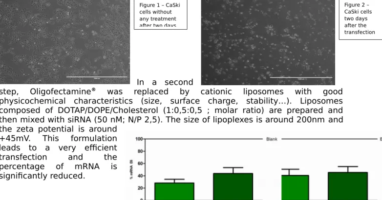
Figure 1 – CaSki cells without any treatment after two days
Figure 2 – CaSki cells two days after the transfection of the three
Please create your abstract using the following template and send this word document to biobarriers2014@mx.uni-saarland.de
Please do not modify the format of this template! Abstract limit is 1 page A4.
NOTE: Please save your abstract file according to the name of the presenting author as a ‘doc’ file (Author A B.doc).
In conclusion, the association of the three siRNA decreases proliferation and induces apoptosis on monolayer cells. Moreover, lipoplexes seems to be an appropriate delivery system and we would like to test their efficiency on a 3D model of cervical lesion.
References
1. Chang, JT-C. et al. Canc. Gene The. 17, 827-836 (2010). 2. Jiang, M. et al. Oncology. 21, 6041-6048 (2002).
3. Rajalingam,K. et al. PLoS One 3(9) : e3102, (2008).
Figure 3 – Extinction of E6 mRNA in CaSki cells, two days after the transfection of anti-E6 siRNA (n=4) Figure 4 – Extinction of E7 mRNA in CaSki cells, two days after the transfection of anti-E6 siRNA (n=4)