Annals of Oncology 23: 1954–1962, 2012 doi:10.1093/annonc/mds112 Published online 9 May 2012
Prevalence and management of cancer-related
anaemia, iron de
ficiency and the specific
role of i.v. iron
M. Aapro
1*, A. Österborg
2, P. Gascón
3, H. Ludwig
4& Y. Beguin
5 1IMO Clinique de Genolier, Genolier, Switzerland;2
Department of Hematology, Karolinska Institutet and Karolinska Hospital, Stockholm, Sweden;3
Department of Haematology-Oncology, Hospital Clínic de Barcelona, University of Barcelona, Barcelona, Spain;4
Department of Medicine I, Center for Oncology and Haematology, Wilhelminenspital, Vienna, Austria;5
Department of Medicine, Division of Hematology, University Hospital Liège, Liège, Belgium
Received 8 February 2012; accepted 27 February 2012
Background:Chronic diseases reduce the availability of iron for effective erythropoiesis. This review summarises clinical consequences of iron deficiency (ID) and anaemia in cancer patients, mechanisms how impaired iron homeostasis affects diagnosis and treatment of ID, and data from clinical trials evaluating i.v. iron with or without concomitant erythropoiesis-stimulating agents (ESAs).
Design:Clinical trial reports were identified in PubMed and abstracts at relevant major congresses.
Results:Reported prevalence of ID in cancer patients ranges from 32 to 60% and most iron-deficient patients are also anaemic. Randomised clinical trials have shown superior efficacy of i.v. iron over oral or no iron in reducing blood transfusions, increasing haemoglobin, and improving quality of life in ESA-treated anaemic cancer patients.
Furthermore, i.v. iron without additional ESA should be evaluated as potential treatment in patients with chemotherapy-induced anaemia. At recommended doses, i.v. iron is well tolerated, particularly compared with oral iron. No serious drug-related adverse effects were seen during long-term use in renal disease and no effect on tumour growth has been observed in trials with anaemic cancer patients.
Conclusions:Reliable diagnosis and treatment of ID are recommended key steps in modern cancer patient management to minimise impact on quality of life and performance status.
Key words: anaemia, chemotherapy-induced anaemia, diagnosis, intravenous iron, iron deficiency, hepcidin
introduction
Iron deficiency (ID) and anaemia are frequent complications in
cancer patients, in particular during treatment with
chemotherapeutic agents [1–3]. ID, even in the absence of
anaemia, may be associated with impaired physical function, weakness, and fatigue that can be ameliorated by iron therapy
[4]. If left untreated, ID can lead to anaemia; thus, the
potential effect of ID on vulnerable populations such as cancer patients should not be underestimated.
Current guidelines on the treatment of cancer-related anaemia recommend restricted usage of erythropoiesis-stimulating agents (ESAs) and reduction/prevention of blood
transfusions [5–8]. Data from several controlled clinical trials
have shown that i.v. iron supplementation enhances response to ESA treatment and can reduce administered ESA doses in
cancer patients [9–14]. Early results from clinical trials on i.v.
iron treatment without concomitant ESAs suggest that initiation of anaemia treatment with i.v. iron alone should be investigated as treatment option for cancer-related anaemia
[15,16].
This review focuses on the clinical consequences of ID and anaemia in cancer patients along with data from clinical trials using i.v. iron highlighting the evolving role of i.v. iron therapy in cancer patient management. In addition, mechanisms behind cancer-related anaemia that influence the correct diagnosis and treatment of ID or the maintenance of iron availability are discussed.
search strategy and selection criteria
Data from appropriate clinical trials were identified byscreening the US National Library of Medicine’s PubMed
database with the search terms‘cancer’, ‘intravenous iron’ or
‘parenteral iron’, and ‘anaemia’, and limiting the search to clinical trials reported in English. Data reported in abstract
form only were identified by manual search through abstracts
at major congresses in thefield.
*Correspondence to: Dr M. Aapro, Multidisciplinary Oncology Institute, Clinique de Genolier, 1, rte du Muids, Genolier CH-1272, Switzerland. Tel: +41-22-366-91-06; Fax: +41-22-366-91-31; E-mail: maapro@genolier.net
© The Author 2012. Published by Oxford University Press on behalf of the European Society for Medical Oncology. All rights reserved. For permissions, please email: journals.permissions@oup.com
at Bibliotheque Fac de Medecine on November 19, 2012
http://annonc.oxfordjournals.org/
prevalence and burden of ID
and anaemia in cancer patients
The high prevalence of anaemia in patients with different cancer types (39% at enrolment and 68% becoming anaemic at least once during the 6-month survey period) has been alreadyshown in the European Cancer Anaemia Survey (ECAS) [1].
Conversely, published data on the prevalence of ID in cancer
patients are scarce (Table1) [2,3,17–19]. Reported prevalence
of ID was highest for colorectal cancer (60%, and 69% of those
were also anaemic) [2]; probably, chronic blood loss may
render patients with colorectal or gastrointestinal cancers more prone to ID and anaemia. Nevertheless, also in other
populations, the prevalence of ID and anaemia was considerably high (29%–46% and 7%–42%, respectively)
[3,17–19].
Currently, data on the impact of ID in cancer patients are
only available in abstract form, suggesting a significant
correlation between low iron status and worse World Health
Organisation (WHO) performance scores [19]. Correction of
ID in a noncancer population ( patients with chronic heart
failure) significantly improved exercise capacity, quality of life,
and disease state independently whether patients were anaemic
or not [20].
More data are available on the impact of anaemia, showing a
65% increased risk of death [21] and close to fourfold higher
average annual health care cost per patient [22]. A causal
relation between anaemia and the risk of death remains to be
confirmed before the final conclusion that correcting anaemia
improves prognosis. Two large analyses demonstrated the relationship between haemoglobin (Hb) levels and physical
performance as well as quality of life in cancer patients [1,23].
In ECAS, patients with the poorest WHO performance scores
(2–4) were more likely to have low Hb levels (P < 0.001). A
direct correlation between quality of life and Hb levels in cancer patients receiving chemotherapy has been shown across
the clinically relevant Hb range of 8–14 g/dl. Accordingly, Hb
increase achieved with i.v. iron supplementation of ESA treatment in patients with chemotherapy-related iron
deficiency anaemia (IDA) was associated with significantly
better effects on energy level, activity, and overall quality of life
(P < 0.0002) [9].
causes and diagnosis of ID and anaemia
impaired iron utilisation in chronic diseaseAnaemia of chronic disease (ACD) and chemotherapy-induced anaemia (CIA) are the major causes of anaemia in cancer patients and can be aggravated by chronic blood loss and nutritional deficiencies (e.g. due to cancer-induced anorexia or resection of gastrointestinal malignancies). In patients with ACD, the availability of iron is affected by hepcidin, the key
regulator of iron homeostasis (Figure1) [24,25]. Increased
hepcidin levels block the ferroportin-mediated release of iron
from enterocytes and macrophages [25]. In the long term, this
can lead to absolute ID (AID, insufficient iron stores) due to
impaired utilisation of nutritional or orally administered iron.
In the short term, this‘hepcidin block’ can result in functional
iron deficiency (FID), a condition under which iron cannot be
efficiently mobilised from stores in the reticuloendothelial
system (RES) [24]. In patients with inflammation, iron release
is reduced to 44% compared with normal subjects (Figure2)
[26]. Thus, even iron-replete patients can experience a shortage
of available iron, especially when exposure to ESAs rapidly
increases red blood cell production [28].
Inflammatory cytokines also inhibit proliferation and differentiation of erythroid progenitor cells and blunt
endogenous erythropoietin production in the kidney [29]. In
addition, reduced sensitivity to erythropoietin, a reduced life span of erythrocytes, solid tumours or metastases infiltrating the bone marrow, and myelosuppressive effects of
chemotherapies can impair normal haematopoiesis
[25,30–32].
diagnostic markers of ID in patients with chronic disease
Serum ferritin, the most commonly assessed marker, generally
reflects the status of iron stores while transferrin saturation
(TSAT), the percentage of hypochromic red cells (%HYPO),
and the Hb content of reticulocytes (CHr) better reflect the
availability of iron [33]. Since serum ferritin, an acute-phase
protein, can be elevated due to inflammation and liver cell
damage, normal or elevated ferritin levels do not necessarily
indicate sufficient iron stores, particularly in cancer patients
Table 1. Reported prevalence of iron deficiency in different cancer patient populations
Tumours/Patients (N) % with ID Definition of ID % with IDA
Kuvibidila, 2004 [3] Prostate (34) 35 SF < 12 ng/mla n/a
32 TSAT < 16%
Beale, 2005 [2] Colorectal (130) 60 SF < 15 ng/ml and/or TSAT < 14% 42b
Steinmetz, 2010 [17] CIA (286) 29 FI > 3.2cor CHr≤ 28 pg 7d
Beguin, 2009 [18] CIA (481) 43 SF < 100 ng/ml and/or TSAT < 20% n/a
Ludwig, 2011 [19] Solid tumours (1053) 46 SF < 30 ng/ml or TSAT < 20% 33
aOr <100 ng/ml in subjects with inflammation. bHb < 12.5 g/dl in men and Hb < 11.5 g/dl in women. c
Morr than 2.0 in subjects with C-reactive protein levels >5 mg/l.
dFI≤ 3.2 (or 2.0) and CHr ≤ 28 pg.
ACD, anaemia of chronic disease; CHr, haemoglobin content of reticulocytes; CIA, chemotherapy-induced anaemia; FI, ferritin index (soluble transferrin receptor/log ferritin); FID, functional iron deficiency; ID, iron deficiency; IDA, iron deficiency anaemia; SF, serum ferritin; TSAT, transferrin saturation.
Volume 23 | No. 8 | August 2012 doi:10.1093/annonc/mds112 |
at Bibliotheque Fac de Medecine on November 19, 2012
http://annonc.oxfordjournals.org/
[34]. Thus, routine blood analysis should also include C-reactive protein (CRP) and alanine aminotransferase (ALT)
to check for inflammation and liver function. Soluble
transferrin receptor levels, recently suggested for allocation of
cancer patients to treatment with ESA alone, iron alone or a
combination thereof [17,35], rather reflect the erythropoietic
activity than the iron status and cannot be used as iron status parameter when erythropoiesis is stimulated, e.g. with ESAs. Therefore, in routine clinical practice, serum ferritin levels
<100 ng/ml probably indicate insufficient iron stores for
successful ESA therapy in patients with cancer, and the combination of low TSAT (<20%) and normal or even elevated
serum ferritin may indicate FID (Figure3) [34].
The potential of TSAT as marker of FID has been shown in patients with lymphoproliferative malignancies, where 39% of patients presented with a TSAT <20% despite detectable iron
deposits in the bone marrow [12]. During the subsequent
clinical study, mean levels dropped in patients receiving only ESA but rose in patients treated concurrently with i.v. iron and ESA. The dysregulation of serum ferritin and TSAT was also shown in a cohort with haematologic and malignant diseases. Twenty-two percent of patients with serum ferritin levels of
100–800 ng/ml and even 24% of those with serum ferritin
≥800 ng/ml had IDA (TSAT <20% and Hb <12 g/dl) [36].
Other markers of iron-restricted erythropoiesis (%HYPO
>5%, CHr <26 pg) reflect the outcome of erythropoiesis,
especially the rapidly changing CHr [33]. However, advanced
equipment and rapid sample processing to avoid expansion of red blood cells are required.
treatment of ID and anaemia
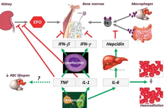
guidelines and regulationsOptions for treating anaemia in cancer patients include blood transfusions, ESA therapy, and i.v. iron supplementation. Goals Figure 1. Hepcidin-mediated blockade of iron homeostasis due to inflammation in anaemia of chronic disease. During inflammation, monocytes release cytokines such as tumour necrosis factor (TNF), interleukin (IL)-1, and IL-6. Red blood cell (RBC) lifespan is reduced possibly through a TNF-dependent mechanism. TNF and IL-1 impair erythropoietin (EPO) production by the kidney and induce lymphocytes to release interferons that in turn inhibit the proliferation and differentiation of erythroid progenitor cells. On the other hand, IL-6 worsens anaemia through expansion of plasma volume. IL-6 also increases hepcidin secretion by the liver, thereby inhibiting iron absorption and iron release from macrophages [24].
Figure 2. Intravenous iron overcoming the iron blockade in patients with chronic disease. Intravenous iron can overcome the iron blockade in patients with chronic disease and elevated hepcidin levels. Iron entering macrophages as senescent RBC (in red) or i.v. iron (in brown) can either be released immediately (and saturate plasma transferrin to a certain degree) or be stored in ferritin. Compared with the 25 mg daily iron release from macrophages in normal individuals, this rate decreases to∼15 mg/day in patients with inflammation [26]. In such patients, stable i.v. iron–
carbohydrate complexes release the iron over a prolonged period of time, so that large iron doses can be injected once, while iron from less stable complexes is released rapidly by macrophages and therefore requires multiple low-dose injections [27].
| Aapro et al. Volume 23 | No. 8 | August 2012
at Bibliotheque Fac de Medecine on November 19, 2012
http://annonc.oxfordjournals.org/
of anaemia treatment are to improve patients’ quality of life
and reduce reliance on blood transfusions [5–8] that are still
associated with a potential risk for transmission of infectious diseases, transfusion reactions, lung injury, and
alloimmunisation [37]. Furthermore, transfusion per se may
increase the risk of mortality and morbidity including stroke, myocardial infarction, acute renal failure, and recurrence of
cancer [38–41].
Transfusion requirements of cancer patients could be
reduced with ESAs [42]; however, haematologic response to
ESAs is limited (30%–75% of treated patients) [43–45].
Moreover, clinical trials, systematic reviews, and meta-analyses have raised concerns that ESAs increase the risk of
thromboembolic events and may increase mortality in patients not receiving chemotherapy, particularly when used off-label
[8]. Accordingly, the European Medicines Agency (EMA)
revised the target Hb values and highlighted that ESA use
should be restricted for clearly symptomatic anaemia [46]. The
US Food and Drug Administration (FDA) restricted the indication for ESAs to anaemic patients undergoing
myelosuppressive chemotherapy unless cure is the anticipated
outcome of chemotherapy [47,48]. FDA has also implemented
a risk evaluation and mitigation strategy requiring training for ESA prescribers and information of patients about
ESA-associated risks [49]. Further limits comprise initiation of
ESAs in patients with Hb <10 g/dl, dose reductions if Hb
increase is≥1 g/dl within 2 weeks, and avoidance in cancer
patients who are not receiving concurrent myelosuppressive
chemotherapy [8].
Conversely, current anaemia treatment guidelines in oncology acknowledge that i.v. iron enhances efficacy of ESAs
in patients with absolute or functional ID [5–8]. The National
Comprehensive Cancer Network (NCCN) suggests consideration of i.v. iron in patients with FID and serum ferritin levels up to 800 ng/ml if TSAT is below 20%; mentioning active infection as only restriction to iron
supplementation [7]. Since iron-replete anaemic patients may
benefit from i.v. iron supplementation only after initiation of
ESA therapy [8], iron status assessment is recommended at
baseline, before each cycle of chemotherapy, and throughout any kind of anti-anaemia therapy to ensure timely
commencement of iron supplementation.
Oral iron supplementation is only recommended in cases of
absolute ID [7]. Although oral iron has been used more
commonly than i.v. iron, it is less effective ( particularly in ESA-treated cancer patients) and associated with
gastrointestinal intolerance and poor compliance [7,50].
supplementation of ESA therapy with i.v. iron Seven randomised controlled clinical trials investigating the
efficacy of i.v. iron supplementation in ESA-treated anaemic
cancer patients have been published between 2004 and 2011
[9–14,51]. Six studies focused on CIA and one on patients not
receiving chemotherapy [12]. All studies except one with an
unusual (off-label) dosing schedule [51] showed significant
benefit of i.v. iron supplementation. Studies excluding patients
with FID at enrolment achieved a 13%–19% absolute increase
in response rate [10,11,13,14]. When patients with FID were
not excluded from enrolment, absolute increase in response rate was even 34%–43% with i.v. iron compared with no iron
[9,12]. Two studies showed a more rapid response associated
with i.v. iron supplementation [12,14]. This supports the
concept that high i.v. iron doses can overcome
hepcidin-mediated reduction of iron release from the RES (Figure2).
In studies including an oral iron arm, i.v. supplementation
significantly improved haematologic response compared with
oral iron, whereas no significant difference was observed
between oral and no iron supplementation [9,13]. Despite the
trials covered different patient populations, i.v. iron formulations, and concomitant chemotherapies,
generalisability of the results has been questioned [8]. In
particular, the wide range of differences in Hb response rates
between treatment and control groups (13%–43%) and a study
that seemed to show no benefit of parenteral iron may have
raised concerns about the heterogeneity across the trials. However, grouping the results from trials that included patients
with FID [9,12] and trials that focused on iron-replete patients
[10,11,13,14] at enrolment showed comparable significantly
improved response rates within each of the two populations. A meta-analysis of eight publications and abstracts on trials comparing i.v. iron with no or oral iron supplementation of ESA therapy (N = 1555; including the seven trials mentioned above) showed a 31% increase in the number of patients who achieved a haematopoietic response [95% confidence interval
(CI) 1.15–1.49] [52]. Similarly, another meta-analysis of i.v.
iron versus no iron supplementation of ESA therapy in seven trials showed a significant 29% increased chance for
haematologic response and a 23% reduction in the risk of
transfusion (95% CI 0.62–0.97) with i.v. iron supplementation.
Comparison of oral versus no iron supplementation in three trials, showed no difference in haematologic response and only
a nonsignificant reduction in transfusion risk [53].
dosing of i.v. iron
In thefirst published trial on i.v. iron supplementation of ESA
therapy that included iron-deficient patients, total iron doses
Figure 3. Criteria for diagnosis of iron deficiency in routine practice. Minimal criteria (black) and optimal (grey) work-up for the diagnosis of iron deficiency and distinction between absolute and functional iron deficiency in routine practise. ACD, anaemia of chronic disease; CHr, reticulocyte haemoglobin content; HYPO, hypochromic red cells; IDA, iron deficiency anaemia; TSAT, transferrin saturation.
Volume 23 | No. 8 | August 2012 doi:10.1093/annonc/mds112 |
at Bibliotheque Fac de Medecine on November 19, 2012
http://annonc.oxfordjournals.org/
up to 3000 mg were given [9]. Infive of the subsequent trials,
planned total iron doses were∼1000 mg (Table2and more
detailed information in supplemental Table S1, available at Annals of Oncology online). One study planned a total dose of 2000 mg iron but the administered mean total iron dose was
only 1169 mg [10]. The maximum single doses and minimum
infusion times of available parenteral iron preparations
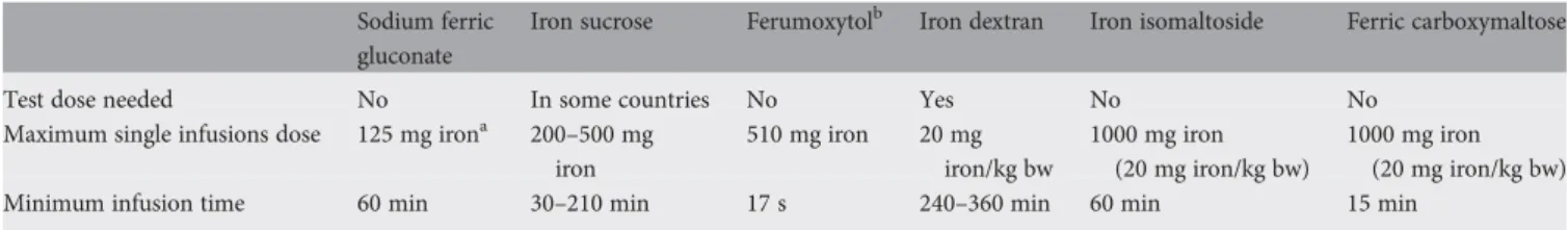
(Table3) depend on their tolerability profiles, mainly
determined by the biochemical properties and the manufacturing process. Stable iron complexes can be administered at high doses of 20 mg iron/kg body weight within 15 min (ferric carboxymaltose) to 6 h (iron dextran). Compounds that release iron at a faster rate should be given at
a lower pace and dose per infusion (Figure2).
The impact of dosing schedules beyond recommendations or even off-label has been highlighted by the only study that
seemed to show no significant benefit of i.v. iron [51,55]. In
that study, sodium ferric gluconate was given in single doses of
187.5 mg, i.e. 50% above the compound’s recommended single
dose and known to be associated with a higher incidence and/ or increased severity of adverse events (AEs). Conversely, the 3-week administration interval in this study resulted in the lowest calculated weekly iron dose among all published studies
(Table2) [51,55]. Post hoc analyses suggest that the response
to sodium ferric gluconate depended on both the i.v. iron
dosage and the hepcidin levels [54,56]. Patients who received
≥750 mg iron achieved an 80% response rate compared with 67% and 65% in the oral iron and placebo group, respectively. Patients with low or medium hepcidin levels (≤64.3 ng/ml) showed better response rates than patients with high hepcidin levels.
reduction of blood transfusions and ESA doses A critical aspect in the evaluation of anaemia treatment options such as i.v. iron is the potential to reduce transfusions and/or ESA dose requirements in addition to relief anaemia symptoms. One study that was large enough to uncover potential differences in transfusion rates (N = 396 patients) showed a significant reduction of transfusion requirements
with i.v. iron versus no or oral iron supplementation [11].
While a prior smaller study suggested a reduction in transfusions compared with the oral or no iron group from
week 4 onwards [13], this large study showed a significant
reduction of blood transfusions in the i.v. iron treatment group irrespectively whether the entire study period was evaluated (P = 0.038) or only the period from week 5 to the end of the
study period (P = 0.005). The only other study on a comparable
scale was stopped prematurely as discussed above [51,55].
Only one of the trials confirming the efficacy of i.v. iron
supplementation reported data on the administered ESA doses
[12,57], showing a dose-sparing effect of i.v. iron from week 5
onwards that reached significance at week 13 (P = 0.029).
Overall, i.v. iron supplementation of epoetinβ reduced the
mean cumulative ESA dose by 18% and the mean ESA dose required at the end of the study (week 15) by 25%. Economic analysis revealed that cost savings due to reduced ESA dosage outweighed any additional costs incurred by i.v. iron
administration, and an 11% cost benefit could be achieved with
i.v. iron supplementation of the ESA regimen [57]. Another
study showed that supplementation of darbepoetin alfa with 400 mg i.v. iron provided approximately the same
improvement of haematologic response and fatigue scores as
ESA dose increase from 300 to 500μg darbepoetin alfa (all
given every 3 weeks) [10].
potential role for i.v. iron asfirst-line therapy for CIA?
Guidelines recommend treatment of underlying causes of anaemia such as ID before initiation of an ESA. However, studies examining i.v. iron as sole anaemia treatment in cancer patients are only just starting to emerge. Two relevant small (N = 44 and 75 patients), controlled, randomised clinical trials have been published. Both studies involved patients with gynaecologic cancers receiving chemotherapy or
radiochemotherapy, and in both, i.v. iron supplementation
significantly reduced the number of required blood
transfusions [15,16]. In one study, significantly higher Hb
levels were observed in the i.v. iron compared with the oral iron group at the end of the study period, although mean Hb levels included data from patients who received transfusions as
well as those who did not [15]. The other study, comparing i.v.
iron versus no anaemia treatment, achieved a lower rate in transfusions despite a higher baseline proportion of anaemic
patients in the study group [16]. Both studies missed to assess
iron status parameters such as TSAT and serum ferritin; thus, the proportion of patients with either functional or absolute ID could not be determined.
In the absence of further randomised controlled clinical
trials, the trial by Steinmetz et al. [17] may be instructive. This
prospective, multicentre parallel group study was designed to investigate the rationale for assigning patients to treatment with either ESA alone, ESA and i.v. iron, or i.v. iron alone
based on a diagnostic plot defined by CHr (anaemia defined as
Table 2. Planned total and weekly i.v. iron doses in published randomised controlled trials on i.v. iron supplementation of ESAs in cancer patientsa
Iron in mg Auerbach, 2004 [9] Hedenus, 2007 [12] Henry, 2007 [13] Bastit, 2008 [11] Pedrazzoli, 2008 [14] Auerbach, 2010 [10] Steensma, 2011 [51,54]
Weekly dose 100 100 and
50b
125 67 125 133 62.5
Total dose 1000–3000 1100 1000 1000 750 2000 937.5
a
More detailed information on patient populations, treatments, and outcomes is available as online supplementary material (supplemental Table S1, available at Annals of Oncology online).
b100 mg iron once weekly from week 0–6 and every second week from week 8–14.
| Aapro et al. Volume 23 | No. 8 | August 2012
at Bibliotheque Fac de Medecine on November 19, 2012
http://annonc.oxfordjournals.org/
CHr≤ 28 pg) and the ferritin index (absolute ID defined as ferritin index >2.0 or >3.2 depending whether CRP levels are >5 mg/ml or not). Anaemic patients without ID received ESA treatment only, patients with FID and anaemia received both i.v. iron and ESAs, and patients with absolute ID anaemia received iron only. Hb response rates (Hb increase >1 g/dl from baseline) were comparable between patients receiving i.v. iron alone and those receiving ESA alone. However, 49% of Hb
responders to ESA developed ID (as defined by the ferritin
index) over the course of the 12-week study. This result, compared with only 19% among patients who received ESA and i.v. iron, suggests that patients identified as iron replete at
baseline could also have benefited from i.v. iron
supplementation [17].
Notably, a multicentre observational study on the use of ferric carboxymaltose in clinical practice showed a 1.4 g/dl median increase in Hb levels over a 12-week study period (median total iron dose 1000 mg) despite most patients (83%)
received ferric carboxymaltose without additional ESA [58].
The efficacy and safety profile of i.v. iron as a monotherapy to treat ID and anaemia has been extensively investigated in patients with other chronic diseases. Several randomised controlled trials have shown that i.v. iron is well tolerated and effective in treating ID anaemia and improving quality of life in
patients with inflammatory bowel disease [59,60]. In patients
with chronic heart failure, i.v. iron treatment significantly
improved the NYHA functional class (New York Heart Association), physical performance, and quality of life in
anaemic and non-anaemic patients [20].
tolerability of i.v. iron in clinical routine
Six of seven randomised controlled clinical trials on i.v. iron supplementation of ESAs in cancer patients did not show a difference in AE rates between i.v. iron and control treatmentarms [9–14]. Individual high doses of sodium ferric gluconate
that exceeded the recommended dose may have been the reason why one trial reported higher AE rates in the i.v. iron
arm [51,55].
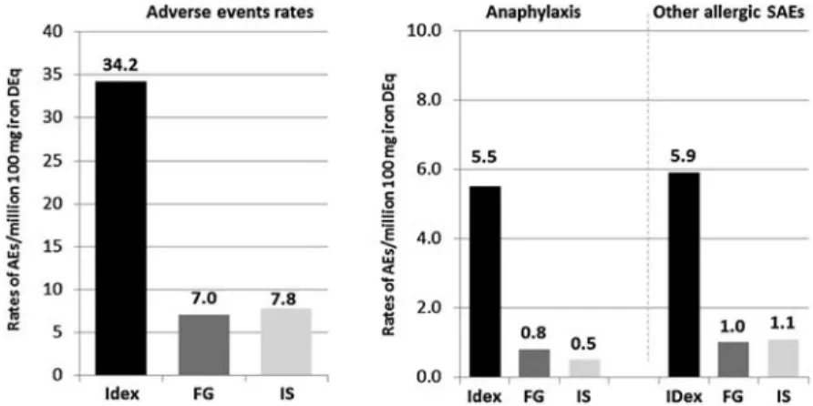
One common prejudice against i.v. iron refers to potential hypersensitivity reactions. However, three analyses evaluating AE reports from 1997 to 2009 have shown that allergic and anaphylactoid reactions, even if rare, are mainly related to iron
dextran preparations (Figure4) [61–63]. Even with
low-molecular-weight iron dextrans (numbers from Europe only),
the rate of anaphylactic reactions is substantially higher compared with iron sucrose or sodium ferric gluconate (15.6, 0.9, and 0.4 per million 100 mg iron dose equivalents, respectively).
The overall rate of AEs per million 100 mg iron dose equivalents in Europe is 68.9 for low-molecular-weight iron dextrans, 12.8 for iron sucrose, and 3.5 for sodium ferric
gluconate [61]. In North America, the AE rate for iron sucrose
is lower (4.4), which may be related to the introduction of copies of iron sucrose in Europe but not in the United States. The therapeutic and toxicological equivalence of these copies is currently under discussion and clinical head-to-head
comparisons between the copies and the reference product will
be needed for clarification [64–66]. Notably, the AE rate for
sodium ferric gluconate is higher in North America compared with Europe (12.6 versus 3.5), which may be related to the higher maximum permitted iron dose (125 versus 62.5 mg in most European countries).
Another frequently raised question asks whether i.v. iron increases the risk of infections. To date, no increased rate of infections was observed in patients receiving i.v. iron for treatment of cancer-related anaemia. A study in haemodialysis patients even reported lower rates of infection-related
hospitalisations (relative risk 0.54, P < 0.001) and mortality (relative risk = 0.61, P = 0.08) among haemodialysis patients treated with i.v. iron compared with a general haemodialysis
population [67]. However, although not specifically
investigated in human studies, animal studies suggest that administration of i.v. iron should be avoided in patients with
active sepsis [68].
limitations to the use of i.v. iron
in clinical practice
One potential limitation to the use of i.v. iron in cancer patients might be the interaction of iron with certain chemotherapies, in particular anthracyclines and
platinum-based therapies [69,70]. In case of anthracyclines, preclinical
data suggest that electron transfer via the superoxide radical
results in the release of ferrous iron (Fe2+) from polynuclear
ferric oxyhydroxide cores (Fe3+). The released ferrous iron can
be reoxidised to ferric iron via the Fenton reaction and thereby
generates the highly reactive hydroxide radical [71]. By this
means, redox cycling of iron, in particular‘labile’
non-transferrin-bound iron, can result in oxidative stress and tissue Table 3. Maximum approved single doses for i.v. administration of iron carbohydrate compounds
Sodium ferric gluconate
Iron sucrose Ferumoxytolb Iron dextran Iron isomaltoside Ferric carboxymaltose
Test dose needed No In some countries No Yes No No
Maximum single infusions dose 125 mg irona 200–500 mg iron 510 mg iron 20 mg iron/kg bw 1000 mg iron (20 mg iron/kg bw) 1000 mg iron (20 mg iron/kg bw)
Minimum infusion time 60 min 30–210 min 17 s 240–360 min 60 min 15 min
aBased on results of a recent clinical trial, NCCN guideline changed the recommendation from 200 to 125 mg iron over 60 min [7]. b
Available only in the United States;
bw body weight; see local summaries of product characteristics for effective updates.
Volume 23 | No. 8 | August 2012 doi:10.1093/annonc/mds112 |
at Bibliotheque Fac de Medecine on November 19, 2012
http://annonc.oxfordjournals.org/
damage. Cardiotoxicity of other chemotherapies seem to be related to other factors such as off-target inhibition of kinases or reduced nitric oxide production rather than interaction with
iron [72,73]. Currently available clinical studies with i.v. iron
in cancer patients reported no signs of drug-related iron toxicity and only non-clinical data are available on this topic. Until the availability of such human data, one should consider separating the administration of cardiotoxic cancer treatments
and give the i.v. iron at thefirst visit after administration of a
potentially cardiotoxic chemotherapy.
Uncertainty on the potential role of iron in tumour
progression largely arises from epidemiological studies showing links between conditions associated with long-term iron overload (e.g. haemochromatosis) and an increased risk of
newly induced cancer [71]. However, these conditions do not
reflect the situation in anaemic and iron-deficient cancer patients who receive i.v. iron over rather short periods of time for repletion of their iron stores. A potential role of iron in tumour progression of existing tumours has been investigated in non-clinical models. However, these models often used excessive iron doses in iron-replete animals or i.p. iron
administration [74,75]. Until now, no animal model of cancer
has been published using i.v. iron administration and thus, the clinical relevance of available non-clinical studies needs to be carefully weighed against the risks that other anti-anaemia treatments, or no treatment present to the patient. Unfortunately, most trials on i.v. iron supplementation of cancer patients were not designed to collect long-term data. One prospective randomised controlled study that is reported in abstract form only monitored patients with lymphoid malignancies who received darbepoetin alfa and i.v. iron following autologous stem cell transplantation. In this preliminary study, 3-year progression-free survival was
independent of i.v. iron treatment [76,77].
concluding remarks
The high prevalence of ID and anaemia in cancer patients suggests that these complications may need more attention in
clinical practice. Current guidelines for treating anaemic cancer patients recommend that ID should be considered as
underlying cause of anaemia before initiating ESA treatment and acknowledge that i.v. iron supplementation is superior to
oral iron. Thorough and regular assessment of cancer patients’
iron status (before each chemotherapy cycle) can ensure that these patients receive the most appropriate supportive treatment for their needs. Among different potential markers of iron status, TSAT has been suggested as reliable marker that allows diagnosis of both absolute and FID without the need for
additional assessments of liver function or inflammatory
parameters.
Published randomised controlled trials show that i.v. iron enhances response rates to ESA therapy and may be effective in reducing ESA doses and blood transfusion requirements, even if long-term safety remains to be examined. Available early
reports on the use of i.v. iron asfirst-line anaemia therapy
suggest that some patients could benefit from i.v. iron even
without concomitant ESA. However, larger randomised controlled studies with long-term follow-up are necessary to
confirm long-term efficacy and safety.
acknowledgements
All authors contributed equally to the literature search, data interpretation and review of the manuscript. YB developed
Figures1,2, and3. Medical writing support was provided by
Walter Fürst (SFL Regulatory Affairs & Scientific
Communication, Switzerland).
funding
This work was supported by an unrestricted grant Vifor Pharma Ltd. (Switzerland) funding medical writing support.
disclosures
Interpretation of the data as well as the review and decision to submit the manuscript for publication have been carried out by all authors independently. MA disclosed membership in Figure 4. Adverse events rates with parenteral iron preparations in Europe and North America (2003–2009). Rates of total adverse events (AEs) as well as anaphylaxis and other allergic serious AEs per 100 mg iron dose equivalents (DEq) in Europe and North America between 2003 and 2009 [61]. Over the entire observation period, the total of anaphylaxis and other serious allergic AEs was 6- to 10-fold higher with iron dextrans (IDex) than other i.v. iron compounds (iron sucrose, IS; sodium ferric gluconate, FG). (Figure adapted from Bailie et al. Drug Research 2011 [61].)
| Aapro et al. Volume 23 | No. 8 | August 2012
at Bibliotheque Fac de Medecine on November 19, 2012
http://annonc.oxfordjournals.org/
speaker bureaus and/or advisory boards, and/or receipt of study grants from Vifor Pharma, Sandoz, Amgen, Roche, Hexal, and Hospira. AÖ disclosed receipt of honoraria for advisory board meetings and conduct of clinical trials of Vifor Pharma. PG disclosed no conflicts of interest. HL disclosed membership in the speaker bureau of Vifor Pharma. YB disclosed membership in the speaker bureau of Vifor Pharma.
references
1. Ludwig H, Van BS, Barrett-Lee P et al. The European Cancer Anaemia Survey (ECAS): a large, multinational, prospective survey defining the prevalence, incidence, and treatment of anaemia in cancer patients. Eur J Cancer 2004; 40: 2293–2306.
2. Beale AL, Penney MD, Allison MC. The prevalence of iron deficiency among patients presenting with colorectal cancer. Colorectal Dis 2005; 7: 398–402. 3. Kuvibidila SR, Gauthier T, Rayford W. Serum ferritin levels and transferrin
saturation in men with prostate cancer. J Natl Med Assoc 2004; 96: 641–649. 4. Krayenbuehl PA, Battegay E, Breymann C et al. Intravenous iron for the treatment
of fatigue in nonanemic, premenopausal women with low serum ferritin concentration. Blood 2011; 118: 3222–3227.
5. Aapro MS, Link H. September 2007 update on EORTC guidelines and anemia management with erythropoiesis-stimulating agents. Oncologist 2008; 13 (Suppl 3): 33–36.
6. Bokemeyer C, Aapro MS, Courdi A et al. EORTC guidelines for the use of erythropoietic proteins in anaemic patients with cancer: 2006 update. Eur J Cancer 2007; 43: 258–270.
7. National Comprehensive Cancer Network Inc. NCCN Practice Guidelines in Oncology; Cancer and Chemotherapy-Induced Anemia—v.2. 2012;http://www. nccn.org/professionals/physician_gls/PDF/anemia.pdf(5 September 2011, date last accessed).
8. Rizzo JD, Brouwers M, Hurley P et al. American Society of Hematology/American Society of Clinical Oncology clinical practice guideline update on the use of epoetin and darbepoetin in adult patients with cancer. Blood 2010; 116: 4045–4059.
9. Auerbach M, Ballard H, Trout JR et al. Intravenous iron optimizes the response to recombinant human erythropoietin in cancer patients with chemotherapy-related anemia: a multicenter, open-label, randomized trial. J Clin Oncol 2004; 22: 1301–1307.
10. Auerbach M, Silberstein PT, Webb RT et al. Darbepoetin alfa 300 or 500 ug once every 3 weeks with or without intravenous iron in patients with chemotherapy-induced anemia. Am J Hematol 2010; 85: 655–663.
11. Bastit L, Vandebroek A, Altintas S et al. Randomized, multicenter, controlled trial comparing the efficacy and safety of darbepoetin alpha administered every 3 weeks with or without intravenous iron in patients with chemotherapy-induced anemia. J Clin Oncol 2008; 26: 1611–1618.
12. Hedenus M, Birgegard G, Nasman P et al. Addition of intravenous iron to epoetin beta increases hemoglobin response and decreases epoetin dose requirement in anemic patients with lymphoproliferative malignancies: a randomized multicenter study. Leukemia 2007; 21: 627–632.
13. Henry DH, Dahl NV, Auerbach M et al. Intravenous ferric gluconate significantly improves response to epoetin alfa versus oral iron or no iron in anemic patients with cancer receiving chemotherapy. Oncologist 2007; 12: 231–242. 14. Pedrazzoli P, Farris A, Del PS et al. Randomized trial of intravenous iron
supplementation in patients with chemotherapy-related anemia without iron deficiency treated with darbepoetin alpha. J Clin Oncol 2008; 26: 1619–1625. 15. Dangsuwan P, Manchana T. Blood transfusion reduction with intravenous iron in gynecologic cancer patients receiving chemotherapy. Gynecol Oncol 2010; 116: 522–525.
16. Kim YT, Kim SW, Yoon BS et al. Effect of intravenously administered iron sucrose on the prevention of anemia in the cervical cancer patients treated with concurrent chemoradiotherapy. Gynecol Oncol 2007; 105: 199–204.
17. Steinmetz HT, Tsamaloukas A, Schmitz S et al. A new concept for the differential diagnosis and therapy of anaemia in cancer patients. Support Care Cancer 2010; 19: 261–269.
18. Beguin Y, Lybaert W, Bosly A. A prospective observational study exploring the impact of iron status on response to darbepoetin alfa in patients with chemotherapy induced anemia. Blood 2009; 114 (Abstr 2007). 19. Ludwig H, Müldür E, Endler G et al. High prevalence of iron deficiency
across different tumors correlates with anemia, increases during cancer treatment and is associated with poor performance status. Haematologica 2011; 96 (Abstr 982).
20. Anker SD, Comin CJ, Filippatos G et al. Ferric carboxymaltose in patients with heart failure and iron deficiency. N Engl J Med 2009; 361: 2436–2448. 21. Caro JJ, Salas M, Ward A et al. Anemia as an independent prognostic factor for
survival in patients with cancer: a systemic, quantitative review. Cancer 2001; 91: 2214–2221.
22. Nissenson AR, Wade S, Goodnough T et al. Economic burden of anemia in an insured population. J Manag Care Pharm 2005; 11: 565–574.
23. Crawford J, Cella D, Cleeland CS et al. Relationship between changes in hemoglobin level and quality of life during chemotherapy in anemic cancer patients receiving epoetin alfa therapy. Cancer 2002; 95: 888–895. 24. Weiss G, Goodnough LT. Anemia of chronic disease. N Engl J Med 2005; 352:
1011–1023.
25. Grotto HZ. Anaemia of cancer: an overview of mechanisms involved in its pathogenesis. Med Oncol 2008; 25: 12–21.
26. Fillet G, Beguin Y, Baldelli L. Model of reticuloendothelial iron metabolism in humans: abnormal behavior in idiopathic hemochromatosis and in inflammation. Blood 1989; 74: 844–851.
27. Van Wyck D, Anderson J, Johnson K. Labile iron in parenteral iron formulations: a quantitative and comparative study. Nephrol Dial Transplant 2004; 19: 561–565.
28. Brugnara C, Chambers LA, Malynn E et al. Red blood cell regeneration induced by subcutaneous recombinant erythropoietin: iron-deficient erythropoiesis in iron-replete subjects. Blood 1993; 81: 956–964.
29. Weiss G. Iron metabolism in the anemia of chronic disease. Biochim Biophys Acta 2009; 1790: 682–693.
30. Groopman JE, Itri LM. Chemotherapy-induced anemia in adults: incidence and treatment. J Natl Cancer Inst 1999; 91: 1616–1634.
31. Tas F, Eralp Y, Basaran M et al. Anemia in oncology practice: relation to diseases and their therapies. Am J Clin Oncol 2002; 25: 371–379.
32. Untch M, Mobus V, Kuhn W et al. Intensive dose-dense compared with conventionally scheduled preoperative chemotherapy for high-risk primary breast cancer. J Clin Oncol 2009; 27: 2938–2945.
33. Wish JB. Assessing iron status: beyond serum ferritin and transferrin saturation. Clin J Am Soc Nephrol 2006; 1Suppl 1S4–S8.
34. Beguin Y. Prediction of response and other improvements on the limitations of recombinant human erythropoietin therapy in anemic cancer patients. Haematologica 2002; 87: 1209–1221.
35. Thomas C, Kirschbaum A, Boehm D et al. The diagnostic plot: a concept for identifying different states of iron deficiency and monitoring the response to epoetin therapy. Med Oncol 2006; 23: 23–36.
36. Ludwig H, Endler G, Hübl W et al. High prevalence of iron deficiency in patients with various hematological and malignant diseases: a single center study in 1989 sequential patients. Haematologica 2010; 95 (Abstr 1819).
37. Vamvakas EC, Blajchman MA. Transfusion-related mortality: the ongoing risks of allogeneic blood transfusion and the available strategies for their prevention. Blood 2009; 113: 3406–3417.
38. Marik PE, Corwin HL. Efficacy of red blood cell transfusion in the critically ill: a systematic review of the literature. Crit Care Med 2008; 36: 2667–2674. 39. Thomson A, Farmer S, Hofmann A et al. Patient blood management—a new
paradigm for transfusion medicine?. ISBT Science Series 2009; 4: 423–435. 40. Rawn J. The silent risks of blood transfusion. Curr Opin Anaesthesiol 2008; 21:
664–668.
41. Amato AC, Pescatori M. Effect of perioperative blood transfusions on recurrence of colorectal cancer: meta-analysis stratified on risk factors. Dis Colon Rectum 1998; 41: 570–585.
Volume 23 | No. 8 | August 2012 doi:10.1093/annonc/mds112 |
at Bibliotheque Fac de Medecine on November 19, 2012
http://annonc.oxfordjournals.org/
42. Bohlius J, Schmidlin K, Brillant C et al. Erythropoietin or darbepoetin for patients with cancer—meta-analysis based on individual patient data. Cochrane Database Syst Rev 2009; CD007303.
43. Gabrilove JL, Cleeland CS, Livingston RB et al. Clinical evaluation of once-weekly dosing of epoetin alfa in chemotherapy patients: improvements in hemoglobin and quality of life are similar to three-times-weekly dosing. J Clin Oncol 2001; 19: 2875–2882.
44. Littlewood TJ, Bajetta E, Nortier JW et al. Effects of epoetin alfa on hematologic parameters and quality of life in cancer patients receiving nonplatinum chemotherapy: results of a randomized, double-blind, placebo-controlled trial. J Clin Oncol 2001; 19: 2865–2874.
45. Ludwig H, Aapro M, Bokemeyer C et al. Treatment patterns and outcomes in the management of anaemia in cancer patients in Europe:findings from the Anaemia Cancer Treatment (ACT) study. Eur J Cancer 2009; 45: 1603–1615. 46. European Medicines Agency. Summary of scientific discussion.http://www.ema.
europa.eu/docs/en_GB/document_library/EPAR_-_Scienti fic_Discussion_-_Variation/human/000332/WC500026146.pdf(5 September 2011, date last accessed).
47. US Food and Drug Administration. Epoetin Alfa (Marketed as Epoetin, Procrit) Label.http://www.accessdata.fda.gov/drugsatfda_docs/label/2010/ 103234s5199lbl.pdf(5 September 2011, date last accessed).
48. US Food and Drug Administration. Darbepoetin Alfa (Marketed as Aransep) Label.
http://www.accessdata.fda.gov/drugsatfda_docs/label/2010/103951s5197lbl.pdf
(5 September 2011, date last accessed).
49. US Food and Drug Administration. FDA Announces New Safety Plan for Agents Used to Treat Chemotherapy-Related Anemia.http://www.fda.gov/NewsEvents/ Newsroom/PressAnnouncements/2010/ucm200471.htm(5 September 2011, date last accessed).
50. Macdougall IC. Strategies for iron supplementation: oral versus intravenous. Kidney Int Suppl 1999; 69: S61–S66.
51. Steensma DP, Sloan JA, Dakhil SR et al. Phase III, randomized study of the effects of parenteral iron, oral iron, or no iron supplementation on the erythropoietic response to darbepoetin alfa for patients with chemotherapy-associated anemia. J Clin Oncol 2011; 29: 97–105.
52. Gafter-Gvili A, Rozen-Zvi B, Vidal L et al. Intravenous iron supplementation for the treatment of cancer-related anemia—systematic review and meta-analysis. Blood 2010; 116 (Abstr 4249).
53. Petrelli F, Borgonovo K, Cabiddu M et al. Addition of iron to erythropoiesis-stimulating agents in cancer patients: a meta-analysis of randomized trials. J Cancer Res Clin Oncol 2012; 138: 179–187.
54. Steensma DP, Sloan JA, Loprinzi CL. Reply to M. Aapro et al. J Clin Oncol 2011; 29: e527–e528.
55. Aapro M, Beguin Y, Birgegard G et al. Too-low iron doses and too many dropouts in negative iron trial?. J Clin Oncol 2011; 29: e525–e526.
56. Steensma DP, Sasu BJ, Sloan JA et al. The relationship between serum hepcidin levels and clinical outcomes in patients with chemotherapy-associated anemia treated in a controlled trial. J Clin Oncol 2011; 29 (Abstr 9031).
57. Hedenus M, Nasman P, Liwing J. Economic evaluation in Sweden of epoetin beta with intravenous iron supplementation in anaemic patients with
lymphoproliferative malignancies not receiving chemotherapy. J Clin Pharm Ther 2008; 33: 365–374.
58. Steinmetz T, Tschechne B, Virgin G et al. Ferric carboxymaltose for the correction of cancer- and chemotherapy-associated anemia in clinical practice. Haematologica 2011; 96 (Abstr 983).
59. Evstatiev R, Marteau P, Iqbal T et al. FERGIcor, a randomized controlled trial on ferric carboxymaltose for iron deficiency anemia in inflammatory bowel disease. Gastroenterology 2011; 141: 846–853.
60. Kulnigg S, Stoinov S, Simanenkov V et al. A novel intravenous iron formulation for treatment of anemia in inflammatory bowel disease: the ferric carboxymaltose (FERINJECT) randomized controlled trial. Am J Gastroenterol 2008; 103: 1182–1192.
61. Bailie GR, Horl WH, Verhof JJ. Differences in spontaneously reported hypersensitivity and serious adverse events for intravenous iron preparations: comparison of Europe and North America. Drug Res 2011; 61: 267–275. 62. Bailie GR, Clark JA, Lane CE et al. Hypersensitivity reactions and deaths
associated with intravenous iron preparations. Nephrol Dial Transplant 2005; 20: 1443–1449.
63. Chertow GM, Mason PD, Vaage-Nilsen O et al. Update on adverse drug events associated with parenteral iron. Nephrol Dial Transplant 2006; 21: 378–382.
64. European Medicines Agency. Reflection Paper on Non-Clinical Studies for Generic Nanoparticle Iron Medicinal Product Applications.http://www.ema. europa.eu/docs/en_GB/document_library/Scientific_guideline/2011/04/ WC500105048.pdf(17 August 2011, date last accessed).
65. Rottembourg J, Kadri A, Leonard E et al. Do two intravenous iron sucrose preparations have the same efficacy?. Nephrol Dial Transplant 2011; 26: 3262–3267.
66. Schellekens H, Klinger E, Muhlebach S et al. The therapeutic equivalence of complex drugs. Regul Toxicol Pharmacol 2011; 59: 176–183.
67. Aronoff GR, Bennett WM, Blumenthal S et al. Iron sucrose in hemodialysis patients: safety of replacement and maintenance regimens. Kidney Int 2004; 66: 1193–1198.
68. Zhang F, Wang W, Tsuji Y et al. Post-transcriptional modulation of iron homeostasis during p53-dependent growth arrest. J Biol Chem 2008; 283: 33911–33918.
69. Baliga R, Zhang Z, Baliga M et al. In vitro and in vivo evidence suggesting a role for iron in cisplatin-induced nephrotoxicity. Kidney Int 1998; 53: 394–401.
70. Minotti G, Menna P, Salvatorelli E et al. Anthracyclines: molecular advances and pharmacologic developments in antitumor activity and cardiotoxicity. Pharmacol Rev 2004; 56: 185–229.
71. Toyokuni S. Role of iron in carcinogenesis: cancer as a ferrotoxic disease. Cancer Sci 2009; 100: 9–16.
72. Hasinoff BB, Patel D, O’Hara KA. Mechanisms of myocyte cytotoxicity induced by the multiple receptor tyrosine kinase inhibitor sunitinib. Mol Pharmacol 2008; 74: 1722–1728.
73. Kamba T, McDonald DM. Mechanisms of adverse effects of anti-VEGF therapy for cancer. Br J Cancer 2007; 96: 1788–1795.
74. Bergeron RJ, Streiff RR, Elliott GT. Influence of iron on in vivo proliferation and lethality of L1210 cells. J Nutr 1985; 115: 369–374.
75. Carthew P, Nolan BM, Smith AG et al. Iron promotes DEN initiated GST-P foci in rat liver. Carcinogenesis 1997; 18: 599–603.
76. Auerbach M, Glaspy J. What is the right balance between iron and erythropoiesis stimulating agents in chemotherapy induced anemia?. Eur J Clin Med Oncol 2009; 1: 7–12.
77. Beguin Y, Maertens J, De Prijck B et al. Darbepoetin-alfa and i.v. iron administration after autologous hematopoietic stem cell transplantation: a prospective randomized multicenter trial. Blood 2008; 112: (Abstr 54).
| Aapro et al. Volume 23 | No. 8 | August 2012
at Bibliotheque Fac de Medecine on November 19, 2012
http://annonc.oxfordjournals.org/