Development and Validation of a New Mouse
Model to Investigate the Role of SV2A in
Epilepsy
Catherine Menten-Dedoyart1 , Maria Elisa Serrano Navacerrada2 , Odile Bartholome1, Judit Sa´nchez Gil1, Virginie Neirinckx1, Sabine Wislet1, Guillaume Becker2,
Alain Plenevaux2, Priscilla Van den Ackerveken1‡, Bernard Rogister1,3‡
1 GIGA-Neurosciences, University of Li ge, Li ge, Belgium, 2 Cyclotron Research Center, University of Li ge, Li ge, Belgium, 3 Neurology Department, CHU, Academic Hospital, University of Li ge, Li ge, Belgium
These authors contributed equally to this work. ‡ These authors also contributed equally to this work. *Bernard.Rogister@ulg.ac.be
Abstract
SV2A is a glycoprotein present in the membranes of most synaptic vesicles. Although it has been highly conserved throughout evolution, its physiological role remains largely unknown. Nevertheless, Levetiracetam, a very effective anti-epileptic drug, has been recently demon-strated to bind to SV2A. At present, our understanding of the normal function of SV2A and its possible involvement in diseases like epilepsy is limited. With this study, we sought to develop a relevant model enabling analysis of SV2A’s role in the occurrence or progression of epilepsy. For this purpose, we generated a floxed SV2A mouse model with conditional alleles carrying LoxP sites around exon 3 by means of a gene-targeting strategy. The SV2A lox/lox mouse line is indistinguishable from wild-type mice. When the recombination was observed in all cells, a model of mice with both SV2A alleles floxed around exon 3 recapitu-lated the phenotype of SV2A KO mice, including seizures. However, the specific invalidation of SV2A in the CA3 hippocampal region was not followed by epileptic seizures or decrease in the epileptic threshold on pentylenetetrazol (PTZ) test. These results demonstrate that the floxed SV2A mouse line has been successfully established. This transgenic mouse model will be useful for investigating SV2A functions related to cell types and developmental stages.
Introduction
The synaptic vesicle protein 2 (SV2) family comprises transmembrane glycoproteins in three paralogs: SV2A, SV2B, and SV2C. These are found in the membranes of neuronal synaptic ves-icles and in secretion vesves-icles of endocrine cells [1]. These proteins contain a highly-glycosy-lated backbone measuring 80kDa. They possess 12 transmembrane regions (TMRs) with N-[2] and C-terminal cytoplasmic sequences and a large intravesicular loop. Comparisons of the different SV2 homologs have revealed that TMRs and cytoplasmic loops are highly conserved,
a11111
Citation: Menten-Dedoyart C, Serrano Navacerrada ME, Bartholome O, Sa´nchez Gil J, Neirinckx V, Wislet S, et al. (2016) Development and Validation of a New Mouse Model to Investigate the Role of SV2A in Epilepsy. PLoS ONE 11(11): e0166525. doi:10.1371/journal.pone.0166525
Editor: Edward E Schmidt, Montana State University Bozeman, UNITED STATES Received: June 30, 2016
Accepted: October 31, 2016 Published: November 18, 2016
Copyright: 2016 Menten-Dedoyart et al. This is an open access article distributed under the terms of theCreative Commons Attribution License, which permits unrestricted use, distribution, and reproduction in any medium, provided the original author and source are credited.
Data Availability Statement: All relevant data are within the paper
Funding: Funded by FRS-FNRS, the Fonds Le´on Fredericq and ARC. The funders had no role in study design, data collection and analysis, decision to publish. This work was funded too with UCB Pharma as partners.
Competing Interests: The funders UCB Pharma had no role in study design, data collection and analysis, decision to publish, or preparation of the
while the intravesicular loop exhibits little homology. Moreover, all SV2 proteins are N-glycosy-lated. In normal adult brains, SV2A is the only form ubiquitously expressed in all brain areas, with the possible exception of the trigeminal and facial nuclei [1]. SV2A is also expressed in the neuroendocrine cells [1,3] and at the neuromuscular junction [4]. SV2B is primarily expressed in the cerebral cortex and hippocampus [1], while SV2C expression is much more restricted to a few specific areas, including the striatum, the substantia nigra, and the hippocampus [5,6].
Although SV2A is a highly-conserved sequence across all of evolution [7], with a well-docu-mented expression profile, its physiological role remains elusive. However, studies have indi-cated its involvement in several processes, such as regulating synaptic vesicle exocytosis and maintaining calcium homeostasis in synaptic vesicles [2,8]. More recently, SV2A was identi-fied as the specific binding partner for Levetiracetam, a second generation antiepileptic drug. Levetiracetam reduces presynaptic glutamate release and is able to modify the natural history of epilepsy in various rodent models [9].
To better characterize the role of SV2A, SV2A-knockout (SV2A KO) mice have been engi-neered. Homozygous SV2A- (-/-) KO mice exhibited lethal seizures at P7 and died quickly after birth, at P15 [2,10]. Heterozygous SV2A (+/-) mice, however, had a normal lifespan [11]. The brain morphology of these mice appeared unchanged [10]. At the functional level, sponta-neous inhibitory postsynaptic current (sIPSC) frequencies were found to be reduced in the CA1 and CA3 regions of the hippocampus of SV2A- (-/-) deficient mice. However, miniature inhibitory postsynaptic current (mIPSC) appeared unchanged, meaning that the protein machinery allowing the vesicles filled with GABA is not affected but the coupling molecular machinery between the action potential reaching the synaptic button and the GABAergic-dependent neurotransmission is. [10,12].
As SV2A-KO models appear unsatisfactory to fully characterize SV2A functions in adult brains, we decided to generate Cre-Lox-based conditional SV2A mice with SV2A silenced in specific brain areas. We herein report our construction of mice with SV2A alleles floxed around exon 3. Once this recombination was observed in all cells, we could recapitulate the phenotype of SV2A-KO mice. We also report the specific deletion of SV2A in the CA3 hippo-campal region of adult mouse brains, demonstrating that the disappearance of SV2A from the CA3 region in these conditions do not lead to the onset of epileptic seizures or a decrease in the epileptic threshold, based on pentylenetetrazol (PTZ) test.
Methods
Animals
SV2A flox/flox mice (generated by Ozgene1) were crossed with either Ubiquitin-creERT2
(Ubc-cre) (Jackson Laboratory1#007001) or Grik4-Cre mice (Jackson Laboratory1
#006474). With the former, we obtained Ubc-Cre/+; SV2A flox/flox pups (hereafter referred to as “Ubc:SV2A-cKO”) and Ubc+/+, SV2A flox/flox pups. When crossed with Grik4-Cre mice (Jackson Laboratory1#006474), we obtained Grik4-Cre/+; SV2A flox/flox mice (hereafter
“Grik4:SV2A-cKO”) and Grik4+/+, SV2A flox/flox pups with a C57BL/6 genetic background. We assessed Grik4:Cre recombinase activity by means of the RosaR26R-Yellow fluorescent protein (eYFP; R26R-YFP) (Jackson Laboratory1#006148) reporter mouse line. The recombi-nation in the latter led to permanent expression of eYFP. Ubc+/+, SV2A flox/flox, and Grik4 +/+, as well as SV2A flox/flox mice were all labeled “wild-type” (WT) as they did not display abnormal phenotypes.
Ubc:cre-ERT2 recombinase activity was induced by Tamoxifen (Sigma- Aldrich1, Bel-gium), dissolved at 30mg/mL in a sunflower oil/ethanol (9:1) mixture, then administered at E18.5 to timed-pregnant mice by oral gavage (180mg/Kg body weight/administration).
manuscript. This does not alter our adherence to PLOS ONE policies on sharing data and materials.
Animal care was in accordance with the declaration of Helsinki and the guidelines of the Belgium Ministry of Agriculture, in agreement with the European Community laboratory ani-mal care and use regulations. Experimental research on the aniani-mals was performed with the approval of the University of Liège ethics committee (Belgium), filed under numbers 1258 and 1753, accepted in 2011 and 2016, respectively.
Genomic Design, Manipulation, and SV2A
lox/loxMouse Generation
mRNA/cDNA sequences corresponding to theSV2A gene were identified on the National Cen-ter for Biotechnology Information database; GenBank accession number NM_022030 and BC046587. FLSniper plasmid was used to construct the targeting vector. This vector was built using three fragments: the 3’ and 5’ homology arms flanking exon 3 of theSV2A gene, gen-erated by polymerase chain reaction (PCR) from C57BL/6 genomic DNA, and the LoxP arm. PGK-Neo-pA-SD-IS was used as a selection cassette, containing the exon 3 sequence flanked by LoxP sites by homologue recombination. This cassette was then inserted upstream ofSV2A’s exon 3 of the targeting vector. The required sequence was contained within bacterial artificial chromosome (BAC) clones RP23-171B14 and RP23-16I15. Intron/exon structure was con-firmed by aligning the mouse mRNA sequence (NM_022030) with the Ensembl1chromosome 3 sequence. Clones with the correct insertion were screened and confirmed by PCR, then linear-ized and electroporated in embryonic stem-cell (ES cell) lines. Notice that the PGK-neo cassette is flancked by FRT sites and can be deleted using FLPe recombinase. Then following southern blotting, homologous recombinant ES cells were injected into C57BL/6J blastocysts. Chimeric mice were crossbred with C57BL/6J WT mice to obtain germline transmission.
Genotyping
DNA was extracted from the mice’s tail tips. Tail samples were digested in buffer solution (0.1M Tris-HCl, 5mM EDTA, 0.2M NaCl, and 0.2% sodium dodecyl sulfate [SDS]) containing proteinase K (16ug/mL Promega1) overnight at 55˚C. Following precipitation with
isopropa-nol, the DNA was washed in a 70% ethanol solution and then suspended in water. PCR was performed using the following primers (Integrated DNA Technologies, Belgium) and cycles: (i)SV2A gene amplification: SV2Alox/loxforward: 5’–TGG GTT GGG CTA CTG TTA GG– 3’,
SV2Alox/loxreverse: 5’–AGT TGG GAA GGA GGC AAG AT– 3’, 35 cycles: 94˚C 30sec, 52˚C 30sec, 72˚C 1min30; (ii) Cre detection: Cre forward: 5’–GCG GTC TGG CAG TAA AAA CTA TC– 3’, Cre reverse: 5’–GTG AAA CAG CAT TGC TGT CAC TT– 3’, Internal control forward: 5’–CTA GGC CAC AGA ATT GAA AGA TCT– 3’, Internal control reverse: 5’–GTA GGT GGA AAT TCT AGC ATC ATC C– 3’, 35 cycles: 94˚C 30sec, 52˚C 30sec, 72˚C 1min30.
Tissue Harvesting and Sections
Mice aged 8 to 12 weeks old were anesthetized using 60mg pentobarbital (Ceva Sante Ani-mal1), then perfused intracardially with 0.9% NaCl followed by 4% ice-cold paraformaldehyde (PFA) solution (in 0.1M phosphate-buffered saline, [PBS]). The mouse brains were then har-vested and post-fixed for two hours at 4˚C in 4% PFA solution, and finally cryopreserved over-night in 30% sucrose and 0.1M PBS. The brains were frozen in isopentane for preservation at -20˚C prior to being sliced into 20μm-thick cryosections.
Immunolabeling
The hippocampus cryosections were first washed in 0.1M PBS then permeabilized and blocked for 30 minutes with 5% normal donkey serum (NDS) and 0.3%triton X-100 (Sigma-Aldrich1)
in 0.1M PBS at room temperature. The same solution containing the primary antibody– (chicken anti-GFP antibody [1/1000, ABCAM1, ab6556] was incubated overnight at 4˚C. After three washes in 0.1M PBS, the sections were incubated for 90 minutes with a FITC-con-jugated secondary antibody (1/500 Jackson Immunoresearch Laboratories1). After three
washes in 0.1M PBS, the sections were incubated with Hoechst (1/5000) for 10 minutes, then dried and mounted in Safe Mount. Image acquisition was performed using a Zeiss Axiovert 10VR microscope (Carl Zeiss1) coupled with FluoView software (Olympus1).
In Situ Hybridization
Following post-fixation with 4% PFA, the hippocampus cryosections were treated with 0.25% acetic anhydride in 0.1 M triethanolamine. The sections were washed with 0.1M PBS and pre-hybridized for 2h with 50% formamide, 5× sodium saline citrate buffer (SSC), and citric acid to pH with 6.1% SDS, 500μg/mL yeast RNA, hybridization cocktail, and 50% formamide (Lucron bioproducts1, Belgium). The 5’-digoxigenin-labeled LNA probes (30nM, final) were
added to the fresh hybridization cocktail and hybridization occurred overnight at 55˚C. The LNA probe /5DIGN/ TGACTGAGAGTGAGATGAGCAGA/3DIG_N/ (Exiqon1)
was designed to target exon 3 ofSV2A in order to distinguish WT from Grik4:Sv2A-cKO and a scramble LNA probe was used as a control. The hybridized sections were then washed twice with 50% formamide, 2× SSC, and 0.1% tween at 50˚C, followed by a single wash in buffer B1 (150mM NaCl, 100mM Tris, pH 7.4) at room temperature (RT). The sections were then treated for 1h at RT in buffer B1 + 10% normal goat serum (buffer B2), followed by overnight incubation at 4˚C with alkaline phosphatase (AP)-labeled anti-digoxigenin antibody (1:2000; Roche Applied science1, Belgium) in buffer B2. After extensive washes with PBS containing 0.1% Tween 20, the sections were exposed to the substrate for AP, nitroblue tetrazolium, and 5-bromo-4-chloro-3-indoyl phosphate (NBT/BCIP; Sigma–Aldrich1). Reactions were blocked by washes with PBS, followed by postfixation in 4% PFA for 20 minutes, then finally rinsed in MilliQ water. The slides were cover-slipped with AquamountTM(Merck1, Germany).
In Vitro Autoradiography
In vitro autoradiography was performed in the Grik4:SV2A-cKO and WT mice aged 8 to 12 weeks. Following brief isoflurane inhalation, the mice were decapitated for careful removal of their brains, which were immediately frozen in 2-methylbutane cooled with dry ice (-29˚C). Coronal sections (20-μm thick) were cut across the hippocampus using a -20˚C cryostat (CM3000, Leica1) then thaw-mounted on glass slides and allowed to air-dry prior to storage
at -80˚C until use. The day of the radiotracer synthesis experiment, the slides were left to reach room temperature then incubated for 20 minutes in Tris PBS (138mM NaCl, 2.7mM KCl, pH adjusted to 7.5) containing 9.25kBq/mL (0.25μCi/mL) of18F-UCB-H. Following incubation,
the slides were dipped in cold buffer (4˚C) for 90 seconds, in distilled cold water (4˚C) for 90 seconds, then dried and placed on phosphor imaging plates for 90 minutes (Kodack1). All films were analyzed by a computer-assisted image analysis system (Personal Molecular Imager, BIORAD1). At the end of the autoradiography protocol, the brain slices were stained with a classic eosin-hematoxylin procedure and the resulting anatomical images digitalized. We used PMOD software to co-register the autoradiographic images with anatomical images and then analyze the data. Regions of interest (ROIs) were manually drawn on the anatomical images to segment the different hippocampus areas (the dentate gyrus and Ammon’s horn CA1, CA2, and CA3) according to a mouse brain atlas. Radiotracer labeling intensity in each ROI was extracted, the results expressed in Radioluminescence Optical Density (ROD).
Micro-Dissection
Cryosections (40μm thick) were first extracted from non-perfused mouse brains of 8-12-week-old mice. Micro-dissection of regions of interest was then performed using an LMD7000 laser microdissecter (Leica-Microsystems1), recovered either in PBS or in lysis buffer (Triton
X-100, 0.1M PBS, 1.5M NaCl, 0.5M EDTA) for DNA or protein extraction, respectively. DNA extraction was performed using a QIAamp DNA Mini kit (Qiagen1) according to the
manu-facturer’s instructions. Protein extraction was performed as described below.
Protein Extraction and Western Blotting
Briefly, the tissues were ground in lysis buffer (Triton X-100, 0.1M PBS, 1.5M NaCl, 0.5M EDTA) and incubated on ice for 15 minutes. Following centrifugation at 10,000 x g, 4˚C, for 10 minutes, the supernatant was collected then conserved at -80˚C. Following protein concen-tration quantification, 25μg of total extracted protein were diluted in 20μl of loading buffer (106mM Tris, 141mM Tris base, 2% LDS, 10% Glycerol, 0.51mM EDTA, 0.22mM SERVA Blue G250, and 0.175mM red Phenol, pH: 8.5) and heated at 70˚C for 10 minutes. Proteins were then separated through a 12% acrylamide commercial gel (4–12% NuPage, Life Technol-ogies1), in MOPS buffer (50mM Tris Base, 0.1% SDS, and 1mM EDTA, pH 7.7), for 15 min-utes at 80V then 90 minmin-utes at 150V. Proteins were transferred onto a PVDF membrane (Roche1) using a semi-dry transfer system (Biorad1) for 2h at 50V in transfer buffer (25mM Tris Base, 192mM glycine, and 20% methanol). The PVDF membrane was then washed three times in TTBS (50mM Tris Base, 150mM NaCl, 1M HCl, 0.1% Tween 20, pH 7.5) and blocked for 1h at RT in TTBS with 5% milk. We then incubated it overnight with SV2A antibody (1:2000, Abcam1) diluted in blocking buffer at 4˚C. Following three washes, the membrane was incubated with a secondary antibody (anti-Rabbit HRP, 1/5000, Abcam1) diluted in blocking solution, for one hour at RT. For quantification, anti- -actin-peroxydase antibody was added (1:10,000, Sigma1) during one hour at RT after three 5 min washes of TTBS. Finally, after three additional washes, it was incubated with ECL substrate (Thermo Fischer Scientific1) and chemiluminescent signals were acquired by means of ImageQuant camera and software (GE Healthcare1).
RNA Extraction and Quantitative Real-Time PCR (qRT-PCR)
Total RNA was extracted from the DG, CA3, and CA1 areas using TRIzol reagent (Invitro-gen1), treated with DNase I (Promega1) prior to reverse transcription, according to the man-ufacturer’s instructions. First-strand cDNA was synthesized from 1μg total RNA by means of M-MLV Reverse Transcriptase (Promega1). The resulting cDNA product was used for real-time PCR in a LightCycler 480 (Roche1) using a SYBR Green Master Mix (Roche1). Gene expression levels were normalized upon the glyceraldehyde-3-phosphate dehydrogenase (GAPDH) housekeeping gene. All amplifications were performed in triplicate and a minimum of three biological replicates were performed. The primer sets purchased from IDT1were: SV2A, forward GTCTTTGTGGTGGGCTTTGT, reverse CGAAGACGCTGTTGACTGAG; SV2B, forward AAACGCGGTGAGCATCTTAG, reverse ACTTCAGAGCCACCATGGAC; SV2C, forward TCTTTGCCTTCCTCTCCTCA, reverse GAACATGCAGAGCCAACTGA.
PTZ Test
The PTZ test (Sigma-Aldrich1) was performed following the recommendations laid down by Suzukiet al. [13]. PTZ was freshly dissolved in sterile PBS and administered by intraperitoneal injection at a dose of50mg/Kg, to an ultimate volume of 200–300μl. The mice were observed
for 10 minutes post-PTZ injection and seizure onset was measured according to the PTZ scale described by Lu¨ttjohann. This scale defines six stages of seizure intensity: sudden behavioral arrest (Stage 1), facial jerking (Stage 2), neck jerks (Stage 3), clonic seizures (sitting) (Stage 4), tonic seizure (lying on belly) (Stage 5), and tonic seizure with wild jumping (Stage 6).
For a positive control, we injected PTZ into twoSV2A-hemizygous mice (SV2A+/-), which died less than 10 minutes after the injection from convulsions and tonic-clonic seizures. Statis-tical analysis was performed by a one-way analysis of variance (ANOVA), and the level of sig-nificance was set at 0.05.
Statistical Methods
All data is reported as mean± standard error of the mean (SEM). Statistical analyses were per-formed using GraphPad InStat software (GraphPad). Student’st-tests were used for paired comparisons and one-way ANOVA, followed by Tukey’s post-hoc test, for multiple compari-sons. The level of significance was set at⇤p <0.05,⇤⇤p <0.01,⇤⇤⇤p<0.001.
Results
Generation of SV2A-Conditional Mice
To generate mice withSV2A conditional deletion, we used a Cre/loxP recombination system. In mice, theSV2A gene is located on chromosome 3 (Ensembl Gene Report ID: ENSMUSG00000038486) and has 13 exons spread over approximately 14kb. A PGK-Neomy-cin selection cassette was inserted upstream of exon 3 to select ES cell containing this construc-tion of interest (Fig 1A). At the end, theSV2A gene contained the exon 3 flanked by LoxP sites that could be deleted using Cre-recombinase (Fig 1A). Stem cells containing the floxed gene were then injected into C57BL/6 blastocysts. Chimeric mice were then crossed with C57BL/6 wild-type (WT) mice to obtain SV2Alox/loxhomozygote mice. When a recombination occurred, the exon 3 deletion introduced an open reading frame shift and produced an early STOP codon that disrupted, at the protein level, the transporter/Major Facilitator Superfamily domain. Wild-type, floxed, and recombined SV2A alleles were genotyped using PCR, which allowed us to clearly identify floxed (2500bp), wild-type (2000bp), and recombined (1500bp) alleles (data not shown). The mice that were homozygous for the floxed allele were viable, fer-tile, and exhibited no obvious phenotype (data not shown).
Ubiquitous Recombination of SV2A
lox/loxLed to Significantly Reduced
SV2A Production and Mimiced the SV2A KO Phenotype
To determine whether SV2A flox/flox mice exhibited efficiently-invalidatedSV2A expression, we crossed SV2Alox/loxmice with Ubiquitin-CreERT2 mice to generate pups (i.e. Ubc: SV2A-cKO). We assessed SV2A expression in brain sections extracted from pups, at postnatal stage 12 (P12), that had received tamoxifen during embryonic stages 18.5 (E18.5). The pups dis-rupted forSV2A exhibited spontaneous seizures at approximately P12. The mice that experi-enced epileptic seizures were smaller and thinner compared to the WT mice (Fig 1B).
Following sacrifice,SV2A gene recombination was revealed by genotyping the DNA extracted from brain extracts (Fig 1C) and tail biopsies (data not shown). As expected, quantitative RT-PCR performed on the RNA brain extracts demonstrated massively reduced SV2A mRNA levels in Ubc:SV2A-cKO mice versus their WT littermates (Fig 1D) (⇤⇤:p <0.01). To confirm
the depletion of SV2A at the protein level, we performed western blotting on brain protein extracts, again observing significantly reduced SV2A protein expression in the Ubc:SV2A-cKO mice versus the WTs (Fig 1E). Altogether, these results demonstrated that theSV2Alox/lox
gene is efficiently recombined by Cre-recombinase, and that the deletion of theSV2A gene’s exon 3, occurring in late embryonic development (E18.5), could mimic the SV2A KO phenotype.
Validation of SV2A Gene Recombination in the Hippocampus of
Grik4-Cre/SV2A
lox/lox/ROSA-EYFP Mice
Previous studies have demonstrated the relationship between seizures and SV2A depletion. Moreover,SV2A–null mice have been reported dying at approximately P15 in status epilepticus [2,10]. With the aims of bypassing early lethality and analyzingSV2A’s functions in epilepsy, we thus established a conditional mouse line carryingSV2A deletion in the hippocampus,
Fig 1. Conditional removal of the SV2A exon 3 in Ubiquitin-positive cells led to seizure. A: Illustration of the homologous recombination strategy used to generate the SV2A lox/lox allele. The SV2A gene is located on chromosome 3, composed of 13 exons. A PGK-Neomycin selection cassette flanked by FRT sites was inserted upstream of exon 3. Following FLP recombinase, the SV2A gene contained the exon 3 flanked by LoxP sites that could be deleted using Cre-recombinase. B: Comparison of P12 WT and conditional SV2A-knockout (Ubc:SV2A-cKO) littermate mice. Note the reduced size of the Ubc:SV2A-cKO mice. C: PCR genotype analysis of WT and Ubc:SV2A-cKO mouse brains (the loxed allele at 2,500bp and recombined allele at 1,500bp). D: SV2A mRNA expression in brains, measured by quantitative real-time PCR from WT and Ubc:SV2A-cKO mice. The level of SV2A mRNA was normalized using the level of GAPDH mRNA. All data is presented as mean SEM,** p 0.01. E: Western blot analyses showing the depletion of SV2A protein in Ubc:SV2A-cKO brains. Actin was used as a control for the proteins loaded in each protein extract. doi:10.1371/journal.pone.0166525.g001
starting after P14 or, in other words, after the earliest and main postnatal stages of neurodeve-lopment. For this purpose, we crossedSV2Alox/loxmice with Grik4-Cre+/-mice [14] that expressed the Cre recombinase controlled by the glutamate receptor ionotropic kainate 4 (Grik4) gene promotor (Grik4:SV2A-cKO). In these mice, Cre/loxP recombination was described manifesting at approximately P14 in the CA3 hippocampus area [14]. Later, at P56, recombination had occurred in nearly 100% of the CA3 pyramidal cells [14]. To assess the Cre recombinase activity, we first crossed the Grik4:SV2A-cKO line with a R26R-YFP reporter mouse line, in which the recombination led to permanent expression of YFP (Grik4-Cre+/
SV2Alox/lox/ROSA-EYFP). As expected, the Grik4-Cre+/SV2Alox/lox/ROSA-EYFP mice specifi-cally expressed YFP in the entire hippocampus, although only the CA3 and dentate gyrus (DG) cell bodies were EYFP-positive (Fig 2A). It should be noted that the recombination also occurred in the cortex and hypothalamus neurons, though at lower frequencies (data not shown).
To assess the specificity of theSV2A recombination in the hippocampus CA3 region, we micro-dissected this region along with the cerebral cortices and performed PCR, following DNA extraction. As expected, theSV2A recombination was only observed in the CA3 region of Grik4:SV2A-cKO mice, as opposed to those of WT animals and the cerebral cortex samples (Fig 2B). We then analyzed the SV2A mRNA expression profile in the hippocampus by per-forming qRT-PCR on RNA extracted from the micro-dissected CA1, CA3, and DG regions of GriK4:SV2A-cKO mice, compared to those of WTs. Interestingly, the highest level of SV2A expression was the CA3 in WTs. Significant levels of SV2A expression were also seen in the DG and, to a lesser extent, CA1. When RNA was extracted from these micro-dissected regions from the hippocampus of GriK4:SV2A-cKO mice, they also revealed a statistically-significant decrease in SV2A mRNA expression in the CA3 area (⇤⇤⇤:p <0.001) and DG (⇤⇤:p <0.01) (Fig
2C), while the expression in the CA1 region remained the same. These results were confirmed byin situ hybridization using an LNA probe targeting an exon 3 sequence of SV2A, proving the strong reduction of SV2A mRNA expression in the CA3 hippocampus area of Grik4: SV2A-cKO mice in comparison with that of the WTs (Fig 2D). All in all, these findings pro-vide epro-vidence of a specific disruption of theSV2A gene leading to a decrease in its mRNA expression in the CA3 and DG hippocampus areas of Grik4:SV2A-cKO mice.
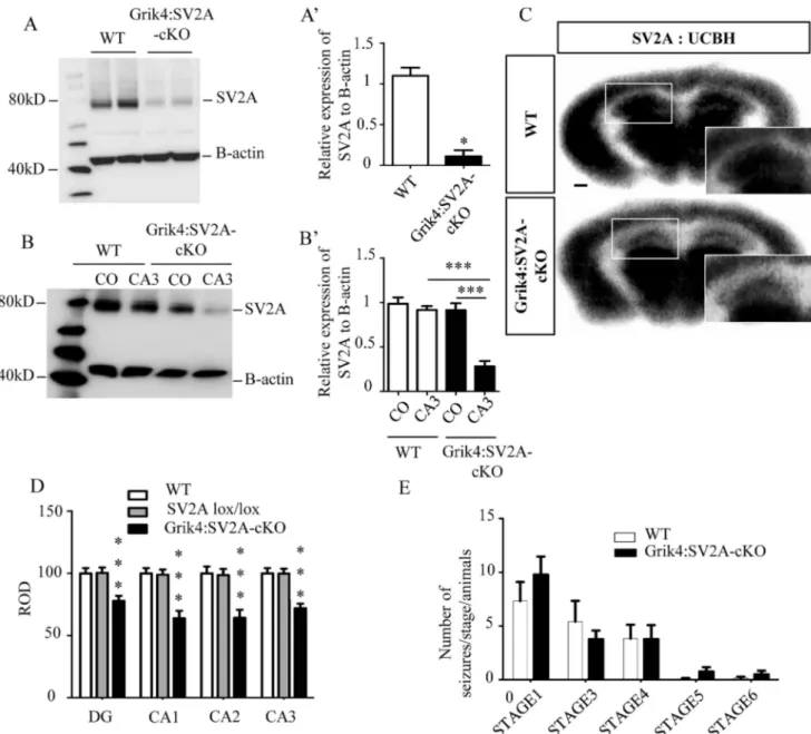
To determine if SV2A protein was still present in the CA3 pyramidal cells of Grik4:SV2A-cKO mice, we performed western blotting on whole hippocampus protein extracts. SV2A expression levels significantly decreased in the hippocampus of the Grik4:SV2A-cKO mice, compared to that of the WTs (⇤⇤⇤:p <0.0001) (Fig 3A and 3A’). To assess the specificity of the
SV2A protein depletion in the CA3 hippocampus region, we micro-dissected this region and the cerebral cortex of adult mice (i.e., Grik4:SV2A-cKO and WT) and performed western blotting. We detected a similar amount of SV2A protein in the CA3 hippocampus region and cerebral cortex of the WT animals. On the other hand, as expected, we observed a stronger reduction of SV2A protein signal in the CA3 hippocampus region than in the cortex in Grik4:SV2A-cKO ani-mals (⇤⇤⇤:p <0.001) or the CA3 hippocampus region of the WTs (⇤⇤⇤:p <0.001)(Fig 3B and
3B’). These results were confirmed byin vitro autoradiography using the18F-UCB-H radioli-gand. This radiotracer has been shown to specifically bind to the SV2A protein [15,16]. Autora-diograms obtained post-incubation using a constant radiotracer concentration enabled us to visualize decreased binding of18F-UCB-H in all hippocampus regions of the Grik4:SV2A-cKO
animals compared to WT littermates (⇤⇤⇤:p <0.001) (Fig 3C and 3D). Significantly, the signal
was roughly the same in the other brain regions. Altogether, these results demonstrated the spe-cific recombination of theSV2A gene in the hippocampus, primarily in the CA3 area, of Grik4: SV2A-cKO mice and, consequently, the strong decrease of SV2A protein in the CA3 region of these animals.
To assess the efficiency of the recombinaison, we evaluated the loss of SV2A protein accord-ing to mRNA expression in Grik4:SV2A-cKO and Ubc:SV2A-cKO mouse line. Both mouse
Fig 2. Specific invalidation of SV2A in the CA3 and DG areas of the adult hippocampus. A: Cre-mediated EYFP reporter expression demonstrating Cre recombination in the CA3 and DG areas of the hippocampus of Grik4-CRE/SV2ALox/Lox/-ROSA-EYFP mice, versus controls (i.e.: SV2Alox/lox/ROSA-EYFP) (Green: e-YFP; blue: Hoechst; scale bar = 250 m). B: PCR genotype analysis
on DNA extracted from the CA3 region or cerebral cortex and microdissected, from WT and Grik4:SV2A-CKO mouse brains (the loxed allele amplicon migrated at 2,500bp and the recombined allele amplicon at 1,500bp). C: SV2A mRNA expression in the microdissected hippocampus (CA3, CA1, DG), measured by quantitative real-time PCR, from WT and Grik4:SV2A-cKO mice. The SV2A mRNA level was normalized using the GAPDH level. The data is presented as mean SEM,** p 0.01, ***p 0.001. D: Conditional removal of SV2A from adult mouse CA3 areas, validated by in situ hybridization using an LNA probe targeting an exon 3 sequence of the SV2A transcript. The arrowhead indicates the hippocampus. Scale bar = 500 m.
lines reduced more than half of mRNA and protein expression (S1A Fig). Interestingly, we observed in Ubc:SV2A-cKO cortex a depletion of SV2A mRNA expression greater than the decrease in protein (S1B Fig). On the other hand, we detected a similar amount of SV2A pro-tein and mRNA in CA3 of the hippocampus of Grik4:SV2A-cKO. This result suggest that both mouse lines are equally efficient to invalidate SV2A expression.
Fig 3. The absence of SV2A protein in the hippocampus does not lower the epileptic threshold. A: Western blot analyses revealing SV2A protein depletion in the hippocampus of Grik4:SV2A-cKO mice in comparison with that of WTs. A’: Quantification of the SV2A proteins extracted from the hippocampus of WT and Grik4:SV2A-cKO mice (n = 3/group, data presented as mean SEM,***p 0,001). B: Western blot analyses demonstrating SV2A protein depletion in the cerebral cortex and CA3 of Grik4:SV2A-cKO mice compared to WT. B’: Quantification of the SV2A proteins (n = 3/group, data presented as mean SEM,***p 0,001). C: Conditional removal of SV2A from adult mice CA3 areas validated by using in vitro autoradiography with the radioligand18F-UCB-H (scale bar = 500 m). D: Semi-quantitation of 18
F-UCB-H signal intensity in the hippocampus (ROD: radioluminescence optical density). E: Number of seizures per animal in those
expressing different behavioral categories post-PTZ injection: sudden behavioral arrest (Stage 1), facial jerking (Stage 2), neck jerks (Stage 3), clonic seizures (sitting) (Stage 4), tonic seizure (lying on belly) (Stage 5), tonic seizure with wild jumping (Stage 6).
Grik4:SV2A-cKO Adult Animals Exhibited Neither Spontaneous
Seizures nor Lowered Epileptic Thresholds
The Grik4:SV2A-cKO mice developed normally; viable and fertile with no obvious phenotype, such as epileptic seizures or increased mortality, compared to the WT littermate animals (data not shown). As theSV2A-null heterozygous mice did not exhibit specific problems, yet had reduced epileptic thresholds [17], we explored the possibility that the Grik4:SV2A-cKO mice could present the same phenotype. We therefore injected adult mice with pentylenetetrazole (PTZ) and analyzed seizure occurrence by means of the PTZ scale, as previously described [13]. Briefly, the PTZ scale defines six stages of seizure onset induced by PTZ injections, from frozen behavior (Step 1) to convulsions (Step 6). No significant difference was observed between the groups (WT and Grik4:SV2A-cKO mice) throughout the six stages of the scale (Fig 3E). We thus concluded that the adult Grik4:SV2A-cKO mice were neither subject to spontaneous epileptic seizures nor characterized by lower epileptic thresholds, despite present-ing reduced levels of hippocampal SV2A protein. Interestpresent-ingly, in human epileptic foci, the down-regulation of SV2A expression is associated with an overexpression of SV2C [18]. We thus investigated if the levels of other proteins of the SV2 family were affected while SV2A is strongly reduced. Using RT-PCR and western blot analyses, we measured the levels of SV2B (Fig 4A, 4B and 4B’) and SV2C (Fig 4C, 4D and 4D’) expression in the hippocampus of WT and GriK4:SV2A-cKO mice. No significant difference was observed between the groups (WT and Grik4:SV2A-cKO mice) for SV2B (Fig 4A, 4B and 4B’) or SV2C expression (Fig 4C, 4D and 4D’). In all, these results excluded the possibility of a compensatory phenomenon relating to a SV2A-depleted hippocampus.
Discussion
Although the exact role of the SV2A protein is still unclear, it is abundantly present in the syn-aptic vesicles of all presynsyn-aptic nerve terminals, as well as in most neuroendocrine secretory granules [1,3,9]. SV2A is the target of an anticonvulsant drug named Levetiracetam (Kep-pra1), and its expression has been reported to be reduced during epileptogenesis in rats, con-firming the role this protein plays in epilepsy [19]. However, as previously stated, the
physiopathological role of SV2A in epilepsy has not yet been clearly defined. In order to gain insight into the physiological role of SV2A, a homozygousSV2A-null mouse (SV2A KO) line was designed [10]. Interestingly and surprisingly, SV2A-KO mice experience severe seizures and weight loss at approximately P7, dying soon after [2,10]. In this context, the classic knock-out approach cannot be used to precisely characterise the role of SV2A, even if, once again, this protein can be linked to epilepsy.
To circumvent the limitations of SV2A-KO mice, we used a conditional gene-targeting strategy using a Cre-loxP system to produce animals that do not express SV2A in specific brain regions. In order to use the Cre-LoxP system to inactivate SV2A, we generated SV2A lox/lox mice using a gene targeting strategy. SV2A lox/lox mice exhibited a normal phenotype associated with a normal level of SV2A expression, thus rendering them indistinguishable from wild-type mice, indicating that insertion of the LoxP sites had no effect onSV2A gene function. When we invalidated SV2A in widespread tissues during late embryogenesis, Ubc: SV2A-cKO pups experienced postnatal growth retardation, severe seizures, and low levels of SV2A expression; the same principal defects observed inSV2A-null pups [10]. The only differ-ence between our model of Ubc:SV2A-cKO mice and the SV2A-KO mice was the age of sei-zure onset, recorded at P12 in our model versus P7 in the SV2A-KO. This small difference could be a result of our model only invalidating SV2A at E18.5, when tamoxifen was injected, while the invalidation was already present in the early embryogenesis steps in the SV2A-KO
model. All things considered, these results validated the suggestion that cre-mediated excision of the floxed SV2A allele is the cause of disruption in theSV2A gene function.
We then performed a zone-specific knockdown of theSV2A gene in the hippocampus, more precisely in the CA3 and DG regions, demonstrating that our strategy was suitable for tissue-specific targetedSV2A gene suppression. In order for us to be able to investigate the pos-sible role of the SV2A protein in epilepsy, we first studied a conditional mouse line carrying SV2A deletion in the hippocampus (i.e., Grik4:SV2A-cKO) [14,20]. The hippocampus is already well-known to be a generator of temporal-lobe epilepsy (TLE), a form found in humans associated with a down-regulation of SV2A expression [21]. Surprisingly, the strong reduction of SV2A protein in the CA3 hippocampal area of Grik4:SV2A-cKO mice was not associated with spontaneous seizures or reduced epileptic threshold. Several hypotheses could be formulated to explain this observation. Firstly, the strong reduction of SV2A expression observed in Grik4:SV2A-cKO mice couldn’t be sufficient to abolish the function of SV2A in the hippocampus. An investigation of the half-live of SV2A could help us to clarify this situa-tion. Secondly, SV2A proteins are detected as early as the first 12 days of embryogenesis (E12),
Fig 4. SV2B and SV2C are not upregulated in the absence of SV2A in the hippocampus. A: SV2B mRNA expression in the hippocampus of WT and Grik4:SV2A-cKO mice, measured by quantitative real-time PCR. The SV2A mRNA levels were normalized using the GAPDH mRNA levels. B: Western blot analyses showing the expression of SV2B protein in the hippocampus of Grik4:SV2A-cKO mice, versus that of WT mice. B’: Quantification of the SV2B proteins extracted from the hippocampus of WT and Grik4:SV2A-Grik4:SV2A-cKO mice. C: SV2C mRNA hippocampus expression in WT and Grik4:SV2A-cKO mice, measured by quantitative RT-PCR. D: Western blot analyses showing the expression of SV2C protein in Grik4:SV2A-cKO mouse hippocampus versus that of WT mice. D’: Quantification of the SV2C proteins extracted from the hippocampus of WT and Grik4:SV2A-cKO mice. (n = 3/group, data presented as mean SEM) doi:10.1371/journal.pone.0166525.g004
including in the hippocampus [18]. It is thus possible that any impact of SV2A on epileptogen-esis observed at approximately P7 in an SV2A-KO mouse is more likely a consequence of SV2A, rather than a role restricted to the developmental stages of the hippocampus. Moreover, continuing with this developmental hypothesis, the expression of SV2A is modulated in the hippocampus at approximately P7, suggesting SV2A’s involvement in the early stages of devel-opment and maturation of synaptogenesis in the hippocampus [18]. The slow disappearance of the SV2A protein in the CA3 and DG regions from P14 to P56 could, in this context, not lead to epilepsy onset, as this disappearance developed too late or too slowly. Thirdly, the Grik4 promoter induced the recombination exclusively in excitatory pyramidal neurons of the CA3 area in Grik4:SV2A-cKO mice. However, SV2A-KO mouse studies have demonstrated thatSV2A deletion results also in the reduced action potential-dependent release of the inhibi-tory neurotransmitter GABA in this hippocampal region. This imbalance between excitainhibi-tory and inhibitory neurotransmission in the hippocampus could be responsible for seizures, at least concerning those observed in SV2A-KO mice [10,12]. The possibility thus exists that GriK4:SV2A-cKO does not affect the neuronal population responsible for epilepsy, as the GABAergic system is preserved. Lastly, in human epileptic loci, the downregulation of SV2A expression is associated with an overexpression of SV2C in TLE patients (Crevecoeur, 2013). Therefore, the invalidation of SV2A solely in the glutamatergic system of the CA3 and DG hip-pocampal regions would not be sufficient to recapitulate the epileptic phenotype.
In conclusion, we have generated and validated theSV2A flox/flox mouse line for condi-tional inactivation of theSV2A gene after crossing with a promising Cre mouse line. This SV2A flox/flox mouse line will offer new opportunities to the research community to investi-gate the role of SV2A in specific tissues and cells.
Supporting Information
S1 Fig. Recombination efficiency and comparison of Ubc:cKO and GriK4: SV2A-cKO. A: Relative expression of SV2A mRNA and protein of Ubc:SV2A-cKO brain and of
Grik4:SV2A-cKO CA3 compared to WT (the expression of SV2A in WT mice was arbitrarily fixed at 100%). B: Ratio of SV2A Protein expression vs SV2A mRNA.
(TIFF)
Acknowledgments
The authors wish to thank A. Colige for sharing the Ubc: cre-mouse lines and L. Nguyen for the RosaEYFP mice. We thank the GIGA-Imaging for their technical support. B.R. and A.P are Senior Research Associates from the Belgian National Funds for Scientific Research (FRS-FNRS). B.R. is funded by grants from the FRS-FNRS, the Fonds Le´on Fredericq, and ARC. This work was funded with UCB Pharma as partners.
Author Contributions
Conceptualization: BR AP.
Formal analysis: CMD MESN VN OB JSG PVdA GB. Funding acquisition: BR AP.
Investigation: CMD MESN VN OB JSG PVdA. Methodology: BR.
Resources: BR AP. Supervision: BR AP.
Visualization: CMD MESN VN OB JSG PVdA BR GB SW. Writing – original draft: CMD PVdA BR.
Writing – review & editing: PVdA BR.
References
1. Bajjalieh SM, Frantz GD, Weimann JM, McConnell SK, Scheller RH. Differential expression of synaptic vesicle protein 2 (SV2) isoforms. The Journal of neuroscience: the official journal of the Society for Neu-roscience. 1994; 14(9):5223–35. Epub 1994/09/01. PMID:8083732.
2. Janz R, Goda Y, Geppert M, Missler M, Sudhof TC. SV2A and SV2B function as redundant Ca2+ regu-lators in neurotransmitter release. Neuron. 1999; 24(4):1003–16. Epub 2000/01/07. PMID:10624962. 3. Buckley K, Kelly RB. Identification of a transmembrane glycoprotein specific for secretory vesicles of
neural and endocrine cells. The Journal of cell biology. 1985; 100(4):1284–94. Epub 1985/04/01. PMID: 2579958; PubMed Central PMCID: PMCPmc2113776.
4. Dong M, Yeh F, Tepp WH, Dean C, Johnson EA, Janz R, et al. SV2 is the protein receptor for botulinum neurotoxin A. Science (New York, NY). 2006; 312(5773):592–6. Epub 2006/03/18. doi:10.1126/ science.1123654PMID:16543415.
5. Mendoza-Torreblanca JG, Vanoye-Carlo A, Phillips-Farfan BV, Carmona-Aparicio L, Gomez-Lira G. Synaptic vesicle protein 2A: basic facts and role in synaptic function. Eur J Neurosci. 2013; 38 (11):3529–39. Epub 2013/10/10. doi:10.1111/ejn.12360PMID:24102679.
6. Janz R, Sudhof TC. SV2C is a synaptic vesicle protein with an unusually restricted localization: anatomy of a synaptic vesicle protein family. Neuroscience. 1999; 94(4):1279–90. Epub 2000/01/07. PMID: 10625067.
7. Bindra PS, Knowles R, Buckley KM. Conservation of the amino acid sequence of SV2, a transmem-brane transporter in synaptic vesicles and endocrine cells. Gene. 1993; 137(2):299–302. Epub 1993/ 12/31. PMID:8299963.
8. Wan QF, Zhou ZY, Thakur P, Vila A, Sherry DM, Janz R, et al. SV2 acts via presynaptic calcium to regu-late neurotransmitter release. Neuron. 2010; 66(6):884–95. Epub 2010/07/14. doi:10.1016/j.neuron. 2010.05.010PMID:20620874; PubMed Central PMCID: PMC2913707.
9. Loscher W, Brandt C. Prevention or modification of epileptogenesis after brain insults: experimental approaches and translational research. Pharmacological reviews. 2010; 62(4):668–700. Epub 2010/11/ 17. doi:10.1124/pr.110.003046PMID:21079040; PubMed Central PMCID: PMCPmc3014230. 10. Crowder KM, Gunther JM, Jones TA, Hale BD, Zhang HZ, Peterson MR, et al. Abnormal
neurotrans-mission in mice lacking synaptic vesicle protein 2A (SV2A). Proc Natl Acad Sci U S A. 1999; 96 (26):15268–73. Epub 1999/12/28. PMID:10611374; PubMed Central PMCID: PMC24809.
11. Lamberty Y, Detrait E, Leclercq K, Michel A, De Ryck M. Behavioural phenotyping reveals anxiety-like features of SV2A deficient mice. Behavioural brain research. 2009; 198(2):329–33. Epub 2008/12/02. doi:10.1016/j.bbr.2008.11.005PMID:19041904.
12. Venkatesan K, Alix P, Marquet A, Doupagne M, Niespodziany I, Rogister B, et al. Altered balance between excitatory and inhibitory inputs onto CA1 pyramidal neurons from SV2A-deficient but not SV2B-deficient mice. J Neurosci Res. 2012; 90(12):2317–27. Epub 2012/08/01. doi:10.1002/jnr.23111 PMID:22847229.
13. Suzuki T, Miyamoto H, Nakahari T, Inoue I, Suemoto T, Jiang B, et al. Efhc1 deficiency causes sponta-neous myoclonus and increased seizure susceptibility. Hum Mol Genet. 2009; 18(6):1099–109. Epub 2009/01/17. doi:10.1093/hmg/ddp006PMID:19147686.
14. Nakazawa K, Quirk MC, Chitwood RA, Watanabe M, Yeckel MF, Sun LD, et al. Requirement for hippo-campal CA3 NMDA receptors in associative memory recall. Science. 2002; 297(5579):211–8. Epub 2002/06/01. doi:10.1126/science.1071795PMID:12040087; PubMed Central PMCID: PMC2877140. 15. Bretin F, Warnock G, Bahri MA, Aerts J, Mestdagh N, Buchanan T, et al. Preclinical radiation dosimetry
for the novel SV2A radiotracer [18F]UCB-H. EJNMMI research. 2013; 3(1):35. Epub 2013/05/08. doi: 10.1186/2191-219X-3-35PMID:23647774; PubMed Central PMCID: PMC3655042.
16. Warnock GI, Aerts J, Bahri MA, Bretin F, Lemaire C, Giacomelli F, et al. Evaluation of 18F-UCB-H as a novel PET tracer for synaptic vesicle protein 2A in the brain. Journal of nuclear medicine: official
publication, Society of Nuclear Medicine. 2014; 55(8):1336–41. Epub 2014/06/18. doi:10.2967/jnumed. 113.136143PMID:24935992.
17. Kaminski RM, Gillard M, Leclercq K, Hanon E, Lorent G, Dassesse D, et al. Proepileptic phenotype of SV2A-deficient mice is associated with reduced anticonvulsant efficacy of levetiracetam. Epilepsia. 2009; 50(7):1729–40. Epub 2009/06/03. doi:10.1111/j.1528-1167.2009.02089.xPMID:19486357. 18. Crevecoeur J, Foerch P, Doupagne M, Thielen C, Vandenplas C, Moonen G, et al. Expression of SV2
isoforms during rodent brain development. BMC neuroscience. 2013; 14:87. Epub 2013/08/14. doi:10. 1186/1471-2202-14-87PMID:23937191; PubMed Central PMCID: PMCPmc3765414.
19. Lynch BA, Lambeng N, Nocka K, Kensel-Hammes P, Bajjalieh SM, Matagne A, et al. The synaptic vesi-cle protein SV2A is the binding site for the antiepileptic drug levetiracetam. Proceedings of the National Academy of Sciences of the United States of America. 2004; 101(26):9861–6. Epub 2004/06/24. doi: 10.1073/pnas.0308208101PMID:15210974; PubMed Central PMCID: PMCPmc470764.
20. Haws ME, Kaeser PS, Jarvis DL, Sudhof TC, Powell CM. Region-specific deletions of RIM1 reproduce a subset of global RIM1alpha(-/-) phenotypes. Genes, brain, and behavior. 2012; 11(2):201–13. Epub 2011/11/23. doi:10.1111/j.1601-183X.2011.00755.xPMID:22103334; PubMed Central PMCID: PMCPmc3268893.
21. Crevecoeur J, Kaminski RM, Rogister B, Foerch P, Vandenplas C, Neveux M, et al. Expression pattern of synaptic vesicle protein 2 (SV2) isoforms in patients with temporal lobe epilepsy and hippocampal sclerosis. Neuropathology and applied neurobiology. 2014; 40(2):191–204. Epub 2013/04/27. doi:10. 1111/nan.12054PMID:23617838.