A Dosimetric Selectivity Intercomparison of HDR
Brachytherapy, IMRT and Helical Tomotherapy
in Prostate Cancer Radiotherapy
Johanne Hermesse
1, Sylvie Biver
1, Nicolas Jansen
1, Eric Lenaerts
2, Nathalie De Patoul
3,
Stefaan Vynckier
3, Philippe Coucke
1, Pierre Scalliet
4, Philippe Nickers
5Background and Purpose: Dose escalation in order to improve the biochemical control in prostate cancer requires the ap-plication of irradiation techniques with high conformality. The dosimetric selectivity of three radiation modalities is compared: high-dose-rate brachytherapy (HDR-BT), intensity-modulated radiation radiotherapy (IMRT), and helical tomotherapy (HT). Patients and Methods: Ten patients with prostate adenocarcinoma treated by a 10-Gy HDR-BT boost after external-beam radiotherapy were investigated. For each patient, HDR-BT, IMRT and HT theoretical treatment plans were realized using com-mon contour sets. A 10-Gy dose was prescribed to the planning target volume (PTV). The PTVs and critical organs’ dose-volume histograms obtained were compared using Student’s t-test.
Results: HDR-BT delivers spontaneously higher mean doses to the PTV with smaller cold spots compared to IMRT and HT. 33% of the rectal volume received a mean HDR-BT dose of 3.86 ± 0.3 Gy in comparison with a mean IMRT dose of 6.57 ± 0.68 Gy and a mean HT dose of 5.58 ± 0.71 Gy (p < 0.0001). HDR-BT also enables to better spare the bladder. The hot spots inside the urethra are greater with HDR-BT. The volume of healthy tissue receiving 10% of the prescribed dose is reduced at least by a factor of 8 with HDR-BT (p < 0.0001).
Conclusion: HDR-BT offers better conformality in comparison with HT and IMRT and reduces the volume of healthy tissue receiving a low dose.
Key Words: Prostate cancer · IMRT · Brachytherapy · Tomotherapy Strahlenther Onkol 2009;185:736–42
DOI 10.1007/s00066-009-2009-5
Ein dosimetrischer Vergleich von HDR-Brachytherapie, IMRT und helikaler Tomotherapie bei der Radiotherapie des Prostatakarzinoms
Hintergrund und Ziel: Eine Dosiseskalation zur Steigerung der biochemischen Kontrollraten beim Prostatakarzinom erfordert die Anwendung von Bestrahlungstechniken, die eine hohe Dosiskonformität ermöglichen. Verglichen wird die dosimetrische Selektivität von drei Bestrahlungsmodalitäten: High-Dose-Rate-Brachytherapie (HDR-BT), intensitätsmodulierte Radiotherapie (IMRT) und helikale Tomotherapie (HT).
Patienten und Methodik: Zehn Patienten mit einem Adenokarzinom der Prostata, die im Anschluss an eine perkutane Ra-diotherapie einen Boost von 10 Gy in Form einer HDR-BT erhielten, wurden untersucht. Für jeden dieser Patienten wurden Bestrahlungspläne für eine HDR-BT, eine IMRT und eine HT unter Anwendung gemeinsamer Konturierungsverfahren erstellt. Für das Planungszielvolumen (PTV) wurden 10 Gy verordnet. Die ermittelten jeweiligen PTV und Dosis-Volumen-Histogramme für die kritischen Organe wurden mittels Student-t-Test miteinander verglichen.
Ergebnisse: Die HDR-BT führt zu höheren mittleren Dosen im PTV mit kleineren Cold Spots als die IMRT oder HT. 33% des bestrahlten Volumens des Rektums erhielten bei der HDR-BT eine mittlere Dosis von 3,86 ± 0,3 Gy im Vergleich zu 6,57 ± 0,68 Gy bei der IMRT und 5,58 ± 0,71 Gy bei der HT (p < 0,0001). Die HDR-BT ermöglicht eine bessere Schonung der Harnblase. Die Dosisspitzen (Hot Spots) an der Urethra sind jedoch bei der HDR-BT höher. Das Volumen des gesunden Gewebes, das 10% der vorgeschriebenen Dosis erhält, wird bei Anwendung der HDR-BT etwa um den Faktor 8 verringert (p < 0,0001).
1Department of Radiation Oncology, Liège University Hospital, Belgium, 2Department of Medical Physics, Liège University Hospital, Belgium, 3Department of Medical Physics, St Luc University Hospital, Brussels, Belgium, 4Department of Radiation Oncology, St Luc University Hospital, Brussels, Belgium, 5Department of Radiation Oncology, Oscar Lambret Center, Lille, France.
Introduction
Many randomized studies have shown that in prostate can-cer radiotherapy, dose escalation significantly improves the rate of biochemical control [24, 27, 28, 39]. Nevertheless, an increasing dose to the prostate is associated with a certain level of toxicity. Moderate side effects still remain relatively frequent even by using a conformal radiation therapy [11, 38]. Different radiation modalities developed in order to improve the conformality of the radiation treatment and to decrease the toxicity are under investigation.
High-dose-rate brachytherapy (HDR-BT) is a precise hypofractionated radiation treatment whose efficacy is well established in prostate cancer [13, 14, 18, 19]. The α/β ratio of prostate carcinoma is still being discussed but well known to be lower than the typical value of 10 Gy of most other solid tumors [4, 9]. So, hypofractionated treatment should be able to increase the therapeutic ratio [8, 35]. This hypofrac-tionation was initially used in HDR-BT in combination with external-beam radiation therapy (EBRT) as demonstrated in a recent randomized phase III trial [14].
Intensity-modulated radiotherapy (IMRT) is also able to safely achieve high dose to the planning target volume (PTV) in prostate cancer. Retrospective studies indicate that dose distributions of IMRT translate into improved rates of disease control and/or lower rates of rectal toxicity [12, 37]. A recent study reported acceptable toxicity and favorable biochemical outcome provided by ultrahigh-dose (86.4 Gy) IMRT for localized prostate cancer [5].
Helical tomotherapy (HT) is an advanced form of con-tinuous helical IMRT with accurate integrated image-guided radiotherapy (IGRT) [34]. This complex rotational method of treatment delivery may improve the dose conformity of a treatment plan compared with the fixed-beam method of IMRT using a limited number of beam directions. First re-ports encouraged this radiation modality [7, 16, 32].
Improvement of treatment conformality in order to spare organs at risk (OARs) sometimes increases the volume of healthy tissues at distance of the PTV receiving low radia-tion doses, with possible higher rates of late side effects such as secondary cancers [2, 3].
We therefore decided to compare the dosimetric selec-tivity of HDR-BT, IMRT and HT on a prostate model with-out taking the impact of fractionation on tumor control and side effects into account.
Patients and Methods
In the beginning of 2007, ten consecutive patients with lo-calized advance prostate adenocarcinoma treated with a 10-Gy HDR-BT boost after EBRT were investigated.
HDR-BT was delivered through eight to ten catheters placed by the same well-trained radiation oncologist ac-cording to a method previously reported [23]. Joint slices of 5 mm thickness each were obtained and transferred to
the contouring software platform (Artiview®, Aquilab,
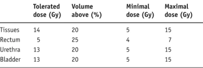
Lille, France). The clinical target volume (CTV) included only the prostate. No further expansion from CTV was applied to generate the PTV. The rectum, bladder and urethra were contoured entirely. The Brachyvision® (ver-sion 8, Varian Medical System, Charlottesville, VA, USA) treatment-planning system (TPS) was used to calculate the treatment for an HDR 192Ir stepping source. At least 95% of the PTV had to be covered by the 10-Gy isodose while 50% could not receive > 150% of the prescribed dose. Dose constraints for OARs are represented in Table 1. The dose optimization by modeling dwell times was done step by step by manually improving a theoretical proposal given at first by the TPS.
For this study, we then transferred, via DICOM RT link, the computed tomography (CT) scan images and all contouring information performed on the Artiview® station
to the Corvus® (Nomos Corp., Pittsburgh, PA, USA) TPS
for IMRT and to Hi-Art® (Tomotherapy inc, Madison, WI,
USA) for HT treatment planning.
Concerning IMRT, a step-and-shoot technique was planned with five 6-MV photon beams (0°, 60°, 120°, 240°, 300°). The PTV was defined as the CTV plus 4 mm in the left-right and anterior-posterior axes and 10 mm in the
cra-Schlussfolgerung: Die HDR-BT führt zu einer günstigeren Dosiskonformität im Vergleich zur HT und zur IMRT und reduziert so das mit einer niedrigen Dosis belastete Volumen gesunden Gewebes.
Schlüsselwörter: Prostatakarzinom · IMRT · Brachytherapie · Tomotherapie
Table 1. First constraints applied to organs at risk in the different treat-ment plans for HDR-BT, IMRT and HT dosimetry. HDR-BT: high-dose-rate brachytherapy; HT: helical tomotherapy; IMRT: intensity-modulated radiotherapy.
Tabelle 1. Dosisbereiche für die Risikoorgane bei den unterschied-lichen Bestrahlungsplänen für die HDR-BT-, IMRT- und HT-Dosimetrie. HDR-BT: High-Dose-Rate-Brachytherapie; HT: helikale Tomotherapie; IMRT: intensitätsmodulierte Radiotherapie.
Tolerated Volume Minimal Maximal dose (Gy) above (%) dose (Gy) dose (Gy)
Tissues 14 20 5 15
Rectum 5 25 4 7
Urethra 13 20 5 15
niocaudal direction. Dose constraints equal to those used for HDR-BT were first applied to the PTV and OARs and were next modified until the lowest doses to critical organs were achieved, while maintaining the initial constraints to the PTV.
HT planning was done according to a standardized class solution with a field width of 25 mm, a pitch of 0.215, and modulation factor of 2. Preliminary constraints for PTV and OARs were identical to those introduced in IMRT and HDR-BT planning. The dose calculation used a total of 18.4 full gantry rotations for the dose spread array of the incident 6-MV beam. Importance and penalty values were adjusted as the dosimetric parameters were modified to ob-tain the lowest doses to critical organs without decrease of the PTV coverage initially planned.
Figure 1. Axial and sagittal views show high conformality of HDR-BT, HT and IMRT. The 10-Gy isodose lines well surround the PTV considered as the prostate for HDR-BT and as the prostate with margins taking the prostate motion (blue line) for both other techniques into account.
Abbildung 1. Die Darstellungen in axialer und sagittaler Schnittebene belegen eine hohe Konformalität von HDR-BT, HT und IMRT. Die 10-Gy-Iso-dose, die das PTV definiert, entspricht dem Prostatavolumen bei der HDR-BT, während für die beiden anderen Bestrahlungstechniken die Bewe-gungen der Prostata (blaue Linie) mitberücksichtigt wurden.
Figure 2. Axial and sagittal views show volume receiving 5 Gy with HDR-BT, IMRT and HT.
Abbildung 2. Mit 5 Gy belastetes Volumen bei der HDR-BT, IMRT und HT (axiale und sagittale Schnittebene).
Figure 3. Volume of healthy tissues receiving 10% of the prescribed dose with the three irradiation techniques.
Abbildung 3. Volumen gesunden Gewebes, das bei den drei Bestrah-lungstechniken mit 10% der verschriebenen Dosis belastet wird.
In order to compare the different treatment plan-ning methods, dose normalization was done to all HDR-BT, IMRT and HT plans to obtain a full coverage of the PTV with the 95% isodose curve. Then, we compared the different PTV and OARs dose-volume histo-gram parameters of the different treatment options using a double-sided paired t-test.
Table 2. Dose received by 95% of planning target volume (PTV), mean and minimal doses delivered to the PTV with HDR-BT, IMRT and HT modality. HDR-BT: high-dose-rate brachytherapy; HT: helical tomothe-rapy; IMRT: intensity-modulated radiotherapy.
Tabelle 2. Dosis für 95% des Planungszielvolumens (PTV), mittlere und minimale auf das PTV eingestrahlte Dosen für das HDR-BT-, IMRT- und HT-Verfahren. HDR-BT: High-Dose-Rate-Brachytherapie; HT: helikale Tomotherapie; IMRT: intensitätsmodulierte Radiotherapie.
Mean for p-valuea
10 patients (Gy) PTV95% HDR-BT 10.07 ± 0.02 p = 0.3 HDR-BT vs. IMRT IMRT 10.01 ± 0.07 p = 0.3 HDR-BT vs. HT HT 10.00 ± 0 p = 0.6 IMRT vs. HT PTV mean dose HDR-BT 16.23 ± 0.49 p < 0.0001 HDR-BT vs. IMRT IMRT 10.47 ± 0.18 p < 0.0001 HDR-BT vs. HT HT 10.41 ± 0.06 p = 0.6 IMRT vs. HT PTV minimal dose HDR-BT 8.97 ± 0.32 p = 0.03 HDR-BT vs. IMRT IMRT 7.93 ± 1.08 p = 0.05 HDR-BT vs. HT HT 8.77 ± 0.31 p = 0.04 IMRT vs. HT
adouble-sided paired t-test
Table 3. Maximal doses delivered to the rectum; doses received by 33%, 20% and 0.5 ml of rectum volume with BT, IMRT and HT. HDR-BT: high-dose-rate brachytherapy; HT: helical tomotherapy; IMRT: in-tensity-modulated radiotherapy.
Tabelle 3. Maximale Dosen am Rektum; Dosen für 33%, 20% und 0,5 ml des Rektumvolumens für das HDR-BT-, IMRT- und HT-Verfahren. HDR-BT: High-Dose-Rate-Brachytherapie; HT: helikale Tomotherapie; IMRT: intensitätsmodulierte Radiotherapie.
Mean for 10 p-valuea
patients(Gy) Maximal rectal dose
HDR-BT 10.14 ± 0.87 p = 0.3 HDR-BT vs. IMRT IMRT 10.45 ± 0.22 p = 0.3 HDR-BT vs. HT HT 10.40 ± 0.24 p = 0.6 IMRT vs. HT D33% rectum HDR-BT 3.86 ± 0.3 p < 0.0001 HDR-BT vs. IMRT IMRT 6.57 ± 0.68 p < 0.0001 HDR-BT vs. HT HT 5.58 ± 0.71 p = 0.02 IMRT vs. HT D20% rectum HDR-BT 4.80 ± 0.35 p < 0.0001 HDR-BT vs. IMRT IMRT 8.08 ± 0.58 p < 0.0001 HDR-BT vs. HT HT 6.87 ± 0.69 p = 0.004 IMRT vs. HT D0.5 ml rectum HDR-BT 7.99 ± 0.74 p < 0.0001 HDR-BT vs. IMRT IMRT 10.21 ± 0.38 p < 0.0001 HDR-BT vs. HT HT 10.04 ± 0.28 p = 0.3 IMRT vs. HT
adouble-sided paired t-test
Table 4. Maximal, mean and minimal doses received by urethra; dose received by 20% of urethra volume with HDR-BT, IMRT and HT. HDR-BT: high-dose-rate brachytherapy; HT: helical tomotherapy; IMRT: intensi-ty-modulated radiotherapy.
Tabelle 4. Maximale, mittlere und minimale Dosen an der Urethra; Dosis für 20% des Urethravolumens für das HDR-BT-, IMRT- und HT-Verfahren. HDR-BT: High-Dose-Rate-Brachytherapie; HT: helikale Tomotherapie; IMRT: intensitätsmodulierte Radiotherapie.
Mean for 10 p-valuea
patients (Gy) Maximal urethral dose
HDR-BT 13.13 ± 0.74 p < 0.0001 HDR-BT vs. IMRT IMRT 11.12 ± 0.27 p < 0.0001 HDR-BT vs. HT HT 10.58 ± 0.13 p = 0.0004 IMRT vs. HT Mean urethral dose
HDR-BT 10.52 ± 0.05 p = 0.6 HDR-BT vs. IMRT IMRT 10.43 ± 0.23 p = 0.6 HDR-BT vs. HT HT 10.42 ± 0.07 p = 0.89 IMRT vs. HT Minimal urethral dose
HDR-BT 5.48 ± 1.56 p = 0.005 HDR-BT vs. IMRT IMRT 7.90 ± 2.46 p < 0.0001 HDR-BT vs. HT HT 9.93 ± 0.38 p = 0.027 IMRT vs. HT D20% urethra HDR-BT 10.62 ± 0.41 p = 0.1 HDR-BT vs. IMRT IMRT 10.46 ± 1.07 p = 0.98 HDR-BT vs. HT HT 10.49 ± 0.12 p = 0.91 IMRT vs. HT
adouble-sided paired t-test
Table 5. Volume of healthy tissues receiving 10% of the prescribed dose. HDR-BT: high-dose-rate brachytherapy; HT: helical tomotherapy; IMRT: intensity-modulated radiotherapy.
Tabelle 5. Volumen gesunden Gewebes, das 10% der vorgeschriebenen Dosis erhält. HDR-BT: High-Dose-Rate-Brachytherapie; HT: helikale Tomotherapie; IMRT: intensitätsmodulierte Radiotherapie.
Mean volume receiving p-valuea
10% of prescribed dose for 10 patients (ml)
HDR-BT 475.25 ± 87.24 p < 0.0001 HDR-BT vs. IMRT IMRT 3,899.43 ± 1,183.24 p < 0.0001 HDR-BT vs. HT HT 5,965.80 ± 1,862.66 p < 0.0001 IMRT vs. HT
Results
The three treatment plans were able to stick to the dosimet-ric criteria and the 10-Gy isodose did systematically sur-round 95% of the PTV while sparing the critical organs. Nevertheless, the dose distribution is different with the three techniques of irradiation (Figures 1 to 3).
Table 2 presents the doses to the PTV with the three different methods. The dose to 95% of the PTV is 10 Gy for all the treatments (p ≥ 0.3) which is in accordance with the designed methodology. The mean dose to the PTV is signifi-cantly increased with HDR-BT (16.23 ± 0.49 Gy; p < 0.0001) in comparison with the other methods while there is no differ-ence between IMRT and HT (10.47 ± 0.18 Gy and 10.41 ± 0.06 Gy, respectively; p = 0.6). Dose distribution inside the PTV is more heterogeneous with HDR-BT. Hot spots are observed around HDR catheters. Cold spots are slightly less with HDR-BT in comparison with HT (8.97 ± 0.32 Gy and 8.77 ± 0.31 Gy, respectively; p = 0.05) and more important in the IMRT planning (7.93 ± 1.08 Gy; p ≤ 0.04).
Rectal doses are represented in Table 3. The maximal dose is similar for the three methods (p ≥ 0.3). The dose deliv-ered to 0.5 ml of the organ is significantly reduced from 10.21 ± 0.38 Gy (IMRT) and 10.04 ± 0.28 Gy (HT) to 7.99 ± 0.74 Gy with HDR-BT (p < 0.0001). 33% of the rectal volume receives a mean dose of 3.86 ± 0.3 Gy with HDR-BT compared to 6.57 ± 0.68 Gy with IMRT and 5.58 ± 0.71 Gy with HT. The differ-ence is in favor of HDR-BT (p < 0.001) even if the protection rate of the rectum offered by HT is higher than with IMRT (p = 0.02).
Likewise, the bladder is better spared with HDR-BT.
The mean dose to 20% of the OAR is 3.49 ± 0.65 Gy with
HDR-BT compared to 7.11 ± 1.13 Gy with IMRT and 6.87 ± 1.09 Gy with HT (p < 0.0001). Urethra irradiation is more heterogeneous with HDR-BT with a maximal dose of 13.13 ± 0.74 Gy and a minimal dose of 5.48 ± 1.56 Gy. For IMRT,
the maximal and minimal doses are 11.12 ± 0.27 Gy and
7.9 ± 2.46 Gy; and for HT 10.58 ± 0.13 Gy and 9.93 ± 0.38 Gy, respectively. The mean dose to the urethra is, however, similar for the three approaches (p ≥ 0.6; Table 4).
The volume of distant tissues from the PTV receiv-ing 10% of the prescribed dose (1 Gy) is 475.25 ± 87.24 ml, 3,899.43 ± 1,183.24 ml, and 5,965.80 ± 1,862.66 ml for HDR-BT, IMRT, and HT, respectively (p < 0.0001; Table 5).
Discussion
This study aimed to compare HDR-BT, IMRT and HT as a way to deliver a fixed dose to the PTV while best sparing the critical organs and healthy distant tissues. Hypofrac-tionation favored by a low α/β value of prostatic adenocar-cinoma was initially used in HDR-BT. It is also applicable to IMRT and HT but remains under investigation [15, 36]. We decided thus to deliver a theoretical normalized dose of 10 Gy for each of the methods to compare only the
dosimet-ric selectivity of these irradiation techniques without taking the impact of hypofractionation into account.
CTV-PTV Expansions
For IMRT and HT, the PTV definition had to consider the intrafraction movements of the prostate and the setup un-certainties before treatment using the most modern IGRT techniques [21, 26]. These prostatic intrafraction motions required a safety margin of 4 mm from the prostate [1, 22]. The longer the treatment duration, the higher the risk of displacement, mainly in the craniocaudal direction [17, 20]. We therefore decided to fix 4 mm as the internal margin in the left-right and anterior-posterior axes and 10 mm in the craniocaudal axis. No additional margin was added to take setup errors into account assuming that patient position is corrected daily applying the most recent IGRT.
Considering HDR-BT, no further expansion from CTV was utilized to generate the PTV because movement of the implant has no marked influence on dose distribution with proper fixation of catheters [25]. Moreover, radiation treat-ment was performed in the 30 min following the dosimetric CT scan while the patient was unable to move.
Dose Distribution Inside the PTV
The goal was not to create a dose painting inside the PTV but to create a fall of the dose outside the PTV as sharp as possible. HDR-BT allows delivering spontaneously higher mean doses to the PTV with smaller cold spots compared to IMRT and HT. This higher mean dose in the central parts of the prostate is more likely to have clinical consequences for tumor control.
Dose Distribution Inside the OARs
The rectal dose was significantly reduced in the HDR-BT approach compared to IMRT and HT approaches. Not only maximal doses were decreased but also the mean rec-tal doses which were recently demonstrated to contribute significantly to the toxicity [31]. HT seems to better spare the rectum as compared to IMRT. If maximal delivered doses are identical with both techniques, doses to 20% and 33% of the organ are lower with HT compared to IMRT (p ≤ 0.02).
The bladder sparing is also greater with HDR-BT in comparison with IMRT and HT. The hot spots inside the urethra are more marked with HDR-BT. This level of dose has, however, not been demonstrated to contribute to in-creased late toxicity. In phase II studies, Martin et al. and Martinez et al. demonstrated the possibility of four 9.5 Gy HDR-BT fractions for the treatment of favorable-stage prostate cancer (equivalent dose per 2 Gy fraction/EQD2 95 Gy or 119.5 Gy according the prostate α/β ratio selected for the calculation: 3 Gy or 1.5 Gy) [18, 19]. The dose to any segment of urethra was limited to ≤ 125% of the pre-scribed dose (EQD2 of 141 Gy). A recent report shows that
doses up to 75.6 Gy and 86.4 Gy are recommended for in-termediate- and high-risk patients, respectively [39]. If we considered a total treatment of HDR-BT with three 10-Gy fractions corresponding to EQD2 of 78 Gy or 98.55 Gy (ac-cording the prostate α/β ratio selected: 3 Gy or 1.5 Gy), the maximal dose received by urethra would be an EQD2 of 127 Gy. Moreover, in our study, the mean urethral dose is identical for the three compared methods (p ≥ 0.6).
Risks of Secondary Cancer
Radiation-induced cancers are an uncommon late compli-cation of radiation therapy [2]. The risk of developing sec-ond primary cancer in irradiated normal tissues increases as radiation dose increases to a maximum at doses around 3–5 Gy. The cell kill effect becomes dominant at higher doses and causes a reduction in survival of transformed cells [29]. For low- and intermediate-risk patients, HDR-BT mono-therapy may be a means of safely delivering higher doses with less secondary radiation-induced cancers in com-parison to IMRT or HT. HDR-BT as monotherapy is not widely established as a standard treatment but is still under investigation [6, 13, 18, 19]. Common schedules of HDR monotherapy proposed three or four fractions of 9.5–10 Gy to the prostate. In this study, the volume of healthy tissue receiving 1 Gy is reduced by a factor 8 or 10 when compared to IMRT and HT data, respectively (p < 0.0001). Schneider et al. showed that dose escalation for prostate radiation treatments relates to an increased risk of secondary tumor induction [30]. Takam et al. demonstrated that among all radiation treatment techniques for prostate cancer, either LDR- (low-dose-rate) or HDR-BT offers a smaller risk of carcinogenis than treatments involving EBRT techniques [33]. These prostate patients are not the most relevant when looking at radiation-induced tumors, because the average age at treatment of these patients is quite high. Neverthe-less, taking the risk of secondary cancer into account, HDR should be specially recommended to younger patients or those with a life expectancy > 10–15 years.
Conclusion
HDR-BT remains thus within the radiation therapy tech-niques offering the highest dosimetric selectivity. The rela-tively less impressive performances of IMRT and HT are partially induced by the need to take a margin for setup and organ motion, even with the best IGRT. Irradiation offering real tracking properties such as CyberKnife could perhaps compete with HDR-BT that is more invasive [10]. However, low dose received by healthy tissues at distance of the PTV should also be quantified in future CyberKnife trials.
References
1. Beltran C, Herman MG, Brian D. Planning target margin calculations for prostate radiotherapy based on intrafraction and interfraction motion using four localization methods. Int J Radiat Oncol Biol Phys 2008;70:289–95. 2. Bhojani N, Jeldres S, Da Pozzo LF, et al. External-beam radiation therapy
in-creases the rate of secondary malignancies relative to radical prostatectomy in men with prostate cancer. J Urol 2008;179:Suppl:113.abstract 318. 3. Brenner DJ, Curtis RE, Hall EJ, et al. Second malignancies in prostate
carcinoma patients after radiotherapy compared with surgery. Cancer 2000;88:398–406.
4. Brenner DJ, Martinez AA, Edmundson GK, et al. Direct evidence that pros-tate tumors show high sensitivity to fractionation (low alpha/beta ratio), similar to late-responding normal tissue. Int J Radiat Oncol Biol Phys 2002;52:6–13.
5. Cahlon O, Zelefsky MJ, Shippy A, et al. Ultra-high dose (86.4 Gy) IMRT for localized prostate cancer: toxicity and biochemical outcomes. Int J Radiat Oncol Biol Phys 2008;71:330–7.
6. Corner C, Rojas AM, Bryan L, et al. A phase II study of high-dose-rate afterloading brachytherapy as monotherapy for the treatment of localized prostate cancer. Int J Radiat Oncol Biol Phys 2008;72:441–6.
7. Cozzarini C, Fiorino C, Di Muzio N, et al. Significant reduction of acute tox-icity following pelvic irradiation with helical tomotherapy in patients with localized prostate cancer. Radiother Oncol 2007;84:164–70.
8. Dasu A. Is the alpha/beta value for prostate tumours low enough to be safely used in clinical trials? Clin Oncol (R Coll Radiol) 2007;19:289–301. 9. Fowler JF. The radiobiology of prostate cancer including new aspects of
fractionated radiotherapy. Acta Oncol 2005;44:265–76.
10. Fuller DB, Naitoh J, Lee C, et al. Virtual HDR CyberKnife treatment for localized prostatic carcinoma: dosimetry comparison with HDR brachy-therapy and preliminary clinical observations. Int J Radiat Oncol Biol Phys 2008;70:1588–97.
11. Goldner G, Bombosch V, Geinitz H, et al. Moderate risk-adapted dose esca-lation with three-dimensional conformal radiotherapy of localized prostate cancer from 70 to 74 Gy. Strahlenther Onkol 2009;185:94–100.
12. Guckenberger M, Flentje M. Intensity-modulated radiotherapy (IMRT) of localized prostate cancer. A review and future perspectives. Strahlenther Onkol 2007;183:57–62.
13. Hoskin PJ, Bownes P, Bryan L, et al. HDR monotherapy brachytherapy for localised prostate cancer. Clin Oncol (R Coll Radiol) 2007;19:Suppl:S7. 14. Hoskin PJ, Motohashi K, Bownes P. High dose rate brachytherapy in
combi-nation with external beam radiotherapy in the radical treatment of prostate cancer: initial results of a randomised phase three trial. Radiother Oncol 2007;84:114–20.
15. Junius S, Haustermans K, Bussels B, et al. Hypofractionated intensity mod-ulated irradiation for localized prostate cancer, results from a phase I/II feasibility study. Radiother Oncol 2007;8:29–39.
16. Keiler L, Dobbins D, Kulasekere R, et al. Tomotherapy for prostate adenocar-cinoma: a report on acute toxicity. Radiother Oncol 2007;84:171–6. 17. Langen KM, Willoughby TR, Meeks SL, et al. Observation on real-time
pros-tate gland motion using electromagnetic tracking. Int J Radiat Oncol Biol Phys 2008;71:1084–90.
18. Martin T, Baltas D, Kurek R, et al. 3-D conformal HDR brachytherapy as monotherapy for localized prostate cancer. Strahlenther Onkol 2004;180: 225–32.
19. Martinez AA, Pataki I, Edmundson G, et al. Phase II prospective study of the use of conformal high-dose-rate brachytherapy as monotherapy for the treatment of favorable stage prostate cancer: a feasibility report. Int J Radiat Oncol Biol Phys 2001;49:61–9.
20. Meijer GJ, De Klerk J, Bzdusek K, et al. What CTV-to-PTV margins should be applied for prostate irradiation? Four-dimensional quantitative assessment using model-based deformable image registration techniques. Int J Radiat Oncol Biol Phys 2008;72:1416–25.
21. Nairz O, Merz F, Deutschmann H, et al. A strategy for the use of image-guid-ed radiotherapy on linear accelerators and its impact on treatment margins for prostate cancer patients. Strahlenther Onkol 2008;184:663–7. 22. Nedervenn A, Langedijk J, Hofman P. Detection of fiducial gold markers for
automatic on-line megavoltage position verification using a marker extrac-tion kernel (MEK). Int J Radiat Oncol Biol Phys 2000;47:1435–42.
23. Nickers P, Thissen B, Jansen N, et al. 192Ir or 125I prostate brachytherapy as
a boost to external beam radiotherapy in locally advanced prostatic cancer: a dosimetric point of view. Radiother Oncol 2006;78:47–52.
24. Peeters STH, Heemsbergen WD, Koper PCM, et al. Dose-response in radio-therapy for localized prostate cancer: results of the Dutch multicenter ran-domized phase III trial comparing 68 Gy of radiotherapy with 78 Gy. J Clin Oncol 2006;24:1990–6.
25. Pieters BR, Van der Grient JNB, Blank LECM, et al. Minimal displacement of novel self-anchoring catheters suitable for temporary prostate implants. Radiother Oncol 2006;80:69–72.
26. Pinkawa M, Pursch-Lee M, Asadpour B, et al. Image-guided radiotherapy for prostate cancer. Strahlenther Onkol 2008;184:679–85.
27. Pollack A, Hanlon AL, Horwitz EM, et al. Prostate cancer radiotherapy dose response: an update of the fox chase experience. J Urol 2004;171:1132–6. 28. Roach M. Dose escalated external beam radiotherapy versus neoadjuvant
androgen deprivation therapy and conventional dose external beam radio-therapy for clinically localized prostate cancer: do we need both? Strahlen-ther Onkol 2007:183:Special Issue 2:26–8.
29. Ruben JD, Davis S, Evans C, et al. The effect of intensity-modulated radio-therapy on radiation-induced second malignancies. Int J Radiat Oncol Biol Phys 2008;70:1530–6.
30. Schneider U, Lomax A, Besserer J, et al. The impact of dose escalation on secondary cancer risk after radiotherapy of prostate cancer. Int J Radiat Oncol Biol Phys 2007;48:475–81.
31. Söhn M, Alber M, Yan D. Principal component analysis-based pattern analy-sis of dose-volume histograms and influence on rectal toxicity. Int J Radiat Oncol Biol Phys 2007;69:230–9.
32. Sterzing F, Schubert K, Sroka-Perez G, et al. Helical tomotherapy. Experiences of the first 150 patients in Heidelberg. Strahlenther Onkol 2008;184:8–14. 33. Takam R, Bezak E, Yeoh E. Risk of second primary cancer following prostate
cancer radiotherapy: DVH analysis using the competitive risk model. Phys Med Biol 2009;54:611–25.
34. Tomsej M. The TomoTherapy Hi.Art System for sophisticated IMRT and IGRT with helical delivery: recent developments and clinical applications. Cancer Radiother 2006;10:288–95.
35. Williams SG, Taylor JM, Liu N, et al. Use of individual fraction size data from 3756 patients to directly determine the alpha/beta ratio of prostate cancer Int J Radiat Oncol Biol Phys 2007;68:24–33.
36. Yuen J, Rodrigues G, Trenka K, et al. Comparing two strategies of dynamic intensity modulated radiation therapy (dIMRT) with 3-dimensional con-formal radiation therapy (3DCRT) in the hypofractionated treatment of high-risk prostate cancer. Radiother Oncol 2008;3:1–10.
37. Zelefsky MJ, Fuks Z, Hunt M, et al. High-dose intensity modulated radiation therapy for prostate cancer: early toxicity and biochemical outcome in 772 patients. Int J Radiat Oncol Biol Phys 2002;53:1111–6.
38. Zelefsky MJ, Fuks Z, Wolfe T, et al. Locally advanced prostatic cancer: long-term toxicity outcome after three-dimensional conformal radiation therapy: a dose-escalation study. Radiology 1998;209:169–74.
39. Zelefsky MJ, Yamada Y, Fuks Z, et al. Long term results of conformal ra-diotherapy for prostate cancer: impact of dose escalation on biochemical control and distant metastases-free survival outcomes. Int J Radiat Oncol Biol Phys 2008;71:1028–33.
Address for Correspondence Johanne Hermesse, MD
Department of Radiation Oncology B35 Domaine Universitaire du Sart Tilman 4000 Liège
Belgium
Phone (+32/43) 667-596, Fax -952 e-mail: jhermesse@chu.ulg.ac.be, johermesse@hotmail.com