Effect of Ser-129 Phosphorylation on Interaction of
␣-Synuclein with Synaptic and Cellular Membranes
*
□SReceived for publication, April 20, 2011, and in revised form, August 11, 2011 Published, JBC Papers in Press, August 17, 2011, DOI 10.1074/jbc.M111.253450 Naomi P. Visanji‡1,2, Sabine Wislet-Gendebien§1,3, Loren W. Oschipok¶, Gang Zhang‡, Isabelle Aubert储, Paul E. Fraser‡**, and Anurag Tandon‡ ‡‡4
From the‡Centre for Research in Neurodegenerative Diseases, University of Toronto, Toronto, Ontario M5S 3H2, Canada,§GIGA Neurosciences, University of Liege, 1 Avenue de l’Hoˆpital, 4000 Lie`ge, Belgium, the¶Brain Research Centre, University of British Columbia, British Columbia V6T 2B5, Canada, the储Sunnybrook Research Institute and Department of Laboratory Medicine and Pathobiology, University of Toronto, Toronto, Ontario M4N 3M5, Canada, and the Departments of **Medical Biophysics and
‡‡Medicine, University of Toronto, Toronto, Ontario M5S 1A8, Canada
Background:The majority of␣-synuclein is phosphorylated at serine 129 in Lewy bodies.
Results:The membrane association of PD-linked mutant␣-synuclein, but not wild-type␣-synuclein, was increased by serine 129 phosphorylation.
Conclusion:Pathological serine 129 phosphorylation regulates membrane accumulation of mutant␣-synuclein.
Significance:The relationship of serine 129 phosphorylation to pathogenic aggregation of normal and mutant␣-synuclein may be governed by distinct effects on phosphoprotein membrane accumulation.
In the healthy brain, less than 5% of␣-synuclein (␣-syn) is phosphorylated at serine 129 (Ser(P)-129). However, within Parkinson disease (PD) Lewy bodies, 89% of␣-syn is Ser(P)-129. The effects of Ser(P)-129 modification on␣-syn distribution and solubility are poorly understood. As␣-syn normally exists in both membrane-bound and cytosolic compartments, we examined the binding and dissociation of Ser(P)-129␣-syn and analyzed the effects of manipulating Ser(P)-129 levels on␣-syn membrane interactions using synaptosomal membranes and neural precursor cells from␣-syn-deficient mice or transgenic mice expressing human␣-syn. We first evaluated the recovery of the Ser(P)-129 epitope following either ␣-syn membrane binding or dissociation. We demonstrate a rapid turnover of Ser(P)-129 during both binding to and dissociation from synap-tic membranes. Although the membrane binding of WT␣-syn was insensitive to modulation of Ser(P)-129 levels by multiple strategies (the use of phosphomimic S129D and nonphosphory-lated S129A␣-syn mutants; by enzymatic dephosphorylation of 129 or proteasome inhibitor-induced elevation in Ser(P)-129; or by inhibition or stable overexpression of PLK2), PD mutant Ser(P)-129 ␣-syn showed a preferential membrane association compared with WT Ser(P)-129␣-syn. Collectively, these data sug-gest that phosphorylation at Ser-129 is dynamic and that the sub-cellular distribution of␣-syn bearing PD-linked mutations, A30P or A53T, is influenced by the phosphorylation state of Ser-129.
Parkinson disease (PD)5is a progressive neurodegenerative disorder characterized by a loss of dopaminergic neurons in the substantia nigra pars compacta and depletion of dopamine in the striatum. The major pathological hallmark in affected brains is the presence of ␣-synuclein (␣-syn)-positive Lewy bodies (LBs) in surviving neurons. Normally,␣-syn is an abun-dant presynaptic protein thought to play a role in synaptic plas-ticity (1, 2). However, multiplication of the gene encoding␣-syn or missense mutations (A53T, A30P, or E46K) are known to cause inherited forms of PD (3). In vitro studies suggest that ␣-syn is natively unfolded in aqueous solution and that expo-sure to lipids stabilizes the amino terminus in an amphipathic ␣-helix that aligns polar and nonpolar residues into opposing orientations (4 – 6). This secondary structure confers the lipid-binding properties for direct membrane interaction such that purified recombinant␣-syn can bind to small diameter artificial vesicles rich in acidic phospholipids (7, 8), to purified synaptic vesicles and membranes (9 –11), and to membranes within intact cells (12).
␣-syn also has the propensity to aggregate and can form amy-loid-like fibrils, which are the main component of LBs (13). Increasing evidence suggests that phosphorylation may be an important regulator of␣-syn oligomerization, fibrillogenesis, LB formation, and neurotoxicity in vivo (14). Immunohisto-chemical and bioImmunohisto-chemical studies suggest that the majority of ␣-syn within LBs from patients with PD and related synucle-inopathies is phosphorylated at serine residue 129 (Ser(P)-129) (15–18). Moreover, alteration of Ser-129 to the negatively charged amino acid aspartate, to mimic phosphorylation, sig-nificantly enhanced␣-syn toxicity in Drosophila, although its toxicity in studies with rodents has been equivocal (14, 19, 20). Several kinases, including casein kinases 1 and 2 (21–23) and members of the G-protein-coupled receptor kinase family (24), *This work was supported in part by Canadian Institutes of Health Research
Operating Grants MOP 84501 (to A. T.), FRN 93603 (to I. A.), and MOP 84527 (to P. E. F.) and an Alzheimer Association grant (to I. A.).
□S
The on-line version of this article (available at http://www.jbc.org) contains
supplemental Fig. 1.
1Both authors contributed equally to this work.
2Supported by a fellowship from the Parkinson Society of Canada. 3Supported by a fellowship from the Canadian Institutes of Health Research. 4To whom correspondence should be addressed: Centre for Research in
Neu-rodegenerative Diseases, University of Toronto, 6 Queen’s Park Crescent West, Toronto, Ontario M5S 3H2, Canada. Tel.: 8880; Fax: 416-978-1878; E-mail: [email protected].
5The abbreviations used are: PD, Parkinson disease;␣-syn, ␣-synuclein;
ANOVA, analysis of variance; LB, Lewy body; NPC, neural precursor cells; CIP, calf intestinal phosphatase.
THE JOURNAL OF BIOLOGICAL CHEMISTRY VOL. 286, NO. 41, pp. 35863–35873, October 14, 2011 © 2011 by The American Society for Biochemistry and Molecular Biology, Inc. Printed in the U.S.A.
at UNIV DE LIEGE-MME F PASLE, on November 7, 2011
www.jbc.org
are capable of phosphorylating ␣-syn at Ser-129. However, recent evidence suggests that a member of the polo-like kinase family, PLK2, is the prime contributor to phosphorylation of ␣-syn at Ser-129 (25, 26). Inhibitory RNA-induced knockdown of PLK2 in human cortical cultures or the knock-out of the plk2 gene in mice reduced the levels of Ser(P)-129␣-syn by ⬎70%, indicating that PLK2 is responsible for the majority of phosphor-ylation of␣-syn at Ser-129 in vivo. Nevertheless, Ser-129 phos-phorylation reduced ␣-syn aggregation in vitro (27), and pharmacological inhibition of kinases associated with phos-phorylation of Ser-129 failed to reduce␣-syn aggregation in a cellular model (28), suggesting that␣-syn aggregation is either inhibited or is independent of Ser-129 phosphorylation.
Because inclusion formation may be linked to nucleation-elongation process initiated on membranes (29), we hypothe-sized that phosphorylation of␣-syn at Ser-129 may affect mem-brane binding. Therefore, we took advantage of in vitro binding and dissociation assays (11, 30) to analyze the effect of phos-phorylation at Ser-129 on the interaction of␣-syn with synaptic membranes. We then tested the effects of manipulating the level of Ser-129 phosphorylation, both pharmacologically and by virus-mediated expression of PLK2, on membrane binding of␣-syn in neural precursor cells prepared from transgenic mice expressing human␣-syn.
EXPERIMENTAL PRECEDURES
Synaptosomal Membrane Preparation
Synaptosomal membranes were prepared as described pre-viously (31). Briefly, murine cerebral cortices were homoge-nized in ice-cold buffer A (320 mMsucrose, 1 mMEGTA, and 5
mMHEPES (pH 7.4)) and centrifuged (1000⫻ g, 10 min). The
supernatant was centrifuged (24,000 ⫻ g, 10 min), and the resulting pellet (P2) was resuspended in buffer A. The P2 frac-tion was loaded onto a discontinuous Ficoll gradient (13, 9, and 5% in buffer A) and centrifuged (35,000⫻ g, 35 min). The 13 to 9% interface, containing intact synaptosomes, was resuspended in buffer B (140 mMNaCl, 5 mMKCl, 20 mMHEPES, 5 mM
NaHCO3, 1.2 mMNa2HPO4, 1 mMMgCl2, 1 mMEGTA, and 10
mMglucose) and centrifuged (24,000⫻ g, 10 min).
Synapto-somes were lysed in buffer C (10 mMHEPES, 18 mMKOAc (pH 7.2)), centrifuged (24,000 ⫻ g, 10 min), and resuspended in buffer D (25 mMHEPES, 125 mMKOAc, and 2.5 mMMgCl2).
Cytosol Preparation
Brains from␣-syn KO mice and mice overexpressing WT, A30P, or A53T␣-syn were homogenized in 85 mMsucrose, 100
mM KOAc, 1 mM MgOAc, and 20 mM HEPES (pH 7.4).
The homogenate was centrifuged (15,000⫻ g, 10 min), and the resulting supernatant was centrifuged (100,000⫻ g, 1 h). The supernatant was dialyzed for 4 h in 145 mMKOAc and 25 mM
HEPES (pH 7.2). Protein concentration was determined by BCA protein assay (Pierce).
Binding/Dissociation Experiments
Synaptosomal membranes were incubated with cytosol for 10 min at 37 °C in the absence or presence of a serine/threonine phosphatase inhibitor mixture (phosphatase inhibitor mixture
3 purchased from Sigma) and 1Mokadaic acid. Membrane
and supernatant were separated by centrifugation (24,000⫻ g, 10 min), and␣-syn levels in the membrane and supernatant fractions were quantified by Western blotting.
Western Blotting
Proteins were separated by electrophoresis using 12% Tris-glycine polyacrylamide gels. Proteins were transferred to nitro-cellulose membranes (Life Sciences) and probed with antibod-ies against ␣-syn (Syn-1, BD Transduction Laboratories, 1:1000), Ser(P)-129␣-syn (clone 11A5, 1:500, Elan Pharmaceu-ticals), GAPDH (Sigma 1:1000), syntaxin (Sigma, 1:1000), tetra-His (Qiagen, 1:1000), and ubiquitin (Dako, 1:1000). Bound HRP-conjugated anti-mouse or anti-rabbit IgG (Sigma) was revealed by chemiluminescence using ECL Plus (GE Health-care) and quantified with a Storm 860 fluorescent imager and ImageQuant software (GE Healthcare).
Isolation of Mouse Whole Brain Neural Precursor Cells
Mouse whole brain neural precursor cells (NPC) were pre-pared according to the method of Ray and Gage (32). Briefly, the brains of six mice, overexpressing human WT, A30P, or A53T ␣-syn under the control of the hamster prion protein promoter (see Ref. 30) were chopped into pieces (⬃2 mm3) before being
incubated at 37 °C in 10 volumes (g/ml) of DMEM containing papain (2.5 units/ml), protease type 1 (1 unit/ml), and DNase I (250 units/ml). The tissue was triturated every 5 min until a smooth consistency was achieved (⬃30 min). Following diges-tion, the cell suspension was mixed with an equal volume of DMEM/F-12/N2 containing 10% FBS. The suspension was then passed through a sterilized 15-m nylon mesh filter and centrifuged (1000⫻ g for 3 min). The pellet was resuspended in DMEM/F-12/N2 containing 10% FBS and mixed with an equal volume of Percoll (9:1 v/v Percoll/PBS) and centrifuged (20,000⫻ g for 30 min at 18 °C). NPC were harvested from the low buoyancy fraction (⬃5 ml above the red blood cell layer). Cells were washed in cold PBS containing 1⫻ antibiotic/anti-mycotic solution (containing penicillin, streptomycin, and amphotericin B) before plating in DMEM/F-12/N2 containing 1⫻ antibiotic/antimycotic solution,FGF (20 mg/ml), EGF (20 ng/ml), and heparin (5 g/ml). Proliferating NPC began to grow in clusters⬃2–3 weeks post-isolation. During this period, half the media were changed weekly, and FGF (20 mg/ml), EGF (20 ng/ml), and heparin (5 mg/ml) were added every 3– 4 days.
Tissue Culture
SHSY5Y Cells—SHSY5Y cells (ATCC) were maintained under an atmosphere of air and 5% CO2at 37 °C. The medium
consisted of DMEM containing 4.5 g/liter glucose, 0.01 mM
sodium pyruvate, 1Mnonessential amino acids, and 10% fetal
bovine serum (Multicell). Cells were cultured in 10-cm plates and passaged at⬃75% confluence.
Maintenance of NPC—NPC were maintained under an atmosphere of air and 5% CO2at 37 °C. The medium consisted
of DMEM/F-12/N2 (Invitrogen) containingFGF (20 mg/ml, Invitrogen), EGF (20 ng/ml, Invitrogen), and heparin (5 mg/ml,
at UNIV DE LIEGE-MME F PASLE, on November 7, 2011
www.jbc.org
Sigma). Cells were cultured in T75 flasks and passaged at⬃75% confluence.
Drug Treatment—BI2536 (Axon Medchem, Groningen, The Netherlands) was dissolved in PBS (10 mM) and stored at
⫺20 °C until use. Epoxomicin (Peptides International, Louis-ville, KY) was dissolved in PBS (1 mM) and stored at⫺20 °C
until use. Both BI2536 and epoxomicin were added to cells in culture for 24 h prior to harvesting.
Nucleofection—pcDNA3 WT, S129A, and S129D␣-syn plas-mids were prepared as described previously (33). 4 ⫻ 106
SHSY5Y cells were resuspended in 100l of transfection buffer (Amaxa Biosystems, Gaithersburg, MD). 4 g of DNA was added, and the cell/DNA mixture was transferred to 1-cm transfection cuvettes (Amaxa Biosystems, Gaithersburg, MD). Nucleoporation was carried out using the Amaxa nucleofector set to a predefined program (A023). Following electroporation, cells were cultured for 48 h prior to harvest.
Virus Production and Infection
Snk/PLK2 was subcloned from pcDNA3.1/V5-His Snk/ PLK2 (Addgene plasmid 16015 (34)) into a pWPI Bicistronic lentiviral vector pWPI/hPLK2WT/Neo. A mutant pWPI/ hPLK2K111M/Neo was created using site-directed mutagene-sis (Stratagene). Based on sequence homology, the K111M PLK2 mutation corresponds to the K82M PLKI sequence lack-ing kinase activity (35). Viral production was mediated by HEK293T cells co-transfected with 3 g of the envelope plasmid pMD2.G (Addgene plasmid 12259), 5g of packag-ing plasmid psPAX2 (Addgene plasmid 12260), and 10 g
of either pWPI/⫺/Neo, pWPI/hPLK2WT/Neo, or pWPI/
hPLK2K111M/Neo using Lipofectamine 2000 (Invitrogen) (36, 37). The lentivirus was harvested 48 h post-transfection, cleared by centrifugation (3000 rpm, 15 min) and filtered through 0.45-m pore size cellulose acetate filters. NPC were dissociated and incubated with lentivirus at for 5 h. Cells were then rinsed in 12/N2 and cultured in DMEM/F-12/N2 containingFGF (20 mg/ml), EGF (20 ng/ml), and hep-arin (5 mg/ml). 48 h post-infection, the infected NPC were selected with G418 (50g/ml) for 2 weeks (38, 39).
Cellular Fractionation
Cells were hypotonically lysed in 10 mMHEPES/KoAc buffer
containing protease inhibitor (Sigma) and phosphatase inhibi-tor mixture (Sigma). Lysates were centrifuged (20,000 ⫻ g, 5 min at 4 °C). The soluble fraction was removed and centrifuged (100,000 ⫻ g 15 min). Proteins in the remaining pellet were extracted in 100 mMNaCl, 50 mMTris, 1 mMEDTA, 1% Triton
X-100 containing protease and phosphatase inhibitors and cen-trifuged (20,000⫻ g, 5 min at 4 °C) to produce the membrane-bound fraction. Protein levels were quantified by Western blot-ting of equal volumes of the membrane and soluble fractions. Immunofluorescence Microscopy
Cells were fixed with 4% formaldehyde for 15 min, permea-bilized with 0.3% Triton X-100, blocked in 100% HHG (1 mM
HEPES, 2% horse serum, 10% goat serum in Hanks’ buffered salt solution), and incubated with primary antibodies (11A5 1:500 and anti-His 1:500) overnight at 4 °C. Cells were then incubated
with fluorescent secondary antibodies (Alexa Fluor 488 or 555 1:500) for 1 h at room temperature before mounting with ProLong威 Gold DAPI mounting media (Invitrogen).
Statistical Analysis
Statistical comparisons were carried out using GraphPad Prizm version 4.0 for Windows. In all cases either a one-way or two-way ANOVA, followed by a Bonferroni post hoc multiple comparison test, was used.
RESULTS
Total␣-syn and Ser(P)-129 ␣-syn in Synaptosomes—The
dis-tribution of total␣-syn and Ser(P)-129 ␣-syn was examined in synaptosomes prepared from transgenic mice expressing human (WT, A30P, or A53T) but not endogenous murine ␣-syn (Fig. 1). The total ␣-syn expression relative to GAPDH was similar between the WT, A30P, and A53T isoforms. How-ever, total A30P Ser(P)-129␣-syn was lower than the corre-sponding levels of both WT and A53T Ser(P)-129␣-syn. A30P Ser(P)-129 ␣-syn was reduced in both the membrane-bound and cytosolic fractions. Thus, similar to unphosphorylated ␣-syn, Ser(P)-129 ␣-syn can also exist as both membrane-bound and cytosolic forms.
Does Phosphorylation at Ser-129 Regulate Binding or Disso-ciation of␣-syn to/from Synaptic Membranes?—The effect of
Ser-129 phosphorylation on␣-syn membrane interaction is poorly understood, and despite the small proportion of Ser(P)-129␣-syn in normal brains, it remains possible that Ser(P)-129 represents an intermediate step for␣-syn membrane associa-tion or dissociaassocia-tion. Therefore, to determine whether phosphor-ylation or dephosphorphosphor-ylation at Ser-129 may play a role in reg-ulating ␣-syn exchange between membrane and cytosolic compartments, we measured the amount of Ser(P)-129␣-syn before and after membrane binding or dissociation in vitro as described previously (11, 30).
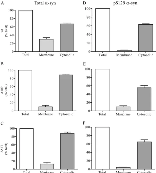
Binding of␣-syn to synaptic membranes was determined by incubating␣-syn⫺/⫺ synaptic membranes with brain cytosol prepared from mice overexpressing the human form of␣-syn (WT, A30P, or A53T) (11). The recovery of total␣-syn and Ser(P)-129␣-syn in the membrane and cytosolic fractions was compared with the starting material. Following incubation with WT␣-syn cytosol, 30 ⫾ 3% of total ␣-syn was recovered in the membrane fraction, and 67⫾ 2% remained in the cytosol (Fig. 2A). Using cytosol prepared from A30P or A53T mice, slightly lower amounts, 10⫾ 4 and 12 ⫾ 5% respectively, were recov-ered on membranes, and the corresponding 90⫾ 2 and 88 ⫾ 3% remained in the cytosol (Fig. 2, B and C). Therefore, recovered
FIGURE 1. Levels of␣-syn and Ser(P)-129 ␣-syn in synaptosomes. Intact synaptosomes were isolated from the brains of transgenic mice expressing the human␣-syn (WT, A30P, or A53T). The levels of total and Ser(P)-129 ␣-syn were visualized by Western blot in intact synaptosomes (Total), the mem-brane fraction (Memmem-brane), and cytosolic fraction (Cytosol).
at UNIV DE LIEGE-MME F PASLE, on November 7, 2011
www.jbc.org
␣-syn matched the quantity of ␣-syn in the starting material. Surprisingly, however, the net recovery of Ser(P)-129␣-syn in the same experiments was substantially less than the total input amount. Only 3⫾ 1 and 63 ⫾ 2% of Ser(P)-129 WT␣-syn were recovered in the membrane and cytosolic fractions, respec-tively (Fig. 2D), totaling 66% of initial Ser(P)-129 WT␣-syn, and suggesting a loss of 34% of Ser(P)-129 WT␣-syn. Similarly, net recovery of both A30P and A53T Ser(P)-129 ␣-syn was also reduced after binding. Cytosolic Ser(P)-129␣-syn was 55 ⫾ 5 and 65⫾ 5%, respectively, and the level recovered on the membrane was only 10⫾ 3 and 4 ⫾ 1% of the starting input amounts (Fig. 2, E and F). These data suggest net loss of phosphorylated␣-syn during the process of binding with the synaptic membrane.
Using another cell-free assay to measure␣-syn movement off membranes (30), we then investigated changes to Ser(P)-129 ␣-syn during dissociation from synaptic membranes prepared from mice overexpressing WT, A30P, or A53T␣-syn into cyto-sol prepared from␣-syn⫺/⫺mice. As illustrated in Fig. 3A, fol-lowing incubation with membranes from WT mice, 7⫾ 1% of ␣-syn dissociated to the cytosol and 92 ⫾ 2% remained
mem-brane-bound. A similar pattern was observed with membranes prepared from both A30P and A53T mice, with 10⫾ 2 and 12 ⫾ 1% found in the cytosol and 88⫾ 2 and 88 ⫾ 2% remaining membrane-bound, respectively (Fig. 3, B and C). Thus, follow-ing incubation with␣-syn⫺/⫺cytosol, a small percentage of WT, A30P, and A53T␣-syn disassociates into the cytosol, with the majority remaining membrane-bound. We then measured the disassociation of Ser(P)-129 ␣-syn from synaptic mem-branes using the same assay. As illustrated in Fig. 3D, following exposure to ␣-syn⫺/⫺ cytosol the level of membrane-bound Ser(P)-129␣-syn was reduced to 52 ⫾ 4% of starting material. However, only 2⫾ 1% of WT Ser(P)-129␣-syn was found in the cytosol. Thus,⬃46% of Ser(P)-129␣-syn was lost during the incubation. A similar loss of Ser(P)-129 was observed with both A30P and A53T ␣-syn with membrane-bound Ser(P)-129 ␣-syn reduced to 40 ⫾ 4 and 45 ⫾ 6%, respectively, and the amount recovered in the cytosol was only 5⫾ 1 and 6 ⫾ 2%, respectively (Fig. 3, E and F). These data also suggest a net loss of phosphorylated␣-syn during its dissociation from the syn-aptic membrane.
FIGURE 2. Ser(P)-129␣-syn binding to synaptic membranes. Cytosol obtained from the brains of transgenic mice expressing the human form of WT, A30P, or A53T␣-syn was incubated with ␣-syn⫺/⫺synaptic membranes for 10 min at 37 °C. The percentage of membrane-bound and cytosolic␣-syn (WT, A30P, and
A53T, A–C, respectively) and Ser(P)-129␣-syn (WT, A30P, and A53T, D–F, respectively) relative to starting material are displayed.
at UNIV DE LIEGE-MME F PASLE, on November 7, 2011
www.jbc.org
The experiments described above required the co-incuba-tion of membrane and cytosol to assess the transfer of␣-syn between fractions. To test whether the loss of the Ser(P)-129 signal is due to degradation or dephosphorylation during incu-bation, we first compared the effects of incubating membrane and cytosol separately. These control experiments demon-strated that there was no loss of either cytosolic or membrane-bound Ser(P)-129␣-syn during incubation. Following a 10-min incubation at 37 °C, the level of membrane and cytosolic Ser(P)-129 was 123⫾ 13 and 122 ⫾ 24% of starting material, respec-tively (p⬎ 0.05; n ⫽ 6) (supplemental Fig. 1A). Thus, loss of the Ser(P)-129 epitope occurred only during co-incubation of membrane and cytosol, suggesting either that components from both fractions are required for the loss of the Ser(P)-129 epitope or that dephosphorylation occurred during the transfer of ␣-syn from one fraction to another. We also assessed whether the loss of Ser(P)-129 ␣-syn could be mitigated by blockade of endogenous phosphatases in the binding reaction mixture. As shown insupplemental Fig. 1B, a serine/threonine phosphatase inhibitor mixture did not affect the recovery of Ser(P)-129␣-syn on membranes or in cytosol. Therefore, we cannot rule out that some of the Ser(P)-129␣-syn may be pref-erentially degraded when membrane and cytosol fractions are combined. Alternatively, it remains possible that the phospha-tase involved may be insensitive to the inhibitor mixture.
Does Enzymatic Dephosphorylation or Mutation of Ser-129 Affect ␣-syn Interaction with Synaptic Membranes?—The
results above suggest that although Ser(P)-129␣-syn levels can be rapidly modulated, phosphorylation does not directly con-trol␣-syn membrane interaction because Ser(P)-129 ␣-syn loss occurred during both the binding and dissociation events and Ser(P)-129␣-syn was not preferentially associated with either the membrane or cytosolic compartments. Therefore, we asked whether a change in phosphorylation state may be required prior to binding to or dissociation from synaptic membranes.
We first measured the effects of enzymatic dephosphory-lation of ␣-syn prior to binding to synaptic membranes. We preincubated cytosol prepared from mice overexpressing WT, A30P, or A53T␣-syn with calf intestinal phosphatase (CIP) to dephosphorylate proteins. We then incubated this untreated or CIP-treated cytosol with␣-syn-deficient membranes and com-pared the binding capacity of the phosphorylated and dephos-phorylated ␣-syn. As we reported previously (11), WT and A53T ␣-syn showed equivalent binding to synaptic mem-branes, and both were higher than A30P␣-syn. However, no significant difference in binding capacity was evident between each phosphorylated versus the dephosphorylated␣-syn (Fig. 4,
Aand B, p⬎ 0.05, two-way ANOVA).
Second, we assessed whether membrane distribution of ␣-syn mutants that mimic dephosphorylated (S129A) or
phos-FIGURE 3. Ser(P)-129␣-syn dissociation from synaptic membranes. Synaptic membranes isolated from the brains of transgenic mice expressing the human form of WT, A30P, or A53T␣-syn were incubated with ␣-syn⫺/⫺cytosol for 10 min at 37 °C. The percentage of membrane-bound and cytosolic␣-syn (WT, A30P, and A53T, A–C, respectively) and Ser(P)-129␣-syn (WT, A30P, and A53T, D–F, respectively) relative to starting material are displayed.
at UNIV DE LIEGE-MME F PASLE, on November 7, 2011
www.jbc.org
phorylated (S129D)␣-syn differed in SHSY5Y cells. Cells were transfected with WT, S129A, or S129D␣-syn, and following a 48-h period were fractionated, and the level of membrane-bound ␣-syn was assessed by Western blot (Fig. 4, C and D). The level of membrane binding was similar for WT (47⫾ 10%), S129A (50⫾ 9%), and S129D (50 ⫾ 7%)␣-syn (p ⬎ 0.05, one-way ANOVA). These data also support the data above that the phosphorylation state of␣-syn at Ser-129 does not influence steady-state membrane binding of␣-syn.
Does Manipulation of Phosphorylation at Ser-129 Affect the Membrane Binding of Endogenous␣-syn?—As an alternative
approach, we manipulated the Ser-129 phosphorylation state of ␣-syn acutely or chronically in NPC isolated from mice expressing WT␣-syn using inhibitors of either PLK2 or the proteasome or by overexpression of PLK2.
Treatment with the PLK2 inhibitor BI2536 (1.0M)
signifi-cantly reduced the level of Ser(P)-129␣-syn relative to GAPDH ⬃6-fold from 7.3 ⫾ 2.5 to 1.2 ⫾ 0.7% (p ⬍ 0.05, one-way ANOVA, see Fig. 5A), but it had no effect on the total amount of membrane-bound␣-syn (Fig. 5B). Conversely, exposure to the proteasome inhibitor epoxomicin (20 nM) significantly
increased the level of Ser(P)-129␣-syn ⬃50-fold from 1.3 ⫾ 0.6 to 65⫾ 18% (p ⬍ 0.05 one-way ANOVA, see Fig. 6A) but had no effect on the levels of membrane-bound ␣-syn (Fig. 6B). In addition, the epoxomicin-induced increase in Ser(P)-129␣-syn was completely blocked by BI2536, independently of the increased protein ubiquitination and with no effect on mem-brane binding of␣-syn (Fig. 6, A–C), indicating that the accu-mulation of Ser(P)-129␣-syn was dependent of PLK2-medi-ated phosphorylation. Following treatment with epoxomicin, Ser(P)-129 levels were elevated such that we could readily detect membrane-bound levels of Ser(P)-129. Of the total
Ser(P)-129␣-syn, ⬃40% was associated with the membranes (data not shown).
To study the long term effects of alterations in levels of Ser(P)-129␣-syn on membrane binding of ␣-syn, we generated NPC stably expressing His6-tagged human PLK2, which phos-phorylates␣-syn only at Ser-129 (26), either as WT or a kinase-dead version bearing a K111M mutation. Expression of the epitope-tagged protein was verified WT and K111M cells by both immunofluorescence (Fig. 7A) and Western blotting (Fig. 7C). To confirm the kinase activity of the exogenous PLK2 expression, Ser(P)-129␣-syn was assessed by immunofluores-cence in control, WT, and K111M cells (Fig. 7A) and Western blotting (Fig. 7, B and C). Compared with the empty vector controls, Ser(P)-129 ␣-syn was elevated by WT PLK2 and reduced by K111M PLK2 (p⬍ 0.01 and p ⬍ 0.05, respectively, one-way ANOVA). Nevertheless, despite the changes in Ser(P)-129, the proportion of membrane-bound␣-syn was similar for the empty vector control (48⫾ 1.4%), WT PLK2 (43 ⫾ 3.4%), and K111M PLK2 (42⫾ 2.7%) (p ⬎ 0.05, one-way ANOVA) (Fig. 7D). Thus, both stable increases and reductions in the extent of␣-syn Ser-129 phosphorylation had no effect on the membrane binding capacity of␣-syn.
Differential Effect of Ser-129 Phosphorylation on the Mem-brane Binding of PD-linked␣-syn Mutants—Our data above
indicate that manipulating Ser-129 phosphorylation has little effect on the binding properties of WT␣-syn, suggesting that Ser(P)-129 is likely not a critical, though short lived, modifica-tion during membrane binding or dissociamodifica-tion. In previous studies, we showed that both A30P and A53T␣-syn have faster membrane dissociation kinetics than WT␣-syn and that both mutants also display more transient association upon binding to synaptic membranes (11, 30). Therefore, we asked whether
FIGURE 4. Binding of dephosphorylated or phosphomimic␣-syn to synaptic membranes. A, cytosol obtained from the brains of transgenic mice express-ing the human form of WT, A30P or A53T␣-syn was incubated with or without CIP for 1 h at 37 °C. The CIP-treated cytosol was then incubated with ␣-syn⫺/⫺ synaptic membranes for 10 min at 37 °C, and the level of membrane-bound␣-syn was quantified. B, representative Western blots showing the level of total (T), cytosolic (C), and membrane-bound (M)␣-syn and Ser(P)-129 ␣-syn are shown. C, SHSY5Y cells were transfected with either WT, S129A, or S129D ␣-syn, and the level of membrane-bound␣-syn was quantified 48 h post-transfection. D, representative Western blots showing the level of membrane-bound (M) and cytosolic (C)␣-syn and syntaxin in untransfected (UT) and transfected (WT, S129A, and S129D) lysates are shown. at UNIV DE LIEGE-MME F PASLE, on November 7, 2011
www.jbc.org
Ser(P)-129 might have any differential effects on the mem-brane-binding properties of either A30P or A53T ␣-syn. Because of the relatively low endogenous levels of Ser(P)-129 ␣-syn, we used epoxomicin to increase acutely the levels of the phosphoprotein. Treatment of NPC expressing WT, A30P, or A53T␣-syn with 20 nMepoxomicin for 24 h elevated total
Ser(P)-129␣-syn levels but had no effect on total ␣-syn expres-sion (data not shown). Surprisingly, there was a striking differ-ence in the distribution of WT and mutant phosphoproteins (Fig. 8A). Although the proportion of Ser(P)-129␣-syn that was membrane-bound in WT␣-syn cells was 38 ⫾ 1.6%, it was significantly higher in A30P and A53T␣-syn cells (79 ⫾ 5.6 and 81⫾ 2.3%, respectively (both p ⬍ 0.001, one-way ANOVA)) (Fig. 8B). These results suggest that increased Ser-129 phos-phorylation may have differential effects on␣-syn depending on the presence of PD-linked mutations.
DISCUSSION
Phosphorylation of␣-syn at Ser-129 has been observed in post-mortem brain tissue from patients with PD, multiple
sys-tem atrophy, and neurodegeneration with brain iron accumu-lation type 1 (15). Under normal physiological conditions,⬃4% of␣-syn is phosphorylated at Ser-129, whereas ⬃89% of ␣-syn is phosphorylated at this residue in LB (15, 40). However, the effects of phosphorylation on the physiological and pathogenic properties of␣-syn in vivo remain unknown. In this study, we used two cell-free assays to measure changes to ␣-synphosphor-ylation during binding to or dissociation from synaptic mem-branes (11, 30). We also analyzed the effects of manipulating Ser(P)-129 ␣-syn levels by enzymatic, pharmacological, or genetic approaches to assess the impact of Ser-129 phosphory-lation on␣-syn membrane binding. Our data suggest that the phosphorylation state of␣-syn at Ser-129 is highly dynamic, but does not appear to influence the binding of WT␣-syn to mem-branes. We also compared the effects of modifying Ser(P)-129 levels in neural cells expressing PD-linked␣-syn bearing A30P or A53T mutations. When Ser-129 phosphorylation in these cells was increased by treatment with the proteasome inhibitor epoxomicin, the phosphorylated␣-syn PD mutants showed an increased association with membrane fractions as compared with WT␣-syn, suggesting the possibility that the Ser(P)-129 modification may differentially affect ␣-syn conformation depending on mutations in the amino-terminal membrane binding domain.
We initially investigated the distribution of␣-syn and Ser(P)-129 ␣-syn in brain synaptosomes prepared from transgenic mice expressing human WT, A30P, or A53T␣-syn but not the endogenous murine␣-syn. Consistent with our previous find-ings (30), we found that the relative amount of synaptosomal ␣-syn in the membrane-bound and cytosolic fractions was sim-ilar for WT, A30P and A53T␣-syn. Experiments in yeast have suggested that A30P ␣-syn has a lower membrane binding capacity than WT and A53T␣-syn (41, 42). However, we have previously shown that cytosolic factors expressed in brain, which yeast may lack, influence the membrane binding ability of recombinant ␣-syn and can indeed increase the binding capacity of A30P␣-syn (11). We also demonstrate that in syn-aptosomes, there was a lower amount of A30P Ser(P)-129 ␣-syn, as compared with WT and A53T, in both the mem-brane-bound and cytosolic fractions. Indeed, these data sup-port those of Zabrocki et al. (29) who also suggest that in yeast reduced Ser(P)-129 A30P␣-syn correlates with reduced mem-brane binding.
As the binding of␣-syn to membranes has been shown to be dynamic (30, 43), we investigated changes in the phosphoryla-tion of␣-syn at serine 129 during exchange between the mem-brane and cytosolic compartments. We found that following binding to synaptosomal membranes, there was a loss of Ser(P)-129␣-syn, for WT, A30P, and A53T. A relative loss of Ser(P)-129␣-syn was also observed during dissociation from syn-aptosomal membranes, suggesting that degradation or dephosphorylation of␣-syn may be involved in the binding and dissociation of␣-syn. Separate incubations of synaptic mem-branes or cytosol did not induce a loss of the Ser(P)-129 epitope, arguing that the loss of the phospho-epitope requires both frac-tions to mix to initiate degradation or dephosphorylation. To shed some light on a role for potential phosphatases, we also included a serine/threonine phosphatase inhibitor mixture in
FIGURE 5. Effect of reducing Ser(P)-129␣-syn on membrane binding of ␣-syn in WT NPC. WT NPC were treated with BI2536 (0.01–10 mM) for 24 h.
A, total Ser(P)-129␣-syn was quantified relative to GAPDH (*, p ⬍ 0.05 cf. 0 mM
BI2536). B, level of membrane-bound␣-syn was quantified and expressed as a percent of membrane-bound␣-syn in untreated cells. Representative West-ern blots showing the level of membrane- bound (M) and cytosolic (C)␣-syn and syntaxin are shown.
at UNIV DE LIEGE-MME F PASLE, on November 7, 2011
www.jbc.org
the binding and dissociation assays. The phosphatase inhibitor mixture failed to block the loss of the Ser(P)-129 signal, raising the possibility that Ser(P)-129␣-syn may undergo preferential degradation or that the phosphatase responsible is incom-pletely inactivated by the inhibitors.
We examined the ability of enzymatically dephosphorylated ␣-syn to interact with synaptic membranes. There was no sig-nificant difference in the membrane binding capacity of␣-syn from control cytosol or that containing no Ser(P)-129␣-syn following exposure to CIP, suggesting that phosphorylation at Ser-129 does not directly affect the ability of␣-syn to interact with synaptic membranes. This is further supported by our results that there is no difference in the membrane binding capacity of WT␣-syn or two Ser-129 mutants of ␣-syn, one with Ser-129 replaced with alanine (S129A), to prevent phos-phorylation, and one with Ser-129 replaced with aspartate (S129D), to mimic the negative electrostatic charge of Ser(P)-129. It has previously been shown that S129A␣-syn increases aggregate formation in Drosophila (14). However, more recent studies comparing the two Ser-129 mutants by adenoviral expression in rat brain had differing results. One reported that both mutants had similar aggregation profiles and toxicity (19), and the other suggested that S129A␣-syn induced more degen-eration than the S129D␣-syn, but with no significant differ-ences in the morphology of␣-syn aggregates between WT and S129A/D mutants (20). These latter findings are consistent
with our results that the membrane binding of S129A and S129D␣-syn is similar, although controversy remains regard-ing the mechanism of toxicity of the S129A and S129D␣-syn mutants.
Interestingly, we were unable to find evidence of expression of PLK2 in synaptosomal preparations nor indeed was there any effect of the polo-like kinase inhibitor BI2536 (44) on Ser(P)-129␣-syn levels in synaptosomes (data not shown). We hypoth-esized that the proteins present in synaptosomes may not rep-resent the full complement of proteins required for modeling ␣-syn phosphorylation and binding dynamics. Therefore, we made use of NPC isolated from the brains of mice expressing WT␣-syn to investigate the effects of manipulating levels of Ser(P)-129␣-syn on membrane binding. Consistent with pre-vious reports (25, 28), 1.0MBI2536 inhibited levels of
Ser(P)-129␣-syn by ⬃87% in cultured cells. This significant inhibition of Ser(P)-129␣-syn levels over a 24-h period demonstrates that the phosphorylation of␣-syn at Ser-129 is an ongoing process. However, we demonstrate that the membrane-bound ␣-syn pool in NPC was insensitive to the loss of Ser-129 phosphory-lation by BI2536. We further investigated the role of ␣-syn phosphorylation at Ser-129 in membrane binding by treating NPC with a proteasome inhibitor that has been reported to increase levels of Ser(P)-129 ␣-syn via stabilization of PLK2 levels (28). Significant increases in Ser(P)-129␣-syn levels by this method also had no effect on membrane binding over a
FIGURE 6. Effect of increasing the level of Ser(P)-129␣-syn on membrane binding of ␣-syn in WT NPC. WT ␣-syn expressing NPC were treated with BI2536 (1 mM) and/or epoxomicin (EPOX) (20 nM) for 24 h. A, level of Ser(P)-129␣-syn relative to GAPDH was quantified (left panel; *, #, and $ ⫽ p ⬍ 0.05 of EPOX-BI2536⫺, EPOX-BI2536⫹, and EPOX⫹ BI2536⫹, respectively) and a representative Western blot (right panel). B, level of membrane-bound␣-syn was quantified and expressed as a percent of membrane-bound␣-syn in untreated cells. Representative Western blots showing the level of membrane bound (M) and cytosolic (C)␣-syn and syntaxin are shown. C, representative Western blot showing the total level of ubiquitinated protein.
at UNIV DE LIEGE-MME F PASLE, on November 7, 2011
www.jbc.org
24-h period. Moreover, additional studies using NPC with sta-ble expression of either WT PLK2 or a K111M mutant with reduced kinase activity produced cell lines with significantly increased or reduced Ser(P)-129 levels, albeit without effects on ␣-syn membrane binding. Thus, it seems that the membrane binding capacity of␣-syn is independent of either acute or chronic increases or decreases in Ser-129 phosphorylation.
Although the early structures of inclusion formation are poorly understood and there remains controversy as to whether ␣-syn is phosphorylated before or after aggregation (27, 28), it is notable that the majority of aggregated␣-syn in LB is phosphor-ylated at Ser-129. Therefore, we also examined the membrane binding capacity of Ser(P)-129␣-syn itself. Because existing levels of Ser(P)-129␣-syn were very low in NPC, we took advan-tage of the fact that a short term exposure to proteasome
inhib-itors induces a substantial increase in Ser(P)-129 modification as shown in Fig. 6. Following epoxomicin treatment of NPC expressing WT, A30P, or A53T␣-syn, we were able to more clearly compare the membrane binding of WT, A30P, and A53T Ser(P)-129 ␣-syn. Interestingly, membrane binding of phosphorylated A30P and A53T␣-syn was significantly higher than for WT Ser(P)-129␣-syn. It is known that both A30P and A53T mutant forms of␣-syn have a higher propensity to aggre-gate (45, 46), and indeed these mutants have been known to be phosphorylated at Ser-129 in Lewy bodies of familial forms of PD (18). Our finding that both A53T and A30P␣-syn preferen-tially accumulate on membranes may underlie the enhanced propensity of these familial PD-related mutants to aggregate.
As phosphorylation of WT␣-syn at Ser-129 appears not to mediate its membrane binding or toxicity, per se, the following
FIGURE 7. Expression of WT and mutant PLK2 in WT NPC. WT NPC stably expressing PLK2 WT or human PLK2 K111M were cultured. A, immunocytochemistry for tetra-His (red) and Ser(P)-129␣-syn (green) was performed in empty vector, PLK2 WT or PLK2 K111M cells (panels i–iii, respectively). B, level of Ser(P)-129 ␣-syn relative to GAPDH was quantified. C, representative Western blots showing the total levels of tetra-His, Ser(P)-129 ␣-syn, and GAPDH are shown. (*, p ⬍ 0.05; **, p⬍ 0.01 cf. vector-infected cells). D, level of membrane-bound␣-syn was quantified. Representative Western blots showing the level of membrane-bound (M) and cytosolic (C)␣-syn and syntaxin are shown.
at UNIV DE LIEGE-MME F PASLE, on November 7, 2011
www.jbc.org
questions arise. Why does this residue undergo phosphoryla-tion and dephosphorylaphosphoryla-tion and why is the majority of␣-syn in LB phosphorylated at Ser-129? Given that Ser-129 is quite dis-tal to the amino-terminal membrane binding region, one pos-sible role for modification of this site may be to regulate inter-actions with binding partners on synaptic vesicles. For example, the site of binding of␣-syn to vesicular synaptobrevin-2 over-laps with the Ser-129 residue (47). Modulation of protein-pro-tein interactions by Ser-129 phosphorylation could thereby impact vesicle mobilization without a direct effect on ␣-syn membrane binding. With respect to pathological Ser(P)-129 ␣-syn accumulation, one hypothesis is that ␣-syn may be phos-phorylated after aggregation as it has a higher affinity for kinases and a lower affinity for phosphatases in this altered conformation (28). Furthermore, studies in transgenic mice and cultured cells suggest that cellular toxicity, including pro-teasomal dysfunction, increases both casein kinase 2 activity and PLK2 leading to enhanced phosphorylation of␣-syn at Ser-129 (23, 25).
In conclusion, our data demonstrate that phosphorylation of ␣-syn at Ser-129 is a continuous cellular process. Although blocking ␣-syn phosphorylation at Ser-129 or enhancing Ser(P)-129 levels, by enzymatic, pharmacological, and molecu-lar biological means, had little effect on the membrane binding of WT ␣-syn under either acute or chronic conditions, we obtain contrasting results with the pathogenic␣-syn mutants, indicating that membrane association of A30P and A53T␣-syn was differentially up-regulated by Ser-129 phosphorylation. This work raises the possibility that the relationship of Ser-129
phosphorylation to the pathogenic aggregation of WT and mutant␣-syn may be governed by distinct effects on phospho-protein accumulation on intracellular membranes.
Acknowledgment—We are grateful to John P. Anderson (Elan Phar-maceuticals) for the generous provision of antibodies recognizing Ser(P)-129␣-syn.
REFERENCES
1. Cookson, M. R., and van der Brug, M. (2008) Exp. Neurol. 209, 5–11 2. Venda, L. L., Cragg, S. J., Buchman, V. L., and Wade-Martins, R. (2010)
Trends Neurosci. 33,559 –568
3. Cookson, M. R. (2005) Annu. Rev. Biochem. 74, 29 –52
4. Chandra, S., Chen, X., Rizo, J., Jahn, R., and Su¨dhof, T. C. (2003) J. Biol.
Chem. 278,15313–15318
5. Eliezer, D., Kutluay, E., Bussell, R., Jr., and Browne, G. (2001) J. Mol. Biol.
307,1061–1073
6. Jao, C. C., Der-Sarkissian, A., Chen, J., and Langen, R. (2004) Proc. Natl.
Acad. Sci. U.S.A. 101,8331– 8336
7. Jo, E., McLaurin, J., Yip, C. M., St George-Hyslop, P., and Fraser, P. E. (2000) J. Biol. Chem. 275, 34328 –34334
8. Kubo, S., Nemani, V. M., Chalkley, R. J., Anthony, M. D., Hattori, N., Mizuno, Y., Edwards, R. H., and Fortin, D. L. (2005) J. Biol. Chem. 280, 31664 –31672
9. Jensen, P. H., Nielsen, M. S., Jakes, R., Dotti, C. G., and Goedert, M. (1998)
J. Biol. Chem. 273,26292–26294
10. Maroteaux, L., and Scheller, R. H. (1991) Brain Res. Mol. Brain Res. 11, 335–343
11. Wislet-Gendebien, S., Visanji, N. P., Whitehead, S. N., Marsilio, D., Hou, W., Figeys, D., Fraser, P. E., Bennett, S. A., and Tandon, A. (2008) BMC
Neurosci. 9,92
12. Fortin, D. L., Troyer, M. D., Nakamura, K., Kubo, S., Anthony, M. D., and Edwards, R. H. (2004) J. Neurosci. 24, 6715– 6723
13. Spillantini, M. G., Crowther, R. A., Jakes, R., Hasegawa, M., and Goedert, M. (1998) Proc. Natl. Acad. Sci. U.S.A. 95, 6469 – 6473
14. Chen, L., and Feany, M. B. (2005) Nat. Neurosci. 8, 657– 663
15. Fujiwara, H., Hasegawa, M., Dohmae, N., Kawashima, A., Masliah, E., Goldberg, M. S., Shen, J., Takio, K., and Iwatsubo, T. (2002) Nat. Cell Biol.
4,160 –164
16. Kahle, P. J., Neumann, M., Ozmen, L., Muller, V., Jacobsen, H., Spooren, W., Fuss, B., Mallon, B., Macklin, W. B., Fujiwara, H., Hasegawa, M., Iwat-subo, T., Kretzschmar, H. A., and Haass, C. (2002) EMBO Rep. 3, 583–588 17. Takahashi, M., Kanuka, H., Fujiwara, H., Koyama, A., Hasegawa, M.,
Miura, M., and Iwatsubo, T. (2003) Neurosci. Lett. 336, 155–158 18. Anderson, J. P., Walker, D. E., Goldstein, J. M., de Laat, R., Banducci, K.,
Caccavello, R. J., Barbour, R., Huang, J., Kling, K., Lee, M., Diep, L., Keim, P. S., Shen, X., Chataway, T., Schlossmacher, M. G., Seubert, P., Schenk, D., Sinha, S., Gai, W. P., and Chilcote, T. J. (2006) J. Biol. Chem. 281, 29739 –29752
19. Gorbatyuk, O. S., Li, S., Sullivan, L. F., Chen, W., Kondrikova, G., Man-fredsson, F. P., Mandel, R. J., and Muzyczka, N. (2008) Proc. Natl. Acad.
Sci. U.S.A. 105,763–768
20. McFarland, N. R., Fan, Z., Xu, K., Schwarzschild, M. A., Feany, M. B., Hyman, B. T., and McLean, P. J. (2009) J. Neuropathol. Exp. Neurol. 68, 515–524
21. Okochi, M., Walter, J., Koyama, A., Nakajo, S., Baba, M., Iwatsubo, T., Meijer, L., Kahle, P. J., and Haass, C. (2000) J. Biol. Chem. 275, 390 –397 22. Ishii, A., Nonaka, T., Taniguchi, S., Saito, T., Arai, T., Mann, D., Iwatsubo,
T., Hisanaga, S., Goedert, M., and Hasegawa, M. (2007) FEBS Lett. 581, 4711– 4717
23. Waxman, E. A., and Giasson, B. I. (2008) J. Neuropathol. Exp. Neurol. 67, 402– 416
24. Pronin, A. N., Morris, A. J., Surguchov, A., and Benovic, J. L. (2000) J. Biol.
Chem. 275,26515–26522
25. Inglis, K. J., Chereau, D., Brigham, E. F., Chiou, S. S., Scho¨bel, S., Frigon, N. L., Yu, M., Caccavello, R. J., Nelson, S., Motter, R., Wright, S., Chian, D., FIGURE 8. Membrane-binding properties of Ser(P)-129 ␣-syn in WT,
A30P, and A53T NPC. NPC (WT, A30P, and A53T) were treated with
epoxomi-cin (Epox) (20 nM) for 24 h. A, representative Western blots showing the level of membrane-bound (M) and cytosolic (C) WT, A30P, and A53T␣-syn, and the corresponding Ser(P)-129␣-syn and syntaxin following the epoxomicin treat-ment are shown. B, level of membrane-bound Ser(P)-129 was quantified by Western blot and shown as a percentage of total␣-syn. (*, p ⬍ 0.05; ***, p ⬍ 0.001 cf. control.)
at UNIV DE LIEGE-MME F PASLE, on November 7, 2011
www.jbc.org
Santiago, P., Soriano, F., Ramos, C., Powell, K., Goldstein, J. M., Babcock, M., Yednock, T., Bard, F., Basi, G. S., Sham, H., Chilcote, T. J., McCon-logue, L., Griswold-Prenner, I., and Anderson, J. P. (2009) J. Biol. Chem.
284,2598 –2602
26. Mbefo, M. K., Paleologou, K. E., Boucharaba, A., Oueslati, A., Schell, H., Fournier, M., Olschewski, D., Yin, G., Zweckstetter, M., Masliah, E., Kahle, P. J., Hirling, H., and Lashuel, H. A. (2010) J. Biol. Chem. 285, 2807–2822 27. Paleologou, K. E., Schmid, A. W., Rospigliosi, C. C., Kim, H. Y., Lamberto, G. R., Fredenburg, R. A., Lansbury, P. T., Jr., Fernandez, C. O., Eliezer, D., Zweckstetter, M., and Lashuel, H. A. (2008) J. Biol. Chem. 283, 16895–16905
28. Waxman, E. A., and Giasson, B. I. (2011) J. Neurosci. Res. 89, 231–247 29. Zabrocki, P., Bastiaens, I., Delay, C., Bammens, T., Ghillebert, R., Pellens,
K., De Virgilio, C., Van Leuven, F., and Winderickx, J. (2008) Biochim.
Biophys. Acta 1783,1767–1780
30. Wislet-Gendebien, S., D’Souza, C., Kawarai, T., St George-Hyslop, P., Westaway, D., Fraser, P., and Tandon, A. (2006) J. Biol. Chem. 281, 32148 –32155
31. Tandon, A., Bannykh, S., Kowalchyk, J. A., Banerjee, A., Martin, T. F., and Balch, W. E. (1998) Neuron 21, 147–154
32. Ray, J., and Gage, F. H. (2006) Mol. Cell. Neurosci. 31, 560 –573 33. Dyllick-Brenzinger, M., D’Souza, C. A., Dahlmann, B., Kloetzel, P. M., and
Tandon, A. (2010) Neurotox. Res. 17, 215–227
34. Burns, T. F., Fei, P., Scata, K. A., Dicker, D. T., and El-Deiry, W. S. (2003)
Mol. Cell. Biol. 23,5556 –5571
35. Lee, K. S., and Erikson, R. L. (1997) Mol. Cell. Biol. 17, 3408 –3417
36. Dull, T., Zufferey, R., Kelly, M., Mandel, R. J., Nguyen, M., Trono, D., and Naldini, L. (1998) J. Virol. 72, 8463– 8471
37. Tiscornia, G., Singer, O., and Verma, I. M. (2006) Nat. Protoc. 1, 234 –240 38. Cenciarelli, C., Budoni, M., Mercanti, D., Fernandez, E., Pallini, R., Aloe, L., Cimino, V., Maira, G., and Casalbore, P. (2006) Neurol. Res. 28, 505–512 39. Li, M., Pevny, L., Lovell-Badge, R., and Smith, A. (1998) Curr. Biol. 8,
971–974
40. Hasegawa, M., Fujiwara, H., Nonaka, T., Wakabayashi, K., Takahashi, H., Lee, V. M., Trojanowski, J. Q., Mann, D., and Iwatsubo, T. (2002) J. Biol.
Chem. 277,49071– 49076
41. Zabrocki, P., Pellens, K., Vanhelmont, T., Vandebroek, T., Griffioen, G., Wera, S., Van Leuven, F., and Winderickx, J. (2005) FEBS J. 272, 1386 –1400
42. Outeiro, T. F., and Lindquist, S. (2003) Science 302, 1772–1775 43. Fortin, D. L., Nemani, V. M., Voglmaier, S. M., Anthony, M. D., Ryan,
T. A., and Edwards, R. H. (2005) J. Neurosci. 25, 10913–10921
44. Steegmaier, M., Hoffmann, M., Baum, A., Le´na´rt, P., Petronczki, M., Krssa´k, M., Gu¨rtler, U., Garin-Chesa, P., Lieb, S., Quant, J., Grauert, M., Adolf, G. R., Kraut, N., Peters, J. M., and Rettig, W. J. (2007) Curr. Biol. 17, 316 –322
45. El-Agnaf, O. M., Jakes, R., Curran, M. D., and Wallace, A. (1998) FEBS Lett.
440,67–70
46. Li, J., Uversky, V. N., and Fink, A. L. (2001) Biochemistry 40, 11604 –11613 47. Burre´, J., Sharma, M., Tsetsenis, T., Buchman, V., Etherton, M. R., and
Su¨dhof, T. C. (2010) Science 329, 1663–1667
at UNIV DE LIEGE-MME F PASLE, on November 7, 2011
www.jbc.org