HAL Id: inserm-02266843
https://www.hal.inserm.fr/inserm-02266843
Submitted on 16 Aug 2019
HAL is a multi-disciplinary open access archive for the deposit and dissemination of sci-entific research documents, whether they are pub-lished or not. The documents may come from teaching and research institutions in France or abroad, or from public or private research centers.
L’archive ouverte pluridisciplinaire HAL, est destinée au dépôt et à la diffusion de documents scientifiques de niveau recherche, publiés ou non, émanant des établissements d’enseignement et de recherche français ou étrangers, des laboratoires publics ou privés.
wiring versus open internal fixation for surgical
treatment of unstable zygomatic bone fractures
Guillaume Giran, Arnaud Paré, Benjamin Croisé, Carine Koudougou, Jacques
Marie Mercier, Boris Laure, Pierre Corre, Helios Bertin
To cite this version:
Guillaume Giran, Arnaud Paré, Benjamin Croisé, Carine Koudougou, Jacques Marie Mercier, et al.. Radiographic evaluation of percutaneous transfacial wiring versus open internal fixation for surgical treatment of unstable zygomatic bone fractures. PLoS ONE, Public Library of Science, 2019, 14 (8), pp.e0220913. �10.1371/journal.pone.0220913�. �inserm-02266843�
Radiographic evaluation of percutaneous
transfacial wiring versus open internal fixation
for surgical treatment of unstable zygomatic
bone fractures
Guillaume GiranID1, Arnaud Pare´2,3, Benjamin Croise´2, Carine Koudougou1, Jacques
Marie Mercier1, Boris Laure2, Pierre Corre1,3, He´lios Bertin ID1,4*
1 Service de Chirurgie Maxillo-faciale et Stomatologie, Centre Hospitalier Universitaire de Nantes, Nantes,
France, 2 Service de Chirurgie Maxillo-faciale et Plastique, Centre Hospitalier Universitaire de Tours, Chambray-lès-Tours, France, 3 Laboratoire de Me´decine Re´ge´ne´ratrice et Squelette (RMeS), Faculte´ de Chirurgie Dentaire, Nantes, France, 4 Laboratoire des Sarcomes Osseux et Remodelage des Tissus Calcifie´ s (Phy.Os), Faculte´ de Me´decine, Nantes, France
*helios.bertin@chu-nantes.fr
Abstract
Introduction
The fixation of unstable zygomaticomaxillary complex (ZMC) fractures can be achieved by open reduction with rigid internal fixation (ORIF) and/or by closed reduction with percutane-ous transfacial Kirschner wire fixation (CRWF). The aim of this study was to tomographically assess the symmetry and the protrusion of the cheekbone with unstable ZMC fractures that had been treated by ORIF vs. CRWF.
Materials and methods
Sixty patients exhibiting a surgically unstable tetrapodal ZMC fracture were included in this multicenter retrospective study. The coordinates of 5 landmarks representing the zygomatic protrusion were comparatively studied on the healthy and on the broken side using preoper-ative and postoperpreoper-ative tridimensional computed tomography (CT) scans or cone beam CT.
Results
No significant difference was found in the zygomatic protrusion irrespective of the surgical technique that was used. The zygomatico-maxillary ansa was found to be the most compli-cated area to reduce, particularly in the frontal plane with both the CRWF and the ORIF technique (p1= 0.001 and p2= 0.0009, respectively). There was no difference in terms of
the level of complications, while the mean duration of the surgery was significantly less for the CRWF group.
Conclusion
With good postoperative radiographic outcomes, the CRWF can be proposed as an alterna-tive or in association with the ORIF technique for fixation of tetrapodal fractures of the ZMC. a1111111111 a1111111111 a1111111111 a1111111111 a1111111111 OPEN ACCESS
Citation: Giran G, Pare´ A, Croise´ B, Koudougou C,
Mercier JM, Laure B, et al. (2019) Radiographic evaluation of percutaneous transfacial wiring versus open internal fixation for surgical treatment of unstable zygomatic bone fractures. PLoS ONE 14(8): e0220913.https://doi.org/10.1371/journal. pone.0220913
Editor: Suresh Agarwal, Duke University School of
Medicine, UNITED STATES
Received: March 11, 2019 Accepted: July 25, 2019 Published: August 15, 2019
Copyright:© 2019 Giran et al. This is an open access article distributed under the terms of the
Creative Commons Attribution License, which permits unrestricted use, distribution, and reproduction in any medium, provided the original author and source are credited.
Data Availability Statement: All relevant data are
within the manuscript and its Supporting Information files.
Funding: The authors received no specific funding
for this work.
Competing interests: The authors have declared
Introduction
Fractures of the zygomaticomaxillary complex (ZMC) are a type of injury that is commonly encountered in maxillofacial surgery, accounting for 24% of all facial fractures [1,2]. Physical assaults, falls, traffic accidents, and sports-related injuries represent the most common etiolo-gies, accounting for 39%, 31%, 11%, and 11% of all ZMC fractures, respectively [2]. Males out-number females in most studies, with a sex ratio of 3.6/1. The mean age of the victims is between 30 and 40 years [2].
Computed tomography (CT) scan and cone beam CT (CBCT) are commonly used for imaged-based preoperative and postoperative evaluation of ZMC fractures. CT imaging with multiplanar and tridimensional (3D) reconstructions allows ZMC fractures to be defined [3]. As it is equally suitable for the examination of bones and as it involves less radiation, CBCT is replacing CT scans, with the exception of fractures of the orbital floor or when there is an asso-ciated brain injury [4].
The zygomatic bone is considered to be a tetrapod in which the four pillars can be broken (i.e., the zygomatic process of the frontal bone, the zygomatic process of the maxillary bone, the orbital rim, and the zygomatico-malar ansa). The treatment of such fracture aims to restore the normal anatomy of these pillars so as to improve the projection of the cheek and the sym-metrical shape of the orbit. Furthermore, the sphenozygomatic suture represents an important site to be reduced, particularly in comminuted high-energy fractures [5]. A conservative treat-ment with simple reduction using a transcutaneous hook (Ginestet) is commonly used for ZMC fractures with minimal displacement. Surgical fixation is indicated for ZMC fractures that involve a significant degree of dislocation [6,7]. Open reduction with rigid internal fixa-tion (ORIF) represents the standard of care to repair unstable zygomatic fractures [8–11] as the bone exposure allows for direct evaluation of the reduction. This technique necessitates various intraoral and/or cutaneous approaches (infraorbital, latero-orbital, coronal) depending on the number and the degree of dislocation [12,13]. There is currently a trend to hide the facial incisions (transconjunctival approach, upper eyelid incision) [14,15]. The osteosynthesis can be carried out by using a wire ligature, screw, or titanium or resorbable miniplate [16]. Because it represents a fast and mini-invasive approach, closed reduction with percutaneous transfacial Kirschner wire fixation (CRWF) can be proposed as an alternative for zygomatic fixation [16].
There are very few reports to date in the literature regarding the treatment of ZMC fractures using CRWF. Depending on the surgeon’s experience, the team, and the type of fracture, these different methods for fixation can be used and sometimes combined as a customized treat-ment. Nevertheless, the reasons for choosing one or the other fixation method are not clear; to our knowledge, there has been no study to date comparing these two fixation methods. The aim of this study was to tomographically assess the symmetry and the projection of the cheek-bone after unstable ZMC fractures treated either by ORIF or by CRWF.
Materials and methods
Data collection
Sixty patients presenting with a unilateral ZMC fracture at the Oral and Maxillofacial Surgery Department of Nantes University Hospital (Center 1) or at the Maxillofacial and Facial Plastic Surgery Department of Tours University Hospital (Center 2) between 2010 and 2017 were included in the study and analyzed retrospectively. All of the patients exhibited a surgically unstable tetrapodal fracture, defined as a type B fracture according to the classification of Zingg et al. [17]. Patients with a fracture of the contralateral ZMC, an isolated zygomatic arch
fracture, a ZMC comminutive fracture, or any combined mid-face fracture were excluded from the study. Patients who did not have preoperative or postoperative CT images were also excluded. The medical charts were reviewed and data documenting their date of birth, the side involved, the mechanism of the fracture, clinical findings, the type of surgery, and the duration of the surgery were compiled.
In this retrospective study, no change to the current clinical practice or randomization was performed. Due to the retrospective nature of this study, it was granted a written exemption from approval by the ethics committee of the Nantes University Hospital, according to Articles L. 1121–1 paragraph 1 and R 1121–2, paragraph 1 of the French Public Health Code.
Surgical technique
Irrespective of the surgical approach used, a close reduction with a Ginestet hook was carried out. Fixation was performed when necessary in case of unstable fractures. The stability was checked manually by applying digital pressure.
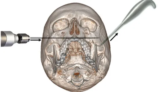
The CRWF was provided at Center 1. Using a power drill, a Kirschner wire (0.071 inches or 1.8 mm in diameter) was introduced into the corpus of the healthy contralateral zygomatic bone, then passed through the maxillary sinus, the nasal septum, and pinned to the inner cor-tex of the broken zygomatic bone (Fig 1). In a few cases, a titanium wire loop was placed on the frontozygomatic suture using a palpebral approach.
The ORIF was performed at Center 2. Depending on the displacement, a subciliary, trans-conjunctival, eyebrow/palpebral, or intraoral approach was performed to expose the infra-orbital rim as well as the frontozygomatic and zygomaticomaxillary sutures. When anatomic reduction was obtained, internal fixation was performed using various types of miniplates (e.g., a straight 5-hole plate for a frontozygomatic fracture, an L-shaped plate for a zygomatico-maxillary fracture, or an arciform plate for the inferior orbital rim).
Fig 1. Representation of the CRWF technique. The broken zygomatic bone (left cheek) is maintained reduced with a
Ginestet hook thanks to an upward pull, while the Kirschner wire is introduced into the corpus of the healthy side (right cheek) and pushed through the midfacial complex up to the broken zygomatic bone. The wire is then left for 3 weeks to allow bone consolidation.
Tomographic evaluation
Preoperative and postoperative CT scans or CBCT were comparatively analyzed. Landmarks were placed manually by the same operator (other than the surgeons), either on sagittal, coro-nal, or axial sequences using Simplant O&O software (Materialise Dental N.V., Leuven, Bel-gium). Five landmarks were drawn on 3D images to assess the ZMC projection. The four classical landmarks Zygomaxillare (Mp), Orbitale (Or), Zygotemporale inferior (Zt), and Zygomaticofrontale (FZS) were chosen to represent each pillar of the zygomatic tetrapod (Fig 2). These anatomical points of the skull base were considered to be stable in light of their use in craniometry analysis, forensic sciences, and anthropology [18]. The fifth landmark, chosen as it is reproducible, was the foramen of the zygomaticofacial nerve (Fzf), which represents the projection of the zygomatic body [19]. These landmarks are listed inTable 1.
An orthonormal coordinate system was constructed as follows (Fig 3):
1. The Z median plane passing through the midpoint of the fronto-nasal suture (MidM), the midpoint of the posterior clinoid process (MidClp), and the foramen caecum (Fc).
2. The X-plane, corresponding to the 3D reconstruction of the C1 line of Delaire cephalomet-ric analysis [20], perpendicular to the Z-plane, and passing through MidM and MidpClp. Fig 2. Representation of the five landmarks used for the cephalometric analysis of the zygomatic bone on 3D images. Fzf, foramen of the zygomaticofacial nerve; FZS, zygomaticofrontal suture; Mp, zygomaxillare point; Zt,
zygotemporale inferior; Or, orbitale; R, right side; L, left side.
https://doi.org/10.1371/journal.pone.0220913.g002
Table 1. The skeletal landmarks used for cephalometric measurements of the zygomatic bone position in 3D reconstructions of the CT scan and CBCT.
Name Definition
Foramen of the zygomaticofacial nerve Fzf Opening of the zygomaticofacial branch of the trigeminal nerve at the center of the zygomatic bone Zygomaxillare point Mp Lowest point of the zygomaticomaxillary suture
Zygotemporale inferior point Zt Lowest point of the zygomaticotemporal suture, at the top of the zygomatic tubercle Zygomaticofrontale suture FZS Cranial suture between the zygomatic bone and the frontal bone
Orbitale Or Lowest point on the inferior edge of the orbit
3. The Y-plane, representing a 3D reconstruction of the C0 line described by Nimersken for the Delaire’s cephalometric analysis [21], constructed perpendicular to Z and X, and pass-ing through MidClp.
The results obtained were the distance, expressed in mm, in an orthonormal basis with three-dimensional coordinates. Landmarks for the zygomatic projection were then studied comparatively between the broken side and the healthy side. The two fixation techniques were then compared in terms of the outcomes obtained in postoperative versus preoperative times.
Statistical analysis
In 20 randomly selected patients, the cephalometric landmarks were positioned by a different operator in order to determine concordance. The methodical error of cephalometric measure-ments was assessed by Dahlberg’s formula (mean square error (S.E2) = d2/2N, where d is the difference between the first and the second measurements and N is the number of double mea-surements) [22].
The statistical analysis was performed using GraphPad Prism 6.0 software (GraphPad Soft-ware, La Jolla, CA, U.S.A.). The parametric function of our series was tested with a Shapiro-Fig 3. Orthonormal coordinate system constructed in the X, Y, and Z planes to determine the corresponding coordinates of the landmarks. The X-plane is the horizontal plane passing through the midpoint of the fronto-nasal
suture (MidM) and of the posterior clinoid process (MidClp). The Y-plane is the vertical frontal plane passing through MidClp. The Z-plane corresponds to the vertical sagittal plane passing through MidClp, MidM, and the foramen caecum (Fc).
Wilk test. Comparative analysis of non-inferiority was carried out with a confidence interval of [-2.5; 2.5], considering that an asymmetry of 2.5 mm was the limit for an acceptable result [23]. The data were secondarily adjusted for age and gender. The quantitative data (i.e., com-parison between the healthy and the broken side, and between the preoperative and the post-operative evaluation) were analyzed using a paired t-test for paired observations and a Mann-Whitney test for non-paired values. A p-value less than 0.05 (p<0.05) was taken to be statisti-cally significant.
Results
Epidemiological data
Thirty patients presenting with a surgical ZMC fracture were reviewed and included in each center. Of the included patients, 48 (80%) were men and 12 (20%) were women. The average age at the time of the surgical procedure was 35.5± 16.4 years at Center 1 and 40.1 ± 18.9 years at Center 2 (Table 2). We noted a significant predominance of the affected side being on the left malar bone (60% versus 40% for the right side). According to the etiologies of the fractures, physical assaults represented 40% of the cases, followed by traffic accidents (22%), falls (20%), and sports-related injuries (18%).
In regard to the clinical findings, 57 patients (95%) had a lack of zygomatic protrusion that was clinically objectivized. The three other patients had a substantial edema or no clinical issues. Fifty-nine patients (98%) presented with an infraorbital nerve (V2) sensitivity disorder. For fourteen patients (23%), their ability to open their mouth was limited. Twelve patients (20%) presented an associated fracture of the orbital floor, 5 in Center 1 and 7 in Center 2, treated by surgical transconjunctival approach. These patients exhibited a binocular diplopia in 5 cases (8%), while one case of oculomotor impairment was noted. Ten patients exhibited facial wounds, preferentially affecting the eyelid, the cheek, and the eyebrows.
In the ORIF group, for 13 patients (43%), only the frontozygomatic suture and orbital rim were approached, and osteosynthesis was carried out using one miniplate in each site. The 17 other patients (57%) needed a third fracture fixation of the zygomaticomaxillary buttress. In the CRWF group, for two patients, the Kirschner wire was associated with a titanium wire loop placed on the frontozygomatic suture using a palpebral approach.
Primary outcome
The mean Dahlberg standard error for the cephalometric measurement was 0.81 mm± 0.38 mm (0.23–1.64).
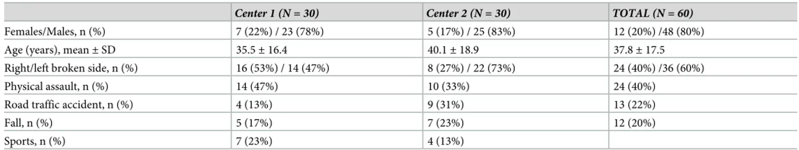
Table 2. Patient characteristics.
Center 1 (N = 30) Center 2 (N = 30) TOTAL (N = 60)
Females/Males, n (%) 7 (22%) / 23 (78%) 5 (17%) / 25 (83%) 12 (20%) /48 (80%) Age (years), mean± SD 35.5± 16.4 40.1± 18.9 37.8± 17.5 Right/left broken side, n (%) 16 (53%) / 14 (47%) 8 (27%) / 22 (73%) 24 (40%) /36 (60%) Physical assault, n (%) 14 (47%) 10 (33%) 24 (40%) Road traffic accident, n (%) 4 (13%) 9 (31%) 13 (22%) Fall, n (%) 5 (17%) 7 (23%) 12 (20%) Sports, n (%) 7 (23%) 4 (13%)
n, number of patients; SD, standard deviation.
The projection of the broken side was compared between the ORIF and the CRWF groups in the preoperative period. No significant difference in the zygomatic coordinates was observed, thus suggesting that the extent of the displacement of the fracture was the same for the two groups.
The difference in the zygomatic protrusion between the healthy and the broken sides was comparatively studied in the two surgical groups (i.e., CRWF vs. ORIF). This revealed no sig-nificant difference, irrespective of the landmark that was studied (Table 3). The Mp point was found to fluctuate the most in the Y (i.e., the MpY point) and X (i.e., the MpX point) planes between the groups, albeit with no statistical significance (mean differences of 0.76± 0.83 mm and 0.60± 0.59 mm, respectively). The FzfY, ZFSY, and OrX landmarks also varied between the two surgical groups. Adjustments for age and gender revealed no significant differences. In the patients treated with CRWF, the landmarks MpZ and MpY were significantly dis-placed postoperatively in the broken side compared to the healthy side, with mean values of 41.51 mm vs. 43.74 mm (p = 0.004) and 42.42 mm vs. 44.82 mm (p = 0.001), respectively (Fig 4).
In the patients treated with ORIF, a significant postoperative variation was found in the position of the MpY point in the broken side versus the healthy side (43.62 mm vs. 46.30 mm, p = 0.0009) (Fig 5).
Secondary endpoints
The duration of the surgery was compared between the two groups, and it was found to be sig-nificantly shorter for the CRWF group than for the ORIF group, at 28.32 min (18–45) vs. 107.7 min (32–202), respectively (p<10−4).
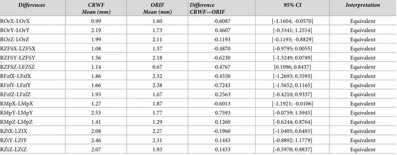
The surgical complications did not differ between the two techniques. Acute maxillary sinusitis was observed in one patient of the CRWF group, and one infection on the maxillary Table 3. Non-inferiority test, comparison of the differences for the zygomatic protrusion between the healthy and the broken sides for the two fixation techniques.
Differences CRWF Mean (mm) ORIF Mean (mm) Difference CRWF—ORIF 95% CI Interpretation ROrX-LOrX 0.99 1.60 -0.6087 [-1.1604; -0.0570] Equivalent ROrY-LOrY 2.19 1.73 0.4607 [-0.3341; 1.2554] Equivalent ROrZ-LOrZ 1.99 2.11 -0.1193 [-0.1193; -0.8829] Equivalent RZFSX-LZFSX 1.08 1.57 -0.4870 [-0.9795; 0.0055] Equivalent RZFSY-LZFSY 1.56 2.18 -0.6230 [-1.3249; 0.0789] Equivalent RZFSZ-LFZSZ 1.14 0.67 0.4767 [0.1096; 0.8437] Equivalent RFzfX-LFzfX 1.86 2.32 -0.4550 [-1.2693; 0.3593] Equivalent RFzfY-LFzfY 1.66 2.38 -0.7243 [-1.5652; 0.1165] Equivalent RFzfZ-LFzfZ 1.93 1.67 0.2563 [-0.4210; 0.9337] Equivalent RMpX-LMpX 1.27 1.87 -0.6013 [-1.1921; -0.0106] Equivalent RMpY-LMpY 2.53 1.77 0.7593 [-0.0759; 1.5945] Equivalent RMpZ-LMpZ 1.41 1.29 0.1260 [-0.6244; 0.8764] Equivalent RZtX-LZtX 2.08 2.27 -0.1960 [-1.0405; 0.6485] Equivalent RZtY-LZtY 2.46 2.31 0.1443 [-0.8892; 1.1779] Equivalent RZtZ-LZtZ 2.07 1.93 0.1433 [-0.5970; 0.8837] Equivalent Confidence Interval: [-2.5; 2.5]. For each landmark studied, R corresponds to the right side, L corresponds to the left side, X/Y/Z correlates with the dimensional coordinates. Or, orbitale point; ZFS, zygomaticofrontale suture landmark; Fzf, foramen of the zygomaticofacial nerve; Mp, zygomaxillare point; and Zt, zygotemporale inferior point.
buttress osteosynthesis was noted in a patient of the ORIF group. These infections were suc-cessfully treated using antibiotic therapy and osteosynthesis removal.
Discussion
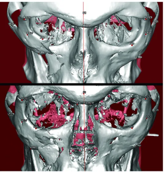
Surgical restoration of protrusion of the cheek can be a challenge with ZMC fractures. When contention is necessary, two surgical techniques can be proposed [16]. The ORIF method is presently the most commonly used technique [8]. The miniplates are widely available in most hospitals, and the current generation of facial surgeons has more experience with such proce-dures. The ORIF method allows open reduction of the different pillars of the zygomatic bone, but for some authors it results in facial scars when a subciliary/eyebrow approach is carried out [24,25]. Transconjunctival and the upper eyelid blepharoplasty incisions allow facial scars to be hidden in the surgical management of zygomatic fractures [14,15]. The Kirschner wire transmalar fixation technique is an old and well-known procedure [26–28]. It is still used by various teams, alone or in association with ORIF techniques, with good surgical outcomes [16]. However, with CRWF, removal of the K-wire under local anesthesia at least three weeks Fig 4. Three-dimensional reconstruction of the CT scan and the cone beam CT in a patient with a right tetrapodal ZMC fracture treated by the CRWF technique, preoperatively (top), and postoperatively (bottom) showing good reduction of the fracture.
after implantation is systematically required. In terms of ORIF, although some authors argue for removal of the osteosynthesis devices, there is no real proof that this is of significant benefit in adults [29,30].
Our study aimed to compare both of the ORIF and CRWF surgical techniques in terms of the zygomatic projection in patients exhibiting type B tetrapodal fractures of the ZMC. We found that, based on the postoperative tomographic assessments, the zygomatic position in the broken side was the same as in the healthy side, irrespective of the surgical technique that was used. The robustness of these findings is boosted by the fact that the preoperative images revealed the same severity of the fracture. These results are concordant with those obtained by Raoulet al. showing a high benefit/risk ratio for the patients in terms of the cost of the
materi-als, the time to perform the surgery, and the degree of scarring in a series of 216 patients treated with CRWF [16]. The K-wire fixation can also be associated with osteosynthesis of the lateral orbital rim to provide a higher level of stability of the reduced zygomatic bone [31]. Other authors have stated that most surgeons find miniplate fixation easier to use than wire osteosynthesis, while more experience is required for K-wire fixation [32]. In our study, the CRWF technique was carried out by twelve different surgeons, most of who were junior sur-geons with limited experience regarding the technique. This procedure is probably much more difficult to teach and to get experience with since the reduction and the repositioning of the bone fragment are not under visual control. The main clinical criterion to assess the quality Fig 5. Three-dimensional reconstruction of the CT scan in a patient with a right tetrapodal ZMC fracture treated by the ORIF technique (lower orbital rim and fronto-zygomatic miniplates), preoperatively (top), and postoperatively (bottom) showing good radiographic outcomes in terms of the bone reduction.
of the reduction remains the symmetry of the broken side compared to the healthy side, as intraoperative cone beam CT is not used in the study centers.
Our findings underscore that the Mp landmark, representing the zygomatico-maxillary ansa, was the most complicated area to reduce, particularly with the CRWF technique. This insufficient reduction can be explained by the occurrence of a comminuted fracture, and hence difficulty with controlling the reduction in this area. The zygomatico-maxillary ansa is in fact anatomically hidden under a soft component (the malar fat pad, the zygomaticus mus-cles, and the masseter) and does not significantly affect the esthetic and functional outcomes.
By using X-, Y-, and Z-planes with the usual anatomical cephalometric landmarks, the model used in this study is readily reproducible. The four landmarks exploited to represent the zygomatic shape are commonly used in anthropometry and anthropology sciences [18]. How-ever, the zygomaticofacial foramen has rarely been described in the literature. Although it can vary anatomically between individuals, it remains easy to process when analyzed in the same patient [19]. The low Dahlberg score found in our work suggests a high reproducibility of the landmark evaluation method. The advent of surface CBCT will help maxillofacial surgeons to measure the projection of the soft parts more objectively, in addition to enhancing the esthetic results of the surgery [33].
No difference was observed between the two techniques in terms of surgical complications, while a reduced operative time was noted with the CRWF technique. The advantages/disad-vantages of CRWF and ORIF are summarized inTable 4.
Our study suffers from some limitations: the first one is that the long-term stability was not comparatively studied for the two surgical techniques. Furthermore, it was based on retrospec-tive data, and the surgical procedures were performed at two different units, by numerous surgeons.
Our study calls for prospective and randomized controlled studies that include a larger sample population to compare osseous and soft tissue projection results for both of the CRWF and ORIF techniques in ZMC fractures.
Conclusion
ZMC fractures are very common in maxillofacial surgery, and they usually require surgical treatment. With good postoperative radiographic outcomes and a shorter operative time, the CRWF can be proposed as an alternative or in association with the ORIF technique for the fix-ation of tetrapodal fractures of the ZMC.
Table 4. Comparison of the advantages and disadvantages expected with the ORIF and CRWF surgical techniques.
Surgical technique Advantages Disadvantages
CRWF Reduced operative time Removal of the wire required under local anesthesia Minimally invasive approach No visual control of the bone reduction
No materials remain after wire removal No approach of the orbital floor Low price of the device
ORIF Visual control of the bone reduction Increased operative time Approach of the orbital floor Facial scar (subciliary approach)
Materials remain
Increased price of the device CRWF, closed reduction with wire fixation; ORIF, open reduction with internal fixation.
Supporting information
S1 Table. Coordinates values, expressed in millimeters, for each patient (from 1 to 30) before the surgical procedure in Center 1 (CRWF group). For each landmark studied, R corresponds to the right side, L corresponds to the left side, X/Y/Z correlates with the three-dimensional coordinates (X, axial plane; Y, coronal plane; Z, sagittal plane). Or, orbitale land-mark; ZFS, zygomaticofrontale suture landland-mark; Fzf, foramen of the zygomaticofacial nerve; Mp, zygomaxillare point; Zt, zygotemporale inferior point.
(DOC)
S2 Table. Coordinates values, expressed in millimeters, for each patient (from 1 to 30) before the surgical procedure in Center 2 (ORIF group). For each landmark studied, R cor-responds to the right side, L corcor-responds to the left side, X/Y/Z correlates with the three-dimensional coordinates (X, axial plane; Y, coronal plane; Z, sagittal plane). Or, orbitale land-mark; ZFS, zygomaticofrontale suture landland-mark; Fzf, foramen of the zygomaticofacial nerve; Mp, zygomaxillare point; Zt, zygotemporale inferior point.
(DOC)
S3 Table. Coordinates values, expressed in millimeters, for each patient (from 1 to 30) after the surgical procedure in Center 1 (CRWF group). For each landmark studied, R corre-sponds to the right side, L correcorre-sponds to the left side, X/Y/Z correlates with the three-dimen-sional coordinates (X, axial plane; Y, coronal plane; Z, sagittal plane). Or, orbitale landmark; ZFS, zygomaticofrontale suture landmark; Fzf, foramen of the zygomaticofacial nerve; Mp, zygomaxillare point; Zt, zygotemporale inferior point.
(DOC)
S4 Table. Coordinates values, expressed in millimeters, for each patient (from 1 to 30) after the surgical procedure in Center 2 (ORIF group). For each landmark studied, R corre-sponds to the right side, L correcorre-sponds to the left side, X/Y/Z correlates with the three-dimen-sional coordinates (X, axial plane; Y, coronal plane; Z, sagittal plane). Or, orbitale landmark; ZFS, zygomaticofrontale suture landmark; Fzf, foramen of the zygomaticofacial nerve; Mp, zygomaxillare point; Zt, zygotemporale inferior point.
(DOC)
Acknowledgments
The authors thank Mrs. Sophie Domingues for her careful reading and correction of the English version of this manuscript.
Author Contributions
Conceptualization: Guillaume Giran, Boris Laure, Pierre Corre, He´lios Bertin. Investigation: Arnaud Pare´, Benjamin Croise´, Carine Koudougou.
Methodology: Guillaume Giran, He´lios Bertin.
Writing – original draft: Guillaume Giran, He´lios Bertin.
Writing – review & editing: Jacques Marie Mercier, Boris Laure, Pierre Corre, He´lios Bertin.
References
1. Boffano P, Roccia F, Zavattero E, Dediol E, UglesˇićV, KovačičZˇ , et al. Assault-related maxillofacial injuries: the results from the European Maxillofacial Trauma (EURMAT) multicenter and prospective
collaboration. Oral Surg Oral Med Oral Pathol Oral Radiol. 2015; 119: 385–391.https://doi.org/10.1016/ j.oooo.2014.12.004PMID:25640305
2. Boffano P, Roccia F, Zavattero E, Dediol E, UglesˇićV, KovačičZˇ , et al. European Maxillofacial Trauma (EURMAT) project: a multicentre and prospective study. J Cranio-Maxillo-fac Surg Off Publ Eur Assoc Cranio-Maxillo-fac Surg. 2015; 43: 62–70.https://doi.org/10.1016/j.jcms.2014.10.011PMID:25457465
3. Furst IM, Austin P, Pharoah M, Mahoney J. The use of computed tomography to define zygomatic com-plex position. J Oral Maxillofac Surg Off J Am Assoc Oral Maxillofac Surg. 2001; 59: 647–654.https:// doi.org/10.1053/joms.2001.23394PMID:11381388
4. Shintaku WH, Venturin JS, Azevedo B, Noujeim M. Applications of cone-beam computed tomography in fractures of the maxillofacial complex. Dent Traumatol Off Publ Int Assoc Dent Traumatol. 2009; 25: 358–366.https://doi.org/10.1111/j.1600-9657.2009.00795.xPMID:19515071
5. Rohner D, Tay A, Meng CS, Hutmacher DW, Hammer B. The sphenozygomatic suture as a key site for osteosynthesis of the orbitozygomatic complex in panfacial fractures: a biomechanical study in human cadavers based on clinical practice. Plast Reconstr Surg. 2002; 110: 1463–1471; discussion 1472– 1475.https://doi.org/10.1097/01.PRS.0000029360.61857.AEPMID:12409765
6. van Hout WMMT, Van Cann EM, Abbink JH, Koole R. An epidemiological study of maxillofacial frac-tures requiring surgical treatment at a tertiary trauma centre between 2005 and 2010. Br J Oral Maxillo-fac Surg. 2013; 51: 416–420.https://doi.org/10.1016/j.bjoms.2012.11.002PMID:23218202
7. Salentijn EG, Collin JD, Boffano P, Forouzanfar T. A ten year analysis of the traumatic maxillofacial and brain injury patient in Amsterdam: complications and treatment. J Cranio-Maxillo-fac Surg Off Publ Eur Assoc Cranio-Maxillo-fac Surg. 2014; 42: 1717–1722.https://doi.org/10.1016/j.jcms.2014.06.005
PMID:25176495
8. Ellis E, Kittidumkerng W. Analysis of treatment for isolated zygomaticomaxillary complex fractures. J Oral Maxillofac Surg Off J Am Assoc Oral Maxillofac Surg. 1996; 54: 386–400; discussion 400–401.
9. Gassner R, Tuli T, Ha¨chl O, Rudisch A, Ulmer H. Cranio-maxillofacial trauma: a 10 year review of 9543 cases with 21 067 injuries. J Cranio-Maxillofac Surg. 2003; 31: 51–61. https://doi.org/10.1016/S1010-5182(02)00168-3
10. Makowski GJ, Van Sickels JE. Evaluation of results with three-point visualization of zygomaticomaxil-lary complex fractures. Oral Surg Oral Med Oral Pathol Oral Radiol Endod. 1995; 80: 624–628. PMID:
8680965
11. Mehrotra S, Anand S. Base of Skull Blocks for Open Reduction and Internal Fixation of Faciomaxillary Fractures. Ann Plast Surg. 2017; 78: 668–672.https://doi.org/10.1097/SAP.0000000000000985PMID:
28195897
12. Evans BGA, Evans GRD. MOC-PSSM CME article: Zygomatic fractures. Plast Reconstr Surg. 2008; 121: 1–11.https://doi.org/10.1097/01.prs.0000294655.16607.eaPMID:18182956
13. Zhang Q-B, Dong Y-J, Li Z-B, Zhao J-H. Coronal incision for treating zygomatic complex fractures. J Cranio-Maxillo-fac Surg Off Publ Eur Assoc Cranio-Maxillo-fac Surg. 2006; 34: 182–185.https://doi. org/10.1016/j.jcms.2005.09.004PMID:16533601
14. Zhang Q-B, Dong Y-J, Li Z-B, Zhao J-H. Minimal incisions for treating zygomatic complex fractures. J Cra-niofac Surg. 2011; 22: 1460–1462.https://doi.org/10.1097/SCS.0b013e31821d1832PMID:21772158
15. El-Anwar MW, Elsheikh E, Hussein AM, Tantawy AA, Abdelbaki YM. Transconjunctival versus subcili-ary approach to the infraorbital margin for open reduction of zygomaticomaxillsubcili-ary complex fractures: a randomized feasibility study. Oral Maxillofac Surg. 2017; 21: 187–192. https://doi.org/10.1007/s10006-017-0617-2PMID:28316023
16. Raoul G, Dujoncquoy J-P, Nicola J, Tison C, Wojcik T, Ferri J. Is transfacial Kirschner wire fixation still indicated in isolated zygomaticomaxillary complex fractures? Retrospective study of 216 cases in CHRU of Lille: epidemiology, therapeutic management, and results. J Craniofac Surg. 2009; 20: 1231– 1239.https://doi.org/10.1097/SCS.0b013e3181acde63PMID:19553833
17. Zingg M, Laedrach K, Chen J, Chowdhury K, Vuillemin T, Sutter F, et al. Classification and treatment of zygomatic fractures: a review of 1,025 cases. J Oral Maxillofac Surg Off J Am Assoc Oral Maxillofac Surg. 1992; 50: 778–790.
18. Caple J, Stephan CN. A standardized nomenclature for craniofacial and facial anthropometry. Int J Legal Med. 2016; 130: 863–879.https://doi.org/10.1007/s00414-015-1292-1PMID:26662189
19. Aksu F, Ceri NG, Arman C, Zeybek FG, Tetik S. Location and incidence of the zygomaticofacial fora-men: An anatomic study. Clin Anat N Y N. 2009; 22: 559–562.https://doi.org/10.1002/ca.20805PMID:
19418451
20. Delaire J, Schendel SA, Tulasne JF. An architectural and structural craniofacial analysis: a new lateral cephalometric analysis. Oral Surg Oral Med Oral Pathol. 1981; 52: 226–238.https://doi.org/10.1016/ 0030-4220(81)90252-8PMID:6945527
21. Nimeskern N, Mercier J-M. [The C0 line: from Delaire’s analysis of craniofacial structure to modeling it on a computer spreadsheet]. Rev Stomatol Chir Maxillofac. 2002; 103: 327–334. PMID:12538916
22. Dahlberg G. Statistical Methods for Medical and Biological Students. Br Med J. 1940; 2: 358–359.
23. Farkas LG, Cheung G. Facial asymmetry in healthy North American Caucasians. An anthropometrical study. Angle Orthod. 1981; 51: 70–77. PMID:6939355
24. Singh MK, Shekar K, Flood T, Greenstein A. Transnasal K-wire to stabilise a complex zygomatic frac-ture: a forgotten method of fixation. Ann R Coll Surg Engl. 2012; 94: e157–158.https://doi.org/10.1308/ 003588412X13171221590494PMID:22613286
25. Larsen OD, Thomsen M. Zygomatic fractures. II. A follow-up study of 137 patients. Scand J Plast Reconstr Surg. 1978; 12: 59–63. PMID:663566
26. Larsen OD, Thomsen M. Zygomatic fractures. II. A follow-up study of 137 patients. Scand J Plast Reconstr Surg. 1978; 12: 59–63. PMID:663566
27. Rohrich RJ, Watumull D. Comparison of rigid plate versus wire fixation in the management of zygoma fractures: a long-term follow-up clinical study. Plast Reconstr Surg. 1995; 96: 570–575.https://doi.org/ 10.1097/00006534-199509000-00008PMID:7638281
28. Fryer MP, Brown JB, Davis G. Internal wire-pin fixation for fracture-dislocation ofthe zygoma. Twenty-year review. Plast Reconstr Surg. 1969; 44: 576–581. https://doi.org/10.1097/00006534-196912000-00008PMID:5352923
29. Pan Z, Patil PM. Titanium osteosynthesis hardware in maxillofacial trauma surgery: to remove or remain? A retrospective study. Eur J Trauma Emerg Surg Off Publ Eur Trauma Soc. 2014; 40: 587– 591.https://doi.org/10.1007/s00068-013-0348-5PMID:26814516
30. O’Connell J, Murphy C, Ikeagwuani O, Adley C, Kearns G. The fate of titanium miniplates and screws used in maxillofacial surgery: a 10 year retrospective study. Int J Oral Maxillofac Surg. 2009; 38: 731– 735.https://doi.org/10.1016/j.ijom.2009.02.016PMID:19303742
31. Soejima K, Sakurai H, Nozaki M, Kitazawa Y, Takeuchi M, Yamaki T, et al. Semi-closed reduction of tri-pod fractures of zygoma under intraoperative assessment using ultrasonography. J Plast Reconstr Aes-thetic Surg JPRAS. 2009; 62: 499–505.
32. Gandi LN, Kattimani VS, Gupta AV, Chakravarthi VS, Meka SS. Prospective blind comparative clinical study of two point fixation of zygomatic complex fracture using wire and mini plates. Head Face Med. 2012; 8: 7.https://doi.org/10.1186/1746-160X-8-7PMID:22390884
33. Li P, Zhou ZW, Ren JY, Zhang Y, Tian WD, Tang W. Accuracy of three-dimensional facial soft tissue simulation in post-traumatic zygoma reconstruction. Int J Oral Maxillofac Surg. 2016; 45: 1665–1670.