Université de Montréal
Rôle du diaphragme au cours de l’expiration chez l’enfant sous ventilation mécanique
Par
Guillaume Emeriaud
Département de médecine sociale et préventive Faculté de Médecine
Mémoire présenté à la Faculté des études supérieures en vue de l’obtention du grade de
Maîtrise en Sciences Biomédicales (M.Sc.) Option recherche clinique biomédicale
Août 2006
V,
Direction des bibliothèques
AVIS
L’auteur a autorisé l’Université de Montréal à reproduite et diffuser, en totahté ou en partie, par quelque moyen que ce soit et sur quelque support que ce soit, et exclusivement à des fins non lucratives d’enseignement et de recherche, des copies de ce mémoire ou de cette thèse.
L’auteur et les coauteurs le cas échéant conservent la propriété du droit d’auteur et des droits moraux qui protégent ce document. Ni la thèse ou le mémoire, ni des extraits substantiels de ce document, ne doivent être imprimés ou autrement reproduits sans l’autorisation de l’auteur.
Afin de se conformer à la Loi canadienne sur la protection des renseignements personnels, quelques formulaires secondaires, coordonnées ou signatures intégrées au texte ont pu être enlevés de ce document. Bien que cela ait pu affecter la pagination, il n’y a aucun contenu manquant.
NOTICE
The author of this thesis or dissertation has granted a nonexclusive license allowing Université de Montréal to reproduce and publish the document, in part or in whole, and in any format, solely for noncommercia? educational and research purposes.
The author and co-authors if applicable retain copyright ownership and moral rights in this document. Neither the whole thesis or dissertation, nor substantial extracts from it, may be printed or otherwise reproduced without the author’s permission.
In compliance with the Canadian Privacy Act some supporting forms, contact
information or signatures may have been removed from the document. While this may affect the document page count, it does not represent any loss of content from the document.
Université de Montréal Faculté des études supérieures
Ce mémoire intitulé:
Rôle du diaphragme au cours de l’expiration chez l’enfant sous ventilation mécanique
Présenté par: Guillaume Emeriaud
A été évalué par un jury composé des personnes suivantes
Philippe Jouvet Président-rapporteur Marisa Tucci Directeur de recherche Christer A Sinderby Co-directeur de recherche Yves Berthiaume Membre du jury
Résumé
Les nouveau-nés et les nourrissons contrôlent de façon active leur volume pulmonaire de fin d’expiration, à un niveau supérieur au volume du système respiratoire au repos. Différents mécanismes ont été évoqués et notamment le freinage expiratoire par la persistance d’une activité diaphragmatique pendant l’expiration, ou activité tonique diaphragmatique. Cependant, la présence de l’activité tonique du diaphragme reste controversée et peu étudiée, et il n’existe notamment aucune étude réalisée sous ventilation assistée.
L’objectif global de notre travail est de mieux comprendre la façon dont les nourrissons contrôlent le volume pulmonaire de fin d’expiration, et comment la ventilation artificielle interagit avec ce contrôle. Nous présentons dans ce mémoire 2 articles qui reprennent les résultats de 2 études basées sur des enregistrements de l’activité électrique du diaphragme de nourrissons intubés et sous ventilation artificielle.
La 1ère étude permet de confirmer la présence du réflexe de Hering Breuer chez ces nourrissons, les cycles ventilatoires administrés par le respirateur entraînant bien une augmentation du temps expiratoire. Cette étude montre également que l’asynchronisme patient-respirateur est majeur, correspondant à plus de la moitié du temps. Dans la 2ème étude, nous avons confirmé le maintien d’une partie de l’activité diaphragmatique jusqu’en fin d’expiration. Cette activité tonique s’accroît lorsque la PEEP est supprimée, suggérant un rôle dans le maintien du volume pulmonaire de fin d’expiration.
Ces résultats permettent de mieux comprendre les interactions entre le respirateur et le nourrisson. Dans le futur, la mesure de l’activité diaphragmatique pourrait permettre d’améliorer la synchronisation patient respirateur et pourrait servir de guide pour l’adaptation du niveau de PEEP chez ces patients.
Mots-clés : nourrisson ; diaphragme ; EMG ventilation artificielle réflexe de Hering-Breuer; expiration ; PEEP.
Abstract
—key words
Infants and neonates actively control the end expiratory lung volume (EELV) above the resting lung volume. The persistence cf diaphragmatic activity during expiration (or tonic diaphragm activity) with an expiratory flow-braking action has been suggested to conttibute to the elevation of EELV. However, this tonic activity of the diaphragm remains controversial and poorly studied. To our knowledge, no studies have been conducted on this topic in mechanically ventilated infants.
The overali objective of this work is to study the roTe cf the diaphragm in the maintenance cf EELV in mechanically ventilated infants, and to evaluate the impact cf mechanical ventilation on its control. Speciflcally, we present 2 studies based on diaphragm electrical activity recorded in intubated infants under mechanical ventilation.
The first study provides evidence that prolonged delivery of assist elicits the Hering Breuer reflex, and promotes an expiratory prolongation in mechanically ventilated infants. We also observed a very important asynchrony between neural timing and the ventilatory timing in the infants studied. In the 2’ study, we observed the presence of tonic diaphragmatic activity in every patient. This tonic activity increased when PEEP was briefly suppressed, suggesting its involvement in the control of EELV.
These results provide interesting information on the interactions occurring between mechanical ventilators and infants. Future studies would be necessary to evaluate the electrical diaphragm activity as a tool te improve infant-ventitator synchrony and to assess its potential implications on the management of PEEP in intubated infants.
Key words: infant diaphragm EMG ; mechanical ventilation; Hering
Table des matières
Page de titre
Identification du Jury ii
Résumé en français iii
Mots-clés iv
Résumé et mots-clés en anglais y
Table des Matières vi
Table des figures viii
Table des tableaux ix
Abréviations x
Remerciements xi
Introduction
I
Revue de la littérature
3
Définitions 3
Contrôle du volume pulmonaire de fin d’expiration chez les
nouveau-nés et nourrissons 3
Enregistrement de l’activité électrique diaphragmatique 8
Objectifs 9
Manuscrïtl
10
Présentation et contributions des différents auteurs 10
Abstract 12
Abbreviations 13
Methods 15 Results 17 Discussion 19 References 26 Tables 36
Manuscrit 2
40
Présentation et contributions des différents auteurs 40
Abstract 42 Introduction 44 Methods 45 Results 48 Discussion 50 Conclusion 56 References 56 Figures 61 Tables 66
Discussion générale
69
Résumé des principaux résultats 69
Difficultés rencontrées et limites de l’étude 70
Implications cliniques 73
Conclusions
74
Liste des figures
Manuscrit J
Figure 1. Definition cf the different quartiles of the fleurai bteath 31 Figure 2. Tracing cf Pao with corresponding fleurai Te in one
representative subject. 32
Figure 3. EAdi—time proflie for group mean data. 33 Figure 4. Patient—ventilator interaction during mandatory breaths. 34 Figure 5. Reiationship between change in neurai Te and time that ventiiator infiation coincides with fleurai expiration. 35
Manuscrit 2
Figure 1. Definition of the periods of anaiysis for each
spontaneous breath. 61
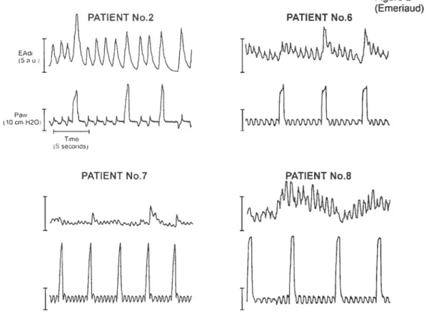
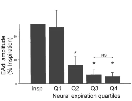
Figure 2. Tracings of diaphragm activity and airway pressure 62 Figure 3. Group mean (± SD) diaphragm eiectrical activity during
inspiration and the expiratory quartiies (n=16) 63 Figure 4. impact of ZEEP on tonic electrical activity of diaphragm (n=8).64 Figure 5. Experimentai record during prescribed PEEP, appiication of zero PEEP, and after prescribed PEEP was re-appiied. 65
Liste des tableaux
Manuscrit J
Table 1. Patient anthropometric data and admission diagnosis36
Table 2. Ventilator settings 37
Table 3. Neural timing and EAdi amplitudes for premandatory spontaneous breaths, mandatory breaths, and postmandatory spontaneous breaths38 Table 4. Factor variability expressed as coefficient of variation for the premandatory spontaneous breaths, mandatory breaths, and
postmandatory spontaneous breaths 39
Manuscrit 2
Table J. Patient anthropometrical data, ventilator settings, diagnosis, and
tonic diaphragm activity 66
Abréviations
AEdi, activité électrique du diaphragme
Paw, Pression des voies aériennes (airway pressure)
PEEP, Pression positive de fin d’expiration (positive end-expiratory pressure)
ZEEP, PEEP à zéro
VACI, Ventilation assistée contrôlée intermittente (équivalent à SIMV en anglais)
Te, Temps expiratoire Ti, Temps inspiratoire
Remerciements
Je désire remercier particulièrement Jacques Lacroix qui, par sa confiance, est à l’origine de ma venue en tant que « fellow > aux soins intensifs
pédiatriques de l’hôpital Sainte-Justine. Je n’oublierai pas son accueil chaleureux, sa passion pour la recherche et son soutien continu.
Un grand merci à Marisa Tucci, qui m’a également accueilli avec chaleur et enthousiasme. Son dynamisme dans les soins, dans la recherche comme dans la vie est un bel exemple. Et un merci particulier d’avoir continué à m’encourager et à m’aider avec toujours autant de disponibilité même de loin après mon retour en France ! Ce mémoire n’aurait pas vu le jour sans cette aide.
Jennifer Beck a contribué à une énorme part du travail présenté dans ce mémoire. Elle est à l’origine des 2 études présentées ici. Merci mille fois de m’avoir fait confiance en me permettant de participer à ce travail, tout en me transmettant une partie de votre grande expérience. J’ai beaucoup apprécié tant votre rigueur scientifique que votre passion pour ce domaine. C’était un plaisir de travailler avec vous.
Je remercie également Christer Sinderby, pour la disponibilité et la patience qu’il m’a accordées pour m’enseigner les principes de la méthode qu’il a progressivement développée et pour la confiance qu’il m’a toujours témoignée.
Je remercie aussi Norman Comtois pour son aide dans la réalisation des enregistrements et des analyses.
Je remercie enfin chaleureusement les parents et les enfants qui ont contribué à la réalisation de ce travail.
Chez le nouveau-né à terme ou prématuré, le volume pulmonaire de fin d’expiration (EELV, pour end expiratory lung volume) est maintenu de façon active à un niveau supérieur au volume de relaxation (Kosch 1984 Stark 1987). Ce dernier, représentant le volume du système respiratoire au repos, est déterminé par les propriétés mécaniques passives de ce système. Ce n’est qu’après l’âge d’un an que I’EELV correspond au volume de relaxation (Colin 1989). Durant les premiers mois, des réflexes adaptatifs doivent donc permettre d’éviter la diminution de I’EELV.
Chez le nouveau-né, différents mécanismes ont été évoqués pour expliquer le contrôle de l’EELV: le mode respiratoire avec une fréquence haute et un temps expiratoire relativement court (Griffiths 1983), le freinage du débit expiratoïre au niveau laryngé (Kosch 1988), ou au niveau diaphragmatique par la persistance d’une activité diaphragmatique pendant l’expiration, ou activité tonique diaphragmatique (Kosch 1984).
Le diaphragme est essentiellement connu comme un muscle inspiratoire actif uniquement durant l’inspiration et au repos lors de l’expiration. Cependant, il a été montré chez des nouveau-nés à terme ou prématurés que l’activité électrique du diaphragme — mesurée par des
électrodes sur la surface du thorax — semblait persister tout au long de
l’expiration, suggérant une activité « tonique» de ce muscle respiratoire (Prechtl 1977 ; Lopes 1981 ; Stark 1987; Eichenwald 1993). Les variations de cette activité tonique étant associées à des variations de I’EELV, Lopes proposa le premier l’hypothèse de l’implication de l’activité diaphragmatique durant l’expiration dans le contrôle de I’EELV chez les nouveau-nés et nourrissons (Lopes 1981). Cependant, cette hypothèse testait controversée, et en outre, aucune étude de l’activité du diaphragme au cours de l’expiration n’a été menée chez des nourrissons intubés et ventilés artificiellement.
L’objectif global des travaux présentés dans ce mémoire est donc d’étudier la façon dont les nourrissons contrôlent leur volume pulmonaire de fin d’expiration, et notamment d’évaluer quelle est la participation de l’activité diaphragmatique dans ce contrôle, et de mieux comprendre l’impact du respirateur sut ce contrôle ventilatoire.
Spécifiquement, après une revue de la littérature qui fera notamment l’état des connaissances actuelles sur le contrôle de I’EELV chez les nourrissons et nouveau-nés, nous présenterons 2 articles le premier article rapporte une étude du retentissement des cycles de ventilation administrés par le respirateur sur le comportement respiratoire des nourrissons. Cela permettra notamment de démontrer la présence et le rôle du réflexe de Hering-Breuer chez ces patients, réflexe pouvant être impliqué dans le contrôle de l’EELV. Dans le deuxième article, qui constitue l’essentiel de notre travail rapporté dans ce mémoire, nous avons étudié de façon quantitative l’activité diaphragmatique durant l’expiration, et étudié l’impact de la ventilation assistée sur cette activité tonique.
Revue de la littérature
Défïnitions
• EELV (end expiratory lung volume) Il s’agit du volume pulmonaire en fin d’expiration.
• Volume de relaxation: volume pulmonaire lorsque le système respiratoire est au repos, c’est à dire qu’il n’y a aucune activité des muscles inspiratoires ni expiratoires, et que les voies aériennes supérieures sont ouvertes.
• Nourrisson : enfant de moins de 2 ans
• Nouveau-né: ce terme s’applique aux enfants de moins de 28 jours.
Particularités du contrôle de l’EELV chez les nouveau-nés et nourrissons
• Niveau de I’EELV
Chez l’enfant plus âgé et l’adulte, le volume pulmonaire de fin d’expiration correspond généralement au volume de relaxation. A l’inverse, les nouveau-nés et les nourrissons (au moins la première année) respirent avec un niveau d’EELV relativement élevé, c’est à dire qu’en fin d’expiration, le volume pulmonaire ne redescend pas jusqu’au niveau du volume pulmonaire de relaxation.
Ce maintien d’un volume pulmonaire relativement haut a été bien démontré dans les années 80, notamment par l’équipe de Kosh et Stark au moyen d’enregistrements respiratoires pléthysmographiques (Kosch 1984; Stark 1987).
L’EELV rejoint le niveau du volume de relaxation après l’âge de 1 an chez la plupart des nourrissons, un peu plus tôt pour certains, mais toujours après 6 mois (Colin 1989).
L’importance du maintien d’un EELV au dessus du volume de relaxation à cet âge peut s’expliquer notamment par le fait que ces nouveau-nés et nourrissons ont un poumon relativement moins compliant et une cage thoracique non rigidifiée, contrairement à l’enfant plus grand. Au repos, le poumon a donc tendance à se rétracter, et cette tendance ne peut être contrée par la rigidité de la cage thoracique. Or, l’établissement puis le maintien d’un volume pulmonaire suffisant est crucial pour permettre une oxygénation optimale. Il a notamment été montré que des chutes de I’EELV (notamment lors d’apnées ou d’irrégularité respiratoire) étaient associées à des désaturations (Poets 1997 ; Stark 1987).
• Mécanismes utilisés pour le maintien de I’EELV
Plusieurs mécanismes sont mis en jeu dans le maintien d’un haut niveau d’EELV par ces nourrissons.
o Le mode respiratoire
Les nouveau-nés et les nourrissons ont une fréquence respiratoire élevée et un temps expiratoire relativement court. Un nouveau cycle inspiratoire peut donc intervenir avant une diminution trop importante du volume pulmonaire. Il est probable que ce mécanisme participe au maintien de I’EELV, comme cela a été suggéré par différentes études (Kosch 1984; Mortola 1984; Griffiths 1983). Cependant, ce mécanisme ne peut pas expliquer à lui seul le maintien de I’EELV (Griffiths 1983) et des mécanismes de freinage expiratoire sont nécessaires.
o Freinage expiratoire
La présence d’un freinage du débit expiratoire a été mise en évidence chez les nouveau-nés et les nourrissons (Kosch 1984 ; Kosch 1988 Mortola 1984). Ce freinage peut se situer à deux niveaux:
Les nouveau-nés exercent un freinage expiratoire en rétrécissant l’ouverture laryngée durant l’expiration. Kosh a notamment pu enregistrer l’activité des muscles crico-aryténoides postérieurs chez certains nouveau-nés (Kosh 1988). L’utilisation de ce freinage est augmentée en présence d’une difficulté respiratoire, ce qui peut entraîner un geignement expiratoire du nouveau-né, un des signes cliniques de la détresse respiratoire néonatale. Par contre, il est évident que ce mécanisme ne peut plus être mis en jeu après une intubation trachéale.
- Freinage expiratoire par le maintien d’une activité diaphragmatique
Le diaphragme est le principal muscle inspiratoire, et son activité
« classique» est cyclique avec une contraction durant l’inspiration et un relâchement avec un repos des fibres musculaires durant l’expiration. Cependant, plusieurs auteurs ont décrit une activité diaphragmatique persistant au cours de l’expiration chez le nouveau-né à terme ou prématuré (sans détresse respiratoire), suggérant une activité <(tonique » du diaphragme (Prechtl 1977 Lopes 1981 ; Stark 1987 ; Eichenwald 1993). Ces auteurs ont donc suggéré que la persistance d’une activité diaphragmatique au cours de l’expiration permettait un ralentissement du débit expiratoire, et donc le maintien d’un haut niveau d’EELV. Lopes a d’ailleurs établi une corrélation entre les changements d’activité tonique diaphragmatique et les changements d’EELV (Lopes 1981).
Cependant, dans ces études, l’activité tonique était généralement enregistrée au moyen d’électrodes de surface, ce qui peut laisser craindre une contamination électrique par l’activité musculaire pariétale abdominale ou thoracique.
En outre, aucune étude n’a été réalisée chez des enfants intubés et ventilés artificiellement. Or l’intubation et la ventilation assistée peuvent avoir plusieurs impacts sur le contrôle de I’EELV et sur l’activité tonique diaphragmatique. La présence de la sonde d’intubation supprime la
possibilité d’une contraction laryngée, et on peut supposer que cela rend le nourrisson plus dépendant de son activité tonique diaphragmatique et que celle-ci va donc augmenter. A l’inverse, si la sonde est de petit diamètre, elle peut entraîner des résistances expiratoires et rendre moins important le freinage diaphragmatique. La ventilation artificielle elle-même peut avoir un impact. Le niveau de pression expiratoire positive (PEEP) a un impact direct sur le niveau d’EELV (Thome 1998). Or le réglage optimal du niveau de PEEP reste un challenge important pour les intensivistes (Gentile 2004), et ce réglage est empirique. Il est donc difficile de savoir si le niveau de PEEP habituellement utilisé est suffisant (ou excessif) pour obtenir un niveau de EELV « physiologique > et donc quel est son impact sur l’activité
tonique diaphragmatique. Enfin, les cycles ventilatoires administrés par le respirateur peuvent modifier la ventilation du nourrisson, notamment par l’activation du réflexe de Hering-Breuer. L’implication de ce réflexe dans la ventilation artificielle pédiatrique n’a été que très peu étudiée.
• Importance du réflexe de Hering-Breuer chez le nouveau-né et le nourrisson
Hering et Breuer (Hering 1868) ont décrit il y a plus d’un siècle sur des animaux nouveau-nés que l’expansion pulmonaire entraînait de façon réflexe une inhibition de l’inspiration et un allongement de l’expiration.
La mise en jeu de ce réflexe a largement été retrouvée dans les études animales (Knox 1973 Sammon 1993 Clark 1972). lI a été montré que la prolongation réflexe de l’expiration pouvait être obtenue par une prolongation de l’inspiration mais aussi par des inflations pulmonaires survenant au cours de la phase expiratoire (Clark 1972; Knox 1973), sauf dans le dernier quartile de l’expiration (Knox 1973). La présence de ce réflexe a également été démontrée depuis de nombreuses années chez le nouveau-né (Cross 1960 Bodegard 1969) puis chez le nourrisson (Rabbette 1991).
La mise en jeu du réflexe de Hering-Breuer a également été décrite dans une situation inverse lorsque le volume pulmonaire de fin d’expiration est artificiellement réduit, par l’application d’une pression nasale continue ou par une distension abdominale, une augmentation de l’activité respiratoire a été observée, chez l’animal (D’Angelo 2002 Meessen 1993) puis chez l’homme adulte sain (Meessen 1994). Ces auteurs suggèrent donc l’implication de ce réflexe à la déflation (Hering Breuer deflation reflex) dans le contrôle de I’EELV.
L’étude de l’impact de ces réflexes sur la ventilation assistée est beaucoup plus limitée. La prolongation de l’expiration a été observée au décours des insufflations artificielles chez des nouveau-nés et des nourrissons (Greenough 1983 ; Giffin 1996), mais sans étude quantitative de ce réflexe.
En outre, l’ensemble des études réalisées chez le nouveau-né ou le nourrisson sont basées sur des temps inspiratoire et expitatoire mesurés de façon très indirecte, sur les courbes de pressions des voies aériennes ou oesophagiennes. Or, il a été démontré récemment que les temps respiratoires mesurés de cette façon étaient insuffisamment précis (Parthasarathy 2000). La voie efférente de ce réflexe est le nerf phrénique et il serait beaucoup plus précis de mesurer les temps respiratoires idéalement au niveau du nerf phrénique (ce qui est impossible en condition clinique) ou au niveau de l’activité électrique du diaphragme (au moyen d’un enregistrement électromyographique). Nous n’avons pas pu retrouver dans la littérature d’étude de ce réflexe chez le nouveau-né ou le nourrisson basée sur ce type d’enregistrement.
Pourtant, l’étude précise de ce réflexe chez le nourrisson sous ventilation assistée serait très importante. En effet, malgré les progrès des respirateurs artificiels, il existe fréquemment un important asynchronisme entre la ventilation spontanée des nourrissons et les cycles respiratoires
administrés par le respirateur. L’asynchronisme peut constituer un stimulus important du réflexe de Hering-Breuer, avec comme conséquence la perturbation de la respiration spontanée du patient et éventuellement l’augmentation de cet asynchronisme.
Enregistrement de l’activité électrique dïaphragmatique
Le faible nombre d’études se rapportant à l’activité tonique diaphragmatique chez le nourrisson et les controverses qui persistent à ce sujet s’expliquent en grande partie par les difficultés d’étude de l’activité électromyographique (EMG) du diaphragme dans des conditions cliniques. La plupart des auteurs utilisent des enregistrements à partir d’électrodes EMG de surface. Cependant, il existe un risque important de contamination du signal recueilli par les électrodes de surface par de l’activité ne correspondant pas au diaphragme, mais aux muscles de la paroi abdominale ou aux muscles intercostaux (Sinderby 1996). Cette contamination est d’autant plus risquée chez le nouveau-né et le nourrisson du fait de sa petite taille.
L’enregistrement par voie oesophagienne présente moins de risque de contamination par les muscles pariétaux, mais il présente également de nombreuses difficultés. La proximité du coeur engendre la nécessité d’un traitement du signal pour supprimer le signal ECG (Sinderby 1997). Surtout, le déplacement important du diaphragme au cours de la respiration (Laing 1988) rend impossible le maintien d’une distance muscle-électrode stable dans le temps si on utilise une seule paire d’électrodes, avec donc une impossibilité d’étudier quantitativement l’activité électrique de façon continue (Beck 1995 Beck 1996).
En utilisant un faisceau de 8-9 paires d’électrodes miniaturisées placées le long d’un tube naso-gasttique standard, et en développant un algorithme de filtrage puis de traitement du signal de ces électrodes (technique de double-soustraction), Sinderby et Beck et al. ont
progressivement mis au point une méthode d’enregistrement EMG peu artéfactée et surtout avec maintien quasi-constant de la distance muscle-électrode, ce qui rend possible l’étude quantitative de ce signal (Sinderby 1997 Beck 1997 Sinderby 1999 Sinderby 1995). De surcroît, cette méthode est très peu invasive et a été utilisée sans problème dans un contexte clinique. Il s’agit donc d’une méthode validée et recommandée pat les sociétés savantes (Aldrich 2002).
Objectifs
Dans les études présentées dans les 2 articles qui suivent, nous avons donc utilisé cette méthode récente et peu invasive pour étudier l’activité diaphragmatique des nourrissons sous ventilation assistée et tenter de répondre à plusieurs questions
- Le
jer
article étudie l’impact de la ventilation assistée sur le mode respiratoire spontanée des nourrissons. Spécifiquement, il étudie l’impact des cycles administrés par la machine sur la durée des temps inspiratoire et expiratoire des cycles spontanés suivants. Ceci permettra de confirmer si le réflexe de Hering-Breuer est activé par les cycles respiratoires artificiels et si l’effet de ce réflexe est prolongé à plusieurs cycles, ou porte simplement sur le cycle suivant. Un objectif secondaire est d’évaluer le synchronisme patient-ventilateur dans un mode supposé synchronisé (ventilation assistée contrôlée intermittente, ou SIMV en anglais).
- Dans le
2ème
article, notre objectif est (1) de développer une méthode standardisée pour quantifier l’activité diaphragmatique au cours de l’expiration, (2) de mesurer cette activité chez des nourrissons sous ventilation artificielle, et (3) d’étudier les variations de cette activité tonique lorsque la PEEP est supprimée, ce qui reproduit une situation où I’EELV est artificiellement réduit.
Manuscrit
I
Prolonged Neural Expiratory Time Induced by Mechanical Ventilation
in Infants
J Beck, M Tucci, G Emeriaud, J Lacroix, and C Sinderby Pediatric Research 55: 747—754, 2004
Contribution spécifique du candidat: Le candidat n’a pas participé à la conception ni à la mise en place initiale de l’étude, car il est arrivé dans cette équipe de recherche alors que l’étude venait de débuter. Il a par contre participé activement au recueil des données et des enregistrements d’activité diaphragmatique. Il a contribué à la discussion des résultats et à l’élaboration du manuscrit dans sa phase de correction.
Contribution des co-auteurs: J Beck est à l’initiative de la conception de l’étude et a réalisé la majeure partie des enregistrements. Elle a également fait l’essentiel des analyses de données ainsi que la rédaction du manuscrit. M Tucci et J Lacroix ont participé à la conception de l’étude et à l’inclusion des patients, ainsi qu’à la discussion des résultats et la correction du manuscrit. C Sinderby est à l’origine de la méthode d’enregistrement et a réalisé l’ensemble des logiciels de recueil de données, Il a supervisé la conception de l’étude ainsi que sa réalisation, et il a participé à la discussion des résultats et à l’élaboration du manuscrit.
Prolonged Neural Expiratory Time Induced by Mechanical Ventilation in Infants
Jennifer Beck, Marisa Tucci, Guillaume Emeriaud, Jacques Lacroix, And Christer Sinderby
Pediatric Intensive Gare Unit, Department cf Pediatrics and Hôpital Sainte Justine Research Center, Université de Montréal, Montreal, Quebec H3T 105 [J.B., M.T., G.E., J.L.J;
Department cf Newborn and Developmental Pediatrics, Sunnybrook and Women’s College Health Sciences Centre, Toronto, Ontarlo M5S 1B2 [JE.];
and Department cf Critical Gare Medicine, St-Michael’s Hospital, Toronto, Ontario M5B
ABSTRACT
Mechanical ventilation may interfere with the spontaneous breathing pattern in infants because they have strong reflexes that play a large role in the control cf breathing. This study aimed to answer the following questions: does a ventilator-assisted breath 1) reduce neural inspiratory time, 2) reduce the amplitude cf the diaphragm electrical activity, and 3) prolong neural expiration, within the deiivered breath? In 14 infants recovering from acute respiratory failure (mean age and weight were 2.3 ±
1.3 mc and 3.95 ± 0.82 kg, respectively), we measured 1) the electrical activity cf the diaphragm with a multiple-array esophageal electrode, and 2) airway opening pressure, while patients breathed on synchronized intermittent mandatory ventilation (mean rate, 11 .2 ± 6.5 breaths/min). We compared neural inspiratory and expiratory times for the mandatory breaths and for the spontaneous breaths immediately preceding and foliowing the mandatory breath. Aithough neural inspiratory time was flot different between mandatory and spontanecus breaths, neural expiratory time was significantly increased (p < 0.001) for the mandatory breaths (953 ± 449 ms) compared with the premandatory and postmandatory spontanecus breaths (607 ± 268 ms and 560 ± 227 ms, respectively). Delivery cf the mandatory breath resulted in a reduction in neural respiratory frequency by 28.6 ± 6.4% from the spontanecus premandatory frequency. The magnitude cf inspiratory electrical activity cf the diaphragm was similar for ail three breath conditions. For the mandatory breaths, ventilatory assist persisted for 507 ± 169 ms after the end cf neural
inspiratcry time. Infant—ventilator asynchrony (both inspiratory and expiratory asynchrony) was present in every mandatory breath and constituted 53.4 ± 26.2% cf the total breath duration.
ABBREVIATIONS
EAdi, electrical activity 0f the diaphragm HB, Hering-Breuer
Pao, airway opening pressure
SIMV, synchronized intermittent mandatory ventilation Te, expiratory time
INTRODUCTION
Newborn infants have strong reflexes that play a large role in the control of breathing (1). The work of Hering and Breuer in 1868 demonstrated in newborn animaIs that expansion of the lung reflexly inhibits inspiration— the H B inspiratoryinhibiting reflex—and promotes expiration—the HB expiratory- promoting reflex (2). With respect to mechanical ventilation in infants, t is reasonable to expect that delivery of ventilatory assistance should reflexly reduce neural Ti and neural inspiratory activity within the breath deiivered. With respect to the HB expiratory-promoting reflex, one would expect an increase in neural Te during the assisted breath,
compared with a spontaneous unassisted breath. The expiratory promoting reflex has been clinically observed in mechanically ventilated infants, but was described with indirect estimates of neural timing (such as airway or esophageal pressure or flow) to gauge its effects (3—5). In addition to the fact that no quantitative evaluation of the reflex was made, the recent work of Parthasarathy etaI. (6) has demonstrated that indirect estimates of respiratory timing are in poor agreement with more direct measurements of neural activity.
Much of the available literature on reflex control cf breathing has been derived from anesthetized healthy animais. Thete is only very limited information on reflex control of healthy awake humans, and virtually no information on reflex control in patients with lung disease undergoing ventilatory support (adult or pediatric). To our knowledge, there are no studies evaluating the influence of ventilatory assist on neural timing and neurai activity in human infants, with direct measurements of respiratory muscle activity.
With this backg round knowiedge, we asked the following questions: In infants, does a single ventilator-assisted breath 7) reduce fleuraI Ti, 2) reduce the diaphragm activation, and 3) prolong neural Te within the
breath delivered? To answer these questions, we studied the EAdi in infants who were breathing on the SIMV mode of mechanical ventilation. Neural timing and inspiratory EAdi were compared for three conditions: 1) the mandatory breaths, 2) the spontaneous breaths immediately preceding the mandatory breath (premandatory spontaneous breath), and 3) the first spontaneous breath after the mandatory breath (postmandatory
spontaneous breath). Our hypothesis was that both the inspiratory inhibiting reflex and the expiratory- promoting reflex would be elicited during the delivery of the mandatory breaths.
METHODS
Subjects. Fourteen patients (11 boys, three girls) with acute respiratory failure were studied. Their mean (±SD) age, postconceptual age, height, and weight were 2.3. ±1.3 mo, 35.8 ±4.5 weeks, 54.3 ±3.4 cm, and 3.95 ±0.82 kg, respectively (Table 1). Seven ofthe 14 patients were born premature. The ethical and scientific committees of Hôpital Sainte-Justine, Montreal, Quebec, Canada, approved the protocol. Written informed consent was obtained from the parents of aIl patients. Patients were mechanically ventilated in the SIMV mode with a relatively 10w rate (rate, 11 .2 ±6.5 breaths/min) and long inflation time (754 ±89 ms). Patients had flot received sedation for at least 6 h before the test. They were orally or nasally intubated and mechanically ventilated with a Draeger Babylog 8000 ventilator (Drager, Lubeck, Germany) with the ventilator settings as
described in Table 2. The ventilator settings had not been adjusted for 12 h before the test. The most sensitive trigger setting (“1”) was used. The mandatory breaths were delivered with a continuous flow of 10 L/min. Measurements. Electrical activity of the crural diaphragm (EAdi) was obtained using nine miniaturized electrodes mounted on a conventional nasogastric (8F) feeding tube (Benlan Inc., Oakville, Ontario, Canada) and spaced 5 mm apart. Pao was measured from a side port of the
endotracheal tube (Sensym Inc., Milpitas, CA, U.S.A.; _350 cm H20). The EAdi and Pao signais were fed into a computer for data acquisition and oniine display. Expired volumes were noted from the ventilator display in nine cf the 14 patients every minute, for both the spontaneous and the mandatory breaths. To achieve this, a stopwatch was used te monitor the time, and every minute, for the 5-min period, one cf the investigators would note the first spontaneous tidai volume dispiayed on the ventiiator monitor (on the Draeger Babylog this is dispiayed breath-by-breath), and then the first mandatory breath. The system for EAdi measurement was approved by Health Canada for investigational Testing cf Medical Devices.
Protocol. After endotracheai aspiration, the feeding tube with the eiectrodes was passed thrcugh the nose or mouth and placed such that the middie cf the array cf electrodes was Iccated at the level cf the crural diaphragm. Eiectrode pcsiticning was achieved by online feedback of the EAdi signaIs (7). Affer insertion and placement cf the catheter, the infants ccntinued te breathe en the prescribed ventiiator settings. Ten minutes after catheter placement, recordings cf EAdi and Pac were obtained for a 5-min period.
Data analysis. EAdi signais were processed with algcrithms as described by Sinderby et aI. (8—10). The root-mean-square of EAdi was calcuiated and used as a measurement cf EAdi signai strength. Timing and
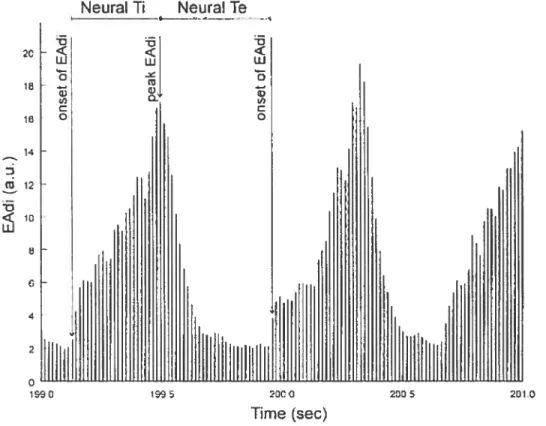
amplitudes cf EAdi were evaiuated for 1) the intermittent mandatory
breaths, 2) the spontaneous breaths immediately preceding the mandatory breath, and 3) the first spontaneous breath after the mandatory breath. Neurai Ti was arbitrariiy defined as the difference in time between the onset cf EAdi and the peak cf the inspiratory EAdi (Fig. 1). Neurai Te was arbitrariiy deflned as the difference in time between the peak cf the
inspiratory EAdi and the onset cf the subsequent inspiratory EAdi (Fig. 1). instantanecus fleurai breathing frequency was caicuiated as 60/(neurai Ti
EAdi from baseline to peak (mean EAdi) and the inspiratory change in EAdi from baseline (baseline-to-peak EAdi) were calcuiated for the three breath conditions. For the mandatory breaths, the relative change in neural Te from the premandatory breath value was expressed as the difference between mandatory breath neural Te and premandatory breath neural Te divided by the premandatory breath neural Te, then multiplied by 100. Statistical analysis. Statistical analysis was performed with commercially available software (Sigmastat, version 2.0; Jandel Scientific, San Rafael, CA, U.S.A.). Data in text and tables are presented as mean ±SD. One-way repeated-measures ANOVA was used to calculate differences in neural Ti, fleurai Te, mean EAdi, and baseline-to-peak EAdi among the three types of breaths. A p < 0.05 was considered to be significant. Linear regression
analysis was used to evaluate the relationship between changes in neural Te and the time of ventilator inflation duting neural expiration. Variability was evaluated by the coefficient of variation, expressed as percent.
RESU LTS
AIl patients tolerated the experimental protocol and insertion of the catheter. The number of breaths analyzed for the 5-min study period ranged between 6 and 25 per minute for the mandatory breaths (and postmandatory breaths), and between 8 and 48 per minute for the premandatory spontaneous breaths. Table 3 presents the neural timing and EAdi amplitude variables for the mandatory and the premandatory and postmandatory spontaneous breaths. Neural Ti was similar for the three breath conditions (p =0.502). In ail subjects, neural Te was significantly increased for the mandatory breaths (953 ±449 ms) compared with the premandatory (607 ±268 ms) and postmandatory spontaneous breaths (560 ±227 ms; p <0.001). There was no difference in neural Te between the premandatory and postmandatory spontaneous breaths. The
28.6 ±6.4% from the premandatory spontaneous neural breathing
frequency (73 ±23 breaths!min; Table 3). Figure 2 displays the tracings of EAdi and Pao for one representative subject (patient 9), and shows how delivery cf a mandatory breath causes an increase in neural Te.
With respect to inspiratory EAdi amplitude, neither mean EAdi nor baseline-to-peak EAdi was significantly different for the three breath conditions (Table 3). Figure 3 shows peak EAdi—time profiles averaged for the group during the premandatory spontaneous breaths (solid une), the mandatory breaths (dashed line), and postmandatory spontaneous breaths (dotted line). Although the diaphragm activation pattern is oversimplified in this schematic (only three points of the diaphragm activation pattern are presented), the figure indicates the similarity in inspiratory timing and inspiratory EAdi amplitude for the mandatory, the premandatory, and the postmandatory breaths, with a clear prolongation of neural Te for the mandatory breaths.
Mandatory volumes (7.33 mL/kg) were significantly greater than spontaneous volumes (3.8 mL/kg; p <0.001).
For the mandatory breaths, the mean delay between the onset of EAdi and the onset of ventilator assistance was 95 (range, 10—170 ms) ±39 ms (range, 5—110 ms). This inspiratory asynchrony (increasing diaphragm activation with no matching ventilatory assist) was equivalent to 29% of the neural Ti. A comparison of neural timing and the ventilator inflation time (which was 754 ±89 ms) for the mandatory breaths revealed that the ventilator continued to inflate the patient for 507 ±169 ms after the end of the neural Ti (i.e. during neural Te). This expiratory asynchrony period represented 53% ofthe neural Te. Taking both the inspiratory and expiratory asynchrony together, we found that asynchrony was present during every mandatory breath and constituted on average 53.4 ±26.2% of the total breath duration. Figure 4 represents these findings schematically.
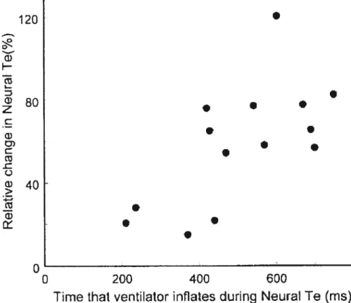
The relative increase in fleurai Te during mandatory breaths (see formula in the “Methods” section) was related to the time that the ventilator inflated during neural Te (i.e. the time that the ventilator continued to inflate after the end 0f neural Ti; i2 = 0.66; p < 0.01 Fig. 5). in contrast, the relative
increase in neural Te during mandatory breaths was not related to the extra volume delivered by the ventilator (i.e. the volume above the spontaneous volume; r2 = 0.42; p = 0.258).
The coefficients cf variation for neural Ti, neural Te, mean EAdi, and baseline-to-peak EAdi are presented in Table 4. There was no significant difference in the coefficients of variation for the premandatory spontaneous breaths, and the mandatory and postmandatory spontaneous breaths.
DISCUSSION
Summary of findings. The results of this study provide evidence for an expiratory prolongation reflex elicited during mechanical ventilation in the SIMV mode in infants. This reflex is confined to the mandatory breath, and does net carry over to the next, postmandatory spontaneous breath. The extent cf expiratory prolongation was related to the time that the ventilator delivered assist during neural expiration. Despite these findings, there was no reduction in the mandatory breath neural Ti nor was there reduction in diaphragm activity when mandatory breaths were delivered. This suggests that the anticipated inspiratory-inhibiting reflex was not elicited by SIMV. During the mandatory breaths, there was significant asynchrony between the infants neural timing and the ventilatory timing.
Critique of methods. To our knowledge, this is the first study to use EAdi for the evaluation of neural timings and diaphragm activation during lung inflation in infants. Consequently, it s important to discuss the reliability cf this technique. The EAdi signal strength is dependent on accurate
electrode positioning (11). With this technology, electrode configuration has been validated (7, 12), and changes in muscle to electrode distance are accounted for (7, 8)—this solution is of critical importance in the EAdi measurements of infants who have large posterior (caudal) dispiacement ofthe diaphragm during tidal breathing (13). The EAUi signal strength is also influenced by factors affecting signal quality, such as cardiac activity, electrode motion artifacts, and noise, technical problems that have been overcome (9, 10, 14). A frequently recurring criticism ofthe measurement of EAUi with esophageal or implanted electrodes during spontaneous breathing is that the signal strength s affected by changes in muscle length or lung volume. This critique is based on studies of evoked diaphragm compound muscle action potentials (15) or outdated
methodology that does flot control for interelectrode distance (16). Using appropriate methodology, EAdi obtained during spontaneous breathing is not artifactually influenced by changes in muscle length, chest wall configuration, or lung volume (17, 18).
The HB expiratory-promoting reflex was originally characterized in animaIs by a prolongation cf the expiratory period (apnea) after maintained lung expansion at end inspiration (2). This “end-inspiratory occlusion”
(sustained inflation) technique is still used today to evaluate the HB expiratorypromoting reflex in infants (19—21). This method has been said, however, to offset the vagally mediated effects by the hypoxic and
hypercapnic stimuli that gradually build during the ventilatory inhibition (i.e.
occlusion) (22, 23). Rapid lung inflation is an alternative approach to evaluate the expiratorypromoting reflex in infants (1), but this method has also been criticized because cf daims that the duration cf the resultant apnea was dependent on the chemical composition of the inflation gas (23). On the one hand, in the present study, the SIMV mode of mechanical ventilation provided the model of rapid lung inflation. [And in this case, we
believe that the chemical composition of the delivered gas
(e.g.
the fraction of inspired oxygen) during the rapid lung inflation (mandatory breaths) was the same as the control breath (premandatory and postmandatoryspontaneous breaths in this model.J On the other hand, because of the poor interaction between the baby’s neural bteathing pattern and the delivery of assist, the SIMV mode can aiso be considered a model ofend inspiratory occlusion (assist was being delivered during neural expiration and an elevated lung volume was maintained). In response to the critique of the method described above, we do flot believe that any hypoxic or hypercapnic stimuli could have influenced the reflex during this period of overinflation (times ranged from 210 to 750 ms).
Comparison with previous findings. In terms of the expiratory promoting reflex, studies have shown that vagal stimulation imposed
during early expiration in anesthetized animaIs prolongs the expiratory time of the perturbed cycle (24, 25). Both Clark and von Euler (26) and Knox (24) showed in animais that the responsiveness of Te prolongation was increased when pulse inflations were delivered further into expiration. It has been demonstrated that there is a timedependence to this response and that when ventilator breaths wete delivered in the last quartile of expiration, Te remained unchanged (24, 27). Our flndings of an augmentation in neural Te in ail infants (range, 14—83%) during the mandatory breaths and the fact that the relative increase in neural Te during the mandatory breath was positively correlated to the duration of ventilatory assist during neural Te (Fig. 5) is consistent with the above findings, and supports the notion that SIMV can induce a vagaily mediated expiratory-promoting reflex. A similar relationship to that described in Figure 5 was recently reported using an indirect mechanical index of muscle activation in mechanically ventilated adult patients with acute lung
injury (28), suggesting that mechanical ventilation can also elicit this reflex in aduits.
Further evidence that the reflex response in neural Te was confined to the mandatory breath was the fact that the effect did flot carry over to the postmandatory spontaneous breath neural Te, which was similar to the premandatory spontaneous breath neural Te. Although the data were flot shown, Satoh et aI. (27) also claimed that single ventilator-delivered bteaths affected the timing only of the perturbed breath, and flot the subsequent spontaneous breath.
Implications for reflex con trol of breathing pattern by lung inflation. Why should the HB expiratory-promoting reflex occur without the
inspiratory-inhibiting reflex? The HB inspiratory- inhibiting reflex is usually studied in infants by comparing inspiratory duration of a control breath (Ti measured from Pao) to the Ti of the inspiratory effort during airway
occlusion at end expiration (29, 30). With surface electromyography of the diaphragm, Witte and Carlo (31) demonstrated in tracheostomized children a clear prolongation in neural Ti (with similar rate of rise of the EAdi) for occluded efforts at end-expiratory lung volume, compared with
spontaneous, unoccluded breaths. A lengthening of the occluded effort presumably reflects the removal cf afferent signais of lung expansion. In effect, this is a loading technique (i.e. occlusion) used to examine the reflex. Mechanical ventilation, which can be considered an unloading technique, should therefote follow the reverse, i.e. that inflation by the ventilator should result in an earlier attainment cf the volume threshold for inspiratoty termination, and a reduction in neural Ti. We expected to observe this effect in our infants, as demonstrated by Younes et aI. (32) in cats during ventilatory assist. However, our data showed neither changes in neural Ti nor a decrease in EAdi amplitude during the mandatcry breaths when compared with premandatory and post mandatory spcntaneous breaths.
The delay between the onset cf fleurai Ti and the onset cf ventilatory support was 95 ms or approximateiy 29% cf neurai Ti. This delay can be explained by 1) the patients neuroventilatory coupling process (deflned as the time between the onset cf diaphragm activation te the onset cf flow generation) (10), 2) the “trigger delay,” which includes the time te
recognize an inspiratory change and send a trigger signal te the ventilator, and 3) the “system delay,” which is the time from the trigger signal until the onset cf positive pressure in the endotracheal tube (approximately 25 ms according te the manufacturers). Considering that eight cf the 14 patients had bronchiolitis and were likely hyperinflated, it could be speculated that the neuroventilatory uncoupling (transformation cf onset cf neural activity into onset cf airway fiow at the endotracheal tube) contributed to the total delay.
The delay we observed constitutes a significant inspiratory asynchrcny between the patient and the ventilatcr; cnly the last 230 ms (approximately 71%) cf neural Ti was associated with ventilator inflation. We suggest, therefore, that there was insufficient time te elicit the inspiratory-inhibiting reflex because the threshold for triggering the inspiratcry-inhibiting reflex progressively increases during inspiration (26).
This timing discrepancy may also explain why lmsand et aI. (33), in adult patients with chronic obstructive pulmonary disease, found no evidence for an inspiratcry-inhibiting reflex during mandatory breaths in SIMV, Le. no instantaneous change in neural Ti and EAdi amplitude. In that study, patients with chronic obstructive pulmcnary disease were studied and the mandatory breaths were pressure-triggered, with trigger levels set at _1 te _2 cm H20. Only two cf the five subjects studied were ventilated with applied positive end-expiratcry pressure. Therefore, there was Iikely a considerable deiay between the onset of neural activity and the onset cf delivered assïst for the mandatory breath in the work cf lmsand et aI. (33). It can be hypcthesized, therefore, that if a synchrcnized mode cf
mechanical ventilation were to be implemented (i.e. matching the initiation and termination of ventilatory assist with the patient’s neural timing), the opposite findings 0f OUC study would have been obtained, i.e. 1) elicitation of the inspiratory-inhibiting reflex, as demonstrated by Younes et aI. (32) with a phrenic nerve—controlled (synchronized) respirator, and 2) no elicitation of the expiratory-promoting reflex (if the ventilator is cycled off neurally).
Further evidence for a conttast between the two components of the HB inflation reflex (i.e. the absence of the inspiratoryinhibiting reflex and presence of the expiratory-prolonging reflex) has been observed in animal studies. Clark and von Euler (26) found that subthreshold inflations, which were able to prolong expiration, were without effect during inspiration. In the present study, however, we obtained volume measurements from the ventilator ifl fine patients. Despite significantly greater volumes for the mandatory breaths (compared with the premandatory spontaneous breaths), we can only speculate that these greater volumes were not sufficiently timed with the patient’s neural inspiration to elicit the
inspiratoty-inhibiting reflex, but could still elicit the expiratory-promoting reflex. In summary, on the basis ofthe literature and the presentfindings, it still remains to be shown that the HB inspiratory-inhibiting reflex is elicited by mechanical ventilation in infants.
Clinical implications. Although flot frequently used in adults (34), SIMV is stili frequently used as a tool for providing support and for weaning from mechanical ventilation in the infant setting (35). It was originally thought that delivery of the mandatory breath would reflexly deactivate the
inspiratory muscles, and that the SIMV mode would allow the combination
0f unassisted breathing with respiratory muscle rest (36). The present
study shows in infants, similar to the findings of Imsand et aI. (33) in adults, that there s no reflex deactivation of the diaphragm during the delivery of the mandatory breaths. Howevet, neither the study of Imsand et aI. (33)
nor the present study used modes of mechanical ventilation in which patient and ventilator timings were matched.
In the present work, infant—ventilator asynchtony was present in every mandatory breath and consisted of 53% of the total bteath duration (inspiratory and expiratory asynchrony combined). Most of the infant— ventilator asynchrony was associated with expiratory asynchrony, in which the ventilator continued to deliver assist despite the fact that diaphragm activation was decreasing. According to the literature, there are severe consequences of expiratory asynchrony, the most important being that active exhalation against positive-pressure inflation develops
pneumothoraces (37), barotrauma (3, 38), and cerebral blood flow fluctuations, which can be associated with intraventricular hemorrhage (39). In extreme cases of expiratory asynchrony, use cf sedation or muscle paralysis is sometimes necessary (38, 40, 41). In the adult literature, it 15 clear that poor off-cycling of mechanical ventilation resuits in an increased work of breathing (42) and patient discomfort (43). The present work therefore highlights the importance of oniine monitoring of diaphragm activation during mechanical ventilation, particuiarly with respect to the adjustment cf cycling off ventilatory assist.
Variability in breathing pattern. There are no reports in the literature concerning the variabiIity cf timing and amplitude of EAdi in mechanically ventilated infants. Te and Ti measured via flow signais in nonmechanicaliy ventilated preterm infants indicate a iarget variabiiity in Te (81%) than Ti
(25%) (44). Dur data show that there was no significant difference in the coefficients cf variation for fleurai Ti and neural Te. Moreover, there were no significant differences cf variability in ail measurements for the three breath conditions (premandatory spcntaneous breaths, mandatory breaths, and postmandatory spontaneous breaths). On the basis 0f the observation of a high variability in diaphragm activation variables, in addition to the instantanecus changes in breathing pattern reflexly caused by
overinflation, one could consider the importance cf recently developed modes cf mechanical ventilation that adapt on a breath-to-breath basis to changes in respiratory drive (e.g. proportional assist ventilation and neurally adjusted ventilatory assist) (45).
Acknowledgments. The authors thank M. Norman Comtois, M. Syivain Morneau, R.T., and Mile Roxanne Trahan, R.N., for their help during the studies. We also thank Dt. Jim Duffin for his comments on the manuscript.
REF ERE N C ES
1. Cross KW, Klaus M, Tooley WH, Weisser K 1960 The response cf the new-born baby to inflation ofthe lungs. J Physiol (Lond) 151:551—565 2. Hering E, Breuer J 1868 Die Selbststeurung der athmung durch den nervus vagus. Sitzber Deut Akad Wiss Wein 57:672—677
3. Greenough A, Morley C, Davis J 1983 Interaction cf spontanecus respiration with artificial ventilation in preterm babies. J Pediatr 103:769— 773
4. Giffin F, Greenough A, Naik 5 1996 The Hering-Breuer reflex in ventilated chiidren. Respir Med 90:463—466
5. Carlo WA, Ambalavanan N 1999 Conventional mechanical ventilation: traditional and new strategies. Pediatr Rev 20:e117—e126
6. Parthasarathy S, Jubran A, Tobin MJ 2000 Assessment cf neural
inspiratory time in ventilator-supported patients. Am J Respir Crit Care Med 162:546—552
7. Beck J, Sinderby C, Lindstrôm L, Grassino A 1996 Influence cf bipolar esophageal electrode positioning on measurements cf human crural diaphragm EMG. J Appi Physiol 81:1434—1449
8. Sinderby C, Beck J, Lindstrom L, Grassino A 1997 Enhancement of signal quality in esophageal recordings of diaphragm EMG. J Appi Physiol 82:1370—1377
9. Sinderby C, Beck J, Weinberg J, Spahija J, Grassino A 1998 Voluntary activation of the human diaphragm in health and disease. J Appi Physiol 85:2146—2158
10. Sinderby C, Navalesi P, Beck J, Skrobik Y, Comtois N, Friberg S, Gottfried SB, Lindstrôm L 1999 Neural control cf mechanical ventilation in respiratory failure. Nat Med 5:1433—1 436
11. Beck J, Sinderby C, Weinberg J, Grassino A 1995 Effects cf muscle-to electrode distance on the human diaphragm electromyogram. J AppI Physiol 79:975—985
12. Sinderby CA, Comtois AS, Thomson RG, Grassino AE 1996 Influence of the bipolar electrode transfer function on the electromyogram power spectrum. Muscle Nerve 19:290—301
13. Laing lA, Teele RL, Stark AR 1988 Diaphragmatic movement in newborn infants.] Pediatr 112:638—643
14. Sinderby C, Lindstrom L, Grassino AE 1995 Automatic assessment of electromyogram quality. J AppI Physiol 79:1803—1815
15. Gandevia SC, McKenzie DK 1986 Human diaphragmatic EMG: changes with lung volume and posture during supramaximal phrenic stimulation. J AppI Physiol 60:1420—1 428
16. Brancatisano A, Kelly SM, Tully A, Loring 5H, Engel LA 1989 Postural changes in spontaneous and evoked regional diaphragmatic activity in dogs. J AppI Physiol 66:1699—1 705
17. BeckJ, Sinderby C, Lindstr5m L, Grassino A 1998 Effects of lung volume on diaphragm EMG signal strength during voluntary contractions. J AppI Physiol 85:1123—1134
18. Sinderby C, Lindstrôm L, Comtois N, Grassino AE 1996 Effects of diaphragm shortening on the mean action potential conduction velocity. J Physiol (Lond) 490:207—214
19. Brown K, Stocks J, Aun C, Rabette PS 1998 The Hering-Breuer reflex in anesthetized infants: end-inspiratory vs. end-expiratory occlusion techniques. J AppI Physiol 84:1437—1446
20. Rabbette PS, Stocks J 1998 Influence cf volume dependency and timing cf airway occlusions on the Hering-Breuer reflex in infants. J AppI Physiol 85:2033—2039
21. Hassan A, Gossage J, Ingram D, Lee S, Milner D 2001 Volume of activation cf the Hering-Breuer inflation reflex in the newborn infant. J AppI Physiol 90:763—769
22. Merazzi D, Mortola J 1999 Hering-Breuer reflex in conscicus newborn rats: effects cf changes in ambient temperature during hypoxia. J AppI Physiol 87:1656—1 661
23. Younes M, Vaillancourt P, Milic-Emili J 1974 Interaction between chemical factors and duration cf apnea following lung inflation. J AppI Physicl 36:190—201
24. Knox 0K 1973 Characteristics cf inflation and deflation reflexes during expiration in cats. J Neurophysiol 36:284—295
25. Sammon M, Romaniuk JR, Bruce EN 1993 Bifurcations cf the
respiratcry pattern produced with phasic vagal stimulation in the rat. J AppI Physicl 75:912—926
26. Clark FJ, von Euler C 1972 On the regulation cf depth and rate cf breathing. J Physiol (Lond) 222:267—295
27. Satch M, Eastwocd PR, Smith CA, Dempsey JA 2001 Nonchemical elimination cf inspiratory motor output via mechanical ventilation in sleep. Am J Respir Crit Care Med 163:1356—1 364
28. Kondili E, Prinianakis G, Anastasaki M, Georgopoulos D 2001 Acute effects of ventilator settings on respiratory motor output in patients with acute lung injury. Intensive Care Med 27:1147—1157
29. Bryan AC, Bryan MH, Kirkpatrick SM, KniIl RL 1976 The use ofairway occlusion in infants. Chest 70:142—145
30. Olinsky A, Bryan MH, Bryan AC 1974 Influence of lung inflation on respiratory control in neonates. J Appi Physiol 36:426—429
31. Witte MK, Carlo W 1987 Prolongation of inspiration during lower airway occlusion in chiidren. J AppI Physiol 62:1860—1 864
32. Younes M, Baker JP, Polachek J, Remmers JE 1978 Termination of inspiration through graded inhibition of inspiratory activity. Adv Exp Med 99:383—395
33. Imsand C, Feihi F, Perret C, Fitting JW 1994 Regulation of inspiratory neuromuscular output during synchronized intermittent mechanical ventilation. Anesthesiology 80:13—22
34. Esteban A, Anzueto A, Alia I, Gordo F, Apezteguia C, Palizas F, Cide D, Goldwaser R, Soto L, Bugedo G, Rodrigo C, Pimentel J, Raimondi G, Tobin MJ 2000 How is mechanical ventilation employed in the intensive care unit? Am J RespirCritCare Med 161:1450—1458
35. Imanaka H, Nishimura M, Miyano H, Uemura H, Yagihara T 2001 Effect of synchronized intermittent mandatory ventilation on respiratory workload in infants after cardiac surgery. Anesthesiology 95:881—888
36. Sassoon C 1994 Intermittent mandatory ventilation. In: Tobin MJ (eU) Principles and Practice of Mechanical Ventilation. McGraw Hill, New York, pp 221—238
37. Greenough A, Milner AD, Dimitriou G 2001 Synchronized mechanical ventilation for respiratory support in newborn infants. Cochtane Database Syst Rev 1 :CD000456
38. Greenough A, Wood S, Morley CJ, Davis JA 1984 Pancuronium prevents pneumothoraces in ventilated premature babies who actively expire against positive pressure ventilation. Lancet 1:1—13
39. Lipscomb AP, Thorburn RJ, Reynolds EO, Blackwell RJ, StewartAL, Blackwell RJ, Cusick G, Whitehead MD 1981 Pneumothorax and cerebral haemorrhage in preterm infants. Lancet 1:414—416
40. Henry GW, Stevens CS, Schreiner RL, Grosfeld JL, Ballantine TV 1979 Respiratory paralysis to improve oxygenation and mortality in large
newborn infants with respiratory distress. J Pediatr Surg 14:761—766 41. StarkAR, Bascom R, Frantz ID 1978 Muscle relaxation in mechanically ventilated infants. J Pediatt 94:439—443
42. Tokioka H, Tanaka T, Ishizu T, Fukushima T, Iwaki T, Nakamura Y, Kosogabe Y 2001 The effect of breath termination criterion on breathing patterns and the work of breathing during pressure support ventilation. Anesth Analg 92:161—1 65
43. Calderini E, Confalonieri M, Puccio PG, Francavilla N, Stella L, Gregoretti C 1999 Patient-ventilator asynchrony during noninvasive ventilation: the role of expiratory trigger. Intensive Care Med 25:662—667 44. Al-Hathlol K, Idiong N, Hussain A, Kwiatkowski K, Alvaro RE,
Weintraub Z, Cares DB, Rigatto H 2000 A study of breathing pattern and ventilation in newborn infants and aduit subjects. Acta Paediatr 89:1420— 1425
45. Navalesi P, Costa R 2003 New modes of mechanical ventilation: proportional assist ventilation, neurally adjusted ventilatory assist, and fractal ventilation. Curr Opin Crit Care 9:51—58
Figure 1. Neural Ti was arbitrarily defined as the difference in time
between the onset cf EAUi and the peak cf the inspiratory EAUi. Neural Te was arbitrarily defined as the difference in time between the peak cf the inspiratory EAdi and the onset cf the subsequent inspiratory EAUi.
2C -t, 4: w I. o w o 16 Neural Ti Neurarre 4: w o o 16 14 12 10 4 o 2000 2005 2010 Tirne (sec)
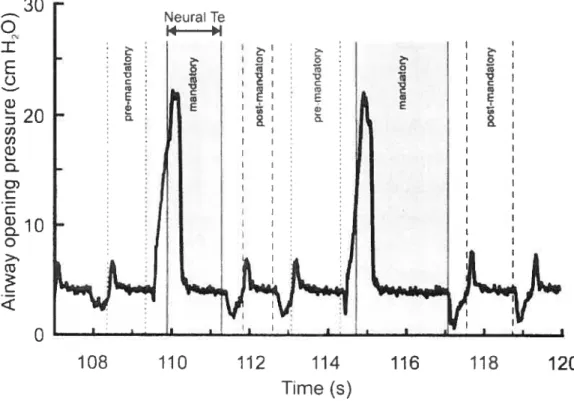
Figure 2. Tracing of Pao with corresponding fleurai Te in one
representative subject. Pao, with shaded areas indicating corresponding fleurai Te period. Premandatory spontaneous breath neural Te is shaded light grayand bound by small dashed lines. Mandatory breath fleurai Te is shaded dark gray and bound by solid unes. Postmandatory spontaneous breath is shaded light gray and bound by long dashed lines. This example shows in one subject (no. 9) the increase in fleurai Te for the mandatory breaths compared with the neurai Te for the premandatory and
postmandatory spontaneous breaths.
30 Neura Te
q
14 I IE
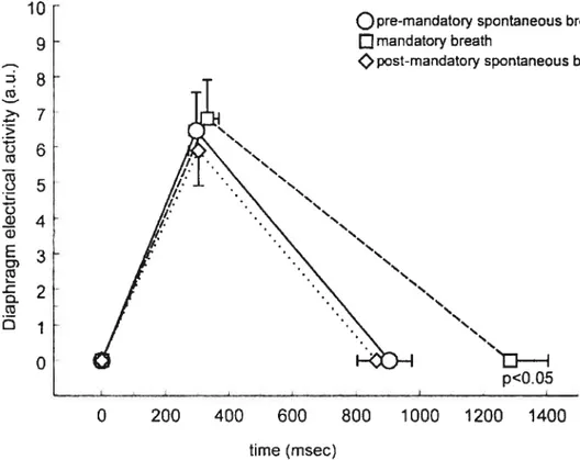
L) C ‘I) o 0 C E o C 108 110 112 114 116 118 120 Time (s)Figure 3. EAdi—time profile for group mean data. Data are presented for group mean values ±SEM. There was no significant difference in neural timing and EAdi amplitude on inspiration for the premandatory
spontaneous breaths (solid une), mandatory breaths (dashed une), or postmandatory spontaneous breaths (dotted une). Neural Te was
prolonged significantly for the mandatory breaths (dashed une). Note that this s flot a representation of the shape of the EAUi recruitment pattern, but simply a representation of three points: the onset cf EAUi, the peak of EAdi, and the onset of the EAdi for the subsequent breath.
10 9 ct > cl) E O. Dj O
Q
premandatory spontaneous breathQmandatory breath
post-mandatory spontaneous breath
p<O.0
0 200 400 600 800 1000 1200 1400
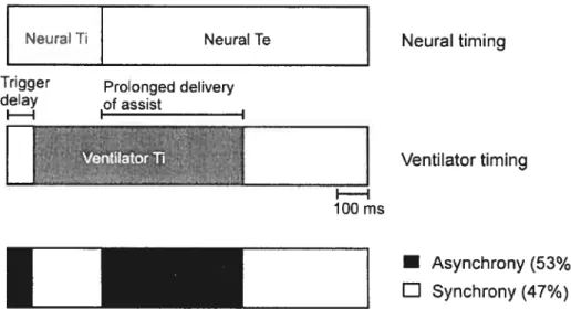
Figure 4. Patient—ventilator interaction during mandatory breaths.
Schematic representation of patient fleurai timing (upper bar) and ventiiator timing (middle bar) during mandatory breaths. Upper bar, neurai Ti (gray area) and fleurai Te (white) for the group data are presented. MidUle bar, periods describing ventiiator timing are dispiayed, including trigger deiay and ventilator Ti. Bottom bar, periods 0f infant—ventiiator synchrony (white) and asynchrony (black).
Neur&T NeuralTe
Trigger Prolonged delivery
delay of assist II I
D
LEEI
•
Asynchrony (53%) L1 Synchrony (47%) Neurai timing Ventilator timing 100 msLE
Figure 5. Relationship between change in neural Te and time that ventilator inflation coincides with neural expiration. Forthe 14 patients studied, figure shows relationship between the relative change in neural Te for the mandatory breaths (from the neural Te cf the preceding
spontaneous breath; y axis) and the time that the ventilator delivers pressure during the neural Te period (x axis).
120 H 180
j4o
eo
O 200 400 600D
36 Th1e 1. ?irr€w irhipn’rrc dQTI md dwi:son Pt:u (me) i M 4 23 5 2 M 2 39 57 S f 1 35 5C 4 M 49 53 5 f 4 35 6 M 4 29 7 M 29 5 S 2.1 05 49 54 3 M 2 33 51 0 f 05 43 5$ 11 M 2 3_ 55 12 M 13 55 13 M 3 35 55 1-M 43 5$D
Ib1e Z T’?r5îarer sr’ 2111V rare ?EE?PI? FtO. Me,ued Per treaù. riz I 13 535 2 13 333 33 3 10 321 35 10 425 55 5 13 433 0 S 522 30 ‘ 15 629 24 2 13 411 20 9 11 517 30 10 5 511 15 lI 13 414 30 11 13 21 30 13 32S 15 1-5 535 25 e0 P130 peal. u pIn:.:v pr;ur. PEEP. pcsune end_expuatr peue 90o. 5:tïc.:D
37M M VI
11H
H-4 H- Ii-H j M HD
I)I)!e 3. Frr flr c.c : ;r:cn: Cfcieiu ?:eL1dltcr:eat! Nr1 Ti INrITe EA3i L:a
.-L:: it:d Eda mii f uipirr:: lelaie peI; f -intc.r
Manuscrit 2
Diaphragm Electrical Activity During Expiration in Mechanically Ventilated Infants
G Emeriaud, J Beck, M Tucci, J Lacroix, and C Sinderby Pediatric Research 59: 705—710, 2006
Contribution spécifique du candidat: Le candidat a participé à la conception et à la mise en place de l’étude. Il a effectué tout le recueil des données cliniques et a participé à tous les enregistrements d’activité diaphragmatique. Il a réalisé la majeure partie du traitement des données recueillies et de l’analyse des résultats. Il a notamment établi la méthode de quantification de l’activité tonique. Il a rédigé la première version de ce manuscrit, puis a participé aux différentes phases de correction.
Contribution des co-auteurs : J Beck est à l’initiative de la conception de l’étude. Elle a participé à la réalisation de tous les enregistrements. Elle a activement participé aux discussions des résultats ainsi qu’à l’élaboration du manuscrit. M Tucci et J Lacroix ont participé à la conception de l’étude ainsi qu’à la discussion des résultats et aux corrections du manuscrit. C Sinderby est à l’origine de la méthode d’enregistrement et a réalisé l’ensemble des logiciels de recueil de données. Il a supervisé la conception de l’étude ainsi que sa réalisation, et a participé à la discussion des résultats et à l’élaboration du manuscrit.
DIAPHRAGM ELECTRICAL ACTIVITY DURING EXPIRATION IN MECHANICALLY VENTILATED INFANTS
Guillaume Emeriaud 1,2 Jennifer Beck ,
MarisaTucci 1,4 Jacques Lacroix
1,4
Christer Sinderby
1
Pediatric Intensive Cate Unit, Department of Pediatrics, Hôpital Sainte Justine, Quebec, Canada, H3T 1C5
2
Pediatric Intensive Care Unit, Department of Pediatrics, Centre Hospitalier Universitaire, Grenoble 38043, France
Department 0f Newborn and Developmental Pediatrics, Sunnybrook and
Women’s College Health Sciences Centre, Toronto, Ontario, Canada M5S 1B2
Hôpital Sainte-Justine Research Center, Université de Montréal, Montreal, Quebec, Canada, H3T 1C5
Department 0f Critical Care Medicine, St-Michael’s Hospital, University of Toronto, Toronto, Ontario, Canada M5B 1W8
ABSTRACT
The presence cf diaphragm electrical activity (EAdi) during expiration is believed to be involved in the maintenance cf end expiratory lung volume (EELV) and has neyer been studied in intubated and mechanically ventilated infants. The aim cf this study was te quantify the amplitude cf diaphragm electrical activity present during expiration in mechanically ventilated infants, and te measure the impact cf removing positive end expiratory pressure (PEEP) on this activity. We studied the EAdi in 16 ready-to-be weaned intubated infants who were breathing on their prescribed ventilator and PEEP settings. In aIl 16 patients, 5 minutes of data were collected on the prescribed ventilator settings. In a subset of 8 patients, the PEEP was briefly reduced te zero PEEP. EAdi was recorded with miniaturized sensors placed on a conventional nasogastric feeding tube. Airway pressure was also measured. For each spontaneous breath, we identified the neural inspiration, and neural expiration. Neural expiration was divided into quartiles Qi, Q2, Q3 and Q4, and the amplitude cf EAdi calculated for each Q1-Q4 represented 95 ± 29 %, 31 ± 15 %, 15 ±
8 ¾, and 12 ± 7 % cf the inspiratory EAdi amplitude (n=16). EAdi for Q3-Q4 significantly increased during zero PEEP, and decreased after reapplication of PEEP (p<O.05) n=8. These findings conflrm that the diaphragm remains partially active during expiration in intubated and mechanically ventilated infants, and that removal of PEEP affects this tonic activity. This could have potential implications on the management of PEEP in intubated infants.
ABBREVIATIONS
EAdi, electrical activity 0f the diaphragm Paw, airway pressure
PEEP, positive end-expiratory pressure ZEEP, zero PEEP
SIMV, synchronized intermittent mandatory ventilation Te, expiratory time
Ti, inspiratory time