Université de Montréal
Overexpression ofNotchl Ectodomain in Macrophages IndHces
Vascular Defects and Promotes Tumor Progression
par Xïujic Li
Programmes de biologie moléculaire Faculté des études supérieures
Thèse présentée à la Faculté des études supérieures En vue de l’obtention du grade de
Philosophi Doctor (Ph.D.) en Biologie Moléculaire
Décembre, 2004
©, Xiujie Li, 2004
o
L)
S(
Université
de Montréal
Direction des bibliothèques
AVIS
L’auteur a autorisé l’Université de Montréal à reproduire et diffuser, en totalité ou en partie, par quelque moyen que ce soit et sur quelque support que ce soit, et exclusivement à des fÏns non lucratives d’enseignement et de recherche, des copies de ce mémoire ou de cette thèse.
L’auteur et les coauteurs le cas échéant conservent la propriété du droit d’auteur et des droits moraux qui protègent ce document. Ni la thèse ou le mémoire, ni des extraits substantiels de ce document, ne doivent être imprimés ou autrement reproduits sans l’autorisation de l’auteur.
Afin de se conformer à la Loi canadienne sur la protection des renseignements personnels, quelques formulaires secondaires, coordonnées ou signatures intégrées au texte ont pu être enlevés de ce document. Bien que cela ait pu affecter la pagination, il n’y a aucun contenu manquant.
NOTICE
The author of this thesis or dissertation has granted a nonexclusive license allowing Université de Montréal to reproduce and publish the document, in
part or in whole, and in any format, solely for noncommercial educational and research purposes.
The author and co-authors if applicable retain copyright ownership and moral rights in this document. Neither the whole thesis or dissertation, nor substantial extracts from it, may be printed or otherwise reproduced without the author’s permission.
In compliance with the Canadian Privacy Act some supporting forms, contact information or signatures may have been removed from the document. While this may affect the document page count, t does flot represent any loss of content from the document.
Université de Montréal
G
Overexpression ofNotchl Ectodomain in Macrophages Induces
Vascular Defects and Promotes Tumor Progression
by Xiujie Li
Programmes de biologie moléculaire f aculty of Medicine
A thesis submitted in conformity with the Requirements for the degree of
Philosophi Doctor (Ph.D.) in Molecular Biology
Décembre, 2004 ©, Xiujie Li, 2004
Université de Montréal Faculté des études supérieures
Cette thèse intitulée
Overexpression ofNotchl Ectodomain in Macrophages Induces
Vascutar Defects and Promotes Tumor Progression
présentée par Xiujie Li
a été évalt;é(e) par un jury composé des personnes suivantes:
Dr. Jean-Philippe Gratton
président-rapporteur
Dr. Paul Jolicocur
difecteur de recherche
Ur. Richard Béliveau
membre dujury
Dr. Michelle P. Bendeck
examinateur externe
représentant du doyen de la FES
Acknowledgments
First and foremost, I am greatly indebted to my supervisor, Dr Paul Jolicocur, for having accepted me as an undergraduate and then as a graduate student in lis lab, for his guidance, financial support, and lis invaluable advice throughout the years. His attitude towards, and commitment to science in general are among the most dynamic and stimulating of the scientific comrnunity in our field. His “high demand for accuracy” enabled me to acquire tremendous knowledge and experience, that will have a positive influence throughout my career, and for which I remain especially grateful.
I thank the Université de Montréal, which has welcomed me as an undergraduate and then as a graduate student for the studies leading to my Ph.D. Particularly, I would like to thank the “Directeur de Programme Biologie Moléculaire”-Dre. Trang Hoang, and lier secretary, Mme Viviane Jodoin, for their help and encouragement during the times of “passage direct”. I thank the Institut de recherches cliniques de Montréal for providing the facilities and support during my studies. Also, I greatly appreciate my committee members, Dr. Norman Marceaux and Dr. Jean-Philippe Gratton, for their wise advice, suggestions, and comments.
Here, I would like to express a few words of appreciation to the following people for their heÏp during my ordeal at the “502” and “50$” laboratory. They made my stay an unforgettable experience: to Benoit and Ginette, for their continuous help as the universal suppliers of raw materials and for their really efficient tecimical assistance; to the two “Isabelle”, Jean-René, Paule, Yan, Viorica, Valérie, Elena, Lin, Karma, Patrick, Marie Eve, Evelyne, Rita, and Femande, for their help with the perpetual supply of mice, tecimical assistance, and materials; to Eziquiel, for lis knowledge that helped me to begin understanding the applications of molecular biology; to Fatiha and Pascale, for their ffiendship and moral support in tirnes of crises; to other members: Tom, Julio, Xiaoduan, Marc, Johanne, Zeher, Dennis, Pavel, Moha, Dragan, Mathieu, Aime, Marie-Chantale, and Soheila, for their companionship; especially to Patrick, who aiways made me laugh with “cinq piastres”. I should flot forget François, with whom I spent the “difficult exam time” during the first two years ofmy studies. My special thanks to Munir who arrived in the lab at the end of my studies, for correcting my thesis (especiaÏÏy the introduction and conclusion) with lis precious time.
To my parents, and Zhongwei
To Paul,
Résumé
Le récepteur Notch est conservé dans un grand nombre d’organismes allant des oursins jusqu’aux êtres humains. Il contient le domaine extracellulaire (N) qui renferme principalement les répétitions de motifs de type EGF ainsi que le domaine intracellulaire 1C) Le signal de Notch est surtout amorcée par une interaction récepteur-ligand. Les signaux transmis par le biais de Notch contrôlent le destin des cellules, allant de l’hématopoïèse à l’angiogenèse, et ils conduisent au cancer et aux maladies vasculaires lors de conditions pathologiques. L’angiogenèse est un des mécanismes rendant compte de la formation de nouveaux vaisseaux sanguins. Elle est régulée de près par l’équilibre entre les signaux pro- et anti-angiogéniques et reste latent à l’état adulte. Lorsque l’équilibre est rompu, l’angiogenèse devient pathologique et soutient alors plusieurs maladies. Les macrophages agissent en tant qu’importants stimuli dans le micro-environnement tissulaire pour réguler l’angiogenèse car, après leur activation, ils peuvent sécréter un grand nombre de facteurs pro- et anti-angiogéniques.
C’est en étudiant la leucémie des cellules T induite par rétrovirus, que l’équipe du Dr Jolicoeur a découvert que Notchi était tronqué par insertion d’ADN viral, créant ainsi
une surexpression des domaines Nl et Niic Nous avons formulé l’hypothèse que Nl, tout comme NIIC, était impliqué dans la formation des tumeurs. Par la suite, nous avons
généré des souris transgéniques (Tg) -CD4C/N1- exprimant NlF dans les cellules T et dans les cellules de la lignée dendritique/macrophage (M0). Contre toute attente, ce fut une maladie vasculaire qui s’est développée chez ces souris Tg, et non une leucémie T cellulaire, On a d’abord observé la maladie vasculaire surtout dans les foies Tg. Suite à une série d’expériences in vivo et in vitro, on a trouvé que la maladie est liée à une angiogenèse aberrante. Nos résultats essentiels indiquent que la surexpression de Nl’ chez les
macrophages induit des malformations vasculaires par le biais d’un mécanisme paracrine. On a ensuite observé la maladie vasculaire dans les utérus des femelles Tg stériles ou moins fertiles. Semblable au cas du foie, les cellules hématopoïétiques (macrophages) jouent un rôle clé dans ce phénotype. Finalement, les souris Tg (CD4C/Nl) furent mêlées à la progression, aux métastases et à l’angiogenèse des tumeurs, toutes détectées avec des modèles de tumeurs différents.
En conclusion, la surexpression de NiEC dans les macrophages induit des maladies vasculaires dans le foie, l’infertilité chez la souris femelle, et encourage tant la croissance des tumeurs que l’angiogenèse tumorale elle-même. Une étude plus poussée des molécules présentes dans les macrophages ciblés par NIEC (requis pour l’angiogenèse et pour la progression des tumeurs) pourrait nous éclairer sur les mécanismes des maladies vasculaires et de la croissance tumorale chez l’être humain, soit somme toute, sur le potentiel thérapeutique de l’anti-angiogenèse et de l’anti-cancer.
Mot-clés: CD4C/Nl, macrophage, vaisseaux, foie, femelle, et tumeur
Summary
Notch receptor is conserved in many organisms, ranging from sea urchins to humans. It contains the extracellular (NEC) domain—mainly including EGF-like repeats—
and the intraceliular (NIC) domain. The Notch signalling is predominantly initiated by a receptor-ligand interaction. The signais transmitted through Notch control celi fate. ranging from hernatopoiesis to angiogenesis, and lead to cancer and vascular diseases in pathologicai conditions. Angiogenesis is one of the mechanisms that accounts for new blood vessel formation. It is tightiy regulated by the balance of pro- and anti-angiogenic signais and is quiescent during adulthood. When the balance is disrupted, angiogenesis becomes pathologic and sustains many diseases. Macrophages act as important stimuli in the tissue microenvironrnent to regulate angiogenesis, since activated macrophages can secrete a large number of pro- and anti-angiogenic factors.
Whiie studying retrovirus-induced T-celi ieukemia, Dr. Jolicoeur’s team found that Notchi was truncated by virai DNA insertion, thus generating overexpression ofNl’ and Niic dornains. We hypothesized that as N1, was involved in tumor formation, so
we further gcnerated Tg mice (CD4C/N1) expressing N1 in T-ceiis and in ceils ofthe macrophage (Mø)/dendritic lineage. Unexpectedly, vascular disease, flot T-cell ieukemia, deveioped in these Tg animals. First, the vascular disease was predominately observed in the Tg liver. By a series of in vivo and in vitro experiments, the disease was found to be involved in an aberrant angiogenesis. Our resuits indicate that overexpression ofNl in macrophages induces vascuiar malformations by a paracrine mechanism. Then, the vascular disease was observed in the uterus of steriie or less fertile Tg femaies. Simiiar to the liver, hematopoietic celis (macrophages) piay a key role in this phenotype. f inaiiy, the
CD4C/N1’ Tg mice implicated in tumor progressiot;, metastasis, and angiogenesis detected with distinct tumor models.
In conclusion, the overexpression ofNl’ in macrophages induces vascular liver disease and female infertility, and promotes tumor growth and angiogenesis. Further study of molecules in the macrophages targeted by NlE required for angiogenesis and tumor progression may provide new insights into the mechanism of human vascular disease and
tumorgrowth, as weÏl as into the therapeutic potential ofanti-angiogenesis and anti-cancer.
Keywords: CD4C/Nl’ Tg mice, Nl, macrophages, vessel, liver, female, and tumor
Table of Contents Titie page Acknowledgments iv Résumé vii Summary ix Table of Contents xi List of figures xv
List of tables xvii
List ofAbbreviations xviii
Preface xxii
Chapter 1: INTRODUCTION 1
1.1. Notch family and fundamental role 2
1.1.1. Notch family and structure 3
1.1.2. Core Notch signaling pathway 3
1.1.2.1. Notch receptors and ligands 4
1.1.2.2. Initiation ofNotch signaling 5
1.1.2.3. The mechanism ofNotch signaling 6
1.1.2.4. Termination ofNotch signaling pathway 10
1.1.3. The fundamental role ofNotch signaling 12
1.1.3.1. Participation in ceil-fate decision 12
1.1.3.2. Induction of terminal differentiation 15
1.1.3.3. Maintenance of an undifferentiated state 15
1.2. Function ofNotch ectodomain 16
1.2.1. Cross-talk withNotch ligands 17
1.2.2. Cross-talkwithotherfactors 1$
1.2.3. Regulation by some factors 19
Fringe 19
O-fucosyltransferase-1 20
1.2.4. Participation in trans-endocytosis 20
1.2.5. function ofthe extracellular domain ofNotch ligands in the Notch signaling 21
1.2.6. Involvement ofNotch ectodomain in the Notch signalling 22
1.3. Physiological role ofNotch signaling 24
1.3.1. Notch and hematopoietic development 24
1.3.1.1. Notch and embryonic hematopoietic stem cells 25
1.3.1.2. Notch and T-cell commitment 25
1.3.1.3. Notch and marginal B-cell development 28
1.3.2. Notch and vascular development 28
1.3.2.1. Arterial/venous specification 29
1.4. Pathological role ofNotch signaling 31
1.4.1. Notch and neoplasms 31
1.4.1.1. T-cell leukemias 31
1.4.1.2. Epithelial tumors 32
1.4.1.3. Basal-cell carcinomas 33
1.4.1.4. Small-cell lung cancer 33
1.4.1.5. Cervical cancer 34
1.4.2. Notch and inherited disease syndromes 34
1.4.3. Notch and human vascular disease. 35
1.4.3.1.AGS 35
1.4.3.2. CADASIL 38
1.5. Endothelial celis, vasculogenesis, and angiogenesis 40
1.5.1. Developmental and physiological conditions 40
1.5.1.1. Endothelial celi developrnent 40
1.5.1.2. Heterogeneity ofendothelial ceils 42
1.5.1.3. Blood vessel formation 43
1.5.1.4. Molecular regulation ofvessel formation 46
1.5.2. Pathological conditions 58
1.5.2.1. Angiogenesis in non-neoplastic disease 59
1.5.2.2. Angiogenesis in neoplastic disease 60
1.6. Macrophages 66 1.6.1. Macrophage development 66 1.6.2. Heterogeneity of macrophages 67 1.6.3. Macrophage functions 6$ 1.6.3.1. Scavengers 6$ 1.6.3.2. Ceil-celi interactions 69 1.6.3.3. Angiogenesis 69
1.7. Liver and angiogenesis 77
1.7.1. Liver development and hematopoiesis 77
1.7.2. Liver structure 79
1.7.3. Functions of liver sinusoidal lining celis (LSECs and KC5) $1
1.7.4. Hepatic Angiogenesis 83
1.7.4.1. Hepatic hemangiomas 83
1.7.4.2. Hepatocellular carcinoma $5
1.7.4.3. Hepatic angiogenesis in liver regeneration and some chronic liver diseases.. 86
1.8. Female reproductive system and angiogenesis $7
1.8.1. Structure and function ofuterus 8$
1.8.2. Uterus angiogenesis 89
1.8.2.1. Macrophages and uterus $9
1.8.2.2. Factors involved in uterus angiogenesis 90
Chapter 2: Rational, Hypothesis, Objectives, and Overview ofthe Research 93
Chapter 3: Overexpression ofNotchl Ectodomain in Macrophages Induces Vascular
Malformations inTg Mice 95 Abstract 96 Introduction 97 Resuits 100 Discussion 114 Acknowledgements 121 Reference List 122
Materials and Methods 127
Figure legends 136
Figures 144
Tables 157
Supplements. 159 Chapter 4: Expression ofthe Mouse Notchi Extracellular Domain in Macrophages Leads
to Sterility in Female Tg Mice 168
Abstract 169 Introduction 170 Resuits 172 Discussion 176 Acknowledgements 179 Reference List 180
Methods and Materials 182
f igure legends 184
Figures 187
Chapter 5: Notchl Ectodomain Expressed in Macrophages Promotes Tumor Progression,
Metastasis, and Angiogenesis 195
Abstract 196 Introduction 197 Resuits 199 Discussion 206 Acknowledgements 210 Reference List 211
Mcthods and materials 213
Figure legends 216
Figures 220
Tables 231
Chapter 6: Conclusions and Perspectives 237
5.1. Involvement ofNotchl ectodomain expression driven by CD4C promoter in vascular
defects ofCD4C/N1’ 1g mice 237
5.2. A remolding of liver vasculatures is induced and transformed into tumor vessels ... 238
5.3. Macrophages, but not T-cells, are reprogrammed by N1ECto induce a severe liver
vascular disease 240
5.4. Tg macrophages induce the liver vascular defects through a paracrine loop 242
5.5. Mechanisms on the vascular defects observed in the 1iver-N1 activated macrophages
via an autocrine loop beyond Notch intracellular domain 242
5.6. Uterus vascular defects observed in sterile CD4C/N l 1g females or less fertile are
induced by hernatopoietic cells (macrophages?) 245
5.7. Involvement ofNl’ expressed in the macrophages in the tumor progression and
metastases 246
5.8. Involvement ofNl Tg expressed in macrophages in tumor angiogenesis and growth 247 5.9. Factors might be related to the tumor progression and metastasis in the 4C/Nl Tg
mice 24$
5.10. Role of macrophages in anti-angiogenesis and anti-mitogenesis 250
Reference List . 254
List of Figures Chapter 1
Figure 1 Notch receptor and ligands 2
Figure 2. Core Notch signaling pathway 4
Figure 3. Diagram of the proteolytic cleavage of !igan-induced Notch activation 5
Figure 4. CSL-dependent Notch signaling Klein et al., 2000; Li and Baker, 2001) 7
Figure 5. Regulation ofNotch intracellular signaling 10
Figure 6. The ffindamental role ofNotch signaling 16
Figure 7. Two distinc insertional mutations ofNotchl as well as two tumor ceil unes
with N1(EC) Mut 23
Figure 8. Defects in vascular remodeling in Notchi-!- and Notch]-/- Notch4-/- mutant
embryos 29
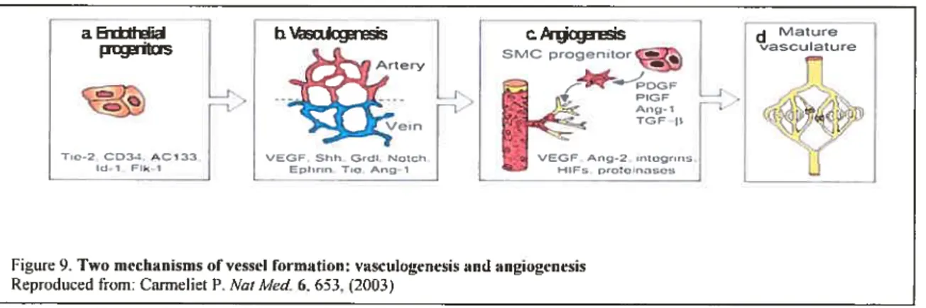
Figure 9. Two mechanisms of vesse! formation: vasculogenesis and angiogenesis 44
Figure 10. Dynamic steps ofnew blood vesse! formation (sprouting angiogenesis) 45
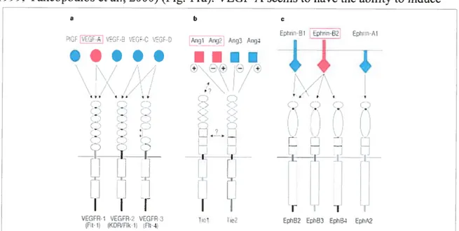
Figure 11. Three families ofvascular growth factors and their receptor interactions 47
Figure 12. $hhIVEGf/Notch in the Arterial Vasculature 55
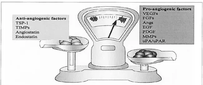
f igure 13. The angiogenic balance 59
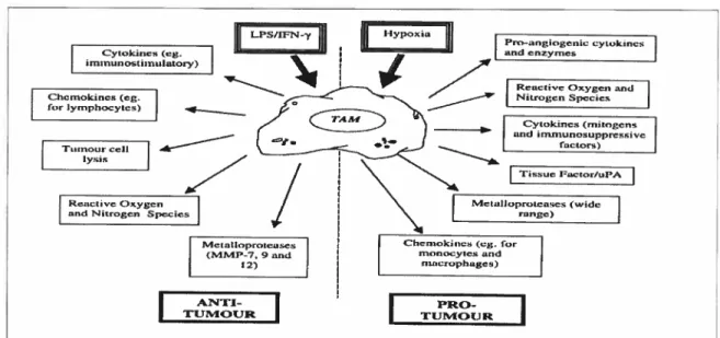
Figure 14. Macrophage production of anti- and pro-angiogenic factors: regulation by
tumour specific signais 75
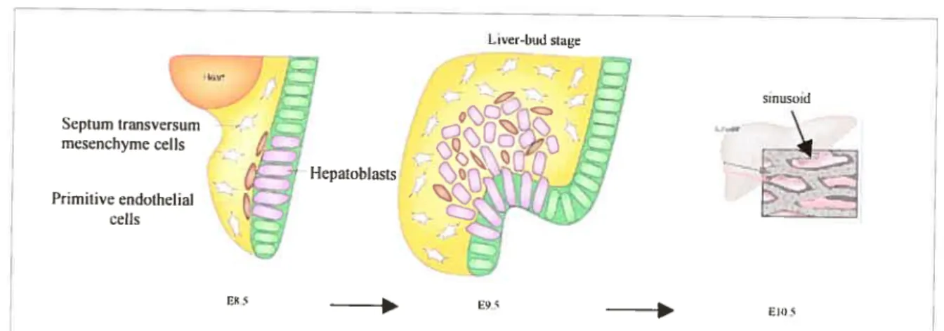
figure 15. Liver sinusoidal development 77
f igure 16. Liver vascular architecture 79
Chapter3 95
Figure t. Structure ofthe N1 and its expression in the Tg mice 144
Figure 2. Liver vascular patterning defects phenotype 146
Figure 3. Aberrant angiogenesis observed in the Tg liver 147
Figure 4. Bon Marrow Transplants (BMT) yielded the liver vascular defects 149
Figure 5. Reproduced liver phenotype in CD4C/N1 X Rag mice 150
Figure 6. A paracrine pathway induces the liver vascular phenotype 151
Figure 7. Macrophages are sufficient to induce the liver vascular phenotype 152
f igure 8. Profile of liver lining cells in the CD4C/N1’ Tg mice 153
f igure 9. Functional abnormalities ofthe Tg L$ECs in the tube formation 154
Figure 10. Enhanced adhesion ofCD4C/N1’ Tg macrophages in vitro 155
Figure 11. Supernatants ofthe Tg macrophages inhibit the growth ofthe nTg LSECs 156
Chapter4 16$
f igure 1. Expression ofNl mRNA and protein in Tg reproductive tracts and an
infertility phenotype in Tg female 187
Figure 2. Developmental studies on the une established from founder 60787 18$
Figure 3. Macro- and micro-scopic analysis ofTg reproductive organs 189
f igure 4. Increased macrophages observed in Tg uteri 190
Figure 5. Tg expression and vascular phenotype are reproduced in the uterus ofthe mice
transplanted with Tg FL cells 191
Figure 6. Histological analysis ofuterus from fL chimeras 192
Figure 7. Notch4 is increased in the Tg uteri. 193
Figure 8. Postulated mechanism leading female fertility 194
Chapter5 195
Figure 1. Experimental design of DEN treatment 220
Figure 2. Macrographs of liver tumors of mice treated with DEN 221
Figure 3. Liver and body weight as well as incidence and number of liver tumors 222
Figure 4. Histological analysis ofhepatocyte tumors ofmice induced with DEN 223
Figure 5. Incidence, numbers, and histological analyses oflung tumors ofmice treated
with DEN 224
f igure 6. Incidence and histological analysis ofkidney tumors of mice induced with
DEN 225
figure 7. Tumor formation in CD4C/Nl Tg mice by transpianting hepatocarcinomas
induced by DEN 226
Figure 8. Prirnary tumor growth observed in CD4C/N1 1g mice transplanted with
C3L5 breast tumor ceils 227
Figure 9. Metastatic turnors observed in CD4C/Nl’ Tg mice transplanted with C3L5
breast tumor celis 228
Figure 10. Faster primary tumor growth observed in Tg-bearing nude mice transplanted
with 378 melanoma celis 230
Chapter 6: Conclusions and Perspectives 237
f igure 1. Model ofN1EC induce liver disease 244
f igure 2. Intra-glomerular metastasis of C3L5 brest tumor celis 247
f igure 3. macrophages and tumor progression and metastasis 250
List of Tables Chapter 1 1 Table 1 66 Chapter3 95 Table.1 157 Table.2 158 Chapter5: 195 Table 1 231 Table 2 232 Table 3 233 Table 4 234 Table 5 235 Table 6 236 xvii
List of Abbreviations AAH: atypical adenomatous hyperpiasia
Ang: angiopoietin
ANGPTL3: Angiopoietin-like protein 3 AGM: aorta-gonad-mesonephros AGS: Alagille syndrome
bFGf: basic fibroblast growth factor bHLH: basic helix-loop-helix BM: bone marrow
BMEs: bovine microendothelial ceils CAM: chicken chorioallantoic membrane
CADASIL: cerebral autosornal-dominant arteropathy with subcortical infracts and leukoencephalopathy
CLPs: common lymphoid progenitors
CFU-GM: colonyforming unit, granulocyte-macrophage COX: cyclooxygenase
CSL: CBF 1 (RBPjk)/Suppressor of hairless/Lag- 1 Dl: Delta
Dlii: Delta-like 1
DMH: 1 ,2-Dimethylhydrazine dihydrochioride DN3: double negative stage 3
Dsh: Dishevelled
DSL: Delta/Senate/Lag-2 ECs: endothelial celis ECM: extracellular matrix EGF: epidermai growth factor
EGfR: epidermal growth factor receptor ER: estrogen receptor
E[spi]: Enhancer of spiit
f C: follicular
FGF: fibroblast growth factor FL: fetal liver
f 1k: fetal liver kinase
Flt:Jms-like tyrosine kinase GLUT- 1: glucose transporter-1
GM-C 5F: granulocyte/macrophage-colony stimulating factor GOM: granular osmophilic materials
HA: hyaluronic acid
HAF: human angiogenic factor HBV: hepatitis B virus
HBx: hepatitis B virus X-protein HCC: hepatocellular carcinoma HCV: hepatitis C virus
HDAC: histone deacetylase HES: Hairy/Enhancer of spiit
HGF/SF: hepatocyte growth factor/scatter factor HHT: hereditary hemorrhagic telangiectasia HIF: hypoxia-inducible factor
HME: human macrophage metalloelastase
HMEC- Ï: human dermal microvascular endothelial celis HNF: hepatocyte nuclear factor
HPV16: human papilloma virus-16 HRE: hypoxia response element FISC: hernatopoietic stem celis
HUVECs: human umbilical vascular endothelial ceils HRMECs: human renal microvascular endothelial celis ICAM: intercellular adhesion molecule
IGF-1: Insulin-like growth factor-1 IFN: interferon
IL: interleukin
NOS: inducible nitric oxide synthase
JAG: Jagged KC: Kupffer ceil
KDR: kinase insert domain-containing receptor LfA-1: lymphocyte function-associated molecule LNR:
LSECs: sinusoidal endothelial celis ofthe liver LYVE- 1: lymphatic vascular endothelial receptor MACS: magnetic celi sorting
MAMLI: Mastermind-like-1
MAPK: mitogen activated protein kinase
MASHÏ: mammalian achaete-scute-homologue 1 MCP: monocyte chemotactic protein
MCP-1: monocyte chemoattactant protein M-C SF: macrophage-colony stirnulating factor
MDF-ECI: macrophage-derived endothelial celi inhibitor MECIF: monocyte-derived endothelial celi inhibitory factor MIP: macrophage inflarnmatory protein
MHC: majorhisto-compatibility complex
MIF: macrophage migration inhibition factor MMPs: matrix metaloproteases
MMTV: mouse mammary tumor virus M01: macrophage index
MSR: macrophage scavenger receptor MZ: marginal zone
MZB: splenic marginal-zone-B celis
NEC Notch extracellular domain
N]C: Notch intracellular domain
NK: natural kili
NLS: nuclear localization sequences NO: nitric oxide
NOS: Nitric oxide synthase
Nrarp: Notch-regulated ankyrin-repeat protein CAR: Ordinary adenomatous hyperplasia OC: oral contraceptives
OFUT1: O-fucosyltransferase 1 PAl: plasminogen activator inhibitor
PD-ECGf: platelet-derived endothelial celi growth factor PDGF: platelet derived growth factor
PDGFR-b: platelet derived growth factor b
PECAM-1: platelet/endothelial celi adhesion molecule-l PE ST: praline, glutamate, serine, threonine-rich
PGE2: prostaglandin E2. PH: Partial hepatectomy P1GF: Placenta growth factor PLNs: peropherial lymphnodes PN$: peripherial neural system P-Sp: para-aortic splanchnopleura P5: presenilins PTB: phosphotyrosine-binding Ser: Seiiate SD: spondylocostal dysostosis SHH: Sonic hedgehog
SKIP: Ski-related Protein
SMRT: Silencing Mediator of Retinoid and Thyroid hormone receptor SOPs: sensory organ precursors
SPARC: secreted protein, acidic, and rich in cysteine. Su(Dx): Supressor ofDeltex
Su(H): Supressor ofHaireless
TAD: transcriptional activator domain T-ALLs: human T-lymphoblast leukaemia TAMs: tumor-associated macrophages
TAN-1: translocation-associated Notch homologue TGF: transforming growth factor
TIMP: tissue inhibitor ofmetaloprotease TNF: tumor necrosis factor
TSC: tuberous scierosis TSP-l: Thrombospondin-1
u-PA: urokinase plasminogen activator
u-PAR: urokinase plasminogen activator receptor VEGF: vascular endothelial growth factor
VEGFR: vascular endothelial growth factor receptor VHL: Von Hippel-Lindau protein
VM: venous malformation
VPF: vascular permeability factor
vSMCs/PCs: vascular smooth muscle cells/pericytes YS: yolk sac
Preface
The present thesis, consisting of six chapters, has as a theme: the overexpression of Notchi ectodomain in macrophages induces vascular defects and promotes tumor progression.
The first chapter is a generai introduction and literature review of ail the reievant works of the research project. It is divided into eight parts. The Notch fundamentai role is rnentioned in the first part. Then the function of Notch ectodomain foliows in the second part, from which the rationaies of the research arise. The third and fourth parts describe the physioiogicai and pathologicai role of Notch, centered on the hematopoietic development and turnors and end with an ernerging roles ofNotch in vascular deveiopment and disease. Knowiedge of vasculogenesis and angiogenesis is reviewed in the fifth section, in which
turnor angiogenesis is introduced. Since macrophages are targeted by our Tg NlF and
since our Tg mice deveioped vascuiar disease, it is reasonable to understand the reiationship between macrophages and angiogenesis. The explanation is to be found in part six. Why are the iiver and uterus the main organs affected by aberrant angiogenesis in our Tg mice? The seventh and the eighth part wiii heip you to iearn more.
Chapter 2 exposes the rationale, hypothesis, objective, and an overview of the project. Chapter 3-5 are the experimental resuits planned to be published in three papers. Chapter 6 presents generai conclusions and perspectives. Each chapter has its own reference list for reader’s convenience. The papers inciuded are the foiiowing:
1. Overexpression ofNotchl ectodomain in macrophages induces vascuiar malformations in Tg mice.
Li, X., Caivo, E.L., Kay, D.G., Chrobak, P., Cool, M., Hanna, Z. and P. Jolicoeur. 2. Expression ofthe mouse Notchi extraceiiuiar domain in macrophages ieads to sterility
in femaie Tg mice
Li, X., Chrobak, P., and P. Jolicoeur.
3. Notchi ectodomain expressed in macrophages promotes tumor progression, metastasis, and angiogenesis.
Li, X. and P. Jolicoeur.
I provided a major contribution earning the first authorship in these papers. The contributions of the coauthors to these publications are acknowledged: Dr. Calvo has collaborated for the first paper at the beginning and has performed hepatectomy. Dr. Kay for his assistance in some pathological works. Dr. Chrobak for participating in bone marrow and fetal liver ccli transplantation. Dr. Cool performed the chimeric experiments. Dr. Haima for designing the CD4C promoter. Other collaborators, but flot coauthors, also contributed to the papers and are gratefully acknowledged. Benoît Laganière for preparing the CD4C/Nl construction and generating the Tg mice. Ginette Massé for manipulating C3L5 turnor celis and for helping to isolate LSECs and to measure subcutaneous C3L5 turnors. Isabelle Corbin, Jean-René Sylvestre and Paule for taking care the mice. Isabelle Labrosse for helping to design some primers for VEGf, angiopoietin, and Ehprin 34. Chunyan Ru and Lin Jia for in situ hybridyzation. Viorica for PECAM-l and Œ-actin immunohistochemistry. Dr. Paul Jolicoeur, acted as my thesis supervisor, thanks to his scientific insight and advice throughout the course of all these studies.
Chapter 1.
INTRODUCTION
Multiceilular organisms arise by a process of progressive change that we cali development. The development of a multicellular organism begins with a single cell. Sidney Brenner (quoted in Wilkins, 1993) has remarked that the animal development proceeds in either of two ways. Most invertebrates are specified predominantiy by the “European style” that is to say the developmentai fate of each ceil is determined by its ancestoral lineage. Conversely, rnost vertebrates are specified predominantly by the “American style” in which there is a great deal of mixing between celis, and ceil fate is determined by its neighbors. Each celi starts off with similar potentiais and develops according to the cell type it interacts with. This type of cell fate determination is called conditional specification, because the fate of a ceil depends upon the condition of ceil-celi interaction.
The role of ceil-ceil interactions in determining the ceil fate is to distinguish two celi types with amplified signals from a celi population. The multiplicity of outcomes arising from the repeated use of such pleiotropic signais depends on the context in which the signals are received. These signals can be caused by sorne chance factors on the celi membrane. Among these chance factors, the most prevalent ones are members of Notch family (Artavanis-Tsakonas et ai., 1999; Mumm and Kopan, 2000). Signais transmitted through Notch control cell fate in a wide array of developmental processes, ranging from neurogenesis to oogenesis (Kimble and Simpson, 1997). Gain- or ioss-of-function ofNotch gene typically results in an increased abundance of ceils adopting one fate at the expense of an altemate fate. In specific contexts, Notch also influences apoptosis, cellular
proliferation, and the organization of tissue boundaries. activities that ftirther contribute to its broad role in morphogenesis, as well as vasculogenesis and angiogenesis (Artavanis
Tsakonas et al.. 1999; Selkoe and Kopan, 2003; Iso et al., 2003). The ceil-ceil interaction controlled by Notch does flot stop at birth and persists throughout adulthood.
1.1 Notch family and fundamental role
In 19l7, Thomas Hunt Morgan and colleagues described a strain of Drosophila with notches, which are absent in the wild type but clearly visible at the border of their
wing blades (Morgan, 1917) (Fig. la). This curious trait was attrïbuted to a partial loss of function (haploinsufficïency) of a genc, which was later named Notch gene. Notch gene, which was first cloned from Drosopliila in the mid-1980s by the teams of Artavanis Tasakonas (Wharton et al., 1985) and Young (Kidd et al., 1986), encodes a receptor with a single transmembrane domain. Notch receptor gcnes are conserved in many organisms, ranging from sea urchins to humans (Radtke and Raj, 2003).
a, Normal wrng ‘Notched”wing I NOTCHI _____________________________ — — — ,... oit,.. N0rC03 -— NOTCK4 — — - -_
figure 1 INotch receptor and Iigands. aJWing blade ofa wild-tvpe Drosophila inetanogaster. andofa mutantwith a partial loss of the 3lotd, gene. b Structure of Notch proteins ami their ligands. Drosophila lias une Noteh receptor (dNotch) ami vertebrates hvp ft)ur (Notchl—4). çIDl Set are two transnwmhrar-pound ligands lbr Notch in Drosophila. The vertebrates possess five Iigands, DLL-l. -3. -4. and JAGI, 2. Reproduced from: Radtke et al.. Nature Reviens cancer 3: 756, (2003)
1.1.1. Notch family and structure
Evolutionary divergence between invertebrates and vertebrates lias been
accornpanied by at least hvo round gene duplications: flues posses a single Notch gene, worms two (glp-1 and lin-12), and mammals four (Notchi, 2, 3, and 4).
At the heart of Notch signaling is the Notch receptor. Although synthesized as a single precursor protein, Notch is cleaved into two parts during its transport to the celi surface and, as a consequence, exists as the heterodimeric receptor (Radtke and Raj, 2003) (fig. lb). The extracellular domain includes many repeats of a protein module ressembling epidermal growth factor (EGf)-like domain and three membrane-proximal Notch-specific repeats (LNR) (Fleming, 1998). The EGF-like repeats mainly participate in ligand binding on an adjacent cell, whereas the LNR prevent signaling in the absence of ligand (Rebay et al., 1991). four functionally distinct, important regions have been identified within the Notch intracellular domain. In N- to C-terminal order, they are the RAM domain and the six ankyrin (also known as CDCÏO) repeats interacting with downstream effectors of Notch pathway, a transcriptional activator domain (TAD), and the proline, glutamate, serine, threonine-rich (PEST) sequence regulating the stability of proteins. Two nuclear localization sequences (NLS) are present prior to, and following, the ankyrin repeats (fig. lb) (Fortini et al., 1993; Struhi et al., 1993; Stifani et al., 1992; Lieber et al., 1993).
1.1.2. Core Notch signaling pathway
The core elements of the Notch signaÏing system include Notch receptors, DSL ligands (Delta and Serrate in Drosophita and vertebrates, Lag-2 in C.eÏegans), CSL DNA
binding proteins (CBF1/RBPjk in vertebrates, suppressor of hairless [Su(H)] in
Drosophita, Lag-1 in c.elegans), and target genes, such as the HES and HERP families of
basic helix-loophelix transcriptional factors (Selkoe and Kopan, 2003; Nakagawa et al., 2000; Fischer et al., 2004) (Fig. 2).
1.1.2.1 Notch receptors and ligands
In Drosophila, a single Notch receptor has 36 EGF-like repeats and one LNR
repeat in the extracellular domain, as well as four distinct regions, RAM, CDC1O, TAD, and PEST domain, in the intracellular part (Radtke and Raj, 2003). Mammals, such as mice and humans, have four Notch receptors encoded by four different Notch genes: Notchi, Notch2, Notch3, and Notch4 (Del Amo et al., 1993; Weinmaster et al., 1992; Lardelli et al., 1994; Uyttendaele et al., 1996). Although the structures of the four Notch receptors are overail very similar, they do show differences in the extracellular and the intracellular parts. The Notchl and Notch2 receptors contain 36 EGF-like repeats in their ectodomain, whereas Notch3 harbors 34 and Notch4 only 29. Additional differences are found within the intracellular domain; specifically, Notchl contains a strong TAD, while Notch2 contains a weak TAD, and no lAD is present in Notch3 and Notch4 (Radtke and Raj, 2003) (Fig. lb).
Whule two Notch ligands, Delta (Dl) and Serrate (Ser), are present in Drosophita, mammals possess five ligands named Delta-like-1, -3, and -4 (DLL1, 3, and 4) (Bettenhausen et al., 1995; Dunwoodie et al., 1997; Shutter et al., 2000) and Jaggedi and
Jagged2 (JAG1 and JAG2), which are Ser-like ligands (Lindsell et al., 1995; Shawber et al., 1996). Each ligand is also a single transmembrane protein that contains EGF-like repeats in the extracellular domain. Different from the Notch receptor, it contains a conserved DSL (DeltaJSerate/Lag-2) domain that can bind to specific EGF-like repeats of the Notch ectodomain on the adjacent cells (Radtke and Raj, 2003). The intracellular domain of the Notch ligands is Iess known. The main structural differences between the members of the ligand family are the number and spacing of the EGF-like repeats and the presence of a cysteine-rich domain, which is located downstream of the EGF-like repeats in Ser, JAG1, and JAG2 (fig. ic).
1.1.2.2. Initiation of Notch signaling
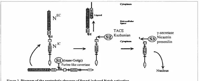
Notch signaling is initiated by a receptor-ligand interaction between two
neighbouring ceÏls, which leads to a couple of successive proteolytic cleavages at three sites—Si, S2, and S3—that in tum liberate the cytoplasmic portion of Notch (N) from the membrane (fig. 3) (Baron et al., 2002; Kopan, 2002; Selkoe and Kopan, 2003).
NEC TACE Kuzbanian y-secretase Nicastrin presenilin Nuc1cu
Figure3. Diagram of the proteolytic cleavage of Iigand-induced Notch activation. Reproduced from: Selkoe et al. Annu. Rev. Neumsci. 26, 565, (2003)
Si cleavage, in the extracellular domain of Notch, occurs constitutively in the trans-Golgi network and is mediated by a furin-like convertase, followed by reassembly of
the fragments to form the heterodimeric Notch receptor at the ceil surface. This event bas been most cÏosely characterized with respect to mammalian Notch, but there is evidence that the fty Notch is similarly processed ($elkoe and Kopan, 2003). $2 cleavage, by a disintegrinlmetalloprotease, occurs in response to ligand binding and releases the majority of the extracellular domain. This cleavage is believed to be mediated by TACE (Henrique et al., 1995) in vertebrates and might be mediated by a related (but distinct) protein Kuzbanian in Drosophila (Pan and Rubin, 1997), although the precise role of the latter protein is stiil controversial. $3 cleavage is caused by the resulting membrane-anchored fragment referred to as Notch extracellular truncation and mediated by y-secretase. A wealth of studies has demonstrated that both Presenilin and Nicastrin are involved in S3 cleavage (Kopan, 2002; Lai, 2002a). This cleavage finally releases N. The mechanism of this particular cleavage is of considerable interest as it is a key event that regulates the nuclear transiocation of NIC and its second life as a transcriptional co-activator. Within the nucleus, the released NIC binds to the transcription factor CSL and finally activates targets ofNotch signaling (Zhou et al., 2000a; Oswald et al., 2001; Fryer et al., 2002).
1.1.2.3. Ihe mechanism of Notch signaling
A well-established mechanism relies on a ligand-induced release of the NIC and the interaction of this fragment with members of CSL family of transcription factors within the nucleus (Lai, 2002b); however, there is increasing evidence that Notch can signal in C$L independent modes (Martinez et al., 2002).
C$L-dependent Notch signaling: The resulting soluble NIC is translocated to the nucleus where it binds to and activates transcription factors: the C$L. C$L proteins bind specific DNA sequences to regulate gene expression. In mammals, HE$ (Hairy/Enhancer of Split) and HERP (HE$-related genes with endothelial specificity) are the direct targets
ofNotch/CSL-dependent signaling (Sasai et al., 1992; Bicknell and Harris, 2004; Iso et aL, 2001). The HES proteins inhibit activfty of other bHLH proteins, such as the Mash-] and
NeuroD (de la Pompa et al., 1997), thereby suppressing transcription of the lineage genes;
however, the interactions between Notch and CSL appear to be more complex and the effects more diverse than originally described. In the absence of the Notch signal, CSL can mediate a repression of gene transcription through the recruitment of the co-repressor protein SMRT (Silencing Mediator of Retinoid and Thyroid hormone receptor). SKIP (Ski-related Protein), and a HDAC (histone deacetylase) (Kao et al., 1998). The binding of NIC displaces the SMRT co-repressor and its associated HDAC enzyme to relieve transcriptional repression (Morel and Schweisguth, 2000; Tani et al., 2001). Furthermore, the Notch ankyrin repeats and TAD-containing regions are involved in recruiting the histone acetylase protein PCAF and GCN5, which may act catalytically to produce an open chromatin conformation (Kurooka and Honjo, 2000). finally. the activator complex recruits Mastermind (Mam), which contains a transcription activator domain (Wu et al., 2000) (fig. 4). The two-state model CSL fiinction allows for variety in the requirements for in vivo transcriptional activation of target genes. for example, while some genes respond simply to NICdependent removal of the repressor function of CSL, other genes require N’ to drive the CSL into an activator complex (Mord and Schweisguth, 2000; Klein et al., 2000; Li and Baker, 2001).
CSL-independent Notch signaling: In Drosophita, the minute analysis ofNotch and Su(H) mutant phenotypes has shown that the Notch phenotype is slightly stronger than that of Su(H) mutant embryos. This suggests that the CSL-dependent signaling pathway does flot mediate ail the functions of Notch (Rusconi and Corbin, 1999; Zecchini et al., 1999). Further support has come from the analysis of a gain-of-function ofNotch alleles that alter adult peripheral nervous system (PN$) development. In this mutant, represented by Ax59
AxM], and Mcd alleles, the sensory bristies do flot develop, thereby suggesting that sensory
organ precursors (SOPs) fail to differentiate. As a resuit, PNS development is not correctÏy initiated. In addition, this phenotype cannot be rescued by removing the Su(H) function (Brennan et al., 1999e; Ramain et aI., 2001). This, in turn, suggests that the increased Notch signaling is occurring via a Su(H)-independent signaling pathway. lit vertebrates, it is less clear whether Notch can signal independently of CSL. Unlike Drosoph lia, it is currently impossible to compare the phenotypes of mice that completely lack the Notch functions with mice lacking CBF1 functions since this requires a generation of mice that lack ail four Notch genes. Nonetheless, several celi culture experiments, in particular on the differentiation of the myogenic ceil une C2C12 into myotube (Nofziger et al., 1999; Kuroda et al., 1999), have provided some evidence for CSL-independent signaling (Martinez et al., 2002). Weinmaster and co-workers have dernonstrated that expressing tntncated forms ofNtc, which cannot activate a CBF1-dependent prornoter, can prevent the C2C12 differentiation even in the presence of a dominant, negative CSL protein (Nofziger et ai., 1999), therefore suggesting a CSL-independent Notch activity. A second une of evidence in support of a CSL-independent signai is that the co-cuiture of C2C12 celis (expressing co-liner forms of Notch) with Jaggedl-expressing celis failed to activate a
CBF 1 -dependent reporter gene but stili prevented their differentiation into myotubes (Bush et al., 2001).
Some molecules are not integral components of the Notch pathway but modulate Notch signal transduction (Egan et al., 1998). These regulators include Wingless, Deltex, and Numb, acting as NotchlCSL-independent signaling.
Wingless is a member of the Wnt family of proteins that regulate rnany developmental processes through the activation of the Frizzled class of ceil surface receptors (Cadigan and Nusse, 1997). Wingless through Fizzled activates the f3-catenin-dependent signaling via the activation of the cytoplasmic protein Dishevelled (Dsh). Evidence ofthe genetic interactions between Wingless and Notch can be seen from the fact that a repressive activity of Wingless can be regulated by Notch (Couso and Martinez, 1994; Rulifson and Blair, 1995; Lawrence et al., 2001), and the Notch activity can be blocked by Dsh (Axelrod et al., 1996). These results provide a molecular mechanism for the inhibitory cross-talk between two pathways. Wingless lias also been shown to inhibit an early Su(H)-independent function of Notch in the selection of muscle founder-celI clusters (Breiman et al., 1999a). Wnt acts to block Notch either directly through its reported binding to NEC (describe later) or, more likely, indirectly by stimulating Dsh to
block GSK3 and!or to recruit Deltex/Notch interaction (Foltz et al., 2002; Wesley and Saez, 2000) (Fig. 5).
The N-terminal region of Deltex mediates binding to the Notch ankyrin repeats (Diederich et al., 1994). Based on its mutant phenotypes and its genetic interaction with Notcli alleles (Xu and Artavanis-Tsakonas, 1990; Gorman and Girton, 1992), Deltex has been identified as a positive regulator of the Notch pathway. Mutations of Drosophila Deltex cause several adult phenotypes similar to Notcli loss-of-function, including wing
notches and thickened veins (Xu and Artavanis-Isakonas, 1990; Gorman and Girton, 1992). As both N- and C-terminal regions of Deltex contain the domains related to ubiquitination (Jackson et al., 2000), Deltex can also act as a repressor of Notch signaling by mediating its degradation (Yun and Bevan, 2003). Other work in mammalian ccli culture has suggested that mammalian Deltex may function on Notch through down regulation of Ras and JNK-dependent signais (Ordentlich et al., 199$) (f ig. 5).
Numb is a phosphotyrosine-binding (PTB) domain protein that binds totwo regions of Notch: the RAM domain and the C-terminus via its PTB. Numb plays a role in the process of asymmetric ce!! division. During neurogenesis in flics, mice, and in avian. the asymmetric distribution of Numb ensures that only one daughter ccli inherits a Numb protein that down-regulates the Notch activity via endocytic pathway, thus biasing the Notch-mediated signaling (Lu et al., 199$; Spana and Doc, 1996; Wakamatsu et al., 1999. Baron et al., 2002) (Fig. 5).
Wïngiess Frizzed / / Notch . Numb . Endocytosis Dish±elled ù&tex f3-catenin Achaete-scute JNK signaling
Figure 5. RegiiJation ofNotch intracelinlar signaling
1.1.2.4. Termination of Notch signalïng pathway
After N1 acts in the nucleus of mitotic ceils, it must be removed, as the daugliters will ofien again rely onNotch signaling to determine their fate (Kopan, 1999). It has been recently demonstrated that the regulation of ubiquitin pathway performed by Sel-10 (Hubard t al., 1997), Noth-regulated 4nkyrjn-repeat protein (Nrarp) (Lamar et al.,
2001), and Neutralized (Lieber et al., 1993) might play a role in the termination ofNotch signaling (Baron et al., 2002), as does the E3 ubiquitin ligase Supressor ofDeltex [Su(Dx)1 (Comell et al., 1999).
Ubiquitination of proteins takes place via a multistep mechanism, with ubiquitin first being covalently linked to an El enzyme before being passed to another covalently linked intermediate, the E2 enzyme, and finally onto the target protein via an E3-ubiquitin ligase-dependent step (Hershko and Ciechanover, 1998). The E3 proteins have been Ïinked to Notch pathway regulation (Baron et al., 2002). The Notch receptor, therefore, is subject to being regulated by ubiquitination at different levels in the signaling pathway.
Sel-10 was originally identified in C.elegans as a negative regulator of Notch signaling and found to bind to the NIC (Hubbard et al., 1997). Recent reports demonstrate
that mammalian homologues of the Sel-10 protein can stimulate ubiquitination of NIC and trigger its proteasome-dependent degradation (Gupta-Rossi et al., 2001; Oberg et al., 2001). The association of mammalian Sel-10 with nuclear N is dependent on a phosphorylation event and also requires the Notch PEST sequence (Rogers et al., 1986). In Drosophila, whether Sel-10 is involved in a proteasome-dependent degradation is not yet known (Schweisguth, 1999).
Nrarp is a small protein encoding two ankyrin repeats (Baron et al., 2002). Nrarp binds to NTC only in presence of Su(H), thereby forming a temary complex. The expression of Nrarp is itself dependent on Notch signaling, suggesting that Nrarp may work in a feedback loop to limit the extent and duration of the Notch signaling. Although flot yet directly implicated in ubiquitination, Nrarp stimulates degradation of the Xenopus NIC (Lamar et al., 2001). So far, the degradation mechanism is not known.
r__
Neutralized is iocaiized at the ce!! membrane and is thought to act upstream of the
release of N, because the expression of the latter rescues the Neutra!ized phenotype
(Lieber et al., 1993). In Drosophila, the Neutralized has been bioiogically shown to be a ubiquitin !igase and to contain a recognizable Ring finger domain (Yeh et al., 2001). Mutation of Neutralized resuits in a disruption of Notch signaling in a certain number of tissues, including lateral inhibition during neurogenesis, but flot ail of the functions of Notch (Lai and Rubin, 200 la; Lai and Rubin, 200 lb).
1.1.3. The fundamental role ofNotch signaling
1.1.3.1. Participation in ceil-fate decision
Notch pathway is already implicated in several processes of development in a
certain number of species. In Drosophila, the Notch protein is indispensable for the determination of numerous types of ce!!s, for example, organogenesis, neurogenesis, and myeogenesis, as well as the development of the wing and eye (Artavanis-Tsakonas et al., 1999; Egan et al., 1998). In mammals, four homologuous proteins are expressed from three-layer of embryos, namely endoderm, mesoderm, and ectoderm. They play a critical role in the differentiation of numerous cell types, including neurogenic, haematopoietic, and endothe!ia! ceils (ECs) (Morrison et al., 2000; Kumano et al., 2003; D’Amore and Ng, 2002; Lawson et al., 2002). The role of Notch pathway in the deveÏopment of mammats has been proved in the mice deficient for Notchi, Notch2, and their ligands (Swiatek et al., 1994; Conlon et al., 1995; Hrabe et al., 1997). Ail the mice have severe problems during their development that cause embryonic and perinata! lethality. for example, in mice lacking Notchi the homozygote embryos deveiop normally until the 9th embryonic day but die before the 115th day. The histological analysis indicated that the majority of the dead
ceils were particularly concentrated in the neuroepithelium of the central nervous system (Swiatek et al., 1994). Consistent with these results, the RNA expression of Notchl was detected in the pre-somite mesoderm at the 7th day of the embryonic development. At the
9th
day, the major site of expression was neuroepithelium, as well as tissues of the neural crest (De! Amo et al., 1992; Reaume et al., 1992). Notchi also plays a role in the developmentaÏ pattem of chickens (Caprioli et al., 2002) and in the differentiation of the oligodendrocytes of rats (Wang et al., 1 99$b). This indicates that the Notch signaling pathway influences a diversity of tissues in various species.
Even though Notch and its ligands are ofien expressed in the same ce!!, the receptor is critica!!y activated by the interaction with the !igand localized on the adjacent ce!!s. The cel!s may be lied to by the homotypic interaction (equipoentential ceils) or by the heterotypic interaction (non-equipotentia! celis) to process different ro!es.
Lateral inhibition between eguivalent ce!!s:
“Lateral inhibition” was first termed by V. B. Wigglesworth to describe a process for the constant density ofbristles in the deve!oping insect (Wigg!esworth, 1940). From the observation of the effective separation between the existing and newly formed bristles, he postulated that an existing bristie inhibits surrounding ceils, such that a new bristle forrning ceil could only arise outside the range ofits inhibitory influence.
The best characterized components of the inhibitory machinery are the members of the Notch-Delta signa!ing pathway in the insect neurob!ast development. In 1990, Simpson proposed a cell-contact-inhibition mode! for Notch-Delta-mediate !atera! inhibition, according to which the epithelia! ceÏls are inhibited directly by the contacting neural celi (Simpson, 1990). Genetic mosaics show that whereas Notch is needed in the ce!!s that are to become neuroblasts, the Delta gene is needed in the adjacent celis that induce the
epidermal phenotype. Later on, a similar model was proposed by Greenwald and Rubin with Lin-12 in C.elegans (Greenwald and Rubin, 1992) in order to explain the spacing pattems of neuroblasts in the proneural clusters of epidermai and fleurai predursors. Jnitiaiiy, ail the cells have equai potentials and signaling; however, when, by chance, there is an event causing one of the ceiis to produce more signals (say, Delta product), this activates the receptors (Notch) on the adjacent cells and, consequentiy, reduces their signaling level. Since the signaling leveis (Notch) on the adjacent ceiis are low, the neighbors of those iow-signaling ceils wiil tend to be, themselves, high-level signalers (Delta). In this way, a spacing of neuroblasts is produced. Inactivating Notch or Delta causes a failure of iateral inhibition resulting in an excess of neural cells at the expense of the epidermal tissues in both embryonic and adult nervous systems. This demonstrates that Notch and Delta are required for the inhibition of the formation of extra neural precursors from a proneurai equivalence group (Heitzier and Simpson, 1991; Parks and Muskavitch, 1993). Among other members of the Notch pathway, Su(H) and E(spi) behave, respectively, as a transducer and downstream nuclear effector of the Notch-mediated inhibition in genetic and biochemical experiments (Schweisguth and Posakony, 1994; Deiidakis and Artavanis-Tsakonas, 1992).
Notch-mediated lateral signaling is not only restricted to neural tissues. It also controls a number of other celi-fate decisions between equivalent ceiis in C.elegans (Wiikinson et al., 1994). In mouse models, Notch is impiicated in a series ofprocesses of lateral inhibition in the generation of somite precursors (Conlon et al., 1995), hematopoietic ceils (Washbum et al., 1997), and others (Lewis, 1998).
Determination of non-eguipotent ceils:
Notch signaling can also occur between two developrnentally-distinct cells referred to as inductive ceil-fate decision (Artavanis-Tsakonas et al., 1995). In this case, Notch and its ligands are expressed exclusively on two different ceil types. The celi expressing the receptor, and therefore the recipient of the Notch signal, is induced to differentiate into a particular celi lineage. for example, a bipotential mouse neural-crest stem ceil can be induced by Notch to adopt a glial-celi fate, as opposed to a neural one by Notch ligands expressed on neuroblasts (Morrison et al., 2000). Mouse thymic epithelial celis expressing ligands for Notchi induce early lymphocyte precursors to adopt the T-cell fate as soon as they enter the thymus, whereas in the absence ofNotchl signaling these precursors take on the B-cell fate as the default pathway (Pear and Radtke, 2003).
1.1.3.2. Induction of terminal differentiation
Tnstead of influencing the choice between two possible ceil fates, Notch signaling
between developmentally-related celi types can induce or enhance a terminal
differentiation. In the aduit mouse-skin keratinocytes, where Dil is flot expressed, Jagged mediated Notch signaling triggers a terminal-differentiation program by inducing early differentiation markers and cell-cycle arrest (Rangarajan et al., 2001). In the human skin, this program is initiated by DII (Loweil et al., 2000).
1.1.3.3. Maintenance of an undifferentiated state
In addition to participating in the binary ceil-fate decision and in the induction of terminal-differentiation, Notch signaling also preserves some ceils, such as stem ceils, in their native state. Notch signaling in the vertebrate nervous system is usually thought to influence the balance between the progenitor celi pool and its progenitor-differentiating
progeny (Lewis, 1996). Gain-of-function studies on chickens and frogs using a dominant
active NIC show that forced Notch signaling prevents progenitors from undergoing
neurogenesis, whereas blocking this pathway leads to excessive neurogenesis and depletion ofthe progenitor pool (Chitnis et al., 1995; Henrique et al., 1997). $imilarly, the exposure of hematopoietic stem ceils to JAG1 increases the proportion of stem celis, as opposed to differentiating ceils; therefore, Notch signaling induces these celis to retain a stem-ceil-like character (Vamum-Finney et al., 1998).
Taken together, the fundamental foie of Notch signaling is mainly to control the celi-fate decision during development and, meanwhile, to take part in the differentiation and the maintenance of stem celis (Fig. 6).
Notch signaling
ceil-fate decision Induction 0f Maintenance of
terminal differentiation an undifferentiated state
Lateral inhibition Determination
between equivalent celis of non-equipotent celis
f igure 6 The fundamental rote ofNotch signating.
1.2. Function ofNotch ectodomain
Since the Notch gene was first cloned (Wharton et al., 1985), one important goal has been to understand the meaning of the repeated modular structure of the ectodomain. Does each EGF-like module have a specific function? Do some modules act simply as a spacer to correct the position of different binding sites? Jnterspecies sequence conservation within EGF repeats 11—13, 23—27, and 3 1—34 of Drosophila Notch suggests that these regions may form functionai subdivisions of the Notch extracellular domain (Wesley, 1999; Wesley and Saez, 2000). Genetic data from Drosophita further suggests that there is a functional diversity within the Notch extracellular domain (Kelley et ai., 1987).
Mutations in different EGF-like repeats show selective effects in different tissues or have positive or negative consequences on the Notch signaling pathway. For instance, a single amino acid substitution in the EGF-like repeat 14 (the spiit allele) affects Notch activation specifically in the eye and in the sensory bristie development, while Abuptex alleles of Notch, clustered within the EGf-like repeats 24—29, produce dominant gain-of-function phenotypes in the sensory bristle development and in the wing veins (Kelley et al., 1987; de Celis and Garcia-Bellido, 1994). It is possible, therefore, that extracellular interactions ofNotch may confer an additional level of regulation, perhaps in a tissue-specific manner. From analysis of the extracellular interactions of Notch, a picture of the functional specification ofthe regions within the extracellular domain begins to emerge.
1.2.1. Cross-talk with Notch tigands
The first region of the Notch extracellular domain to be defined as having a fttnctionally distinct role was the binding site of Drosophila Notch for its two ligands: Delta and Serrate. Using a co-culture of the Drosophila-S2 ceils (expressing ligand or receptor) (Rebay et al., 1991), the EGF-like repeats 11 and 12 were both necessary and sufficient for binding of Notch to both Delta and Serrate on adjacent ceils; this is called a trans-interaction. Furthermore, through a point mutation within the EGF-like repeat 12 in Drosophila, the importance of this region has been established in vivo due to an abolition
ofthe ligand-dependent Notch signal (de Celis et al., 1993).
Apart ftom the trans-interacting complex, other forms of interactions of Notch with Delta and Serrate ligands may also regulate Notch activity. Cis-interaction between Notch and its ligands within the same ceil has been identified (Fehon et al., 1990; Jacobsen et al., 1998) but does not appear to activate the receptor in an autocrine manner. Instead, it has been proposed that the cis-interaction mediates a dominant, negative activity of the ligand
on the receptor by reducing the capability to receive a signal from the adjacent celis when the ligand is in excess. Another level of ligand-receptor interaction may corne from the proteolytic processing of the ligands. Proteolytically cleaved and secreted forms of ligand have been identified, raising the question as to whether or not this might allow action of the ligand at a distance from where it is expressed (Klueg et al., 199$;
Qi
et al., 1999).1.2.2. Cross-talk with other factors Scabrous
In Drosophita, Scabrous has been shown to bind to the Notch receptor through the EGF-like repeats 19—26 (Poweli et al., 2001). It is a secreted fibrinogen-related protein interacting with the Notch extracellular domain. Its mutation causes a tissue-specffic phenotype in the eye and sensory bristles (Mlodzik et al., 1990). Overexpression of Scabrous inhibits Notch signaling in the Drosophila eye and wing (Lee et al., 2000; Poweil et al., 2001). On the other hand, the ectopic Scabrous expression blocked the ligand dependent Notch activity but flot that of constitutively-active Notch intracellular domain. This fact suggests that Scabrous acts at the level where the ligand binds to the receptor (Lee et al., 2000). Intriguingly enough, in the S2-cell culture, $cabrous stabilizes Notch at the ceil surface (Powell et al., 2001). How this might lead to a modulation of the Notch activity remains unrevealed but tethering of Notch to the extracellular matrix (ECM) might offer one possible mechanism for modulating its and/or other’s activity.
Wingless
Recent reports confirm the capability of Wingless to directly bind the Notch extracellular domain. Wingless binds to two in vivo forms ofNotch, i.e., a full-length and a N-terminal truncated forrn, through a site lying within EGf-like repeats 19—36 (Wesley, 1999). These data are consistent with a region of Drosophila Notch, which, when deleted,
alters the observed phenotypic interactions resulting from co-expression of the Notch extracellular domain and the Wingless (Brennan et al., 1999b). In ccli cultures, the association of Wingless with Notch invoked a transcriptional response that differed from that elicited by Delta (Wesley and Saez, 2000). While Delta-dependent signaling was associated with the accumulation of soluble Notch intracellular domain, no such accumulation followed an exposure to Wingless, implying a distinct signaling mechanism (Wesley and Saez, 2000).
1.2.3. Regulation by some factors
Fringe
Fringe acts within the Golgi as an N-acetylglucosaminyltransferase and adds GlcNac group O-linked fucose to specific EGF-like repeats in the Notch extracellular domain (Bruckner et al., 2000; Moloney et al., 2000). Several conservcd O-linked fucose sites close to EGF-like repeats 24—26 are critical for Serrate- but not for Delta-dependent Notch signaling (Lawrence et al., 2000). This has been shown to play a kcy role in special regulations of Notch signaling at the compartment boundaries during pattem formation. Expression of fringe results in inhibition of Serrate-dependent Notch signaling but flot Delta-dependent signaling (Fleming et al., 1997; Panin et al., 1997). The differential effect of Fringe on Serrate and Delta may be due to differences in how the ligands interact with the receptor in the productive signaling complex. When Fringe is expressed in the sarne tissues as Serrate, Serrate will only signal to Notch in the ceils lying adjacent to the Fringe expressing territory. This is a key step in setting up a spatially restricted zone of Notch signaling at the compartment boundaries. It is interesting to note that the EGF-like repeats 24—26 also lie within a segment of the Notch extracellular domain where the Abruptex
gain fttnction Drosophila Notch alleles are clustered (Kelley et al., 1987). The Abruptex mutations are thought to make Notch resistant to the dominant, negative effects of cis interacting ligands and also to confer resistance to the consequences of the Fringe activity (de Celis and Bray, 2000).
O-fucosyltransferase-1
O-fucosyltransferase-1 (OFUT1 in Drosophila) is another protein that regulates glycosylation of the Notch extracellular domain. The transfer of fucose to Notch by this protein is necessary for the fringe to function (Shi and Stanley, 2003; Panin et aÏ., 2002).
Down-regulation of OFUI1 by RNA interference in soluble extracellular domain of
Notch-secreting ceils inhibits both Delta-Notch and Serrate-Notch binding, demonstrating a requirernent for O-linked fucose for efficient binding ofNotch to its ligands. Conversely, overexpression of OFUT1 in cultured cells increases Serrate-Notch binding but inhibits Delta-Notch binding (Okajima et al., 2003), opposing the influence of Fringe on Notch ligand binding. Mouse embryos lacking O-fucosyltransferase-1 die at midgestation with severe defects in somitogenesis, vasculogenesis, cardiogenesis, and neurogenesis. O fucosyltransferase-1 is, therefore, an essential core member of Notch signalling pathways in mammals (Shi and Stanley, 2003).
1.2.4. Participation in trans-endocytosis
Membrane receptors are passively recycled or actively eliminated by endocytosis (Robinson, 1994). This process is very important to regulate the ceil signal transduction. A Ïink between endocytosis and Notch signaling was proposed based on the phenotype of the Drosophila shibire mutant (Seugnet et al., 1997). Using antibodies to the Notch extra- and intra-cellular domains of Drosophita, it has been shown that these two domains can traffic
independently in vivo. In the pupal eye, for example, the Notch extracellular domain undergoes trans-endocytosis from the pigment ceil (where it is expressed) into the adjacent Delta-bearing cone celis. Both the separation ofNEC and N and the trans-endocytosis of
NEC into the cone celis were disrupted by a shibire temperature-sensitive mutation. It was also observed that Delta and Notch become co-localized at the cone ceil-pigment celi junction. Since shibire is required for Notch signaling, and since endocytosis-defective mutations of Delta fail to signal, it was concluded that the trans-endocytosis mechanism plays a part in the generation of the ligand-dependent cleavage site. This might result in a conformational change (Parks et al., 2000). On the other hand, it lias been shown that the halves of the furin-processed Notch receptors are held together non-covalently. The possibility, therefore, arises that, in some circumstances, signaling could be activated by the physical removal of the Notch extracellular domain without further extracellular cleavage (Rand et al., 2000). The trans-endocytosis model of the Notch activation implies that the generation of the Notch signal requires a membrane-tethered ligand and may explain why soluble-secreted forms of Delta and Serrate act antagonistically on the Notch activity (Qi et al., 1999; Hukriede et al., 1997).
1.2.5. Function of the extracellular domain of Notch ligands in the Notch sïgnaling
While most research was focused on the constitutive intracellular domain ofNotch signaling, a few groups were involved in the functional characterization of the extracellular domain of the Notch ligands. Sun and Artavanis-Tsakonas first proved that secreted fomts of Delta and Serrate act as antagonists of Notch signaling in DrosophiÏa (Sun and Artavanis-Tsakonas, 1996; Sun and Artavanis-Tsakonas, 1997). They examined the function of secreted forms of Delta and Serrate, named as D1S and SerS, by expressing
them under different promoters in the Drosophita developing eye and wing. The phenotype associated with the expression ofboth secreted forms mimics loss-of-function mutations in the Notch pathway. Consistent with these resuits, Fleming’s group found that the soluble form of Serrate, called BD°, acts as a general antagonist of Notch activation (Hukriede et al., 1997); however, Artavanis-Tsakonas and workers showed that a soluble extracellular fragment of Delta (Dl’) has an apparent agonistic function in the Notch signaling pathway (Qi et al., 1999). D1S and Dl are stntcturally not identicai, which could explain their opposing functions.
In Drosophila, both soluble ectodomains of Delta and Jagged are implicated in the activation of Notch signaling. Since the extracellular fragments of Notch receptors and Notch ligands are constituted of the same EGF-like repeat, it is reasonable to hypothesize that the Notch soluble ectodomain may also play an important role in the Notch (or other) signaling pathway.
1.2.6. Involvement of Notch ectodomain in the Notch signalling
In our iaboratory, Girard et al. (1995) showed that the Notchl extracellular domain (N19 was highiy expressed in almost ail the Notchl-rearranged thymomas ofMMTV’/c myc Tg mice infected by Mo-MuLV (Moloney Murine Leukemia Virus) (Girard et al., 1996; Girard and Jolicoeur, 1998). Jnitially, they intended to identify some of the collaborators of c-myc to induce thymomas in the MMTVD/cmyc Tg mice using provirus insertional mutagenesis. They found that quite a high proportion (52%) of these tumors contained Notchi mutations. furthermore, they revealed that the provims was mainly inserted upstream of the exon coding for the transmembrane domain of Notchi. These
mutations lcd to a high expression of truncated Notchl intracellular domain (Ni) and
fuii-length Notchi RNAs and proteins. Interestingiy enough, in aimost ail these Notchi rearranged tumors, high ieveis ofNicouid be detected by Western biotting.
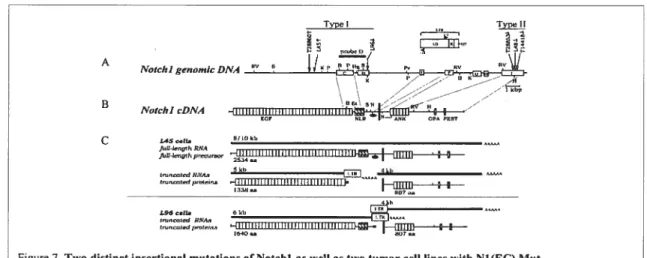
Later on, following Girard’s observations, Hoemann et al. found that two distinct Notchi insertionai mutations were invoived in these thymomas (Hoemann et ai., 2000) (fig. 7). The first type ofproviral insertion, named “type I,” is in genomic regions
Type Typej
I Nf A
Notcl, I genonuc DATA
B Notcl,1 cDNA C L450e1b 8110kb 5kb 4kb tm,watcdRNA.,
,
iHHHtUHiiHHHHHil’Ifl__.++—figure 7. Two distinct insertional mutations ofNotchl as well as two tumor ccli unes with N1(EC) Mut. Reproduced from: Hoemann CD et al. Mot Cet! Biot. 11, 3831, (2000)
coding for sequences between the 34thi EGF-iike repeat and the transmembrane domain
(Fig. 7A and 7B). The second kind of insertion, termed “type II,” is within an 800-nucieotide span at the C-terminal region of Notchi. “Type I” insertion comprises the majority of mutations. The extracellular fragment produced from this specific insertion was calied Ni (EC)MLt (fig. 7A and 7B). It was demonstrated that the Notchi ectodomain could also be generated by normal processing of wiid-type Notchi precursors and was named Ni(EC)wt. in order to analyze the putative Ni(EC)Mt proteins, two tumor celi lines, L96 and L45 (fig. 7C), were selected, each harboring distinct type I insertions that produced both intra- and extraceilular domains (as detected by Western biotting) (Girard et ai., 1996). N1(EC) Mut in the L96 celi line contains 36 EGF-iike repeats and a LNR domain, whereas L45 solely contains the 36 EGF-iike repeats. It was found that the soluble extraceliuiar fragments of Notchi, Nl(EC)Mut, were different from the processed