Université de Montréal
Exploring a role for a Par3/CaMKII protein complex in photoreceptor cell polarity
and ciliogenesis
Par Yulia Ezhova
Programme de biologie moléculaire Faculté de médecine
Mémoire présenté en vue de l’obtention du grade de Maîtrise en biologie moléculaire
Mai, 2020 © Yulia Ezhova, 2020
Table of contents
Résumé………..………..p. vi-vii Abstract………..………..….…p. viii-ix List of Tables.………...p. x List of figures…..………..………p. xi-xii List of abbreviations……….……p. xiii-xiv Acknowledgements………...p. xv Contribution of Authors………p. xvi-xvii
Chapter 1 - Introduction………...p. 1-17 1.1. Cell polarity……….………p. 1-2 1.2. Establishment of polarity in epithelial cells………..….……..p. 2-3 1.2.1. Regulation of cell polarity by Par complex……….……….……….p. 3-4 1.2.2. The polarity complex – Crumbs………..….…p. 4 1.2.3. Scribble protein complex………..…...p. 4-5 1.2.4. Polarity in the central nervous system………..………..p. 5-6 1.3. The mouse retina as a model system to study polarity in the central nervous system………...p. 6 1.3.1. Developmental origin of the retina……….…….…..p. 6-10
1.4. Photoreceptor ciliogenesis……….………..……..p. 10-13 1.5. The role of CaM Kinase family in the CNS……….………p. 13-17 Hypothesis and aims………..p. 17 Chapter 2 – Material and Methods………..………..…p. 18-29 2.1. Cloning……….…p. 18 2.2. Derivation of mice primary embryonic fibroblast………..p. 18-19 2.3. Induction of cilia growth in MEF cells……….p. 20 2.4. Cells transfection………..………p. 20 2.5. Protein detection by Western blot……….……p. 20-21 2.6. Fluorescent immunolabeling………p. 21 2.6.1. Immunohistochemistry………p. 21 2.6.2. Immunocytochemistry………..p. 21-22 2.7. Plasmid electroporation into the eye………..p. 22 2.8. RNA extraction and cDNA synthesis……….p. 22 2.9. In vivo system/Mice line………p. 22-23 2.10. Co-Immunoprecipitation……….…..…p. 23-24 2.11. Immunoprecipitation Mass-Spectrometry……….………….…..p. 24-25 2.12. Short hairpin RNA generation………..p. 25 2.13. Statistic analysis……….p. 25
2.14. Quantitative analysis of the images………p. 25-26 Chapter 3 - Results………p. 30-53 3.1. Identification of CaMKIID localization in the adult mouse retina………..…p. 34-35 3.2. CaMKIID localisation in Par3 cKO mouse line………..………p. 36-38 3.3. Identification of Par3 and CaMKIID proteins interaction in-vitro and in vivo….p. 39-40 3.4. Effect of CaMKIID loss-of-function on ciliogenesis in serum-starved mouse embryonic fibroblasts model (MEF)……….…….…p. 40-43 3.5. Role of CaMKIID loss-of-function on photoreceptor cells in mouse retina………..p. 43-45 3.6. Expression of dominant-negative (K43A) and constitutively active (T287D) forms of CaMKIID affects the outer, but not the inner segment length of PRs.…………..……….p. 45-47 3.7. Distribution of photoreceptors nuclei.………p. 48-49 3.8. Identification of new CaMKIID interactors in the developing and adult retinas by Mass Spectrometry analysis………..…...p. 49-53 Chapter 4 - Discussion……….…..….p. 54-63 4.1. The localisation of CaMKIID in the mouse retina………..……….………p. 54-55 4.2. CaMKIID is capable of interaction with Par3 in vitro and in vivo……….…p. 55-56 4.3. Effect of CaMKIID downregulation on ciliogenesis in MEFs model and mouse retina………..….p. 56-57 4.4. CaMKIID is required for the maintenance of the outer segment length of photoreceptors………..…..p. 57-58
4.5. Constitutively active CaMKIID promotes the PRs nuclei mislocalisation at the basal part of the retina……….………...…p. 58-59 4.6. Identified interaction of CaMKIID with Kif7, annexin2 and rod arrestin might play a role in OS formation of PRs………..……….….p. 60-62 Conclusion……….………p. 62-63 References………...p. 64-80
Résumé
Le traitement et la propagation de l’information nerveuse repose sur une distribution asymétrique de récepteurs et d’émetteurs à la surface de chaque neurone. Ce cloisonnement en domaines sous-cellulaires distincts est également appelé polarité cellulaire. Dans la rétine, la perte de polarité des photorécepteurs peut entraîner des dystrophies rétiniennes telle que l'amaurose congénitale de Leber, mais les mécanismes moléculaires impliqués restent flous. Un complexe protéique impliqué dans l'établissement de la polarité cellulaire, hautement conservé de C. elegans aux mammifères, est le complexe PAR. Localisé au niveau de la région sous-apicale des cellules polarisées, le cœur de ce complexe est constitué des protéines de la famille partitioning defective Par3 / Par6 et de la protéine kinase C atypique aPKC. Bien que largement étudié dans les cellules épithéliales, le rôle du complexe Par dans les neurones de mammifères reste mal compris. Nos résultats indiquent que l'inactivation conditionnelle (cKO) de Par3 dans la rétine de souris en développement interfère avec la croissance polarisée du cil photosensible à la pointe apicale des cellules photoréceptrices (PR), conduisant finalement à une dégénérescence des PRs. Pour découvrir comment Par3 pourrait réguler la ciliogenèse des PRs, nous avons immunoprécipité Par3 à partir d'extraits rétiniens de souris et effectué une analyse par spectrométrie de masse. Nous avons trouvé un ensemble de protéines appartenant à la famille des calcium-calmoduline-dépendantes de la protéine kinase II (CaMKII) comme partenaires potentiels de Par3 dans la rétine. Les CaMKII figurent parmi les protéines les plus abondantes du système nerveux central où elles constituent 1 à 2% des protéines totales. Alors que des études approfondies ont démontré l'importance de CaMKII dans la potentialisation et la dépression à long terme (LTP et LTD), et l'arborisation des dendrites, son rôle dans la polarité cellulaire reste inconnu. En utilisant des versions étiquetées de Par3 et CaMKIID, nous avons validé leur interaction in vivo et in vitro par co-immunoprécipitation. Nous avons mis en évidence une localisation de CaMKIID dans la région ciliaire des PR, suggérant que Par3 pourrait recruter CaMKIID à la membrane apicale des cellules PR, où il pourrait être impliqué dans la ciliogenèse. Pour explorer cette hypothèse, nous avons étudié si les formes dominantes négatives ou constitutivement actives de CaMKIID pouvaient avoir un impact sur la formation des cils des PRs.
La surexpression des deux formes mutantes au cours du développement des PRs a entrainé un raccourcissement des segments externes, semblable à ce que nous avons observé dans les rétines Par3 cKO. Cette étude montre qu'un complexe de protéines CaMKIID / Par3 pourrait réguler l’établissement et le maintien de polarité des PRs, suggérant l’implication ce complexe dans le contrôle de la polarité neuronale de l’ensemble du système nerveux central.
Mots-clés : polarité cellulaire, système nerveux, rétine, photoréceptrices, ciliogenèse, Par3,
Abstract
Cell polarity is an essential property of adult neurons, which rely on asymmetric distribution of receptors and transmitters for proper signal propagation and cell function. In the retina, loss of photoreceptor (PR) polarity can lead to retinal dystrophies such as Leber Congenital Amaurosis, but the molecular mechanisms involved in regulating PR polarity remain unclear. A highly conserved protein complex involved in the establishment of cell polarity from C. elegans to mammals is the Par complex. Localized at the subapical region of polarized cells, it is composed of the “partitioning defective” PDZ domain-containing proteins Par3/Par6 and the atypical protein kinase C (aPKC). Although extensively studied in epithelial cells, the role of the Par complex in mammalian neurons remains poorly understood. Our unpublished results indicate that conditional inactivation (cKO) of Par3 in the developing retina interferes with the polarized growth of the photosensitive cilium at the apical tip of PR cells, eventually leading to PR degeneration. To uncover how Par3 might regulate ciliogenesis in PR cells, we immunoprecipitated Par3 from mouse retinal extracts and carried out mass spectrometry analysis. We found a cluster of calcium/calmodulin-dependent protein kinase II (CaMKII) proteins as potential Par3-interacting partners in the retina. CaMKII is one of the most abundant proteins found in the central nervous system, where it constitutes 1-2% of total proteins. While extensive studies have demonstrated the importance of CaMKII in long-term potentiation (LTP), long term depression (LTD) and dendrite arborisation, its role in cell polarity remains unknown. Using tagged versions of Par3 and CaMKIID, we validated their interaction in vivo and in vitro by co-immunoprecipitation. Interestingly, we found that CaMKIID localizes to the ciliary region of PRs, suggesting that Par3 might recruit CaMKIID at the apical membrane of PR cells, where it could be involved in ciliogenesis. To explore this hypothesis, we investigated whether dominant-negative or constitutively active forms of CaMKIID could impact cilia formation in PRs. Interestingly, overexpression of both mutant forms of CaMKIID during PR development resulted in shortening of the photosensitive cilia (outer segments), similar to what we observed in Par3 cKO retinas. This study suggests that a CaMKIID/Par3 protein complex regulates the establishment of PR cell
polarity, raising the possibility that this complex may be generally involved in controlling neuronal polarity throughout the nervous system.
List of Tables
Chapter 2
Table 1. List of generated plasmids Table 2. List of primary antibodies Table 3. List of secondary antibodies
List of figures
Chapter 1
Figure 1. Retina structure.
Figure 2. Schematic diagram of photoreceptor structure. Figure 3. Protein trafficking in photoreceptors.
Figure 4. CaMKII structure
Chapter 2
Figure 5. Experimental outline for the isolation of mouse embryonic fibroblasts (MEF) on
embryonic day (E) 13.5 by in vitro culture.
Figure 6. Par3 conditional knock out (Par3 cKO) mouse line generation.
Chapter 3
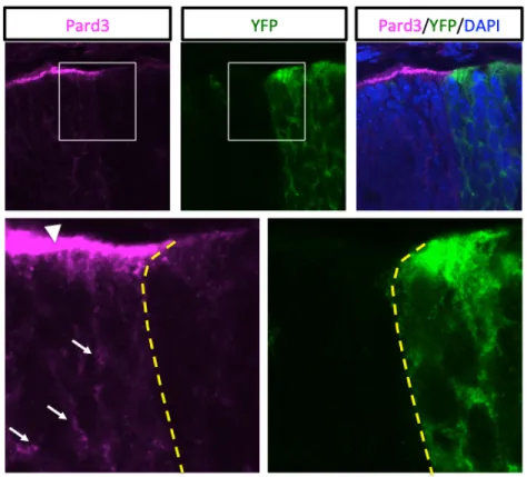
Figure 7. Pard3 gene deletion in Par3 cKO mouse line at the peripheral retina. Figure 8. The localization of Par3 protein expression in embryonic and adult retinas.
Figure 9. Par3 function in developing and adult retinas is essential to maintain retinal structure
and integrity.
Figure 10. Top 10 most enriched Par3 interacting proteins in the neonatal (P0) and adult (P30)
mouse retinas.
Figure 11. The localization of CaMKIID in PRs.
Figure 12. Localization of CaMKIID in developing wild type retinas. Figure 13. CaMKIID loss of localization in Par3 cKO mouse retinas.
Figure 14. Validation of CaMKIID and Par3 protein interaction in HEK293 and in developing retina
Figure 15. Loss of function of CaMKIID does not affect ciliogenesis in Mouse Embryonic Fibroblasts
(MEFs).
Figure 16. CaMKIID KD does not appear to affect ciliogenesis of PRs.
Figure 17. The proper function of CaMKIID is required for the OS growth of PRs.
Figure 18. Overexpression of constitutively active form of CaMKIID changes the localization of
nuclei in PRs.
Figure 19. Identification of CaMKIID interacting proteins in the mammalian retinas in P10 and
P30.
Chapter 4
List of abbreviations
CaMKIID : Calcium/calmodulin dependent protein kinase type II Pard3 : partitioning defective 3 homologCCS : Cosmic calf serum PR : photoreceptors IS : inner segment OS : outer segment ONL : outer nuclear layer
OLM : outer limiting membrane PFA : paraformaldehyde
shRNA : short hairpin RNA PEI : Polyethylenimine BSA : bovine serum albumin NP-40 : Nonidet P40
RT : room temperature IFL : immunofluorescence IP : immunoprecepitation WB : western blot
LTP : long term potentiation RGC : retinal ganglion cell aPKC : atypical protein kinase C AP : anterior-posterior axis
C.elegans : Caenorhabditis elegans VZ : ventricular zone
SVZ : subventricular zone KO : knock-out
IFT – intraflagellar transport GFP : Green Fluorescent Protein
RYFP : Rosa locus - Yellow Fluorescence Protein PhK : Phosphorylated kinase
Pals1 : Protein associated with Lin seven 1 PATJ : Pals1-associated tight junction protein DLG : Discs-large
Lgl : Lethal-2-giant larvae CNS : Central nervous system IHC : immunohistochemistry ICC : immunocytochemistry
Acknowledgement
I would like to thank my supervisor, Dr Michel Cayouette, for allowing me to work in his laboratory and giving me a very interesting project. Dr Michel Cayouette provided me not only the guidance toward my project but also helped me to grow as an enthusiastic researcher. It was a pleasure to be a member of his team and work for two years with very enthusiastic and intelligent people, who I consider as my second family.
Thanks to the Molecular Biology Program in the Faculty of Medicine (University of Montreal) for admitting me into the study program. Thanks to Faculté des études supérieures et postdoctoral (FESP), University of Montreal and to the Montreal Clinical Research Centre (IRCM) foundation bursaries for funding the courses, research project and for giving me the opportunity to study in one of the best universities worldwide. Thanks to IRCM for hosting me and letting me conduct my Master’s project and use their facilities.
I would like to thank my committee members Dr Frédéric Charron and Dr David R. Hipfner for accepting on being part of my committee, and for their scientific suggestions on my research project. Thanks to Dr Martin Sauvageau for accepting to be a member of my thesis seminar. Thanks to Dr Bruno Larrivée for accepting to be a member of the jury.
I would like to give a special thanks to all laboratory members, with whom my studies were more enjoyable. To Dr Michael Housset, Christine Jolicoeur, Dr Sepideh Abbasi, Michel Fries, Maude Vinette, Thomas Brown, Dr Maeva Langouet, Camille Boudreau-Pinsonneault, Awais Javed, Sarah Hales, Pedro L. Franca, Dr Marine Lacomme, Dr Ko Currie, Valérie Lavastre, Kathy Tam and Eva Yuan, it was the greatest pleasure to meet every one of you and work with you. Especially, I want to thank Dr Michael Housset for all your guidance, scientific and technical support during the course of my project and Christine Jolicoeur for your countless hours and attention during the experimental troubleshooting.
I would like to thank my husband and my family for the patience and support that you gave me. You have been my source of inspiration during my master studies.
Contribution of Authors
This project was carried out under the supervision of Dr Michel Cayouette, and with the contribution of Yulia Ezhova (myself) and Dr Michael Housset.
1. Cellular Neurobiology Research Unit, Montreal Clinical Research Institute (IRCM), Montreal, QC, Canada
2. Molecular Biology Program of the Faculty of Medicine, University of Montreal, Montreal, QC, Canada
3. Division of Experimental Medicine, McGill University, Montreal, QC, Canada 4. Department of Medicine, University of Montreal, Montreal QC, Canada
Chapter 1
1.3.1. The schematic diagram presented in Figure 1 was used from the article by Purves et.al., 2001 (Purves et al., 2001).
1.3.1. The schematic diagram presented in Figure 2 and in section 1.4. Figure 3 were used from the article by Ramamurthy and Cayouette, 2009 (Ramamurthy & Cayouette, 2009).
1.5. The schematic diagram presented in Figure 4 was taken from the article by Lisman et. al., 2002 (Lisman, Schulman, & Cline, 2002).
Chapter 2
2.2. The diagram for the isolation of primary mouse embryonic fibroblasts presented in Figure 5 was used from ThermoFisher ©, as per their instruction manual
2.8. The diagram for the mouse line crosses presented as Figure 6 was made by Dr Michael Housset
In this section, Dr Michael Housset made Figures 7, 8, 9, 10 representing previous findings in our laboratory, under the supervision of Dr Cayouette.
3.4. The diagram for the cell transfection presented in Figure 15 was taken from the article Yang et.al., 2017 (Yang, Zhou, Li, Fu, & Sun, 2017).
Chapter 1 – Introduction
Cell polarity is an important property of all eukaryotic cells required for the proper establishment and maintenance of tissues and cellular processes. One important example is the retina, a tissue that absorbs and transmits light to the brain, through its highly polarized laminar architecture defined by a network of appropriately positioned neuronal cells. All vertebrate retinas are composed of three layers of nerve cell bodies and two layers of synapses (Varshney, Hunter, & Brunken, 2015). Although several polarity complexes have been identified, the mechanism of how they establish polarity in the retina has not been elucidated. In general, in the central nervous system (CNS), polarity proteins not only help maintain the tissue morphology, but also contribute to axon extension and dendrite formation, essential for neuronal connections and functional circuitry. Errors in establishing cell polarity are often the cause of photoreceptor death, leading to retina degeneration (Omri et al., 2010; Rich, Figueroa, Zhan, & Blanks, 1995; Stuck, Conley, & Naash, 2012). Thus, we were interested in studying the role of the well-known polarity complex Par during mouse retina development.
To understand the molecular basis of Par3 function in the developing retina, we performed immunoprecipitation and mass spectrometry analysis of retinal extracts and identified proteins that interact with Par3. Among those, in this study, I focused on understanding the functional importance of Par3 and CaMKII (isoform D) interaction for the retinal post-natal development and maintenance in the mouse.
1.1. Cell polarity
Cell polarity is a fundamental feature of all unicellular and multicellular organisms during their development, and it is a reflection of the formation of physically and chemically distinct domains within the cells and tissues (Allam, Charnley, & Russell, 2018; Szu-Yu Ho & Rasband, 2011). Polarity is essential in mediating a variety of cellular processes such as cell division, differentiation, adhesion, protein trafficking and cytoskeletal formation (Arimura & Kaibuchi, 2007; Assémat, Bazellières, Pallesi-Pocachard, Le Bivic, & Massey-Harroche, 2008; Pruyne,
2007). This is accompanied by changes in the cell shape and structure and it is driven by the associated polarity proteins. There are 3 main types of cell polarity: 1) apico-basal polarity (ABCP), also called epithelial polarity (J. Chen & Zhang, 2013; Tepass, 2012); 2) planar cell polarity (cell organisation in the specific direction in the plane of the cell sheet) (Sebbagh & Borg, 2014; Stephens et al., 2018; Wansleeben & Meijlink, 2011); and 3) front-rear cell polarity that is involved in cell migration (May-Simera & Kelley, 2012; Mayor & Etienne-Manneville, 2016; Yassin & Russell, 2016). Different cell types display a specific type of polarity and this is critical for their formation, migration, lamination and maintenance (Allam et al., 2018; Assémat et al., 2008; Rodriguez-Boulan & Powell, 1992; Singh & Solecki, 2015; Stern, 2006).
To segregate fate determinants, cells use apical-basal or the planar polarity of the surrounding tissue to determine the plane of the cell division during cytokinesis. As a result, the cell can be divided asymmetrically or symmetrically. During the asymmetric division, the fundamental aspect is a production of two daughter cells with a different cellular fates (Prehoda, 2009; Yamashita, Yuan, Cheng, & Hunt, 2010), and it occurs when the plane of division is perpendicular to the apico-basal axis. These cells can be recognized by differences in their size, morphology, gene expression pattern, or the number of subsequent cell divisions undergone by the daughter cells (Knoblich, 2008). As a result, this type of division contributes to an increase of the cell diversity within a tissue where one daughter cell self-renews to maintain the progenitor pool, whereas the other differentiates to populate and maintain tissue homeostasis (Campanale, Sun, & Montell, 2017; Knoblich, 2008; Rose & Gönczy, 2014). In the retina, for example, an asymmetrically dividing progenitor cell can give rise to two neurons of different fates (Chiu et al., 2016; Kechad et al., 2012). On the other hand, a symmetric cell division is when the two daughter cells adopt the same fate as a result of symmetric segregation of the fate determinants (Fraschini, 2020) and it takes place when the division plane is along the apico-basal axis. In the developing retina, for example, the dividing progenitor or neuron cells can produce a new progenitor or a neuron cell, respectively.
1.2. Establishment of polarity in epithelial cells
Three major protein complexes are involved in the establishment of the apical basal polarity in the epithelial cell. The Crumbs complex is required for the establishment of apical membrane; the
Scribble protein complex regulates the establishment of the baso-lateral membrane, and the Par complex is involved in the regulation of apical-lateral membrane border (Assémat et al., 2008; F. & M., 2012; Tepass, 1996; Tepass & Knust, 1993).
1.2.1. Regulation of cell polarity by Par complex
The Par protein complex is evolutionary conserved and was first described in C. elegans. Key polarity determinants were identified through a genetic screen for mutants that affected asymmetric sizes of daughter cells during the first division of C. elegans embryo. Using this screen, the first members of the “partitioning-defective” family genes were discovered, and their protein products were shown to accumulate at one of the two cell poles before the first cell division (Kemphues, Priess, Morton, & Cheng, 1988). The identified genes were found to play a fundamental role in establishing the anterior-posterior axis in the C. elegans zygote. Those were the so-called Bazooka (the orthologue of Par3 in D. melanogaster) and its paralogue Par6, belonging to the group of proteins containing PDZ-domains, and the atypical protein kinase C (aPKC), a serine/threonine protein kinase (P. O. Humbert, Dow, & Russell, 2006), and the cell division control protein42 (CDC42).
Early work has shown the importance of the Par complex in the D. melanogaster epithelium, where it regulates and maintains apical-basal polarity. Apart from binding to the proteins from the Par complex, Par3 binds to numerous other proteins through its three central PZD-domains and binding motifs in its C- and N- tails (Harris, 2017). However, its active binding to aPKC/Par6 is not necessarily required for all polarity processes in D. melanogaster. aPKC-dependent phosphorylation can exclude Par3 from the aPKC/Par6 complex in D. melanogaster epithelial cells (Ellenbroek, Iden, & Collard, 2012; Horikoshi et al., 2009; Morais-de-Sá, Mirouse, & St Johnston, 2010). The Par3 complex is shown to interact with other polarity complexes, such as Crumbs, to regulate the membrane identity in epithelial cells (Thompson, Pichaud, & Röper, 2013), but also with the Scribble complex, to control the dendrite morphogenesis, stem cell division and T-cell polarity (P. O. Humbert et al., 2006).
Loss of cell polarity can have a deleterious effect on the tissue structure. Together with a loss of cell proliferation control, it is a hallmark of a complex disease, such as cancer (P. Humbert, Russell,
loss of Pard3 in radial glial progenitors (RGPs) causes severe brain cortex malformations, changes in neuronal subtype composition and massive heterotopia (Liu et al., 2018).
1.2.2. The polarity complex - Crumbs
Epithelial cell polarity is regulated by proteins complexes, such as Par and Crumbs that interact with each other directly, and determine the apico-basal axis, positioning and stability of the cell-cell junctions at the apical-lateral side in invertebrates (Bazellières, Aksenova, Barthélémy-Requin, Massey-Harroche, & Le Bivic, 2018). The Crumbs protein complex has been identified in
Drosophila melanogaster embryo and it consist of Crumbs, Pals1 (Protein associated with Lin
seven 1) and Pals1-associated tight junction protein (PATJ) (Ellenbroek et al., 2012). In mammals, the Crumbs protein family consists of four members, Crb1, Crb2, Crb3A and Crb3B. The CRB protein is a transmembrane protein that has a large extracellular domain with epidermal growth factor (EGF) and laminin-globular domains, a single transmembrane domain, and an intracellular tail with PDZ protein-binding motif (Alves, Pellissier, & Wijnholds, 2014; Tepass, Theres, & Knust, 1990) that allows it to interact with Pals 1 and PATJ (Makarova, Roh, Liu, Laurinec, & Margolis, 2003). In epithelial cells, the CRB3 expression is most abundant and its role is to establish a link of the apical membrane with the tight junction (Makarova et al., 2003; Margolis, 2018).
There is a large body of data in the literature regarding the importance of CRB3 in epithelial polarity. Overexpression of CRB3 was demonstrated to cause abnormal overgrowth of the apical surface and imperfection in tight junctions (Lemmers et al., 2004; Roh, Fan, Liu, & Margolis, 2003). Likewise, in Drosophila, the alteration in the Crumbs3 gene expression leads to tissue overgrowth (C. L. Chen et al., 2010; Lu & Bilder, 2005; Sotillos, Díaz-Meco, Caminero, Moscat, & Campuzano, 2004), which is directly linked with an aberration in the Hippo pathway (Elbediwy, Vincent-Mistiaen, & Thompson, 2016). On the other hand, knockdown of Crumbs3 in Madin-Darby canine kidney (MDCK) cells causes defects in cilia formation without affecting polarity or tight junctions (S. Fan et al., 2004).
1.2.3. Scribble protein complex
Scribble is a multidomain scaffolding protein complex comprising of Scribble (Scrib), Discs-large (Dlg) and Lethal-2-giant larvae (Lgl) (Stephens et al., 2018). It is involved in several biological
processes such as cell proliferation and migration, neuronal development, asymmetric cell division, the establishment of cell polarity and integrity maintenance.
The scribble complex was first identified in D. melanogaster for its role in apico-basal polarity and epithelial integrity. Dlg, Lgl and Scrib were identified as tumour suppressor genes (P. Humbert et al., 2003), and studies have shown that mutations in these genes lead to a disruption of the cell polarity, cell junctions and induce an uncontrolled cell proliferation (Yamanaka & Ohno, 2008). This complex in D. melanogaster and C. elegans appears to share conserved functions.
Scribble is a protein of the LAP (LRR and PDZ domain) protein family, that contain leucine-rich repeats (LPPs) (Bryant & Huwe, 2000) and a multi-PDZ domain that is important for the protein-protein interaction (Fanning & Anderson, 1999). The main role of Scribble protein-protein is to facilitate the key molecular interactions that are associated with the maintenance of apical-basal polarity, asymmetric cell division, cell proliferation and migration (Bonello & Peifer, 2019). Dlg is a member of the MAGUK (membrane-associated guanylate kinase) family and consist of two PDZ domains, the function of which is to bind the extreme carboxy-terminal cytoplasmic tail of transmembrane proteins in a sequence-specific fashion and it has a role in junction formation and cell signalling (Anderson, 1996). A characteristic feature of Lgl, on the other hand, is that it has at least 4-5 WD40 motifs involved in the protein-protein and receptor-ligand interactions during signal transduction (Croze et al., 2000; Li & Roberts, 2001), cell cycle regulation (Ohtoshi, Maeda, Higashi, Ashizawa, & Hatakeyama, 2000) and cytoskeleton assembly (Baek, 2004; Su, Mruk, Wong, Lui, & Cheng, 2013).
1.2.4. Polarity in the central nervous system
The complexity of the mature central nervous system (CNS) is a result of a tight balance between cell proliferation and differentiation throughout development (Costa, Wen, Lepier, Schroeder, & Götz, 2008). It is a great example demonstrating a high degree of tissue and cell polarity. Neurogenesis in mammals begins at the early embryonic stage from a pseudostratified neuroepithelium (Götz & Huttner, 2005), and it heavily relies on polarity to differentiate cells into a variety of neuronal subtypes, to migrate to specific cortical layers and maintain synaptic contacts with other neurons for communication (Rodriguez-Boulan & Powell, 1992; Singh &
of progenitor cells, the newborn neurons reacquire polarity and bipolar morphology, extend the axonal process, migrate, and finally extend their dendritic tree (Namba et al., 2015). Their polarized morphology with dendrites and axons ensures a proper flow of information, on one end receiving and the other transmitting electrical currents. As the polarity proteins are one of the key regulators supporting the architecture of the cellular asymmetry, loss of polarity in neurons could be an underlying cause for developing neuronal diseases and their degeneration. For example, disturbances in the synapses can lead to developing neuropsychiatric disorders, such as schizophrenia, and polymorphism in the Pard3 gene was associated with increased sensibility do develop this disorder (Kim, Lee, Park, Kim, & Chung, 2012). Altogether this makes the CNS an excellent model to study the key regulators of cell polarity formation and maintenance, and here the mammalian retina with its well-defined and polarized architecture can be particularly useful.
1.3. The mouse retina as a model system to study polarity in the central
nervous system
The CNS is composed of the brain, retina and spinal cord, whereas the peripheral nervous system includes the spinal nerves that branch from the spinal cord and the autonomous nervous system (Purves et al., 2001; Sharma & Majsak, 2014). The CNS is highly complex and it is a distinctive feature of all vertebrates where the billions of neurons operate in a highly coordinated way (Centanin & Wittbrodt, 2014).
1.3.1. Developmental origin of the retina
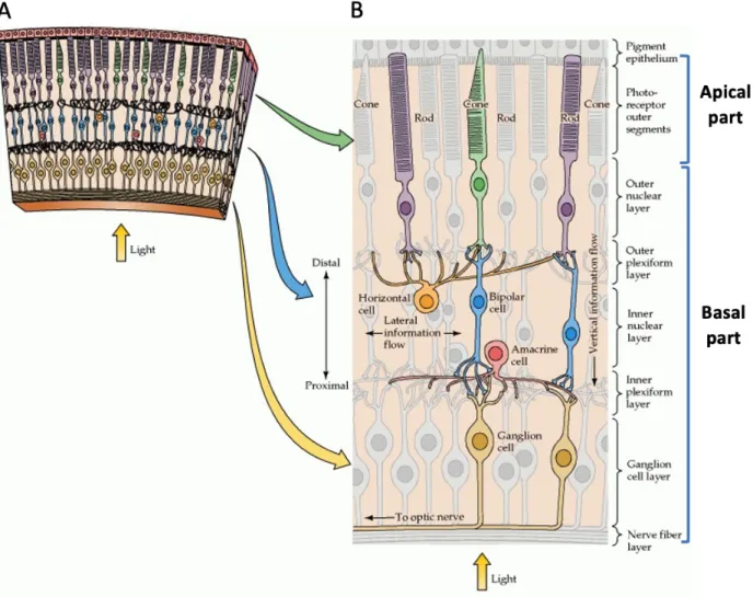
The neural retina is the most accessible part of the vertebrate CNS and it is an excellent system to study neurogenesis, at both molecular and cellular levels (Centanin & Wittbrodt, 2014). The pioneers in the vertebrate retinal studies were Ferrucio Tartuferi and Santiago Ramon y Cajal more than 100 years ago, who first described the structure of retina (R. H. Masland, 2001; Ramón y Cajal, 1892). The vertebrate retina is a multilayered tissue, approximately 200 µm thick (in the case of the mouse) located at the posterior part of the eye (Richard H. Masland, 2012). It is composed of eight major different cell types, distributed into three main layers and interconnected by synapsis in the plexiform layers (Figure 1A). Seven cell types (ganglion cells,
amacrines, bipolars, horizontals, rods, cones, and Müller glia) arise from a pool of retinal progenitor cells (RPCs), whereas astrocytes are produced in the brain and migrate into the retina through the optic nerve. RPCs are organized in a neuroepithelium, where each RPC contacts neighbouring RPC in both apical and basal laminae (Centanin & Wittbrodt, 2014). Notably, before neurogenesis the vertebrate embryonic retina is a sheet of epithelial cells, called pseudostratified neuroepithelium, where establishment and maintenance of apicobasal polarity are regulated by Par, Crumbs and Scribble complexes (Malicki, 2004). The studies on zebrafish and medaka revealed that mutations in the genes that regulate the apico-basal polarity cause severe retinal disorganisation (Herder et al., 2013; X. Wei & Malicki, 2002).
In mouse, the retina is made of more than 60 different cell subtypes, where each of them has a specific role in the vision process (Richard H. Masland, 2012). They are generated sequentially during eye development and the beginning of the retina tissue formation starts during the early embryonic day 9 (E9) when the RPCs undergo symmetric and asymmetric divisions (Heavner & Pevny, 2012). While the early-born retinal neurons are ganglion cells, horizontal, amacrine interneurons, and cone photoreceptors, the late-born ones are rod PR, bipolar interneurons, and Müller glia (Figure 1B) (Heavner & Pevny, 2012). In terms of their function, the light-detecting cells are rod and cone, projecting neurons are retinal ganglion cells (RGC) and interneurons (amacrine, horizontal and bipolar). The amacrine cells process the received information from outside of the eye to transmit it to the PRs, horizontal and glial cells (Müller glia). Horizontal cells provide feedback to PRs and bipolar cells, and these are subdivided into rod bipolar and cone, respectively. Both types of bipolar cells transfer PRs output to all amacrine cells and RGC (Heavner & Pevny, 2012; Richard H. Masland, 2012; Sanes & Zipursky, 2010; Wässle, Puller, Müller, & Haverkamp, 2009).
Among mammals, the composition of the retina is conserved, but the total number of cell-types can vary from one species to another. The human retina, for example, contains approximately 6 to 7 million PRs in total, out of which, rod PRs make 95 %, and cones 5% (Mahabadi & Al Khalili, 2019). In diurnal mammals, the total cone cells number can vary from 8 % to 95 % (Ahnelt & Kolb, 2000; Peichl, 2005). For example, the pig retina is rod dominated where only up to 20 % of cells
are cone cells (Hendrickson & Hicks, 2002). In contrast, nocturnal species has a rod-dominated retina, where the ratio of the rod to cone is 12.4 to 1 (van der Merwe et al., 2018).
Figure 1. – Retina structure adopted from (Purves et al., 2001). (A) Section of the retina with cellular
organisation. (B) Schematic representation of retinal architecture: light detection rod and cone photoreceptors, projection neurons – retinal ganglion cells (RGC) and interneurons – bipolar, amacrine and horizontal cells.
Rod and cone PR cells are neurons that present a remarkable level of sub-compartmentalization of the cell body, related to their specialized function of light detection and contain two different light-sensitive proteins. Rods contain rhodopsin (R. H. Masland, 2001; Sanes & Zipursky, 2010), and the 2 types of cones in mice, the M and S cones are with similar structure and functions and contain opsins (Richard H. Masland, 2012; Thoreson & Dacey, 2019). Rod and cone PRs are active
during different times of the day. While the cones are responsible for colour (photopic) vision during daylight, the rods are accountable for night (scotopic) vision. Additionally, rods have a slow speed response, compared to the rapid one in cones (Mahabadi & Al Khalili, 2019).
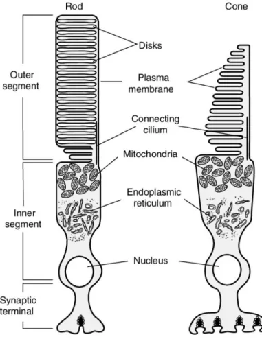
PR cells are composed of the inner segment (IS), where the biosynthesis machinery resides to produce the vital important proteins, the outer segment (OS), where the phototransduction processes occur, and the nucleus. The IS and OS are connected between each other by a microtubule structure, named connecting cilium (Figure 2). All produced proteins from IS pass through the connecting cilia to build up the OS of PRs. The OS of PRs consist of stacks of membranous discs, which contain opsin that forms a chromophore when bound to the 11-cis retinal visual pigments necessary for phototransduction. The IS contains most of the cellular organelles and the protein machinery where all proteins are produced and transferred to the OS through the connecting cilia. The connecting cilium is a specialized non-motile cilium, which is an evolutionally conserved structure that has multiple functions in the developing and mature organisms, for example, sensory function (Sedmak & Wolfrum, 2011). Disruption of cilia is associated with several human disorders such as retinal degeneration, hearing impairment, polycystic kidney and liver, hydrocephalus and dyskinesia (Badano, Mitsuma, Beales, & Katsanis, 2006; Rothschild, Francescatto, Drummond, & Tombes, 2011).
Figure 2. – Schematic diagram of the structure of the photoreceptor. Figure adapted from (Cote,
2019). The phototransducing outer segment (OS) is connected to the inner segment (IS) by the connecting cilium.
1.4. Photoreceptor ciliogenesis
Cilia are a microtubule-based small organelle protruding from the cell surface that play the role of sensory organelles, which help to interpret various environmental signals. Motile cilia were discovered by Antony van Leeuwenhoek in 1670 in protozoa (Dobell & Leeuwenhoek, 2011), and the primary cilia in late 19th century by the Swiss anatomist, KW Zimmerman (Zimmermann, 1898). Cilia are made of a microtubule cytoskeleton that forms the ciliary axoneme that grows from and continues the ninefold structure of the centriole (Satir & Christensen, 2007). Cilia is an evolutionarily conserved structure, and its size can vary from 250 nm in diameter and 100-400 nm in length (Roman, Garrido-Jimenez, Diaz-Chamorro, Centeno, & Carvajal-Gonzalez, 2019).
In the animal kingdom, there are different types of cilia, such as motile cilia (9+2) in which nine doublet microtubules surround a central pair of singlet microtubules (Bayless, Navarro, & Winey, 2019), non-motile (primary) cilia (9+0) in which a central pair is missing and nodal cilia, with (9+0) structure that also misses the central pair but has outer dynein arms (ODA) (Satir & Christensen, 2008). Motile cilia are multifunctional organelles that have a function of transporting the extra-cellular fluid. However, immotile (primary) cilia are known for sensing extra-extra-cellular cues to the cell (Bayless et al., 2019; Hua & Ferland, 2018). Malformations of the cilia lead to many human disorders such as primary ciliary dyskinesia, Meckel syndrome, Joubert syndrome, and retinal degeneration, referred to ciliopathies (Reiter & Leroux, 2017). Evolutionally conserved across vertebrates, immotile cilia are exerting a wide range of functions in different organs. For example, the role of cilia in the olfactory epithelium is to detect odorants (Kaupp, 2010). In the kidney epithelium and the ear, primary cilia act as mechanosensors, detecting the fluid flow (Praetorius & Spring, 2003). In PR cells, they generate the light-sensitive OS (Baylor, Lamb, & Yau, 1979), with mutations of cilium genes causing PR cell degeneration.
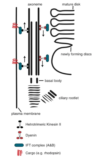
PRs genesis is a long developmental process that begins at the early embryonic stage and ends postnatally where the cone precursors are born at E14, while rod progenitors are born after birth (Morrow, Furukawa, & Cepko, 1998; Rachel, Li, & Swaroop, 2012). The first step of ciliogenesis occurs postnatally when the basal body docks at the cell cortex, with generation and extension of the rod axoneme (Sedmak & Wolfrum, 2011). Basal body ultrastructure reveals a symmetrical array of nine microtubules in a triplet arrangement of A, B and C tubules (Baehr et al., 2019). Soon after birth, the mother centriole acquires a Golgi-derived ciliary vesicle that mediates docking to the cell membrane (Sorokin, 1962). After basal body docking to the cell membrane, A and B tubules arise from basal body forming the proximal axoneme which matures to the connecting cilia (Baehr et al., 2019). The primary cilia of all ciliated cells lack an in situ biosynthesis machinery, meaning that all molecular components forming the cilium are synthetized in the cell prior to transport to the cilium (Ramamurthy & Cayouette, 2009). The formation and maintenance of cilia requires intraflagellar transport (IFT) which refers to the anterograde and retrograde movement of IFT components within the axoneme (Figure 3) (Krock & Perkins, 2014; Rosenbaum & Witman,
2002), initially studied in the unicellular organism Chlamydomonas (Kozminski, Johnson, Forscher, & Rosenbaum, 1993; Pedersen, Geimer, & Rosenbaum, 2006).
Both cone and rod PRs develop an OS by growing the connecting cilium at the end of the apical part of the cell (Ramamurthy & Cayouette, 2009). OS is comprised of a stack of membranous disks that contain a high density of opsin (Röhlich, 1975; Young, 1967). The process of disk membrane formation is maintained in fully mature PRs, with approximately 10 % of the whole length of the OS renewed every 24 hours, demonstrating that protein trafficking initiated during maturation of the OS, is prolonged in mature PRs (Young, 1967). For instance, every minute, 2000 molecules are transported from the IS to the OS of a mature mouse rod PR cell (Insinna & Besharse, 2008). The distinctive feature in the morphology of PRs is that in cone PRs, the disc membranes are continuous with, whereas in rod PRs the discs are separated from the plasma membrane (Ramamurthy & Cayouette, 2009). The first discs formation of the mammalian rod PRs, occurs around P9 by the membrane evagination (Burgoyne et al., 2015; Ding, Salinas, & Arshavsky, 2015). By P21, the OS of mouse PRs is fully mature.
Figure 3. – Protein trafficking in photoreceptors. Figure taken from (Ramamurthy & Cayouette,
2009). Schematic representation the intraflagellar transport (IFT) in the vertebrate photoreceptor. The kinesin II and dynein demonstrate the anterograde and retrograde transport within axoneme. The motor proteins transport the cargo e.g. rhodopsin with IFT complex.
1.5. The role of CaM Kinase family in the CNS
Calcium/calmodulin-dependent protein kinase (CaM Kinase) was identified in nervous tissue and requires for its activation a heat-stable protein factor as well as Ca2+ (Huttner & Greengard, 1979).
Later on, CaM was identified as a regulator of myosin light chain (Yagi, Yazawa, Kakiuchi, Ohshima, & Uenishi, 1978) and phosphorylase kinase activity (PhK) (Cohen et al., 1978). Current nomenclature is based on the loading of brain extract into a fractionation column, and includes CaMK I to IV (Yamauchi & Fujisawa, 1983).
Calcium/calmodulin-dependent protein kinase type II (CaMKII) is activated by Ca2+ and it is essential for the regulation of gene expression, cell cycle control, neurotransmitter synthesis, synaptic plasticity, long term potentiation (LTP) and long term depression (LTD) (Bayer & Schulman, 2019b; Küry et al., 2017; Puram et al., 2011). CaMKII has many isoforms, which are encoded by four different genes. Each CaMKII gene generates different splice variants depending on the region of expression (Bayer, Koninck, & Schulman, 2002). CaMKII is ubiquitously expressed in many regions of the brain and it exceeds 1-2 % of the total amount of proteins. The synaptic activity in the brain is a critical component of learning and memory, and its impeded function leads to many neurological and psychiatric disorders (Bliss, Collingridge, & Morris, 2014; Grant & Silva, 1994). Apart from the brain, CaMKII is important in kidney development and stabilisation of cilium in the pronephric kidney (Bayer & Schulman, 2019a; Küry et al., 2017; Rothschild et al., 2011).
All Calcium/calmodulin-dependent protein kinase (CaMK) isoforms are closely related to each other. CaMKII family contains seven members: four CaMKII isoforms are encoded by different genes, two PhK (phosphorylase kinase) and CASK (Bayer & Schulman, 2019b). CaMKII a, b, g and d are highly homologous and demonstrate differential but overlapping expression patterns in different tissues, brain regions and developing stages. CaMKII a and b subunits are mostly expressed in the brain (especially in hippocampus and neocortex) and they form dodecameric structure containing either one or both subunits (Lisman et al., 2002). On the other hand, CaMKII g and d isoforms are expressed ubiquitously in early developmental stages (Bayer, Löhler, Schulman, & Harbers, 1999; Bayer & Schulman, 2019a). Recently, identification of de novo rare 19 CaMKIIA and CaMKIIB mutations were shown to affect neuronal migration and cause intellectual disability (Küry et al., 2017).
Each CaMKII isoform contains two regulatory domains, catalytic and autoinhibitory (Figure 4A). The catalytic/regulatory domain contains ATP- and substrate binding sites, as well as site of interaction with anchoring proteins. Catalytic and autoinhibitory domains bind through T and S sites forming a “gate” that regulates protein activity. In the presence of Ca2+/calmodulin, these domains dissociate leading to the kinase activation. Once the T site is phosphorylated the gate cannot be closed even after Ca2+/calmodulin dissociation from the enzyme (Figure 4B) (Lisman et al., 2002). Furthemore, CaMKII can be activated autonomously by autophosphorylation at Thr286 site (Figure 4B). Autophosphorylation occurs as an inter-subunit reaction within the holoenzyme, and it requires 2 molecules of calmodulin. Ca2+/calmodulin activates the “kinase” subunits and presents effectively the “substrate” subunit for autophosphorylation (Hanson, Meyer, Stryer, & Schulman, 1994). Finally, its activity can be also regulated through the NMDA (N-methyl-D – aspartate) receptor binding to the T site (Lisman et al., 2002).
Figure 4. – CaMKII structure was taken from (Lisman et al., 2002). (A) Schematic represents the
different protein regulatory domains. (B) Schematic represents inactive and active forms of protein.
In the vertebrate retina, CaMKII isoforms distributions are not described during development, and poorly studied in the adult retina. CaMKIIG is ubiquitously expressed in the entire retina while CaMKIID was present in bipolar and all amacrine cells (Tetenborg et al., 2017). Moreover, the nuclear isoform of CaMKIIaB is highly expressed not only in midbrain and diencephalon but also
in the developing retina regulating ganglion cells survival response (W. Fan, Li, & Cooper, 2007). No function for CaMKII proteins was described in the retina to date.
Given that the role of Calcium/calmodulin-dependent protein kinase II isoform D (CaMKIID) in the mammalian retina is poorly studied, its potential interaction with Par3 allowed us to hypothesise that the Par3 protein complex may recruit CaMKIID to initiate ciliogenesis in the photoreceptor cells.
Hypothesis and aims
Previous work in our lab has shown that conditional ablation of partitioning defective 3 (Pard-3/Par3) gene in retinal progenitor cells of the developing mouse retina leads to a severe disruption of lamination of the retina associated with a defective formation of the apical domain of PRs, leading to their degeneration (unpublished data). To understand the molecular basis of Par3 function in the developing retina, several potential Par3 interacting protein partners were identified by mass spectrometry on retinal protein extracts immunoprecipitated with an antibody directed against the Par3 protein. Interestingly, a cluster of CaMKII was identified among the most abundant Par3 interacting partners, with the isoform D being the most enriched.
The aim of this project is to identify the role of CaMKII in the retinal neurons in order to further elucidate the mechanism by which the loss of the polarity determinant Par3 leads to degeneration. As we identified CaMKIID as a potential binding partner of Par3, we hypothesised that Par3 may require CaMKIID interaction in order to initiate ciliogenesis in PRs. To test this hypothesis, I aimed to characterize the localization of CaMKIID in vivo in adult mouse retina and compare with its localization in Par3 conditional knock out retinas (Aim 1). Then, to validate the protein-protein interactions, I used over-expression models in vitro (Aim 2). By inducing CaMKIID downregulation with shRNAs my goal was to assess the physiological relevance of this protein in vivo in mouse P0 retinal progenitors (Aim 3). Finally, I wanted to understand whether mutations in CaMKIID catalytic/regulatory domain (leading to a constitutively active protein form) and ATP binding domain (leading to a formation of a dominant-negative form) can have any changes in the photoreceptors structure and localization in the retina (Aim 4).
Chapter 2- Material and Methods
2.1. Cloning
CaMKIID coding sequence (Table 1) was cloned in different mammalian expression vectors using Gateway system (Thermo Fischer) together with In-Fusion HD system (Clontech Cat.No.638909) and validated by both sequencing and Western Blot (ATCC; CRL-11268). For amplification of the mus musculus coding sequence of the CaMKIID, we used extracted retinal total RNA from retinas of C57B6J mice retrotranscribed into cDNA. The primers,
FOR-GGGGACAAGTTTGTACAAAAAAGCAGGCTTAATGGCTTCGACCACCACC,
REV-GGGGACCACTTTGTACAAGAAAGCTGGGTTTTAGTTGATGGGTACTGTGG were used for the CaMKIID gene amplification. All primers were designed in SnapGeneâ software and then synthesized by IDTä (Integrated DNA Technologies). For the Gateway approach to 5’ of both Forward and Reverse primers were added the sequence of attB1 and attB2 sites respectively. PCR fragment was extracted after electrophoresis migration in 1% agarose gel using the Invitrogen Gel extraction kit (Thermo Fisher Scientific Cat.No.K210012) and cloned into Entry Vector pcr8-GW-TOPO by using GatewayTM BR Clonase Enzyme Mix (Thermo Fisher Scientific Cat.No.11789013). Entry vector containing attL1 and attL2 sites can be recombined with Destination Vector by using Gatewayä LR Clonase Enzyme Mix (Thermo Fisher Scientific Cat.No.11791019). Desired constructs were transformed into DH5a E. coli cells for the copy amplification. The transformed cells were heat-shocked, grown in shaking incubator in S.O.C media (Invitrogen) for 1h at +370C, then plated onto LB-agarose plates with proper antibiotics and incubated overnight at 370C. The following day, colonies were picked up for further screening by the restriction enzymes digestion strategy designed in SnapGeneâ.
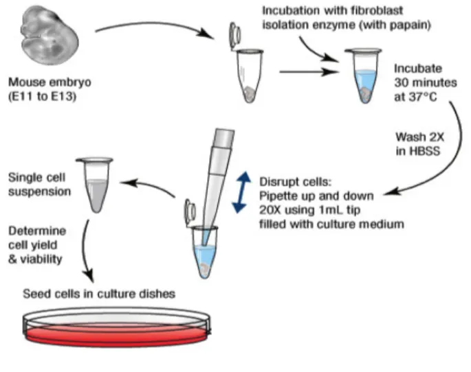
2.2. Derivation of mice primary embryonic fibroblast
Primary Mouse Embryonic Fibroblasts (MEFs) were derived from CD1 mouse embryos at embryonic day 13.5 (E13.5). Briefly, the embryos were taken out from the uterus, separated from placenta and yolk sacs. The individual embryo was placed into cold sterile PBS1x, decapitated, desolated and blood and liver tissue were removed. Embryos were treated with 2,5%
Trypsin/EDTA and triturated by up and down mechanical homogenization for cellular dissociation, followed by addition of DMEM complete medium for Trypsin neutralisation. Cells were then pelleted by centrifugation at 180g for 5 minutes. Cellular pellets were resuspended in fresh DMEM complete medium and cells were plated in 10cm petri dish pre-treated in 1:1 poly-L-lysin: Sterile H2O solution (Sigma Andrich, Cat.No.P4707-50ml) and then 0.1% bovine gelatine solution. The cells were cultured in DMEM, 10% heat-inactivated Cosmic Calf Serum, 1% penicillin-streptomycin, 1% MEM Non-Essential Amino acids (NEAA), 1% sodium pyruvate at 370C with 5% CO2. After 4 passages, cells were used for experiments (Figure 5).
Figure 5. – Experimental outline for the isolation of mouse embryonic fibroblast (MEF) on an embryonic day (E) 13.5 by in vitro culture (ThermoFisher Scientific protocol).
2.3. Induction of cilia growth in MEF cells
To induce ciliogenesis in MEF cells, 24h after shRNA transfection, the medium with 10 % serum was replaced by medium containing 1 % serum (starvation medium). Later on, after 24h of starvation, MEFs were fixed in 4% PFA for 10 minutes at RT and stained with the cilium marker, acetylated tubulin. The enumeration of the number of ciliated cells was done using light fluorescent microscopy in four experiments, and the n varied from 20 to 80 cells per group.
2.4. Cells transfection
To validate our cloning, we transfected HEK293 cells with our plasmids (Table 1). Cells were seed in 6-well plates at the density of 300 x 103 and transfected after 24h using Polyethylenimine, PEI (Polysciences, Inc. Cat. No.23966-1) as a transfection reagent (5% (1mg/ml) PEI, 95% Opti-MEM; 0.5-2 ug DNA). The transfected cells were left at +370C and 5% CO2 for 24h. After transfection cells were collected in PBS1x and lysed with NP-40 (Nonidet P40 Substitute Sigma-Aldrich Cat.No. 74385) lysis buffer (Tris-HCL (pH 7.6), 150mM NaCl, 1% NP-40 with Complete Protease Inhibitor Cocktail (Roche REF.11836153001)).
2.5. Protein detection by Western blot
At postnatal day 0(P0), P10 or P30, mice were euthanized by CO2 asphyxia and enucleated. Eyes were dissected individually in cold PBS1x to isolate the neural retina from the eyecup. Isolated retinas were sonicated using 5 pulses of 5 seconds at the low output (2) in cold NP-40 protein lysis buffer (Tris-HCL (pH7.6), 150mM NaCl, 1% Np-40) with Complete Protease Inhibitors Cocktail (Roche)). Proteins lysate were centrifuged at full speed (13K) for 15 minutes to remove non-dissolved proteins and followed by quantification using the Bradford protein assay (BioRad Laboratories). Between 20 and 100 µg of retina lysates in 1X laemli buffer were loaded in 6.5% Acrylamide gels (BioRad Laboratories). After electrophoresis migration, proteins were transferred onto Low-Fluorescence PVDF membranes using Transblot Turbo (BioRad Laboratories). Membranes were blocked in blocking solution (5% dry milk/TBS-T (10mM Tris, pH8; 150mM NaCl and 0,05 % Tween20)) for 1h at room temperature (RT). Membranes were incubated with primary antibodies overnight at +40C in 1% blocking solution. On the next day, the membrane was washed
in TBS-T solution and incubated for 1h at RT with the appropriate HRP-conjugated secondary antibodies (Table 2) in 1% of blocking solution. After 3 washes of the membrane, HRP activity was visualised on ChemiDoc (BioRad Laboratories) by chemiluminescence using the ECL (Fisherscientific Cat. No.45000875) or ECL Prime kit (Fisherscientific Cat. No.45002401). Protein levels were normalised against the level of housekeeping proteins such as glyceraldehyde 3-phosphte dehydrogenase (Gapdh) or Beta-actin (Actb) using ImageJ software.
2.6. Fluorescent immunolabeling
2.6.1. Immunohistochemistry (IHC)
PFA or TCA fixed mouse eyes were embedded in Tissue-Tek® O.C.T.™ Compound, frozen rapidly using liquid nitrogen and kept frozen at -80° C until sectioned. Frozen eyes were cross-sectioned using a cryostat in slices of 14-18 µm and fixed on treated slides (Denville Ultra Clear Microscope Slides Cat. No.M1021). After slices were dried, slides were rinsed in PBS 1X to remove excess embedding medium from the slice. Slides were then blocked by incubation in blocking solution (1% BSA and 0,2 % triton in PBS 1X) for 1h at RT. Slides were incubated at RT overnight with primary antibody (Table 1) diluted in blocking solution. The next day, the sections were washed with PBS1x and incubated with secondary antibodies coupled to Alexa fluorophore (Table 2) diluted 1/1000 in blocking solution for 1 hour at RT. Finally, after 3 washes, slides were stained with Hoechst (Invitrogen Cat. No.H3570) in dilution 1/10000 and mounted using Mowiol.
2.6.2. Immunocytochemistry (ICC)
Mouse embryonic fibroblast (MEFs) cells were seeded on pre-treated sterile glass coverslips in 24-well plate at density 50 x 103 and cultured in DMEM, 10% Cosmic Calf Serum (CCS), 1% penicillin-streptomycin, 1% MEM Non-Essential Amino acids (NEAA), 1% sodium pyruvate. The glass coverslips were treated with poly-L-Lysin (Sigma Andrich, Cat.No.P4707-50ml) for 30 minutes at RT and 0,1% bovine gelatine solution. Twenty-four hours after seeding, cells were transfected with pSIREN plasmid constructs using Lipofectamine 3000 (Invitrogen cat. No.
L3000015). 24 hours after transfection cells were fixed with PFA 4% for 10 minutes at RT and
triton in PBS 1X) for 1h at RT. Cells were then incubated with primary antibody diluted in blocking solution (Table 2). The following day cells were rinsed 3 times with PBS1x and incubated with secondary antibody coupled to Alexa fluorophore (Table 3) diluted in 1/1000 in blocking solution for 1 hour at RT. Finally, for labelling the cell nuclei Hoechst was used (Invitrogen Cat.No.H3570) at a dilution 1/10000 in PBS1X and mounted with Mowiol.
2.7. Plasmid electroporation into the eye
P0-P1 CD1 pups were anaesthetized using ice for 2-3 minutes. One to 3 µg of plasmids (Table 3) were delivered in the subretinal space of pups using a glass pipette and then electroporated using electrode pad pulsed 5 times in a unipolar direction (50 ms duration, 950ms Interval, 80 Volts). Pups recovered from surgery under a heat lamp and returned to their mother. After 21 days, mice were euthanized, the retinas were collected and fixed in PFA 4% for 30 minutes at RT, gradually equilibrated in 10 % and 20 % sucrose, and embedded in Tissue-Tek® O.C.T.™ Compound (Sakura Cat.No.4583) and frozen rapidly using liquid nitrogen. Retinas were stored at -80 0C until further processing.
2.8. RNA extraction and cDNA synthesis
Total RNA was extracted from adult mice eyes P120 (postnatal day120) using RNEasy Mini Kit (Qiagen Cat. No.74134). cDNA synthesis was performed by using SuperScriptäIV VILO master mix with EZ DNAse (ThernoFisher Cat.No.11766050) on 3 µg of total RNA and stored at -800C. For RT- PCR, 35ng cDNA (equivalent RNA) was used per reaction.
2.9. In vivo system/Mouse lines
The animal experiments were performed in agreement with the Canadian Council on Animal Care (CCAC) guidelines and with the IRCM Animal Care Committee and ethical rules. Crossing 3 different mouse lines generated conditional Pard3 knockout mouse line:
-Rosa Yellow fluorescent protein (RYFP) mouse line (Jackson Laboratory) is conditionally expressing the YFP protein under the endogenous Rosa promoter.
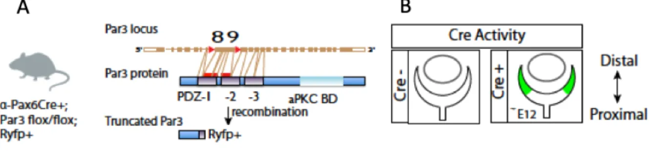
-The alpha-Pax6 Cre-Ires-GFP (MGI: 3052661) mouse line is a transgenic mouse line expressing Cre in peripheral progenitor cells from embryonic day 10.5 (E10.5) (Figure 6B).
-The conditional Par3 knockout mouse line, in which Par3 exons 8 and 9, coding for part of Pard3 domains 1 and 2 are flanked by loxP sites (Floxed) (Figure 6A). Cre recombination generates a shift in the open reading frame, leading to the production of a truncated Pard3 protein. It was generated from the C57BL/6N-Atm1Brd mouse line UC Davis (MGI: 2135608) in which the FRT sites where previously recombined using a mouse line expressing Flippase.
Figure 6. – Par3 conditional knock out (Par3 cKO) mouse line generation. Par3 cKO mouse line was
generated by crossing three different mouse lines: alpha Pax6Cre+; Pard3fl/fl and RosaYFP. The alpha Pax6Cre+ mouse contains a loxP-STOP-loxP Rosa YFP cassette and it was crossed with Pard3fl/fl to induce the Padr3 gene deletion from progenitor cells at the peripheral retina. (A) Schematic presentation of different domains of Pard3 gene. (B) Schematic represents the localisation of Cre expression at the peripheral part of the embryonic retina (E12).
2.10. Co-Immunoprecipitation
After transfection, HEK293T cells were lysed in NP-40 buffer plus complete inhibitors, sequentially quantified using Bradford protein assay. Immunoprecipitation was performed by using the superparamagnetic beads Dynabeadsâ Protein G (Thermo Fisher Scientific Cat. No.1000D3). Briefly, protein G dynabeads were coupled with appropriate primary antibodies in different dilutions (Table 1) in PBS1X-0.05% Tween for 1 hour at +40C. One milligram of total protein extract was added to the beads and incubated overnight at +40C with IpH buffer (50mM Tris pH 8.0, 150 mM NaCl, 5mM EDTA, 0.1% NP-40). Next day, beads were rinsed 3 times with IpH buffer
using the MagnaBind magnet (Pierce). After washes, beads were resuspended in 1x Lameli buffer and boiled for 3 minutes at 950 C. Sequentially samples were loaded in an acrylamide gel.
2.11. Immunoprecipitation Mass-Spectometry (IP-MS)
At postnatal day 10 and 30 (P10 and P30) mice were euthanized by CO2 asphyxia and enucleated. Eyes were dissected individually in cold PBS1x to isolate the neural retina. Isolated retinas were sonicated using 5 pulses of 5 seconds at the low output (2) in cold NP-40 protein lysis buffer (Tris-HCl (pH7.6), 150mM NaCl, 1% Np-40) with Complete Protease Inhibitors Cocktail (Roche). Protein lysates were centrifuged to remove non-dissolved proteins and followed by quantification using the Bradford protein assay (BioRad Laboratories). Immunoprecipitation was performed by using the superparamagnetic beads Dynabeadsâ Protein G (Thermo Fisher Scientific Cat. No.1000D3). Briefly, protein G beads were coupled with appropriate primary antibodies in different dilutions (Table 1) for 1 hour at +40C. One milligram of total protein extract was added to the beads and incubated overnight at +40C with IpH buffer (50mM Tris pH 8.0, 150 mM NaCl, 5mM EDTA, 0.1% NP-40). Next day, beads were rinsed 3 times with IpH buffer using the MagnaBind magnet (Pierce), which were then replaced by freshly made cold 50mM Ammonium Bicarbonate (Sigma Aldrich Cat.No.A6141) buffer. The on-beads proteins were digested by trypsin overnight at +370C and washed several times with different solutions following the manufacturer’s instructions. After elution in 10% ammonium hydroxide/90% methanol (v/v), samples were dried with a Speed-vac, reconstituted under agitation for 15 min in 12 µL of 2%ACN-1%FA and loaded into a 75 μm i.d. × 150 mm Self-Pack C18 column installed in the Easy-nLC II system (Proxeon Biosystems). The peptides were eluted with a two slope gradient at a flow rate of 250 nL/min. Solvent B first increased from 1 to 38% in 105 min and then from 38 to 86% B in 25 min. The HPLC system was coupled to Orbitrap Fusion mass spectrometer (Thermo Scientific) through a Nanospray Flex Ion Source. Nanospray and S-lens voltages were set to 1.3-1.7 kV and 50 V, respectively. The capillary temperature was set to 225 °C. Full scan MS survey spectra (m/z 360-1560) in profile mode were acquired in the Orbitrap with a resolution of 120,000 with a target value at 1e6. The most intense peptide ions were fragmented in the HCD cell and analysed in the linear ion trap with a target value at 2e4 and normalized collision energy at 28 V. A MS3 scanning was performed upon detection of a neutral loss of phosphoric acid (48.99, 32.66 or 24.5 Th) in HCD MS2 scans. The
duty cycle was set to 3 seconds and target ions selected for fragmentation were dynamically excluded for 30 sec after 3 MS/MS events.
The peak list files were generated with Proteome Discoverer (version 2.3) using the following parameters: minimum mass set to 500 Da, maximum mass set to 6000 Da, no grouping of MS/MS spectra, precursor charge set to auto, and the minimum number of fragment ions set to 5. Protein database searching was performed with Mascot 2.6 (Matrix Science) against the Uniprot Mus musculus protein database (April 15th, 2015). The mass tolerances for precursor and fragment ions were set to 10 ppm and 0.6 Da, respectively. Trypsin was used as the enzyme allowing for up to 1 missed cleavage. Cysteine carbamidomethylation was specified as a fixed modification, and methionine oxidation and phosphorylation S/T/Y as variable modifications. Data interpretation was performed using Scaffold (version 4.8).
2.12. Short hairpin RNA generation
shRNAs against murine CaMKIID were designed by using InvivoGEn’s siRNA Wizard software. Oligonucleotides were produced by IDTä (Integrated DNA Technologies). The efficiency of shRNAs was tested in HEK293 together with the overexpression of the gene of interest (CaMKIID) by using (jetPRIME®, DNA and shRNA transfection reagent VWR-114-07 CA89129-922). Immunoblotting analysis was used to assess the potency of shRNA-mediated knockdown against CaMKIID.
2.13. Statistical analysis
All statistics were performed with GraphPad Prism Version 8ã. For the multiple comparisons, a Tukey's and Dunnett’s tests were applied. For the comparison of three and more groups, one-way and two-one-way ANOVA was applied. Statistical significance was defined when P< 0.05.
2.14. Quantitative analysis of the images
To quantify the number of ciliated MEF cells after serum starvation we used a DM6000 (Leica) microscope. The cells were cultured, fixed and immunostained with appropriate antibodies as described in section 2.5.2. After that, transfected cells of each condition were randomly selected,
counted on the presence of cilium and the numbers were converted in percentage of the total number of counted cells.
The imaging of retinal sections was performed with the confocal microscope SP8 (Leica). To measure the length of the IS and OS of PRs, the retina sections were stained with appropriate primary and secondary antibodies as described above. The length of the IS and OS of GFP positive PRs were individually measured using Volocity® Version 6.0.
To analyse the apico-basal distribution of PRs nuclei, we used ImageJ applying the FIJI macro to divide the outer nuclear layer (ONL) of the retina in three equal compartments (apical, middle and basal). Nuclei were counted separately in each compartment and the number was converted in percentage.
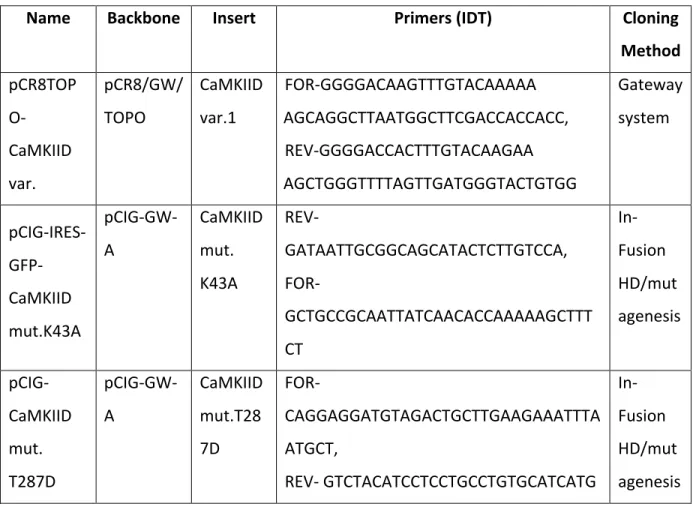
Tableau 1. – List of generated plasmids
Name Backbone Insert Primers (IDT) Cloning Method pCR8TOP O-CaMKIID var. pCR8/GW/ TOPO CaMKIID var.1 FOR-GGGGACAAGTTTGTACAAAAA AGCAGGCTTAATGGCTTCGACCACCACC, REV-GGGGACCACTTTGTACAAGAA AGCTGGGTTTTAGTTGATGGGTACTGTGG Gateway system pCIG-IRES- GFP-CaMKIID mut.K43A pCIG-GW-A CaMKIID mut. K43A REV-GATAATTGCGGCAGCATACTCTTGTCCA, FOR-GCTGCCGCAATTATCAACACCAAAAAGCTTT CT In-Fusion HD/mut agenesis pCIG-CaMKIID mut. T287D pCIG-GW-A CaMKIID mut.T28 7D FOR-CAGGAGGATGTAGACTGCTTGAAGAAATTTA ATGCT, REV- GTCTACATCCTCCTGCCTGTGCATCATG In-Fusion HD/mut agenesis
pSIREN- shRNA-CaMKIID pSIREN- RetroQ-Zsgreen CaMKIID variant1 FOR- GATCCGGATCTGTCAACGCTCTACTGTTTCAA GAGAACAGTAGAGCGTTGACAGATCTTTTTT GCGGCCGCG, REV- AATTCGCGGCCGCAAAAAAGATCTGTCAACG CTCTACTGTTCTCTTGAAACAGTAGAGCGTTG ACAGATCCG Oligo A annealin g
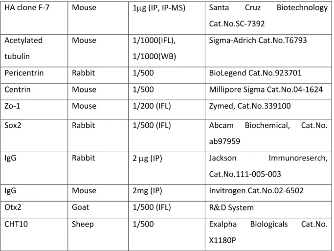
Tableau 2. – List of primary antibodies
Antigen Species Dilution Sources
CaMKIID Rabbit 1/1000 (IFL), 1/500 (WB)
2µg (IP)
LsBio Cat.No. LS-C329304
Ninein Goat 1/100 (IFL) Santa Cruz Cat.No. SC-50142
Na/P-ATPase Alpha 3
Mouse 1/100 (IFL) Novus Biologicals Cat. No. NB300-540SS
Rhodopsin Rabbit 1/500 (IFL) GeneTex Cat. No. GTX129910
Par3 Rabbit 1/1000(WB),
1/500(IFL), 2µg (IP)
Millipore Sigma Cat. No. 07-330
GFP Rabbit 1/5000 (WB), 2µg
(IP)
Thermo Fisher Scientific Cat.No.A11122
GFP Chicken 1/1000 (IFL) Abcam Cat.No.ab13970
GAPDH Mouse 1/2000 (WB) Millipore Sigma Cat.No. MAB374
B-actin Mouse 1/1000 (WB) Sigma-Adrich Cat.No.A5441
Myc (9E10) Mouse 1/500(IFL), 1/1500 (WB), 0.7µg (IP)
HA clone F-7 Mouse 1µg (IP, IP-MS) Santa Cruz Biotechnology Cat.No.SC-7392 Acetylated tubulin Mouse 1/1000(IFL), 1/1000(WB) Sigma-Adrich Cat.No.T6793
Pericentrin Rabbit 1/500 BioLegend Cat.No.923701
Centrin Mouse 1/500 Millipore Sigma Cat.No.04-1624
Zo-1 Mouse 1/200 (IFL) Zymed, Cat.No.339100
Sox2 Rabbit 1/500 (IFL) Abcam Biochemical, Cat.No.
ab97959
IgG Rabbit 2 µg (IP) Jackson Immunoreserch,
Cat.No.111-005-003
IgG Mouse 2mg (IP) Invitrogen Cat.No.02-6502
Otx2 Goat 1/500 (IFL) R&D System
CHT10 Sheep 1/500 Exalpha Biologicals Cat.No.
X1180P IFL-immunofluorescence, WB – western blot, IP - immunoprecipitation
Tableau 3. – List of the secondary antibodies
Fluorochrome Species Dilution Sources
Anti-rabbit HRP Goat 1/10000 (WB) Jackson
Immunoresearch,Cat.No.111-035-144
Anti-mouse HRP Goat 1/10000 (WB) Jackson
Immunoresearch,Cat.No.115-035
Protein A, HRP conjugate
Goat 1/5000 (WB) Millipore Sigma Cat.No.18-160
Anti-mouse AlexaFlour488
Donkey 1/1000 Thermo Fisher Scientific
Cat.No.A-21202 Anti-mouse
AlexaFlour555
Donkey 1/1000 Thermo Fisher Scientific
Cat.No.A-32773 Anti-mouse
AlexaFlour647
Donkey 1/1000 Thermo Fisher Scientific
Cat.No.A-31571 Anti-rabbit
AlexaFlour488
Donkey 1/1000 Molecular Probes Cat.No.
A21206 Anti-rabbit
AlexaFlour555
Donkey 1/1000 Molecular Probes Cat.No.
A31572 Anti-rabbit
AlexaFlour647
Donkey 1/1000 Thermo Fisher Scientific
Cat.No. A31573 Anti-chicken
AlexaFlour488
Donkey 1/1000 Thermo Fisher Scientific