Role of Metallothioneins in Irradiated Human
Rectal Carcinoma
Hanifa Bouzourene,1,M.D. Pascal Chaubert,1,M.D. Sandra Gebhard,1,M.D. Fred T. Bosman,1,M.D., Ph.D. Philippe Coucke2,M.D.1Institute of Pathology, Lausanne, Switzerland. 2Department of Radiation-Oncology, Centre
Hos-pitalier Universitaire Vaudois, Lausanne, Switzer-land.
Address for reprints: Hanifa Bouzourene, M.D., Insti-tute of Pathology, 25 Rue du Bugnon, CH 1010 Lausanne, Switzerland; Fax: ⫹011-41-21-314-72-07; E-mail: hanifa.bouzourene@chuv.hospvd.ch Received October 23, 2001; revision received Feb-ruary 13, 2002; accepted April 5, 2002.
BACKGROUND. Metallothioneins (MT) are low-molecular weight, metal-binding proteins that play a role in cellular proliferation and differentiation, as well as in cellular defense mechanisms. They act as scavengers of free radicals produced by irradiation. A number of in vitro and in vivo studies have linked overexpression of cellular MT with tumor cell resistance to radiation. This is the first study that investigates whether MT expression is involved in the radioresistance of rectal carcinoma.
METHODS.Using a mouse monoclonal antibody, MT expression was analyzed by immunohistochemistry on surgical samples (n⫽ 85) from 85 patients with locally advanced rectal carcinoma who were treated preoperatively with a hyperfraction-ated and accelerhyperfraction-ated radiotherapy schedule and on tumor biopsies (n ⫽ 13) obtained before treatment. The potential correlations between MT expression and pathologic variables and survival were examined.
RESULTS.MT were expressed strongly in both the cytoplasm and nucleus of tumor cells in 7 biopsy and 42 surgical samples. A comparison of MT expression in biopsy and surgical specimens showed that MT expression did not change after irradiation in most cases. Against all expectations, MT were expressed more frequently in tumors from responders than in those from the nonresponders (P⫽ 0.02). There was no correlation between MT expression and tumor stage, histology after radio-therapy, or survival.
CONCLUSION.These findings do not support the hypothesis that MT overexpression at the end of radiotherapy is a marker for radiation resistance. Cancer 2002;95: 1003– 8. © 2002 American Cancer Society.
DOI 10.1002/cncr.10780
KEYWORDS: metallothionein, rectum, cancer, radiotherapy, in vitro.
F
ailure to control local tumor growth and recurrence is due to the acquired or inherent radioresistance of the tumors. Over the last decade, there has been increasing interest in biologic markers to stratify cancers according to their response to chemotherapy or ra-diotherapy. Even if several factors are known to cause resistance to chemotherapeutic drugs and radiation, there have been only a few reports on the prediction of response to radiotherapy. Metallothio-neins (MT) represent a group of low-molecular weight cysteine-rich intracellular proteins that bind and detoxify heavy metal ions.1,2 Syn-thesis of MT is induced in a variety of normal and tumor tissues by these metal ions, as well as by endogenous factors such as glucocor-ticoids, cytokines, and vitamin D.3,4MT are involved in a transient response to any form of stress or injury, providing a cytoprotective mechanism against the potentially damaging effects of alkylating agents and oxygen-derived free radi-cals, particularly those produced by irradiation.1,5Therefore,
tion of MT synthesis may be one of the most impor-tant defense mechanisms against DNA-damaging agents.6 –13Supporting this hypothesis, several groups have found that an increased level of MT in cultured cells or xenograft tumors is associated with resistance to ionizing radiation.6,11,12,14 However, there is no study demonstrating such a correlation in human pri-mary tumors treated with only neoadjuvant radiother-apy.
By using immunohistochemistry, MT are detect-able at a basal level in a variety of normal and tumoral human tissues.15–20Increased expression is found fre-quently in patients with colorectal adenomas and ad-enocarcinomas,21,22 hepatocellular carcinomas,23 tes-ticular embryonal carcinomas,24 thyroid tumors,25 transitional cell carcinomas of the bladder,26 and breast carcinomas.27
Our goal was to determine whether MT expression has any prognostic value for tumor response and sur-vival in patients with locally advanced rectal carcino-mas treated with preoperative hyperfractionated and accelerated radiotherapy (HART).
MATERIALS AND METHODS
Patients
This study included 105 consecutive patients with lo-cally advanced rectal carcinoma (T3–T4, N0, or N1 or any T but N1) who were eligible for Trial 93-01. These patients were treated with HART at the Institute of Pathology, Lausanne, between 1992 and 1998.28,29This protocol was reviewed by the local ethics committee. Informed consent was obtained from all patients. Sat-isfactory immunostaining with MT antibody was achieved in 85 of these 105 patients. Subsequent anal-ysis was limited to these patients. They ranged in age from 28 to 85 years (median age, 63 years) and the male-to-female ratio was 1:2. Before the start of treat-ment, all patients underwent a complete clinical ex-amination, blood count, assessment of renal and liver function, and a carcinoembryonic antigen assay. His-tologic confirmation of malignancy was available for all patients but pretherapeutic biopsies were retriev-able for only 34 patients. Distant metastatic disease was excluded by chest X-ray, abdominal ultrasound, and thoracoabdominal computed tomography (CT) scan. The assessment of the local extension of the tumor was based on digital rectal examination, com-pleted by rectal ultrasound and CT scan. In total, 54 patients were staged T3, 14 were N1, and 30 were T4, 6 of whom were N1 and 1 of whom was T2N1. All patients received the same radiotherapy schedule, which was delivered with a linear accelerator with a minimal energy of 6 MV (1.6 Gy twice a day, with 6 hours as the interfraction interval). The total dose was
41.6 Gy in 26 fractions administered over 2.5 weeks. The interval between the end of radiotherapy and surgical resection was kept as short as possible. Sur-gery was usually performed within 6 days (the median was 5 days).
Macroscopic Examination of Surgical Specimens
The surgical specimens were opened through the an-terior wall and fixed in 10% buffered neutral formalin for 24 hours. The whole tumor and attached mesorec-tum were sliced serially in 3– 4-mm slices and the whole tumor was included for histologic examination. For assessment of perirectal lymph nodes, adipose tissue was removed after tumor sampling and cleared in a Carnoy solution for 24 hours.
Samples
In 21 of 34 pretherapy biopsies, the material was fixed in sublimated formol. The remaining 13 biopsies and surgical specimens were fixed in 10% neutral formalin for 12–24 hours at room temperature. Tumor samples were embedded in paraffin. From the tissue blocks, 4-m thick sections were obtained and stained with hematoxylin and eosin according to standard proce-dure. For each case, one paraffin block with normal mucosa adjacent to the carcinoma was selected for the detection of MT protein expression by immunohisto-chemistry.
Histologic Assessment
All 85 irradiated rectal tumors were reviewed by the same pathologist (H.B). The tumors were classified according to the World Health Organization criteria of intestinal carcinoma30 and staged according to the TNM classification.31 Tumor regression was graded according to the presence of residual tumor cells and the extent of fibrosis. Grade 1 was defined by the absence of residual cancer and fibrosis extending through the layers of the rectal wall, Grade 2 was characterized by the presence of residual cancer cells and fibrosis, and Grade 3 was characterized by the absence of any tumor regression.
Immunohistochemistry
Four-m thick tissue sections were mounted on amin-opropylmethoxysilane-coated glass slides, deparaf-finized in xylol, taken through to absolute alcohol, blocked for endogenous peroxidase with 1% H2O2in methanol (45 mn), and rehydrated through graded alcohols. They were subjected to microwave oven heating for 15 minutes in 10 mM citrate buffer, pH 6.0, and rinsed in Tris-buffered saline (TBS; Tris 0.05 M, NaCl 0.9%, pH7.6). To reduce nonspecific binding, they were incubated for 10 minutes in normal goat
serum (Pel-Freez Biologicals, Rogers, AK) 1:30 in TBS. After a 30-minute incubation with the primary mono-clonal antibody (MoAb; mouse monomono-clonal anticlone E9 Metallothionein, Dako, Glostrup, Denmark) diluted 1:500 in TBS containing 5% nonfat dry milk (TBS-nfdm), the sections were incubated for 30 minutes with biotinylated horse antimouse immunoglobulins (Vector, Burlingame, CA) diluted 1:400 in TBS-nfdm. They were also incubated for 30 minutes in ABC-peroxidase complex solution (Vector) prepared ac-cording to the manufacturer’s instructions. Peroxidase activity was revealed with 5-5, diaminobenzidine as chromogen and the sections were counterstained in Mayer’s acid-free hematoxylin. As a negative control, the primary MoAb was replaced by a mouse hybrid-oma supernatant of similar isotype (IgG1).
Immunohistochemical expression of MT was not assessable on tissue biopsies fixed in sublimated for-mol. In the remaining biopsies (n ⫽ 13) and surgical specimens (n ⫽ 85), immunoreactivity for MT was evaluated semiquantitatively by two observers using a double-headed microscope (H.B, S.G) without knowl-edge of the tumor stage or the clinical data. Staining for MT in the normal rectal mucosa was used as a positive control (Fig. 1). Absorptive epithelial cells bordering the luminal surface and especially the basal part of the crypts showed MT positivity, generally lo-calized in the paranuclear region and cell membranes and less frequently in the nuclei. If no crypt staining was obtained after two attempts, the sample was elim-inated from the study for technical reasons. In tumor cells, MT immunoreactivity was observed in the cyto-plasm and/or in the nuclei. Positive tumor nuclei and cytoplasm were scored separately as follows: 0⫽ less than 5% of immunostained cells; 1⫽ 5–30% of positive cells; 2 ⫽ 30–60% of positive cells; and 3 ⫽ greater
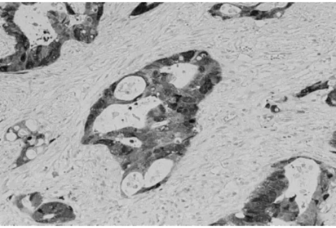
than 60% of positive cells (Fig. 2). The spindle-shaped stromal cells, intermingled with neoplastic glands, sometimes showed staining for MT.
Follow-Up and Statistical Analysis
Follow-up was available for all patients at the date set for collecting data. Overall survival (OS), disease-free survival (DFS), and local control were the endpoints for evaluating the prognostic significance of MT stain-ing. All statistical analyses were conducted using JUMP 3.0 software (SAS Institute, Cary, NC). A P value less than 0.05 was statistically significant. The corre-lation between MT and pathologic factors such as tumor stage, histology, and tumor regression was de-termined by a Pearson chi-square test. Survival curves were estimated according to the Kaplan–Meier method for both cytoplasmic and nuclear MT positiv-ity. The significance between groups for survival curves was estimated by the log rank test.
RESULTS
Pathologic Findings
Correlations between MT expression and histologic parameters in 85 surgical specimens of rectal carci-noma treated with preoperative radiotherapy are sum-marized in Table 1. Of the 85 rectal carcinomas, one was pT1 (1%), 16 were pT2 (19%), 56 were pT3 (66%), and 12 were pT4 (14%). Regional lymph node metas-tases were found in 49 patients (58%). Tumors were classified into well (27%), moderately (42%), and poorly differentiated adenocarcinomas (12%), and mucinous carcinomas (19%). None of the 85 tumors showed complete tumor regression (Grade 1). Partial tumor regression (Grade 2) was observed in 68
pa-FIGURE 1. Nontumoral rectal mucosa showing low-level metallothionein (MT) immunostaining. Some absorptive epithelial cells bordering the luminal surface and crypts show MT positivity.
FIGURE 2.Islets of carcinomatous cells from a rectal carcinoma specimen treated preoperatively with radiotherapy showing cytoplasm and/or nuclear metallothionein immunoreactivity.
tients (80%) and tumor regression (Grade 3) was ab-sent in 17 cases (20%).
Immunohistochemistry confirmed MT expression in 7 and no MT expression in 6 of the 13 tumor biopsies performed before treatment. In the seven specimens expressing tumors, the immunostaining score varied from 1 to 3. In surgical specimens, MT expression was present in 42 carcinomas and absent in 43. The cytoplasmic and nuclear distribution of MT expression and their corresponding scores are sum-marized in Table 2. In 38 of the 42 positive cases, the MT protein was expressed both in the cytoplasm and in the nucleus and the expression score was most frequently 1 (tumor cells with MT expression⬍ 30%). In two cases, MT were expressed only in the cytoplasm
and in two cases, only in the nucleus. When compar-ing MT expression in biopsies and surgical specimens, the MT expression score was identical in five patients and lower in five patients. In three cases, MT expres-sion was present in the biopsies, but not in the surgi-cal specimens.
MT were expressed in 37 of 68 tumors with partial tumor regression (Grade 2), but only in 5 of 17 tumors with no tumor regression (Grade 3; P ⫽ 0.02). There was no statistical correlation between MT expression and tumor stage, tumor grade, and differentiation (Ta-ble 1).
Survival Analysis
The median actuarial OS and DFS period was 52 months. The actuarial local recurrence rate at 2 and 5 years was 6.4% and 7.6%, respectively. During the follow-up (median ⫽ 40 months), 29 patients devel-oped distant metastasis and 8 patients develdevel-oped local recurrence. Forty patients died, 32 of whom died of their carcinoma.
There was no correlation between MT expression and local control, DFS, and OS by univariate analysis. To increase the number of patients per group, the individual scores of MT expression were also com-bined. Using this approach, MT expression also did not correlate with the above-mentioned endpoints.
DISCUSSION
The lack of reliable criteria to predict the outcome after preoperative radiotherapy for individual patients with advanced rectal carcinoma is a major problem. The precise mechanism of radioresistance in experi-mental and human studies is still unknown, and prob-ably multifactorial. Much of the damage induced by ionizing radiation is caused by the oxygen-derived free radicals produced by the radiolysis of water in cells. In recent years, MT have emerged as an important factor in tumor development and progression because of the high Cu/Zn-containing proteins present in patients with malignancies.1,5,8,32–36In addition, MT have been implicated in the transient response to any form of stress or injury such as ionizing radiation and alkylat-ing agents, providalkylat-ing a cytoprotective mechanism against the potential damaging effects of oxygen-de-rived free radicals.1,2,37– 40 Many studies support the hypothesis that MT, acting as hydroxyl radical scaven-gers, lead to radioresistance in normal and tumor cells.6,7,9,11–13,39However, if experimental studies have linked MT overexpression and radioresistance, there is no clinicopathologic study reporting a relationship in patients with rectal carcinoma treated with neoadju-vant therapy. The aim of this study was to evaluate by immunohistochemistry the possible correlation be-TABLE 1
Correlations between MT Expression and Histologic Parameters in 85 Surgical Specimens of Rectal Carcinoma Treated with Preoperative Radiotherapy Tumor staging MT negative No. (%) MT positive No. (%) pT1 0 (0) 1 (1) pT2 7 (8) 9 (11) pT3 30 (35) 26 (31) pT4 8 (9) 4 (5) Nodal staging pN0 18 (21) 21 (25) pN1 27 (32) 19 (22) Tumor differentiation
Well differentiated carcinoma 13 (15) 10 (12) Moderately differentiated carcinoma 17 (20) 19 (22) Poorly differentiated carcinoma 6 (7) 4 (5)
Mucinous carcinoma 9 (11) 7 (8) Tumor regression Complete (Grade 1) 0 (0) 0 (0) Partial (Grade 2) 32 (38) 36 (42) Absent (Grade 2) 12 (14) 5 (6) MT: metallothionein. TABLE 2
Cytoplasmic and Nuclear MT Immunoreactivity in a Series of 85 Surgical Specimens with Rectal Tumors Treated with Preoperative Radiotherapy Nuclear MT Cytoplasmic MT Total (%) 0 1 2 3 0 43 1 2 0 46 (54) 1 2 15 4 2 23 (27) 2 0 3 6 1 10 (12) 3 0 0 0 6 6 (7) Total (%) 45 (53) 19 (22) 12 (14) 9 (11) 85 (100) MT: metallothionein.
tween MT expression and pathologic factors and sur-vival in a series of patients with cancer treated with preoperative radiotherapy.
There are four isoforms of MT (I, II, MT-III, and MT-IV) expressed in mammalian tissues. The MT-I and MT-II isoforms are the most predominant isoforms and are expressed ubiquitously in most mammalian organs and are regulated coordinately, whereas the MT-III and MT-IV isoforms are expressed specifically in the brain and in squamous epithelia. In the current study, we analyzed by immunohistochem-istry the expression of MT in a series of rectal carci-nomas treated by radiotherapy before surgical resec-tion. Using an antibody that recognizes both MT-I and MT-II isoforms, MT immunoexpression was detected in normal rectal mucosa. Cytoplasmic and/or nuclear MT expression was also present in 42 of the 85 irradi-ated tumors. Comparing tumor biopsies taken before radiotherapy and tumor samples obtained from sur-gical specimens, we did not observe a significant in-crease in MT expression after irradiation. In three cases, MT expression in the tumor was higher before radiotherapy than after irradiation. There was no cor-relation between MT expression and tumor stage, tu-mor grade, or histologic types. In addition, MT expres-sion was observed more frequently in the tumors of responders than in those of nonresponders. This find-ing was not associated with a poor prognosis. There-fore, our observation does not support the hypothesis that MT protect human rectal carcinomas against ion-izing radiation.
These results contradict the data reported in some experimental studies. Therefore, we reviewed critically some of the most relevant in vitro and in vivo studies that examined the role of MT in the radioresistance of tumors or tumor cell lines. Renan and Dowman12 observed that various tumor cell lines display in-creased levels after irradiation but that only some acquire radioresistance. This result indicates that if MT induction is one of the mechanisms involved in protection against radiation, this mechanism is cell line specific. However, in Renan and Dowman’s study and in other in vitro experiments, the induction of MT expression was improved by various treatments with heavy metals, bismuth compounds, or salt before ir-radiation, conditions that do not occur in vivo.11,12,41 Different experimental approaches have demon-strated the dose–radiation dependence of MT expres-sion.42Koropatnick et al.39observed that the MT con-tent in rodent or human cells did not increase after a single exposure to X-rays. In mouse fibrosarcoma cells, multiple or fractionated moderate ␥ radiation doses are more effective in inducing MT synthesis than a single large dose.9MT synthesis is also induced
by X irradiation in transplanted murine tumors (fibro-sarcomas) in a dose-dependent manner.43 However, the doses of ionizing radiation used to induce MT in these experimental conditions are much higher than the doses used in a clinical setting. Therefore, it is difficult to extrapolate these results to clinically rele-vant dose levels and fractionation irradiation.
Two in vivo studies on transgenic mouse models have also led to the conclusion that MT do not provide any protective role against ionizing irradiation. Lohrer and Robson44reported that transfection of tumor cell lines with a human MT gene does not provide a de-fense mechanism against irradiation. In vivo studies using transgenic mouse models that overexpress MT also indicate that MT do not protect the animal from irradiation damage.45Conrad et al.46found no differ-ence in radiosensitivity between wild-type and MT -/-mice.
Conclusion
Most of the published reports on the induction of MT after exposure to ionizing radiation are on rodent xenograft tumor models or on cells in culture and at radiation dose levels and fractionation that are not clinically relevant. Therefore, it is difficult to extrapo-late these experimental results to humans. Our results do not provide any evidence of a potential role for MT as a radioprotector in rectal tumors in humans under-going preoperative irradiation.
REFERENCES
1. Kagi JHR. Overview of metallothionein. Methods Enzymol. 1991;205:613– 616.
2. Webb M, Cain K. Functions of metallothionein. Biochem
Pharmacol. 1982;37:1601–1607.
3. Hamer DH. Metallothionein. Annu Rev Biochem. 1986;55: 913–951.
4. Masters BA, Kelly EJ, Quaife CJ, Brinster RL, Palmiter RD. Targeted disruption of metallothionein I and II genes in-creases sensitivity to cadmium. Proc Natl Acad Sci USA. 1994;91:584 –588.
5. Kagi JHR. Overview of metallothionein. Methods Enzymol. 1991;205:613– 616.
6. Bakka A, Jonsen AS, Endresen L, Rugstad HE. Radioresis-tance in cells with high content of metallothionein.
Experi-mentia. 1982;38:381–383.
7. Cai L, Cherian MG, Iskander S, Leblanc M, Hammond RR. Metallothionein induction in human CNS in vitro: neuro-protection from ionizing radiation. Int J Radiat Biol. 2000; 76:1009 –1017.
8. Cherian MG, Ferguson PJ. Metallothionein in cytotoxicity and genotoxicity of metals. In: Hadjiliadis ND, editor. Cyto-toxic, mutagenic and carcinogenic potential of heavy metals related to human environment, NATO ASI Series, 2. Envi-ronment 26. Dordrecht, The Netherlands: Kluwer, 1997:217– 230.
9. Eichholtz-Wirth H, Reidel G, Hietel B. Radiation-induced transient cisplatin resistance in murine fibrosarcoma cells associated with elevated metallothionein content. Br J
Can-cer. 1993;67:1001–1006.
10. Kelley SL, Basu A, Teicher BA, Hacker MP, Hamer DH, Lazo JS. Overexpression of metallothionein confers resistance to anticancer drugs. Science. 1988;241:813– 815.
11. Matsubara J, Tajima Y, Karasawa M. Metallothionein induc-tion as a potent means of radiainduc-tion protecinduc-tion in mice.
Radiat Res. 1987;111:267–275.
12. Renan MJ, Dowman PI. Increased radioresistance of tumor cells exposed to metallothionein-inducing agents. Radiat
Res. 1989;120:442– 455.
13. Sato M, Bremner I. Oxygen free radicals and metallothio-nein. Free Radic Biol Med. 1993;14:325–337.
14. Satoh M, Miura N, Naganuma A, Matsuzaki E, Imura N. Prevention of adverse effects of␥ irradiation by metallothio-nein induction by bismuth subnitrate in mice. Eur J Cancer
Clin Oncol. 1989;25:1727–1731.
15. Nishimura N, Nishimura H, Toyama C. Localization of me-tallothionein in female reproductive organs of rat and guinea pig. J Histochem Cytochem. 1989;37:1601–1607. 16. Clarkson JP, Elmes M, Jasani B, et al. Histological
demon-stration of immunoreactive zinc metallothionein in liver and ileum of rat and man. Histochem J. 1985;17:343–352. 17. Banerjee D, Onosaka S, Gherian MG. Immunohistochemical
localization of metallothionein in cell nucleus and cyto-plasm of rat liver and kidney. Toxicology. 1982;24:95–105. 18. Suzuki T, Umeyama T, Ohma C, et al.
Immunohistochemi-cal study of metallothionein in normal and benign prostatic hyperplasia of human prostate. Prostate. 1991;19:35– 42. 19. Zhang XH, Jin L, Sakamoto H, TaKenaka I.
Immunohisto-chemical localization of metallothionein in human prostate cancer. J Urol. 1996;156:1679 –1681.
20. Jasani B, Elmes ME. Immunohistochemical detection of me-tallothionein. Methods Enzymol. 1991;205:95–107.
21. Giuffre` G, Barresi G, Sturniolo GC, Sarnelli R, D’Inca` R, Tuccary G. Immunohistochemical expression of metallo-thionein in normal human colorectal mucosa, in adenomas and in adenocarcinomas and their associated metastases.
Histopathology. 1996;29:347–354.
22. Ofner D, Maier H, Riedmann B, et al. Immunohistochemical metallothionein expression in colorectal adenocarcinoma: correlation with tumour stage and patient survival. Virchows
Arch. 1994;425:491– 497.
23. Sternram U, Ohlsson B, Tranberg KG. Immunohistochemi-cal expression of metallothionein in resected hepatic pri-mary tumors and colorectal carcinoma metastases. APMIS. 1999;107:420 – 424.
24. Kontozoglou TE, Banerjee D, Cherian G. Immunohistochemi-cal loImmunohistochemi-calization of metallothionein in human testicular embry-onal carcinoma cells. Virchows Arch. 1989;415:545–549. 25. Nartey N, Cherian MG, Banerjee D. Immunohistochemical
localization of metallothionein in human thyroid tumors.
Am J Pathol. 1987;129:177–182.
26. Bahnson RR, Banner BF, Ernstoff MS, et al. Immunohisto-chemical localization of metallothionein in transitional cell carcinoma of the bladder. J Urol. 1991;146:1518 –1520. 27. Haerslev T, Jacobsen K, Nedergaard L, Zedeler K.
Immuno-histochemical detection of metallothionein in primary breast carcinomas and their axillary lymph node metasta-ses. Pathol Res Pract. 1994;190:675– 681.
28. Coucke PA, Sartorelli B, Cuttat JF, et al. The rationale to switch from postoperative hyperfractionated accelerated
ra-diotherapy to preoperative hyperfractionated accelerated radiotherapy in rectal cancer. Int J Radiat Oncol Biol Phys. 1995;32:181–188.
29. Coucke PA. On behalf of the participants to trial 93-01: curative resection rate after hyperfractionated accelerated radiotherapy (HART) immediately followed by surgery in locally advanced rectal cancer. Radiother Oncol. 1997;43: S45.
30. World Health Organization. International histological clas-sification of tumours. Histological typing of intestinal tu-mours, 2nd ed. Berlin: Springer-Verlag, 1989.
31. Sobin LH, Wittekind CH. UICC TNM classification of malig-nant tumours, 5th ed. New York: John Wiley & Sons, 1997. 32. Oyama T, Take H, Hikino T, Lino Y, Nakajima T. Immuno-histochemical expression of metallothionein in invasive breast cancer in relation to proliferative activity, histology and prognosis. Oncology. 1996;53:112–117.
33. Abdel-Mageed AB, Agrawal KC. Anti-sense down-regulation of metallothionein induces growth arrest and apoptosis in human breast carcinoma cells. Cancer Gene Ther. 1996;3 (Suppl):93.
34. Izawa JI, Moussa M, Cherian MG, Goig G, Chin JL. Metallo-thionein expression in renal cancer. Urology. 1998;52:767–772. 35. McCluggage WG, Maxwell P, Hamilton PW, Jasani B. High metallothionein expression is associated with features pre-dictive of aggressive behaviour in endometrial carcinoma.
Histopathology. 1999;34:51–55.
36. Meskel HH, Cherian MG, Martinez VJ, Veinot LA, Frei JV. Metallothionein as an epithelial proliferative compartment marker for DNA flow cytometry. Mod Pathol. 1993;6:755–760. 37. Deng DX, Cai L, Chakrabarti S, Cherian MG. Increased ra-diation-induced apoptosis in mouse thymus in the absence of metallothionein. Toxicology. 1997;36:254.
38. Basu A, Lazo JS. A hypothesis regarding the protective role of metallothioneins against the toxicity of DNA interactive an-ticancer drugs. Toxicol Lett. 1990;50:123–135.
39. Koropatnick J, Leibbrand M, Cherian MG. Organ-specific metallothionein induction in mice by X-irradiation. Radiat
Res. 1989;119:356 –365.
40. Shiraishi N, Yamamozo H, Takeda Y, et al. Increased metal-lothionein content in rat liver and kidney following X irra-diation. Radiat Res Toxicol Appl Pharmacol. 1986;85:128 – 134.
41. Miura N, Satoh M, Imura N, Naganuma A. Protective effect of bismuth nitrate against injury to the bone marrow by gamma irradiation in mice: possible involvement of induc-tion of metallothionein synthesis. J Pharmacol Exp Ther. 1998;286:1427–1430.
42. Cai L, Satoh M, Tohyama C, Cherian MG. Metallothionein in radiation exposure: its induction and protective role.
Toxi-cology. 1999;132:85–98.
43. Shibuya K, Satoh M, Muraoka M, Watanabe Y, Oida M, Shimizu H. Induction of metallothionein synthesis in a transplanted murine tumor by X irradiation. Radiat Res. 1995;143:54 –57.
44. Lohrer H, Robson T. Overexpression of metallothionein in CHO cells and its effects on cell killing by ionizing radiation and alkilating agents. Carcinogenesis. 1989;10:2279 –2284. 45. Liu J, Kimler BF, Liu Y, Klaassen CD. Metallothionein-I
transgenic mice are not protected from gamma-radiation.
Toxicol Lett. 1999;104:183–187.
46. Conrad CC, Grabowski DT, Walter CA, Sabia M, Richardson A. Using MT(-/-) mice to study metallothionein and oxyda-tive stress. Free Radic Biol Med. 2000;28:447– 462.