Université de Montréal
Early lactation extended therapy against Staphylococcus
aureus intramammary infections in heifers
par Sophia Skoulikas
Département de sciences cliniques Faculté de médecine vétérinaire
Mémoire présentéà la Faculté de médecine vétérinaire en vue de l’obtention du grade de maître ès sciences (M. Sc.)
en sciences vétérinaires option sciences cliniques
Avril 2017
RÉSUMÉ
Les infections intra-mammaires (IIM) chez les taures causées par Staphylococcus aureus (S. aureus) sont difficiles à traiter en suivant les protocoles habituels. Des observations récentes suggèrent qu’un traitement prolongé de pirlimycine serait le protocole le plus efficace pour traiter les infections à S. aureus en Amérique du Nord. L<utilisation de milieux de culture à la ferme, tels que les plaques PetrifilmTM Staph Express Count (STX), peut aider à la détection précose des IIM. L’objectif principal de cette étude est d’évaluer le taux de guérison des IIM causées par S. aureus et identifiées grâce aux plaques STX, chez les taures en début de lactation, après un traitement prolongé de pirlimycine. Le objectif secondaire est d’évaluer les caractéristiques des Petrifilm quand celles-ci sont utilisées pour un protocole de détection précoce des IIM chez les taures en début de lactation. Les échantillons de lait ont été récoltés chez des taures (n=946) dans les premiers jours suivant la parturition (moyenne= 5 jours), et parmi celles-ci, les taures ayant une IIM causées par S. aureus (n=72) ont été divisées en deux groupes de façon aléatoire. Le groupe recevant le traitement (n= 55 quartiers de 39 taures) a reçu 50 mg de pirlimycine en infusion intra-mammaire dans les quartiers affectés pendant 8 jours consécutifs, tandis que le groupe contrôle (n=43 quartiers de 33 taures) n’a reçu aucun traitement. Un taux de guérison de 64% a été obtenu pour les quartiers mammaires dans le groupe traitement; ce taux est statistiquement supérieur à celui obtenu dans le groupe contrôle (33%). Les quartiers traités avec de la pirlimycine étaient 3,6 fois plus susceptibles d’être guéris que ceux du groupe contrôle. La proportion de faux positifs rencontrée en utilisant les Petrifilm était de 38% et le genre bactérien le plus souvent cultivé était du staphylocoque. Notre étude démontre qu’un traitement prolongé de pirlimycine mis en place peu de temps après la parturition permet d’atteindre un haut taux de guérison chez les taures laitières. L’utilisation des STX pour la détection des IIM causées pas S. aureus chez les taures peut résulter en des traitements inutiles vu le haut taux de faux positifs.
Mots-clés : Staphylococcus aureus, infections intra-mammaires, taures, Pétrifilm, pirlimycine,
ABSTRACT
Intramammary infections (IMI) in heifers caused by Staphylococcus aureus (S. aureus) are challenging to treat using standard protocols. Recent evidence suggests that an extended treatment protocol with pirlimycin is the most effective way to treat S. aureus IMI in North America. Using on farm culture methods with the PetrifilmTM Staph Express Count (STX)
plates can help with the early detection of IMI. The primary objective of this study is to evaluate the cure rate of an extended pirlimycin treatment on heifers in early lactation positive for S. aureus IMI identified using the STX plates. The secondary objective was to assess Petrifilm characteristics when used in a protocol for early lactation detection of infected quarters in heifers. Milk samples were collected from heifers (n=946) in the first few days of calving (mean= 5 days). Heifers with a laboratory-confirmed IMI caused by S. aureus (n=72) were randomly allocated in two groups. The treatment group (n=55 quarters from 39 heifers) received a sterile intramammary infusion of 50 mg of pirlimycin for 8 consecutive days in the infected quarters; the control group (n=43 quarters from 33 heifers) received no treatment. Mammary quarters treated showed a statistically significant cure rate of 64% compared to the control group (33%). Quarters treated were 3.6 times more likely to be cured then the control group. With the STX, a total of 38% of S. aureus positive quarters were identified as other staphylococci with standard culture. The study reveals that a high cure rate for S. aureus IMI can be achieved in dairy heifers if an extended treatment protocol is used soon after calving. Use of Petrifilm for identification of S. aureus infected heifers could lead to many unnecessary treatments because of false positive results.
Keywords : Staphylococcus aureus, intramammary infections, heifers, Petrifilm, pirlimycin,
TABLE OF CONTENTS
RÉSUMÉ ... i ABSTRACT ... ii TABLE OF CONTENTS ... iii LIST OF TABLES ... i Tables present in the literature review ... i LIST OF FIGURES ... ii LIST OF ABBREVIATIONS AND ACRONYMS ... iii ACKNOWLEDGEMENTS ... vi INTRODUCTION ... 1 LITERATURE REVIEW ... 3 Mastitis ... 3 Intramammary infections ... 3 Clinical and subclinical mastitis ... 4 Pathogens That Cause Udder Infections ... 5 Environmental Pathogens ... 5 Contagious Pathogens ... 6 Staphylococcus aureus ... 7 Coagulase-negative staphylococci ... 9 Heifer Mastitis ... 11 Prevalence and Incidence ... 11 Prevalence ... 11 Incidence ... 14 Impacts (Economic/ Productivity Losses) ... 15 Risk Factors ... 17 Diagnosis Of Mastitis ... 19Introduction to Different Diagnostic Techniques ... 19 Indirect Methods ... 19 Direct Methods ... 21 Culture-dependent methods ... 22 Treatment Of Heifer Mastitis ... 29 Antibiotic Therapies ... 29 Pre-partum heifer treatment ... 31 Cure rates ... 31 Milk yield ... 37 Somatic Cell Count ... 38 Post-Partum Heifer Treatment ... 39 Extended Pirlimycin Treatment ... 40 Factors Affecting Cure Rates ... 41 Spontaneous Cure Rates ... 41 Conclusion ... 42 ARTICLE ... 45 Abstract ... 46 Introduction ... 47 Material And Methods ... 49 Herd and Heifer Selection ... 49 Sampling and Petrifilm Bacteriologic Procedures ... 50 Standard Bacterial Culture Procedures ... 52 Treatment Groups ... 52 Follow-Up ... 53 Cure Definitions ... 53 Use of Composite versus Quarter Milk Samples ... 54 Statistical Analysis ... 54 Results ... 55 Heifer-Level cure rate ... 55 Quarter level cure rate ... 56 Petrifilm diagnostic characteristics ... 57 Quarter vs Composite Samples ... 58
Discussion ... 58 Conclusion ... 62 Acknowledgements ... 62 Conflict OF INTEREST ... 63 References ... 64 Tables ... 67 GENERAL DISCUSSION ... 69 Antimicrobial resistance ... 71 Petrifilms ... 73 Costs ... 74 Evaluation of the project ... 75 Future research ... 76 GENERAL CONCLUSION ... 78 Bibliography ... 79
LIST OF TABLES
Tables present in the literature review
Table I. Prevalence of intramammary infections in dairy heifers caused by Staphylococcus
aureus and coagulase negative staphylococci in the pre-partum period. ... 13
Table II. Prevalence of intramammary infections in dairy heifers caused by Staphylococcus
aureus and coagulase negative staphylococci after calving. ... 14
Table III. Cure rates following pre-partum antibiotic treatment on heifers with intramammary infections caused by different pathogens ... 35 Table I. Cure rate at the quarter- and heifer-level for 72 heifers and 98 quarters enrolled in a randomized controlled trial evaluating effect of an eight day intramammary pirlimycin treatment for treating intramammary infections due to Staphylococcus aureus in early lactation. ... 67 Table II. Colonies shape on the Petrifilm Staph Express as a function of bacterial species involved in 369 isolates from milk samples coming from dairy heifers in early lactation. ... 67 Table III. Size of DNase reaction zones on the Petrifilm Staph Express plate as a function of bacterial species involved in 210 milk samples coming from dairy heifers in early lactation. ... 68 Table IV. Comparing the agreement between composite samples and quarter samples for the detection of Staphylococcus aureus using Petrifilm from milk samples coming from dairy heifers in early lactation. ... 68
LIST OF FIGURES
Figure 1. Staphylococcus aureus survival mechanisms during infection (Liu, 2009) ... 8 Figure 2. Routine bacteriological method for identifying Staphylococcus aureus from a milk sample at the Faculté de médecine vétérinaire of the Université de Montréal ... 23
LIST OF ABBREVIATIONS AND ACRONYMS
CFU : Colony forming unit
CMT: California Mastitis Test
CNS : Coagulase negative Staphylococcus
DHI : Dairy herd improvement
DIM : Days in milk
DNA : Deoxyribonucleic acid
DNase : Deoxyribonuclease
E. coli : Escherichia coli
FAO Food and Agriculture Organisation
FDA : Food and Drug Administration
IIM : Infections intra-mammaires
IMI : Intramammary infection
IRCM : Incidence rate of clinical mastitis
µL: Microlitre
mL : Millilitre
MALDI-TOF MS The matrix assisted laser desorption ionization-time of flight mass spectrometry
PCR : Polymerase chain reaction
NMC : National Mastitis Council
RCT : Randomized control trial
S. aureus : Staphylococcus aureus
Se : Sensitivity
Sp : Specificity
STX : PetrifilmTM Staph Express Count plate
I dedicate this work to my grandfather Γιώργιος Βενιέρις. Χορίς εσένα, δεν θα είτανε δυνατόν. Ευχαριστώ για την υποµονή σου και τις διδασκαλίες σου. Σ’αγαπω.
ACKNOWLEDGEMENTS
I would like to thank all those who have made this project possible. Firstly, I would like to thank my supervisor Jean-Philippe Roy, whose support, good humour, and patience was immeasurable from the very beginning of this project. I would equally like to thank my co-supervisor for his guidance and help especially with the epidemiology aspect of this project. Both are irreplaceable, amazing individuals who have brought me much enlightenment in the field of bovine medicine. I thank Dr. Denis Haine for all of his insight in statistical analysis. I am grateful for the members of my advisory committee: Dre. Marjolaine Rousseau and Dre. Olivia Labrecque who have given me useful feedback using their exceptional knowledge in their field.
A special thank you is warranted to the Clinique vétérinaire du Centre du Québec who’s ideas and motivation for udder health was what inspired this project. Thank you Dre Line Simoneau, Dr. Jean-Yves Perreault and especially Dr. Jean Hébert for all the hard work and always being available for my questions.
I would like to thank all the individuals involved in the bacteriology laboratory at the Faculté de médecine vétérinaire and the laboratory CDEVQ who took part in analysing the Petrifilms and milk cultures and particularly Dre. Julie-Hélène Fairbrother who was always available to answer any of my microbiology questions.
I would like to thank the University of Montreal and Zoetis for making this study possible with their generous financial contribution.
Finally, I would like to thank my friends, and especially my family, for all the support they have given me during my years as a DMV/MSc. student. There are no words to express my appreciation for my family’s patience throughout my studies; without them, this would not have been possible. A shout out is warranted to my friend Colin Laferrière, who helped me understand the wonders of statistics and R. Last but not least, I’d like to thank Geoffrey Judge for always being there to push me and support me when times got tough; nothing is impossible with you around.
INTRODUCTION
Staphylococcus aureus (S. aureus) is a contagious pathogen that causes persistent intramammary infections (IMI). Its prevalence in dairy heifers, in the pre- and post-partum periods, can range between 0 and 15% (Fox, 2009). This bacterium has many virulent factors that make it difficult to treat and contribute to its persistency. Staphylococcus aureus is impossible to eradicate because it is part of the cows natural skin flora.
Intramammary infection with S. aureus has detrimental economic effects on the producer and the health of the herd. The increased somatic cell count (SCC) associated with this pathogen decreases the quality of milk that the animal produces as well as the quantity of milk produced. Its contagious nature makes it very important to identify and treat infected individuals rapidly. It is equally important to implement strict hygiene protocols to avoid the transfer of S. aureus from one animal to the next. S. aureus IMI is a costly disease and it also decreases the life span of the animals because of early culling. Early identification is key to proper management of this microorganism. On-farm culture methods, such as the PetrifilmTM Staph Express Count plate, are an inexpensive quick diagnostic tool that can be used by veterinarians and producers alike in order to make a rapid decision on antibiotic treatment for a given animal
With the recent scrutiny on antibiotic usage in agriculture, it is important to treat bacterial infections appropriately by implementing a protocol on infected quarters only, based on culture results, with a specific dose in order to keep a sufficiently high concentration of antibiotic in the mammary gland. There have been various studies on cows and heifers
positive for S. aureus IMI in order to determine the best antibiotic treatment. To date, the best cure rate results were obtained when using an extended 8-day pirlimycin treatment on
evaluating treating S. aureus infected heifer quarters that have been diagnosed within the first few days of calving.
LITERATURE REVIEW
Mastitis
Intramammary infections
Intramammary infection (IMI) is defined as the presence of microorganisms in the mammary gland, often based on results from bacteriological milk culture (Berry and Meaney, 2006). Researchers have shown that heifers are quite susceptible to IMIs; in fact, > 50% of quarters can be infected before parturition (Fox et al., 1995). The quarter-prevalence of IMIs varies between 29 and 75% before parturition (De Vliegher et al., 2012; Fox et al., 1995; Trinidad et al., 1990b). The number of quarters harbouring an IMI decreases to 12-57% after calving (De Vliegher et al., 2012). Various pathogens can cause IMI and they are discussed in more detail below.
In order to diagnose a quarter as being infected, we most often rely on milk sample bacteriological culture, an imperfect method yielding various sensitivity (Se) and specificity (Sp) depending on the pathogen investigated and the IMI definition chosen. Unfortunately, there is no gold standard to identify infected and non-infected quarters. A consensus was recently reached in order to develop an IMI definition when interpreting routine milk bacteriological culture of three samples collected at weekly intervals (i.e. a pseudo-gold standard): 2 out of 3 consecutive samples positive for the same organism or a single sample harbouring at least 10 CFU/ 10 µL of milk (Dohoo et al., 2011b). Later on, Se and Sp of different IMI definitions were computed for single sample analyses (Dohoo et al., 2011a). A more detailed account of standard bacteriological culture is described in the diagnosis section.
Clinical and subclinical mastitis
Mastitis is defined as the inflammation of the mammary gland, which can result from any type of trauma to the udder, but more frequently from bacterial infection (Harmon, 1994). In most cases, mastitis is caused by bacteria and their toxins that invade and damage milk producing tissue (Jones and Bailey, 2009). The inflammatory response is the defense mechanism of the animal in which polymorphonuclear cells invade the site of infection and release inflammatory mediators in order to eliminate the infection (Jones and Bailey, 2009). Mastitis can be further categorized as being subclinical or clinical (Olde Riekerink et al., 2008).
Subclinical mastitis is defined as inflammation of the mammary gland without visible physical symptoms such as a modification of milk appearance, visible inflammation of the quarters, or systematic signs (Radostits et al., 2007). In addition, subclinical mastitis can cause reduced milk production (Djabri et al., 2002; Halasa et al., 2009). More often, an elevated somatic cell count (SCC) will be used as a screening test to diagnose subclinical mastitis. Somatic cells are a mixture of leukocytes (major percentage) and milk producing cells that are shed from the udder (a minor percentage) (Radostits et al., 2007). With inflammation, the leukocytes will increase in number in the milk. Since udder inflammation is most likely due to a subclinical infection, the SCC helps, veterinarians/ researchers determine if an infection is present within the udder. Therefore, this method of looking at SCC will help us determine indirectly if there is an infection present. The threshold value that is acceptable varies between regions. Confirmation of the presence of an IMI is done using routine milk culture. Somatic cell count can be determined semi-quantitatively by tests such as the California Mastitis Test (CMT), or quantitatively using laboratory-based or hand-held cell counter devices (Harmon, 1994; Radostits et al., 2007). These will be described further in the diagnosis section (section 4).
Conversely, clinical mastitis is defined as inflammation of the mammary gland with visible signs of inflammation like abnormal milk (e.g. flakes in the milk, watery milk), redness, pain, swelling of the udder, and systemic signs such as fever, anorexia and lethargy (Harmon, 1994). Clinical mastitis can be further divided in three categories, according to severity, which can be used to determine treatment regimen. Grade 1 is defined as abnormal milk only (e.g. flaking, curdled), grade 2 is the presence of abnormal milk and inflammation of the udder, and grade 3 is all of the above in addition to systemic signs (Roberson, 2003).
Pathogens That Cause Udder Infections
Environmental Pathogens
The 5-point National Mastitis Council (NMC) control plan is an udder health program that has been put into place in order to minimize the incidence of infection by contagious pathogens (Ruegg, 2012). This plan was then extended to the 10-point plan in order to encompass the control of environmental pathogens as well. The prevalence of environmental pathogens that affect heifer quarters can range between 4 and 10% prior to calving and 4-13% after calving (Fox, 2009). There are many environmental pathogens that cause mastitis such as coliform bacteria (Escherichia coli and Klebsiella spp.) and environment streptococci (Streptococcus uberis and Streptococcus dysgalactiae) to name a few. These bacteria are present in the cow’s environment and most are opportunistic microorganisms, hence their limited capabilities of survival in the udder (Ruegg, 2012). However, one must not be misled by the term environmental pathogens since some strains of these microorganisms may behave like contagious IMI and spread from infected quarters to other quarters. Since these bacteria are mostly present in the environment of the cow, proper management and cleanliness of the cow’s environment
can greatly reduce the incidence of IMI caused by environmental pathogens (Ruegg, 2012).
Environmental management is the best way to control the incidence of environmental pathogen-associated mastitis. The moisture and the type of bedding can influence the amount of bacteria present in the immediate cow’s environment (Hogan et al., 1989; Zdanowicz et al., 2004). Even though approximately 40% of environmental IMIs can be spontaneously eliminated (Hogan and Smith, 1987), it is still important for the producer to have proper management practices such as providing adequate manure removal, decreasing cow density in the herd, ensuring proper ventilation, and having general farm cleanliness. These practices are all important aspects to follow in order to decrease the incidence of IMI caused by environmental pathogens.
Contagious Pathogens
The major reservoir for contagious pathogens is the udder of the infected cow. These pathogens are transmitted from cow to cow during the milking period (Harmon, 1996b). The method of transmission is usually by milking machines, farmers hands or contaminated towels or materials (Harmon, 1996b). There are many contagious pathogens that can infect the cow’s udder. However, the ones that are the most detrimental to the udder’s health are Staphylococcus aureus (S. aureus), Streptococcus agalactiae, and Mycoplasma spp. With proper management and mastitis control programs, S. agalactiae can be easily eradicated in most dairy herds (Radostits et al., 2007). Clinical mastitis cases caused by M. bovis do not respond well to therapy and usually culling is the best way to deal with this pathogen (Radostits et al., 2007). Nevertheless, mastitis cases caused by M. bovis are relatively rare in the Northeast USA and Canada (Radostits et al., 2007). Infections caused by S. aureus, however, are much more prevalent in Canada than M. bovis and can be difficult to treat.
Finally, because of the impossibility to precisely speciate coagulase negative staphylococci (CNS) using routine milk bacteriological culture, this large heterogeneous group of microorganisms is more often consider as a whole. Nevertheless, some CNS species probably act more like contagious pathogens and others like environmental pathogens (Vanderhaegehen et al., 2014). Due to their larger prevalence in heifers in early lactation, the following sections will focus mostly on S. aureus and CNS IMI.
Staphylococcus aureus
In many dairy herds, S. aureus can be the most prevalent bacterial cause of mastitis (Olde Riekerink et al., 2008; Waage et al., 1999). In most cases, it causes subclinical mastitis due to the bacteria’s virulent factors enabling it to evade the immune system.
S. aureus has evolved in such a way that it causes persistent infections of the udder. Firstly, it needs to invade the teat canal in order for the bacteria to adhere to the mammary epithelia cells (Trinidad et al., 1990b). The teat canal is considered part of the primary defense system against IMI (Trinidad et al., 1990b). Keratin, which is found near the orifice of the teat canal, is known for its inhibitory effects on certain microorganisms. However, some pathogens, such as S. aureus, can survive and colonize keratin, and thus can damage the milk secreting epithelium (Nickerson, 2009; Trinidad et al., 1990b). In addition, such colonization can serve as a reservoir for contagious pathogens that cause mastitis (Nickerson, 2009). The risks of infection increase when there are lesions or damage to the teat skin and teat canal keratin (Dufour et al., 2012a). The ability of this bacteria to adhere to epithelial cells allows it to stay within the gland during milking instead of being washed out (Middleton, 2013). Moreover, the bacteria have developed many defense mechanisms in order to decrease neutrophil phagocytosis, which is the primary defense mechanism in the mammary gland (Middleton, 2013). Some other
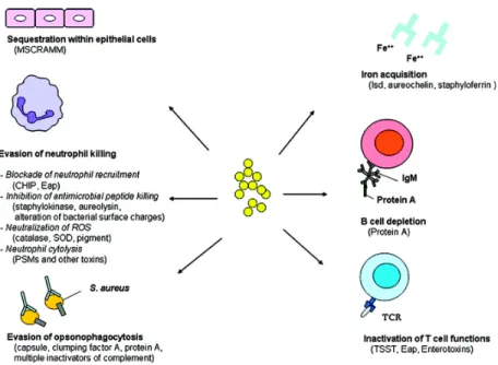
virulent factors include hiding within phagocytic cells, the capacity to resist oxidative bursts, adhering to epithelial cells within the teat canal, and by avoiding the immune system’s Toll-Like Receptors (Zecconi and Scali, 2013). Most of the virulent factors are shown in figure 1 taken from Liu’s review of the pathogenesis of S. aureus (Liu, 2009). Not only does S. aureus evade the immune system with its capsules and proteins, but the concentration of opsonizing antibodies in the milk are low as well (Sutra and Poutrel, 1994). These factors all contribute to the chronicity seen with S. aureus IMI. These virulence mechanisms can lead to persistent IMI, which is why S. aureus can be very detrimental economically for the producer.
Figure 1. Staphylococcus aureus survival mechanisms during infection (Liu, 2009)
Unlike environmental pathogens, S. aureus main reservoir is the mammary gland, thus transmission occurs via milking units, producers’ hands, towels, suckling calves, and any other sources that can potentially transmit the bacteria from one udder to the next (Harmon, 1996a). In addition, infected replacement heifers may also contribute to the introduction of the pathogen within a herd (Middleton, 2013). Nevertheless, the most common source is the milk of the lactating cows (Roberson et al., 1998). Moreover, various studies demonstrated that flies might contribute to the transmission of S. aureus from cow to cow by acting as vectors (Nickerson et al., 1995; Piepers et al., 2011). Herds
that have stringent fly control programs have lower risks of mastitis and IMIs caused by contagious pathogens like S. aureus (Nickerson et al., 1995).
S. aureus is a challenging pathogen to control (Barkema et al., 2006) and it is known to cause IMI that are difficult to cure (Barkema et al., 2006; Oliver and Mitchell, 1983; Roberson et al., 1994). Cure rates for IMI caused by S. aureus depend on various factors such as the SCC, the duration of infection, resistance to antimicrobials, the bacterial colony counts, and the number of quarters infected (Barkema et al., 2006). The different cure rates observed for different treatments will be discussed in a later section (Treatment of Heifer Mastitis). In addition, different strains of S. aureus may have different shedding patterns, which makes it harder to identify and, consequently, harder to control (Sears et al., 1990). Sears et al. (1990) stated that S. aureus with a low shedding cycle has a high risk of false negatives when a single sample is taken (Se of 74.5%). However, methods such as duplicate sampling, centrifugation and increasing the inoculum volume can increase the Se up to 94.2% (Zecconi, 2010). Regardless, shedding patterns should always be taken into consideration when identifying S. aureus to avoid false negative results and to allow us to have a better control over this pathogen.
Coagulase negative staphylococci
Many studies have shown that CNS are the most prevalent pathogens in dairy cows immediately prior to and at calving (Barkema et al., 1999; De Vliegher et al., 2012; Fox, 2009; Nickerson, 2009; Oliver et al., 1992; Oliver et al., 2003; Pankey et al., 1991; Pyörälä and Taponen, 2009; Sampimon et al., 2009). Some studies showed that even though CNS are frequently isolated in many mastitis cases, not many were found to cause severe clinical mastitis (Lam et al., 1997; Makovec and Ruegg, 2003; Piepers et al., 2010; Pyörälä and Taponen, 2009; Supre et al., 2011).
The prevalence of CNS-associated subclinical mastitis in heifers at parturition differs between studies; the percent of mammary quarters infected range between 5.2 and 39.0% (De Vliegher et al., 2012). The prevalence of CNS mastitis in the post-partum period in primiparous cows can be as high as 27.8% compared to 12.3% in the second week of lactation (Matthews et al., 1992). The prevalence of CNS in pre-calving heifers can reach up to 55% in some cases (Trinidad et al., 1990b). The most common species isolated in heifers is Staphylococcus chromogenes and Staphylococcus hyicus (Matthews et al., 1992; Trinidad et al., 1990b). The differences seen across studies may be due to the fact that there are many factors that will influence the prevalence of certain species of CNS in dairy heifers. For instance, factors such as management procedures, virulence of specific species or strains of CNS, and resistance of the pathogen against the immune system all play a role in the incidence of infection (Barkema et al., 2006). The fact that CNS IMI definitions often differed between studies can also explain an important part of this variation (Dufour et al., 2012a).
The SCC is often slightly elevated right after parturition compared to later on throughout lactation and it was initially thought to be physiological in heifers (Dohoo, 1993; Harmon, 1994). However, many studies show that a large percentage of heifer quarters have IMI at the time of parturition, which would explain the increased SCC in early lactation (Fox et al., 1995; Nickerson et al., 1995; Trinidad et al., 1990b). These studies show that elevated SCC above a certain threshold during early lactation is not physiological, but rather an indication of IMIs (Barkema et al., 1999; De Vliegher et al., 2004). The SCC of heifers often gradually decreases in the first two weeks after parturition (Dohoo, 1993). As mentioned, many studies concluded that CNS are commonly isolated in primiparous heifers during the peri-partum period with prevalence ranging from 20 to 55% (Fox et al., 1995; Oliver et al., 1992; Oliver and Mitchell, 1983; Pankey et al., 1991; Trinidad et al., 1990b) and this prevalence decreases after calving without treatment (Fox et al., 1995; Oliver et al., 1992; Oliver and Mitchell, 1983). These results suggest that CNS IMI have a high probability of being eliminated spontaneously after parturition, which would correlate with the gradual decrease of SCC seen in some heifers.
Heifer Mastitis
Prevalence and Incidence
The prevalence of mastitis is defined as the percentage of a given population that is affected with mastitis at a given point in time (Dohoo et al., 2009). The incidence rate of mastitis is the number of new mastitis cases over a period of time for a given population (Dohoo et al., 2009). The prevalence is used in order to evaluate the cases of subclinical mastitis that affect the herds on a given day (i.e. a snapshot). Measuring the incidence rate provides additional useful information and is an important, if not the most important, predictor for future herd prevalence (Dufour et al., 2012a). The incidence is an important tool for mastitis control programs; it is often reported as the number of cows (or quarters) infected per cow-years at risk.
Prevalence
The prevalence of IMI in heifers before parturition has been shown to be quite elevated ranging from 30% and reaching up to 97% in some cases. (Fox et al., 1995; Middleton et al., 2005; Oliver et al., 1992; Oliver and Mitchell, 1983; Trinidad et al., 1990b). In addition, prevalence of S. aureus infected quarters in the pre- and post-partum period can range from 1 to 15% in heifers in North America (Tables 1 & 2) (Andersen et al., 2010; Fox et al., 1995; Middleton et al., 2005; Myllys, 1995; Oliver et al., 1992, 1997; Owens et al., 2001; Pankey et al., 1991; Roberson et al., 1994; Trinidad et al., 1990b). The large variation seen between studies may be explained by the fact that many factors can influence the prevalence of IMI such as season, location, time of gestation, contact between suckling calves and heifers, fly-control programs, number of infected quarters, teat abrasions, and udder edema (Fox et al., 1995).
In a Louisiana study, four herds were observed in order to determine the prevalence of mastitis in unbred and gravid heifers (Trinidad et al., 1990b). The study was conducted on Jersey heifers (n=116) in which teat canal keratin (n = 461) and secretion samples (n= 370) were collected and analyzed. Trinidad et al. found that 93.1% of heifers and 70.7% of quarters had teat canal colonized by pathogens such as S. aureus (16.8% of quarters), Staphylococcus chromogenes (42.9% of quarters), Staphylococcus hyicus (25.2% of quarters), other staphylococci species (5.7% of quarters), as well as various streptococci species.
Trinidad et al. demonstrated that 96.9% of heifers and 75% of quarters had an IMI (Trinidad et al., 1990b) which is in concordance with other findings as well (Oliver et al., 2004). Trinidad et al. (1990) found that S. aureus was isolated in up to 37.1% of Jersey heifers and 14.9% of quarters. Twenty-nine percent (29%) of S. aureus infected heifers showed clinical signs of mastitis (Table I). This is in contrast with Oliver’s study in which, S. aureus was responsible for only 8% of IMI in Jersey heifers (Oliver et al., 2004). Nevertheless, the prevalence of S. aureus IMI in Holstein heifers was found to be 30% by Oliver’s group. In this study, the included cattle of both breeds (Jersey and Holstein) originated from different herds, therefore, the differences between breeds in pathogen causing IMI may have been due to differences in herds and management practices, and thus, the authors couldn’t conclude that these variations were related to breed differences.
Some studies have shown that there is a difference in the prevalence of IMI between seasons. The prevalence of IMI has been shown to be the greatest during the summer months after parturition, but it doesn’t seem to be the case for heifers of breeding age (Fox et al., 1995).
Table I. Prevalence of intramammary infections in dairy heifers caused by
Staphylococcus aureus and coagulase negative staphylococci in the pre-partum
period.
Study Location Number of
Heifers Number of quarters Quarters infected by S. aureus (%) Quarters infected by CNS (%) Trinidad et al., 1990b LA, USA 94 370 14.9 52.9 Oliver et al., 1992 TN, USA 115 460 1.7 52.8 Fox et al., 1995 USA 1,583 4,950 2.8 21.8 Oliver et al., 1997 TN, USA 82 314 3.2 55.1 Middleton et al., 2005 MO, USA 183 663 3.9 37.3 Roy et al., 2007 QC, Canada 428 2,140 10.3 59.3
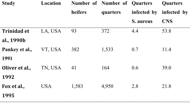
Table II. Prevalence of intramammary infections in dairy heifers caused by
Staphylococcus aureus and coagulase negative staphylococci after calving.
Study Location Number of
heifers Number of quarters Quarters infected by S. aureus Quarters infected by CNS Trinidad et al., 1990b LA, USA 93 372 4.4 53.8 Pankey et al., 1991 VT, USA 382 1,533 0.7 11.4 Oliver et al., 1992 TN, USA 41 164 0.6 39.0 Fox et al., 1995 USA 1,583 4,950 2.8 21.8
Incidence
Dutch researchers have conducted a study in the Netherlands to estimate the incidence of subclinical mastitis in dairy heifers in early lactation using SCC (in the first 100 days in lactation) (Santman-Berends et al., 2012). After questioning 189 farmers, they estimated that the incidence of subclinical mastitis in dairy heifers was 25.5% in the first 100 days in milk (DIM) and correlated a higher incidence with various risk factors. They have shown that housing close-to-calving heifers with lactating cows could decrease the incidence of subclinical mastitis. The authors belie that outing the heifers with lactating cows before calving, allows them time to transition and is less stressful then if they are transferred after calving. Additionally, conventional milking systems rather than automatic milking systems also decrease the incidence of subclinical mastitis in dairy heifers (Santman-Berends et al., 2012).
There are no studies that focus on the incidence rate of clinical mastitis in heifers. There is a Canadian study that compares incidence rates of clinical mastitis in dairy cows. They mentioned that the incidence rate of clinical mastitis (IRCM) was higher in heifers than in cows in the first two weeks of lactation: approximately 118 IRCM vs. 100 IRCM per 100-cow years, respectively (Olde Riekerink et al., 2008). Another study conducted in the Netherlands also demonstrated that the incidence rate of clinical mastitis in the first 2 weeks of lactation was over 30% in heifers as compared to 13% in older animals (Barkema et al., 1998).
Impacts (Economic/ Productivity Losses)
The major problem associated with elevated SCC and mastitis is the milk production losses caused by either low quality milk or a decrease in milk production. In addition, there are economical losses due to the costs for the diagnosis and for the treatment of the infection. There is an exponential growth of the mammary parenchyma that occurs during heifers’ first gestation, especially during the last trimester. Infections that take place during the growth of the mammary gland can have deleterious effects on future milk yield (Nickerson et al., 1995; Trinidad et al., 1990a). Milk yield in unbred and primiparous heifers was found to be 18% less in infected quarters compared to non-infected quarters (Nickerson et al., 1995). Experimental S. aureus IMI in heifers yielded a large amount of inter-alveolar connective tissue deposition instead of secretory tissue in epithelial and luminal areas (Trinidad et al., 1990a). This histological change may explain the low milk yield seen in heifers with IMI because the scar tissue formation potentially interferes with the development of milk producing cells.
A study conducted in Belgium on 117 496 heifers, demonstrated that heifers with SCC over 200 000 cells/ml after calving (i.e. between 5 to 14 days in milk) also had higher SCC throughout the first lactation (De Vliegher et al., 2004). The researchers did not look at the pathogens responsible for the increase in SCC, but their study emphasized that more than 27% of 14 766 dairy heifers had an elevated SCC in early lactation, which
suggests an IMI in the peri-partum period. Once again, this emphasizes the fact that heifers are at great risk of having IMIs early on in their productive life and that this can affect the SCC and their general health throughout their first lactation and possibly in subsequent lactations.
Increased SCC, which is associated with IMI, is coupled to decreased milk productivity especially during the first lactation (De Vliegher et al., 2005). In various studies it has been shown that infections by S. aureus yielded SCC of up to 9.2 x 10⁶ cells/ml in heifers (Nickerson et al., 1995). In addition, heifers with an elevated SCC in early lactation have an SCC that remains elevated throughout the first lactation (De Vliegher et al., 2004). The increased SCC is due to the large amount of leukocyte infiltration seen with S. aureus IMI (Nickerson et al., 1995). Elevated SCC prior to and after calving will affect milk-producing tissue and subsequently affect future milk yield (De Vliegher et al., 2005). De Vliegher et al. (2005) looked at 14 243 Belgian dairy heifers and showed that heifers with 50 000 cell/mL produced 0.26 kg more milk per day than heifers with a first test day SCC between 51 000-200 000 cells/mL. They also deemed that the difference was on average 1.44 kg/day for heifers in the lowest SCC class (< 50 000 cells/mL) compared with heifer in the highest SCC class (> 1 000 000 cells/mL) (De Vliegher et al., 2005). These findings stress the importance of proper udder health management during the pre- and peri-partum periods in order to control IMI and subsequently to avoid drops in milk production.
Another study, however, did not show a significant difference in milk production in S. aureus or CNS infected quarters in the first month of lactation (Paradis et al., 2010). The authors of this later study believed that the lack of a significant difference might have been due to the small numbers of IMI observed and the regrouping of mild and severe infections into one category. Looking at the production differences in severe cases may have yielded different conclusions.
The losses associated with decreased milk yield and costs of treatment can be detrimental to a producer. An increased risk of early culling is also associated with IMI
(Myllys and Rautala, 1995). In the United States, the treatment of IMI in heifers has been shown to be economically beneficial if milk prices were above $0.029/kg USD (Oliver et al., 2003). In this study, heifers with an IMI were treated with an intramammary infusion of 200 mg cephapirin. The treatment costs were $15.60 USD per heifer and the milk production averaged 5 195 kg for untreated heifers and 5 726 kg for treated heifers. The authors took into account the costs of treatment and the revenue obtained by the increased milk production and concluded that prepartum antibiotic treatment was indeed economically beneficial by increasing the net revenue by $200.64 USD per heifer (Oliver et al., 2003).
Risk Factors
It is important to determine risk factors associated with heifer mastitis since this disease has been shown to cause major health issues and negatively impacts milk quality. Knowing the risk factors would be beneficial for producers and veterinarians in order to improve management and to prevent IMI in heifers.
There has been some research stating that horn flies may be a risk factor for the spread of contagious pathogens (Fox et al., 1995; Gillespie et al., 1999; Owens et al., 2002; Owens et al., 1998; Piepers et al., 2011). The examination of DNA profiling of S. aureus isolated in horn flies and S. aureus isolated from heifers revealed that there is a high possibility that the transmission of S. aureus can occur by horn flies (Gillespie et al., 1999). Another group found that pour-on insecticide reduced the prevalence of IMI (Owens et al., 2002) suggesting that controlling flies in the herd can reduce risk of IMI. It was also shown that exposure of non-infected heifers with horn flies colonized by S. aureus can result in IMI acquisition (Owens et al., 1998).
As mentioned previously, season and location may be factors that influence the prevalence of IMI. Fox et al. (1995) looked at numerous herds from different states and the largest prevalence after parturition was seen in Louisiana (58%). The same study also
demonstrated that the prevalence was increased during the winter months. The differences seen across studies suggest that management has an impact on IMI prevalence.
Udder edema is a factor to consider when dealing with clinical mastitis in heifers (Compton et al., 2007; Waage et al., 2001). Udder edema has been shown to increase the chances of heifer pre-partum clinical mastitis by 80% (Compton et al., 2007). It is thought that udder edema impairs the flushing effect and, therefore, causes an increased concentration of the pathogen in the udder; thus, leading to clinical mastitis (Compton et al., 2007). In addition, udder edema is thought to affect the local blood circulation impeding pathogens’ clearance by the immune system (Waage et al., 2001).
Teat abrasions or scabs have been shown to have a correlation with increased IMI. The prevalence of IMI caused by S. aureus was shown to be almost twice as high (7% to 12%) in heifer quarters with scabs or abrasions compared to healthy quarters (Owens et al., 2001). Likewise, another group showed that 70% of heifers with teat abrasions had an IMI compared to only 40% of heifers without any scars or abrasions (Nickerson et al., 1995).
During the final trimester of gestation, prevalence of IMI in heifers by environmental pathogens seems to be at its highest compared to other stages of gestation (Fox et al., 1995)In the spring, S. aureus and CNS infected 5.6% and 20.6% of heifers, respectively, in their first trimester. This number increases to 8.5% and 37.7% in their last trimester of gestation. The fact that the prevalence is increased during the end stages of gestation may be explained by the physiology of the mammary gland. During the last trimester, the mammary gland is at a point where its growth rate is at its maximum, which would make the mammary gland more susceptible to bacterial infections (Trinidad et al., 1990a).
Milking procedures play a role in good managing practices as well. It is highly suggested that workers should wear gloves during the milking procedures, use automatic
milking unit take-offs, and apply a post-milking teat disinfectant (Dufour et al., 2011). In addition, appropriate triage should be implemented by milking clinical mastitis cases and animals with high SCC last.
Based on the different studies on risk factors, there are different levels in which we can intervene in order to reduce the prevalence and incidence of mastitis caused by a contagious pathogen. Therefore, implementing fly control programs, good cow and personnel hygiene, and constant udder examinations are some of the many ways in which a producer can minimize prevalence of mastitis within the herd.
Diagnosis Of Mastitis
Introduction to Different Diagnostic Techniques
Culture and proper identification of microorganisms causing heifer mastitis is imperative for a proper mastitis control plan. Identifying the infection and the bacteria that caused the infection can help veterinarians decide on the appropriate intervention for the heifer (Sears and McCarthy, 2003).
Indirect Methods
There are indirect methods for evaluating udder health. One of the methods mentioned previously is the SCC test that measures the concentration of inflammatory cells (neutrophils, macrophages, and lymphocytes) and desquamated epithelial cells present in the milk sample. This test can be done using an automated electronic counter, which will measure cell concentration (Radostits et al., 2007).
The California Mastitis Test (CMT) is an indirect test that can determine the SCC semi-quantitatively. The test consists of taking milk samples from quarters into a four-plate container and adding a reagent. The reagent contains a detergent that will react to DNA and also has a pH indicator. The CMT is a very fast, reliable and inexpensive test where the results can be read within 15 seconds. Scores from 0-3 can be given depending on the thickness of the milk and reagent solution (Radostits et al., 2007). A score of 0 or negative is given when the mixture remains homogenous and a score of 3 is given when the mixture is a gelatinous mass. The score of + or A is added if the reagent is alkaline (purple) or acidic (yellow: pH <5.2), respectively (Levesque, 2004). The CMT is a common test used cow-side in order to assess the individual cow’s milk quality. The Se (66.7%) and the Sp (54.8%) of the test to identify an IMI in early lactation caused by a major pathogen are relatively modest (Sargeant et al., 2001). Still, this test is important for screening animals in order to determine whether they have an IMI or, following a SCC elevation, to identify which quarter is affected. Since it is only an indicator of inflammation, bacterial cultures are still necessary to identify pathogens involved. There are a few limitations to the test such as the fact that it does not provide an exact value of SCC and the interpretation is dependent on the experience of the person executing the test (CBMRN, 2015).
The measurement of electrical conductivity of the milk is an indirect method for the detection of subclinical or clinical mastitis. The test is based on the sodium and chlorine ion concentrations in the milk. With an infection, cells are damaged and this will release ions present within the cell into the interstitial space. The ions released will cause the concentration of those ions to increase in the milk (will end up in the milk ducts via diffusion) and consecutively, increase the electrical conductivity of the milk. The electrical conductivity test requires looking at every quarter and making a comparative analysis between quarters since there are no threshold values to compare to. This test measures the injury of the udder and not necessarily the immune response, as does the
CMT (Radostits et al., 2007). Hence this test would be better for the detection of clinical mastitis and less useful for the detection of IMI without substantial udder injury.
Out of the three indirect tests described above, the SCC is the most widely used tool for the detection of inflammatory cells. The method is precise and can be used to evaluate individual cows and, sometimes, individual quarters.
Direct Methods
Polymerase Chain Reaction
Polymerase-Chain Reaction (PCR) is a method of diagnosis that is not commonly used because of its costs, despite its very high analytical sensitivity to detect microorganisms present in a milk sample. In one study, pathogens were identified in 70% of milk samples from clinical mastitis using standard bacterial culture whereas pathogens were identified in 92% of milk samples from clinical mastitis cases using real-time PCR (Keane et al., 2013). However, PCR results can be quite difficult to interpret. The technique has a very high analytical sensitivity and can detect the presence of just one bacterium, and the presence of solely one bacterium does not necessarily mean there is an infection; the presence can be due to contamination from the teat skin, or the sampler’s hands. Furthermore, PCR detects not only living bacteria but dead ones (i.e. DNA remnants) too, which can falsify the results. Lastly, PCR requires primers that will only detect one strain. Many primers can be added to a tube sample, however each additional primer is charged to the client. If this technique were to be used to detect many bacteria, the amount of primers required can become very costly.
Culture-dependent methods
Routine bacterial milk culture
Standard aerobic bacterial culture of bovine milk samples is commonly used in the diagnosis of mastitis. Bacterial culture is currently the gold standard for the identification of pathogens because blood agar is capable of supporting the growth of almost any microorganism (Sears and McCarthy, 2003). The inoculate volume recommended is 0.01 mL of milk, which is to be streaked onto a blood agar plate (NMC, 2004). The blood agar plate is then incubated at 37 °C with 5% CO2 for 24 to 48 hours
before examination.
Once colonies are observed on the plate, their color, form, and haemolytic capacities are evaluated. S. aureus colonies, for instance, are usually creamy, white, about 3-5 mm in diameter, and have a double zone of hemolysis (NMC, 2004). This distinct haemolytic zone is composed of a complete beta-hemolysis around the colony and a broader alpha-haemolytic zone surrounding the prior zone (NMC, 2004). However, very rarely, some S. aureus species may be non-haemolytic and further tests are required to identify them. These tests consist of a Gram-stain, catalase tests, and the coagulase test (NMC, 2004). Figure 2 shows a flow chart of the method to identify S. aureus from a milk sample at the Faculty of Veterinary Medicine of the Université de Montréal.
Figure 2. Routine bacteriological method for identifying Staphylococcus aureus from a milk sample at the Faculté de médecine vétérinaire of the Université de Montréal
In order to obtain higher Se for S. aureus detection, various techniques are used. One group suggests that milk samples should be from fresh or frozen pre-milking samples or frozen post milking samples (Godden et al., 2002). The reason for a higher Se when the milk is frozen may be due to the fact that freezing has the potential to lyse the phagocytic cells and release the pathogen for better detection (Villanueva et al., 1991). Godden’s group show that there is no difference in Se when taking a frozen pre-milking sample compared to a frozen post-milking sample (Godden et al., 2002) even though some speculate that a post-milking sample will be less likely to be contaminated due to the teat canals being washed out during the milking process (Sears et al., 1991). In addition to this finding, another research group state that the best results for S. aureus detection were obtained when freezing the milk sample in an incubation broth (Sol et al.,
2002). Centrifugation of quarter milk samples has been shown to increase the number of S. aureus positive samples by 94% (Zecconi et al., 1997). Moreover, Silva’s group (2005) found that they obtained the highest Se when they centrifuged the milk sample prior to incubation.
Despite all these findings, a more recent research group showed contradictory results (Artursson et al., 2010). This Swedish group investigated eight methods for the isolation of S. aureus in 204 quarter-samples throughout 41 dairy herds. These methods included: a standard method of incubation on blood agar, an enrichment method where the milk was added to an equal volume of nutrient broth, a larger volume of 0.1mL instead of the standard 0.01mL, a sedimentation method where the sample was centrifuged, a freezing method, a freezing combined with an incubation method where the thawed milk samples where incubated for about a day before culturing, and an incubation method where the fresh milk sample was incubated for about a day before culturing it. The only methods that seemed be better at identifying the microorganism was the freezing/incubation method and the fresh milk incubation method. The authors state that the only reason that the freezing with incubation resulted in a higher Se is mainly because of the incubation technique. This technique allows S. aureus to multiply to greater numbers in the milk before culturing on a plate and is thought to be an important method to detect infections when there is a low S. aureus concentration (Artursson et al., 2010).
There is always the question on whether one milk sample is sufficient in order to diagnose the quarter as infected or if multiple milk samples are required. The United States FDA guidelines suggest duplicate milk samples should be used in series to correctly identify an IMI, though it has been suggested that a single sample is enough to identify most contagious pathogen (Dohoo et al., 2011a; Erskine and Eberhart, 1988). In Dohoo et al. study (2011a), they suggested that a triplicate sample had the best combination of Se and Sp, however it was not significantly different to the Se and Sp of a single sample. Erskine and collaborators showed that when comparing single and
multiple milk samples, there was a very high agreement between the samples for contagious pathogens like S. aureus (94.2%) compared to the agreement seen with environmental pathogens such as coliforms (55.6%).
When analyzing the milk culture results, one has to take into account the Se and Sp of the standard culture. If we were to use multiple samples, analyzing the results in series (both samples need to be positive for a pathogen to deem a quarter as infected), would lower the Se but increase the Sp (Dohoo et al., 2011a). The opposite holds true if we were to interpret multiple samples in parallel (only one sample needs to be positive for the quarter to be deemed infected). Sensitivity would be increased and Sp decreased.
When defining IMI based on culture results, it is important to understand that the Se can vary depending on whether composite or quarter milk samples are being used. Composite milk samples have relatively low Se, which may be due to the fact that milk from the infected quarter is diluted with milk from the other healthy quarters (Lam et al., 1996; Reyher and Dohoo, 2011). However, when using single quarter milk samples for confirming experimentally induced S. aureus infections, Se of 75% can be achieved. Sensitivity would increase with parallel interpretation of two (Se=94%) or three (Se=98%) consecutive cultures (Sears et al., 1990). The same researchers showed that the Se could approach 100% with three samples interpreted in parallel when investigating naturally occurring S. aureus infections in cows with high shedding cycles.
There are some disadvantages to using routine bacterial culture to identify a microorganism. An important one is the time required to get results (Sargeant et al., 2001). Pathogens are rarely speciated based on colony appearance alone; other tests are usually conducted, which can delay the process of obtaining a definitive result and for treating the infected animal. Moreover, there are chances of contamination due to either non-proper collection of the milk sample or even human laboratory error. Once again, this would delay the time for the results to come in. There is also the fact that many clinical mastitis cases yield no growth in culture media. Reasons for no growth can be
due to infections by slow-growing bacteria or by bacteria that cannot grow on regular medium. For instance, Mycoplasma is a microorganism that does not grow easily on blood agar and requires other specific culture media (Sears and McCarthy, 2003). However, since infection by the Mycoplasma pathogens are rare, there are other more plausible reasons for the no growth. Sometimes it can be due to the fact that there are very few pathogens present in the milk and techniques with higher sensitivities should be used such as PCR (Botaro et al., 2013; Keane et al., 2013; Taponen et al., 2009). There is also the possibility of cyclical shedding patterns which can result in no growth if a sample was taken during the low shedding period (Sears et al., 1990). Lastly, there is the possibility that the pathogen was spontaneously eliminated by the host or even if an antibacterial treatment was already put in place prior to milk collection (Sears and McCarthy, 2003).
Bacterial culture and microbiological techniques are the most common and reliable way to identify mastitis causing microorganisms. Data analysis and the sampling method used are dependent on the objective pursued. As with any technique, there are pros and cons. Despite the lag times associated with bacterial culture, the efficiency, accuracy, and relatively low price associated with these techniques make it a favourable choice for identifying the pathogen causing an IMI.
The MALDI-TOF MS
The matrix assisted laser desorption ionization-time of flight mass spectrometry (MALDI-TOF MS) is a new tool for microbial identification and diagnosis. The principals are based on the conversion of peptides into ions by addition or loss of one or more protons without affecting the integrity of the sample (Singhal et al., 2015). Once the sample is charged, the protons are accelerated to a fixed potential and they are separated from each other on the basis of their mass-to-charge ratio (Singhal et al., 2015). With the MALDI-TOF-MS, this ratio is measured by determining the amount of time it is required to travel the flight tube (Singhal et al., 2015). This will eventually give a peptide mass
fingerprint, which is used to identify microbes. The proteins used are mostly the ribosomal complexes within the microorganisms, which are species specific (Barreiro et al., 2010). This system allows for the identification of microorganisms from its genus to its species and, potentially, its strain (Singhal et al., 2015). This new diagnostic technique is faster at identifying infectious organisms then the conventional diagnostic techniques (Barreiro et al., 2010). The results obtained by the MALDI-TOF MS can be as quick as 24 hours which is a great improvement from the average of 5 days with standard bacterial culture (Barreiro et al., 2010). With quick results, a proper antibacterial protocol can be put in place to treat animals that have bacterial infections quicker.
On farm culture methods
The 3M PetrifilmTM is a ready made dehydrated culture medium used to quickly identify specific microorganisms (3M, 2010). Different plates can be used to identify different microorganisms: the Aerobic Count plate is used to detect aerobic bacteria, the Petrifilm E. coli/Coliform Count plate is used for the detection of coliforms, the Rapid Coliform Count plate is for the quick detection of coliform bacteria (i.e. 4-24 hours), and finally the Petrifilm Staph Express Count (STX) plate is a medium selective for staphylococci.
The STX plate is made of a culture medium that contains a chromogenic modified Baird-Parker medium (3M, 2010). The inoculum volume required is 1 mL of sample, which is incubated for 22-24 hours at 37 °C. The presence of S. aureus in the milk sample is indicated by the presence of red-violet colonies on the STX (3M, 2010). The company suggests that observing red-violet colonies may be sufficient to diagnose the sample as positive for S. aureus. If there are no red-violet colonies, a Petrifilm Staph Express disk is available to detect DNase reactions specific to S. aureus. The disk has toluidine blue-O stain to facilitate the detection of the reaction. Once the disk is placed on
the STX plate, it is incubated for 1-2 hours at 37°C. If S. aureus is present, a pink halo (i.e. a zone of DNase reaction) is observed surrounding the colony.
The inoculum volume necessary for the Petrifilm plate is 1 mL. This volume is 100 times greater than the volume used in standard bacterial culturing (0.01 mL). It is expected that the increase in volume for the Petrifilm plates increase the Se of this technique as compared to the standard routine culture. Research has demonstrated that this larger volume actually impedes the proper reading of the plate in some cases (i.e. too many CFU), making it difficult to correctly interpret the results (Wallace et al., 2011). This later group compared Petrifilm results to standard bacterial culture results and also compared non-diluted and diluted (1:10) milk samples. Wallace et al. (2011) demonstrated that for postpartum milk samples, the highest Se for the STX plate was observed with diluted milk samples (93.7%). The greater Se for diluted samples holds true for elevated SCC samples and clinical mastitis samples as well. The dilution allows for better reading and interpreting the results. Too many colonies on one plate can hinder the reader’s capabilities from correctly identifying S. aureus colonies. However the 1:10 dilution is still 10 times higher than the standard bacterial culture volume required (0.01 mL) and this still potentially leads to a greater Se than the standard test.
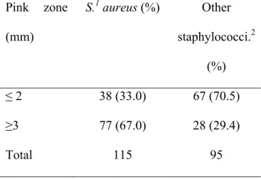
A study evaluating the STX plate for the isolation of S. aureus from milk was conducted in 2005 (Silva et al., 2005). The group conducted many experiments to assess the STX plate. In the first experiment, they compared results obtained from the Petrifilm to standard microbiological procedures (gold standard). When comparing the Petrifilm to other microbiological techniques, Silva et al. (2005) found that the Se for detecting S. aureus was much higher (87.5%) compared to the standard techniques (65.0%). The second experiment’s objective was to compare results from composite and quarter milk samples on the Petrifilm. No significant difference was observed. They also investigated the inter-reader agreement of the STX. They demonstrated that the probability of obtaining positive S. aureus results increased when there was a distinct pink zone surrounding the colony (Se increased to 98.5%). In addition, Silva’s group determined
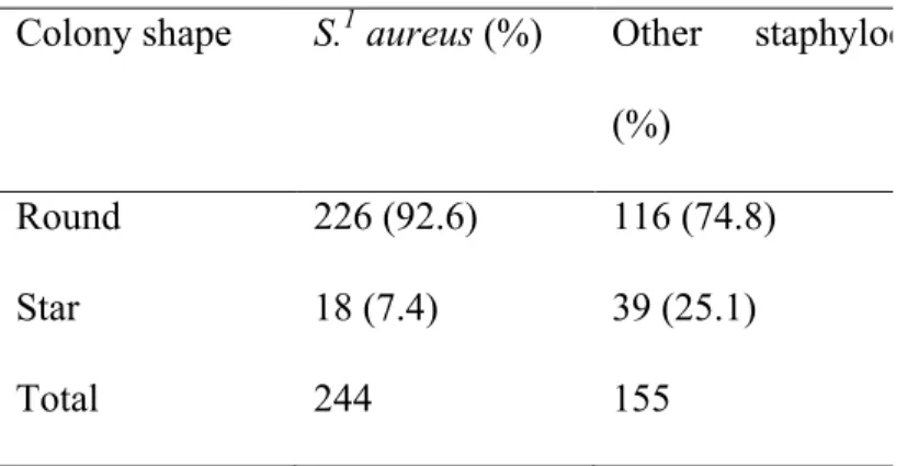
that the reader influenced the STX interpretation since the reader’s assessment varied in number of colonies, size, and recognition of the pink zone (Silva et al., 2005). Conversely to manufacturer’s recommendations, the group suggested that the DNase disk is absolutely necessary for the confirmation of the presence of S. aureus. They showed that out of 35 plates that were considered to have red-violet colonies prior to the DNase disk, only eight were confirmed to be S. aureus.
The main advantage for using the STX plate is that the results can be obtained within 22-24 hours (Silva et al., 2005). This is very favourable because, the faster the results are obtained, the faster a proper intervention or control program can be established. The rapidity of the results is comparable to the MALDI-TOF MS. The main advantage the STX would have over the MALDI-TOF MS would be the fact that no expensive equipment is required for the STX. The increased inoculum volume also increases the Se for S. aureus detection. The increased Se may help diagnose infections when the bacterial concentration is relatively low (Lam et al., 1996). Proper training and experience are, however, essential for dealing with the plates since the reader’s interpretation can affect the Se of the STX plate.
Treatment Of Heifer Mastitis
Antibiotic Therapies
Infection of the mammary gland with S. aureus can cause damage to the milk producing tissue, which can have long term detrimental effects on milk productivity if not treated right away (Owens et al., 2001). Previous studies showed that delaying treatment of dairy heifers infected with a major pathogen often result in persistent infections, since the rate of spontaneous cure for these is quite low (Owens et al., 1991, 1994; Trinidad et
al., 1990b). Immediate treatment of IMI caused by major pathogens in heifers during early lactation is a better option then delaying treatment to drying-off in order to avoid losses in milk production, in addition to preventing chronic carriers in a herd..
Various antimicrobial therapies can be used against mastitis causing pathogens. The preferred route of administration is usually intramammary infusion. Parenteral and subcutaneous injections of antibiotics often result in relatively lower antimicrobials concentration in the mammary glands compared to intramammary infusion (Nickerson, 2009). This is mostly explained by antimicrobials failure to transfer from blood to milk (Nickerson, 2009). Quite a few approved commercial antimicrobial products formulated specifically for intramammary infusion in lactating and dry cows are available to Canadian veterinarians and have beneficial effects on cure rates at least for some pathogens.
Intramammary infections caused by S. aureus are relatively difficult to eliminate compared to other microorganisms. Many studies were conducted to investigate use of various antimicrobials (Owens et al., 2001; Roy and Keefe, 2012; Watts and Salmon, 1997) and vaccines (Nickerson et al., 1999; Smith et al., 2006) for treatment or prevention of S. aureus IMI, respectively. In the following paragraph, these studies will be looked at in greater detail.
Use of antimicrobials in animal production has become a concern for public health because of the emergence of resistant strains. The use of antimicrobial agents creates a selective pressure for microorganisms to acquire multi-resistant genes (Tenover, 2006). Mutations in genes and the exchange of antimicrobial resistant genes from one bacterium to the next are mechanisms by which resistance to antimicrobial agents can be acquired. Using the appropriate drug at the proper dosage and for the right duration is imperative in order to reduce the risk of antimicrobial resistant strains to emerge (Tenover, 2006). It is therefore very important to base treatment protocol on