Université de Montréal
Globin Gene Expression: Role of Transcription Factors
par
Alireza Fotouhi Ghiam
Programmes de biologie moléculaire Faculté de médecine
Mémoire présenté à la Faculté de médecine
en vue de l’obtention du grade de Maîtrise ès Sciences (M.Sc.) en biologie moléculaire
Août, 2009
ii
Université de Montréal Faculté des études supérieures
Ce mémoire intitulée :
Globin Gene Expression: Role of Transcription Factors
présentée par : Alireza Fotouhi Ghiam
a été évaluée par un jury composé des personnes suivantes :
Dr. Dindial Ramotar Président-rapporteur Dr. Eric Milot Directeur de recherche Dr. Chaim Shustik Membre du jury
iii
Résumé
La dérégulation de l’expression génétique est une base pathophysiologique de plusieurs maladies. On a utilisé le locus du gène β-globine humain comme modèle pour élucider le mécanisme de régulation de la transcription génétique et évaluer son expression génétique durant l’érythropoïèse. La famille des protéines ‘E’ est composée de facteurs de transcription qui possèdent plusieurs sites de liaison au sein de locus du gène β-globine, suggérant leur rôle potentiel dans la régulation de l’expression de ces gènes. Nous avons montré que les facteurs HEB, E2A et ETO2 interagissent d’une manière significative avec la région contrôle du Locus (LCR) et avec les promoteurs des gènes de la famille β-globine. Le recrutement de ces facteurs au locus est modifié lors de l’érythropoïèse dans les cellules souches hematopoitiques et les cellules erythroides de souris transgéniques pour le locus de la β-globine humain, ainsi que dans les cellules progénitrices hématopoïétiques humaines. De plus par cette étude, nous démontrons pour la première fois que le gène β-globine humain est dans une chromatine active et qu’il interagit avec des facteurs de transcriptions de type suppresseurs dans les cellules progénitrices lymphoïdes (voie de différentiation alternative). Cette étude a aussi été faite dans des souris ayant une génétique mutante caractérisée par l’absence des facteurs de transcription E2A ou HEB.
Mots-clés: Expression, Facteur de Transcription, Génétique, Gène, Hématopoïèse, Lignée
iv
Abstract
Aberrant gene expression is an underlying pathophysiology in many disease conditions. Lineage-specification and -commitment is tightly dependent on lineage-specific transcription factors to regulate the expression of target genes. Using human β-globin locus as a model, we investigated how the transcriptional machinery is set and regulated during erythropoiesis and how it impacts globally on gene expression. Class I bHLH proteins are important transcription factors whose binding sites are frequently clustered throughout the β-globin gene locus, suggesting their role in globin gene regulation. We showed that, in hematopoietic progenitor (HPC) and erythroid cells (EryC) of the transgenic mouse for human β-globin locus and human HPC cells (CD34+); HEB, E2A and ETO-2 significantly interact with locus control region (LCR) and promoters of globin genes, and their relative ratio is altered during erythropoiesis. For the first time, we found that in other hematopoietic lineages, human β-globin locus is in active chromatin and interacts with transcription factors involved in repression. Strikingly and consistent with the expression of globin genes, we characterized transcription factors involved in open chromatin configuration and basal level of globin gene expression in lymphoid progenitor cells. Further, with the genetic power of E2A and HEB knockout mice, our findings were clarified in mutant backgrounds.
Keywords: Globin, Gene Expression, Hematopoiesis, Lineage Specification, Transcription
v
Table des matières
Résumé……… iii
Abstract……….. iv
Table des matières………. v
Liste des tableaux……….. viii
Liste des figures………. ix
Liste des sigles et des abréviations………... xi
Remerciements………... xv
Introduction………... 16
Hematopoiesis………...17
Hematopoietic Stem Cell (HSC) ………..18
Lineage specification of HSCs……… 19
Erythropoiesis……….. 20
Hemoglobin Synthesis………. 21
Hemoglobinopathies……….……22
Thalassemia. ………...22
Sickle Cell Disease. ……… .23
Human β-globin locus………. 24
Globin gene expression………... 25
Globin gene expression and LCR……… 26
Globin gene expression and transcription factors………...29
Transcription factors and Erythropoiesis……… .30
Helix-Loop-Helix (HLH) proteins……….. 33
Helix-Loop-Helix (HLH) proteins: Regulators of transcription in eukaryotic organisms...33
Classification and structure………. 33
E proteins and hematopiesis……… 36
E proteins in lymphoid-lineage………... 37
vi Applications of study………... .42 Molecular application……….. .42 Clinical application………... .42 Research Proposal………...42 Rational………...42 Hypothesis………...45 Objectives………... .45
Materials and Methods………...47
Mouse models ………...48
Line 2 mouse………... 48
(E2A +/- ln2 +/+) and (HEB +/- ln2 +/+) mice………... 48
Cell Sorting………... 49
Murine erythroid cells (Ter-119+) and Murine HPCs (Ly-6C-CD31highc-Kit+)……….. 49
Murine Lymphoid Progenitors (LPs) (Lin-c-Kit+Sca1+IL-7Rα+)………. 50
Human primitive progenitor cells (CD34+)……… 50
Genotyping………... 51
Chromatin Immunoprecipitation (ChIP) assay……… 52
Real-time Polymerase Chain Reaction (Q-PCR/qPCR)……….. 53
Real-time Reverse Transcriptase Polymerase Chain Reaction (RT-PCR)……….. 54
Western blot………. 54
in vitro clonogenic assays……… 55
Results………. 56
Chapter One: Lineage-Specific Transcription Factors in Multipotent Hematopoietic Progenitors: A Little Bit Goes a Long Way………... 57
HEMATOPOIESIS AND LINEAGE SPECIFICATION………... 59
POTENTIATION AND GENE PRIMING IN HPCs………. 61
β-GLOBIN GENE POTENTIATION: THE ROLE OF LINEAGE-SPECIFIC TFs……. 63
vii
CONCLUDING REMARKS ………68
Chapter One Supplementary section……… 69
Chapter two: The basic helix-loop-helix transcription factors E2A and HEB are involved in globin gene expression………... 71
Identification of factors bound to the human β-globin locus in erythroid cells and murine HPCs………... 72
Identification of factors bound to the human β-globin locus in human primitive progenitor cells………... 76
Identification of factors bound to the human β-globin locus in murine lymphoid progenitor (LP: Lin-c-KitlowSca1lowIL-7Rα+) cells………... 80
Identification of factors bound to the human β-globin locus in fetal erythroid cells with E2A and HEB knock-out background………... 85
Discussion………... 88
Human β-globin locus in erythroid cells and murine HPCs……… 90
Human β-globin locus in human primitive progenitor cells……… 91
Human β-globin locus in murine lymphoid progenitor cells (LPs)……… 92
E2A and HEB are associated with lineage- differentiation and commitment………. 94
Human β-globin locus in E2A and HEB knock-out fetal erythroid cells……… 95
viii
Liste des tableaux
Table 1- Role of Transcription factors in Erythropoiesis Table 2- Classification of bHLH proteins
Table 3- The ratio of LP (lymphoid progenitor) cells/Total Bone Marrow was calculated by limiting dilutions using the LDA program
ix
Liste des figures
Figure 1- Hematopoiesis in humans
Figure 2- Hematopoiesis and stromal cell differentiation Figure 3- Hemoglobin structure
Figure 4- Types of normal hemoglobin Figure 5- Structure of human β-globin locus
Figure 6- Structure of a MyoD basic-helix-loop-helix (bHLH) transcription factor Figure 7- Model of Ldb1 complexes in uninduced MEL cells
Figure 8- Genetic regulatory networks (GRNs) programming hematopoietic stem cells and erythroid lineage specification
Figure 9- One representative experiment of genotyping
Figure 10- Chromatin immunoprecipitation (ChIP) assays on Mac-1+, B220+, and common lymphoid progenitor (CLP) cells
Figure 11- Chromatin immunoprecipitation (ChIP) assays on common myeloidprogenitor (CMP), wild type erythroid (EryC), and EKLF knock-out erythroid (EKLF KO) cells Figure 12- Model of TFs recruitment at the huβ-globin locus in HPCs and EryCs
Figure 13- Chromatin immunoprecipitation (ChIP) assays on human CD34+ multipotent HPCs cells
Figure 14- Analysis of hematopoietic cells from ln2 bone marrow mice with Ter119 and Ly-6C/CD31/c-Kit expression
Figure 15- Chromatin immunoprecipitation (ChIP) assays on murine erythroid cells (EryC, Ter119+) and murine hematopoietic progenitor cells (HPC, Ly-6C-CD31highc-Kit+) Figure 16- Purification of human hematopoietic progenitor cells (CD34+)
Figure 17- Chromatin immunoprecipitation (ChIP) assays on human CD34+ multipotent HPCs cells
Figure 18- Purification of murine lymphoid progenitor (LP: Lin-c-KitlowSca1lowIL-7Rα+) cells
x
Figure 20- ChIP analysis of histone acetylation and interaction of E2A, HEB and ETO-2 proteins with the human β-globin locus in LP (Lin-c-KitlowSca1lowIL-7Rα+) cells from adult ln2 mice
Figure 21- ChIP assays on fetal liver eythroid cells (13.5 dpc) with wild-type and knock-out backgrounds
xi
Liste des sigles et des abréviations
Ab: antibody
AGM: aorta-gonad-mesonephros region AML: acute myeloid leukemia
APC: allophycocyanin
BFU-E: burst forming units-erythroid bHLH: basic helix-loop-helix
CD: cluster of differentiation CFC: colony-forming cell
CFU-E: colony-forming units-erythroid CFU-G: colony-forming units-granulocyte
CFU-GEMM: colony forming units-granulocyte-erythroid-macrophage-megakaryocyte CFU-GM: colony forming units-granulocyte-macrophage
ChIP: chromatin immunoprecipitation CLP: common lymphoid progenitor CMP: common myeloid progenitor CO2: carbon dioxide
CTD: C-terminal domain Ct: threshold cycle
DIVA: DIgitalized VAntage dpc: day post coitus
EDTA: ethylenediaminetetraacetic acid EKLF: erythroid kruppel-like factor EKLF KO: EKLF knock-out
EO: eight twenty-one EPO: erythropoietin EryC: erythroid cell ES: embryonic stem
xii FAB: French-American-British
FACS: fluorescence-activated cell sorter FBS: fetal bovine serum
FITC: fluorescein isothiocyanate FL: fetal liver
FOG-1: friend of GATA-1
G-CSF: granulocyte colony-stimulating factor GD: gestational day
GM-CSF: granulocyte monocyte-stimulating factors GMP: granulocyte macrophage progenitor
GRN: genetic regulatory network GTF: general transcription factor H3Ac : histone H3 acetylation Hb: hemoglobin
HDAC: histone deacetylase HEB: Hela E-box binding protein HLH: helix-loop-helix
HPC: hematopoietic progenitor cell HS: hypersensitivity sites
HSC: hematopoietic stem cell Huβ: human β
Id: inhibitor of differentiation Igh: immunoglobulin heavy chain IFN-α: interferon-α
K4: lysine 4 K9: lysine-9 Kb: kilobase
xiii Ln2: line 2
LP: lymphoid progenitor
LTBMC: long term bone marrow culture mSin3: mammalian Sin3
MEL: murine erythroleukemic
MEP: megakaryocyte erythroid progenitor MRF: muscle regulatory factor
MTG16: myeloid transforming gene chromosome 16 MTGR1: myeloid transforming gene related protein-1 MW: molecular weight
NaB: sodium butyrate NO: nitric oxide
NuRD: nucleosome remodeling and deacetylation O2: oxygen
p21: CDKN1A; cyclin-dependent kinase inhibitor 1A Pax6: paired box protein 6
PBS: phosphate-buffered saline PCR: polymerase chain reaction PE: phycoerythrin
PEV: position effect variegation PIC: preinitiation complex
QRT-PCR: quantitative real time polymerase chain reaction RBC: red blood cell
RT-PCR: Reverse Transcriptase Polymerase Chain Reaction SCF: stem cell factor
SCL: stem cell leukemia
SDS/PAGE: SDS-polyacrylamide gel
xiv TFN: transcription factor network
THP: kidney-specific Tamm-Horsfall TSA: trichostatin A
xv
Remerciements
I would like to express a deep sense of gratitude to my supervisor, Dr. Eric Milot, for constant and generous support, encouragement, guidance and understanding. He is a professional in the field and may serve as the beautiful example to follow.
I would also like to thank Dr. Stefania Bottardi who helped me with getting practical experience on my project. Her discussions and corrections have greatly improved the quality of my work.
I would like to extend my deepest gratitude, love and affection to my beloved parents, Mahboobeh and Vahid, for loving me, believing in me and wishing the best for me. I owe all my achievements to them and it is to them that I dedicate this work.
Finally, I would also like to thank my beloved wife, Bahareh, for her kindness, care and countless sacrifices. She was a real support throughout my thesis writing.
Last but not least, I would like to share this moment of happiness with my brother, Arashk. May this realization encourage him to pursue his education in medicine and science.
17
Background
Blood consists of: • Red cells • White cells • Platelets • PlasmaI
Hematopoiesis
Hematopoiesis is the formation and development of blood cells. Sites of hematopoiesis include the bone marrow, liver, spleen, lymph nodes and thymus. The blood cells have the particular ability for persistent production which demands tight regulatory system. Pathological processes interfering with normal production can lead to an excess (hyperplasia; e.g., leukemia) or an inadequate number of cells (hypoplasia; e.g., anemia, thrombocytopenia, or leukopenia).1
Hematopoietic Stem Cell (HSC)
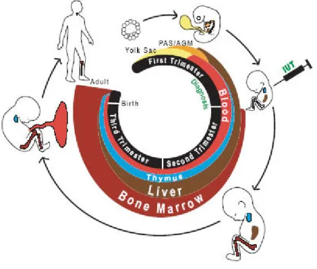
Hematopoiesis begins at embryonic period when blood islands are formed in the yolk sac in the third week of gestation. Blood islands are the source of primitive blood cells till these cells migrate to liver and spleen. These organs are the main sites of hematopoiesis from six weeks to seven months, when eventually the bone marrow becomes the center of hematopoiesis. After birth, the bone marrow is the only source for production of blood cells and hematopiesis takes place in the marrow of nearly all bones. Reaching adulthood, hematopiesis becomes confined to the bone marrow of central skeleton and the proximal
I
18
ends of long bones (Figure-1), in which all blood cell types are derived from pluripotent stem cells, termed as hematopoietic stem cells (Figure-2).2
Figure 1- Hematopoiesis in humans. Anatomical location of hematopoiesis change during human
development.3
Stem cells have been found in practically every tissue. Like other stem cells, HSCs are characterized by two clonal properties: self-renewal, the hallmark property of stem cells, is the production of more stem cells with the maintenance of an undifferentiated state, and the second is the extensive proliferation and differentiation capacity to generate differentiated progeny to commit to one specific cell line (lineage-specification). Excess or inadequate production of hematopoietic cells will end in various disease states.4
19
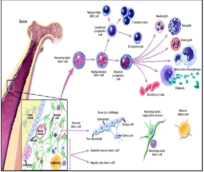
Figure 2- Hematopoiesis and stromal cell differentiation.5
Lineage specification of HSCs. HSCs produce a variety of differentiated cell
lineages, depending on intrinsic cell programming and the micro-environmental signals.6 The stem cell plasticity and lineage commitment are shown to be regulated by a sequential expression of hematopoietic genes and are the result of transcription control in concert with chromatin remodeling and epigenetic modifications. Lineage specification and cellular maturation begins by alteration in cellular gene expression profile, which commits the cell to a specific lineage, and continues by the establishment of lineage-specific gene
20
expression. All these decisions are entirely made by temporal and spatial activation of certain lineage-specific genes and repression of the others. Commitment to a given lineage is mediated by many ubiquitous and lineage-specific transcriptional regulatory proteins that activate specific gene expression programs and extinguish expression of lineage-inappropriate genes.7,8
Erythropoiesis
Erythropoiesis is the process of red blood cell (RBC) formation and development. Red cell precursors pass through several stages in the bone marrow to produce mature red cells (erythrocytes). As development progresses, at each stage, cells contain less RNA and more hemoglobin (Hb) in the cytoplasm. The cell becomes smaller, and the nucleus becomes more condensed and eventually is lost, when the cells are released into circulation as reticulocytes. After 1-2 days, reticulocytes lose their RNA and shape into non-nucleated biconcave discs, namely mature red cell (erythrocyte).9
Hemoglobin Synthesis
The characteristic red color of blood is from hemoglobin (Hb). Hemoglobin is the main protein in the red blood cells that carries oxygen (O2) from lungs to the rest of the
body and returns carbon dioxide (CO2) from the tissues to the lungs. This critical
performance is governed by the biconcave shape of RBCs, providing a large surface area for oxygen and carbon dioxide exchange, and by high affinity of Hb for oxygen and carbon dioxide in lungs and body tissues, respectively.10 Hemoglobin is also involved in transportation of a third gas, Nitric oxide (NO), which is important in regulation of blood pressure by vasodilation and increasing blood flow.11
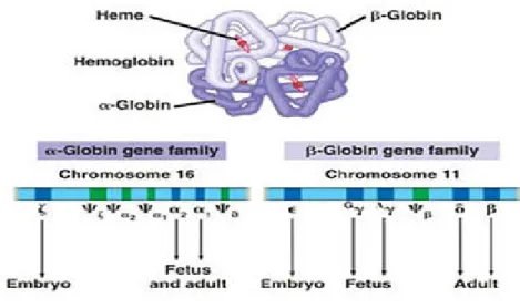
Different types of hemoglobin are produced during development from embryonic period to adult life. Each hemoglobin molecule consists of two α-like (141 amino acids) and two β-like (146 amino acids) chains forming a tetramer. Each globin chain tightly enfolds a non-protein heme moiety in a “pocket”, consisting of a single iron atom (Fe2+) at
21
the center held in a heterocyclic protoporphyrin IX ring with an optimal position for reversible oxygen binding. Four molecules of oxygen can therefore bind to and be transported by one hemoglobin molecule.12
The “blueprint” for hemoglobin synthesis exists in two tightly linked loci, α-like globin genes clustered on chromosome 16II and the β-like globin genes clustered on chromosome 11, with four genes encoding each polypeptide chain during development (Figure-3).13
Figure 3- Hemoglobin structure. hemoglobin is a hetero-oligomeric protein contains two α and two
β subunits arranged with a quaternary structure.14
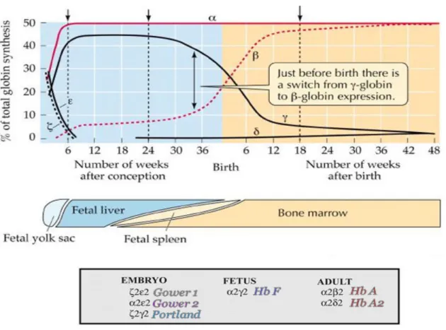
At least six different types of hemoglobin molecules are formed, in steps, following the sequential expression of α- and β-globin gene clusters in process of human development (Figure-4). Hb A (α2β2) is the major hemoglobin in adults with about 97% while two other
types, Hb A2 (α2δ2) and Hb F (α2γ2), are found only in small amounts, 1.5-3.2% and <1%,
respectively. HbF (α2γ2) predominates during most of gestation period.15
II
The α-like cluster consists of two α-globin genes and a single copy of the ζ gene. These genes are similarly arrayed in the order that they are expressed during ontogeny.
22
Figure 4- Types of normal hemoglobin. Different normal hemoglobin variants involve genes both
from the alpha and beta gene clusters. Hemoglobin A (α2β2) is the normal hemoglobin that exists after birth. Hemoglobin A2 (α2δ2) is a minor component (< 3%) of the hemoglobin found in red cells after birth. Hemoglobin F (α2γ2) is the main hemoglobin during fetal development.16
Hemoglobinopathies
Hemoglobinopathies, hemoglobin disorders, are genetic defects that results in qualitative (sickle cell disease) or quantitative (thalassemias) change in the hemoglobin molecule.17
Thalassemia. Thalassemia is a hereditary underproduction of either the alpha or
beta globin chains of the hemoglobin molecule resulting in a hypochromic, microcytic anemia. Gene deletion results in variable levels of disease. There are four genes coding for
23
the alpha chain of hemoglobin. There can be deletions of one, two, three or all four genes. Beta thalassemia can be mutated in either one or two genes.18
The clinical presentation of these disorders is dependent on the number of abnormal genes. In alpha thalassemia one gene deleted yields a normal patient. Individuals with two genes deleted have a mild anemia while those with three genes deleted have more profound anemia where beta chains form tetrads, namely hemoglobin H. Four-gene-deleted alpha thalassemia patients die in utero secondary to gamma chain tetrads called hemoglobin Barts. In one-gene-deleted beta thalassemia (thalassemia minor, thalassemia trait), there is a mild anemia with marked microcytosis. Patients with thalassemia major are homozygous for mutations of both genes coding for the beta hemoglobin gene. These patients with beta thalassemia major, also known as Cooley anemia, become severely symptomatic starting at six month of age when the body would normally switch from fetal hemoglobin to adult hemoglobin. They show severe symptoms of growth failure, hepatosplenomagaly, jaundice, and bony deformities secondary to extramedullary hematopoiesis. Later in life, they are symptomatic from hemochromatosis, cirrhosis, and congestive heart failure from chronic anemia and transfusion dependence. In beta thalassemia, there is an increased level of hemoglobin F and A2. Those with alpha thalassemia will have normal amounts of
hemoglobin F and A2.
Thalassemia trait of both the alpha and beta types do not require specific treatment. Beta thalassemia patients require blood transfusions once or twice a month accompanying with iron chelating therapies with Deferasirox as the standard of care.19 A small number of patients can be treated with hematopoietic cell transplantation.20
Sickle Cell Disease. Sickle cell disease is an autosomal recessive hereditary disease.
Hemoglobin S is due to a substitution of a valine for glutamic acid as the sixth amino acid of the beta globin chain. Almost all of those with the trait are asymptomatic. Those with sickle cell disease (SS) typically have mild to moderate anemia with irreversibly sickled cells and recurrent painful crises. Elaborate therapeutic modalities are beyond the scope of
24
what is neither necessary nor relevant to know for this thesis. Bone marrow transplantation can be curative but still be considered experimental at this time.21
Human β-globin locus
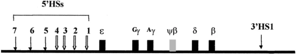
The human β-globin locus consists of five functional genes; ε, Gγ, Aγ, δ and β, organized in the order of their developmentally timed expressionIII (Figure-5). These genes reside within ≈ 50 kilobases (kb) of chromosomal DNA in the transcriptional activation during ontogeny and are expressed in cells of erythroid cells.22 Important regulatory sequences flank each gene: promoter elements at immediate upstream, and enhancers as well as silencers located in vicinity or at distance in the locus.23 Transcription of the β-globin locus undergoes two sequentially programmed switching during development from ε to γ at six weeks of gestation and from γ to δ/β shortly after birth.24
A powerful set of enhancer elements, namely β-locus control region (β-LCR), exists at 5' upstream of ε gene. Human β-globin LCR consists of five developmentally stable, DNase I hypersensitivity sites (HSs) and is located 6-30 kb upstream of the ε geneIV.25 Susceptibility to digestion with DNase I indicates that these regions are, in fact, accessible to transcription and chromatin remodeling factors at the time of gene expression.26 The enhancer activities of 5'HS2, 3 and 4 resides in core elements (200-300 bp) of individual HS sites composed of a wide array of binding sites for ubiquitous and lineage-specific transcription factors. 5'HS1-4 are erythroid-specific and 5`HS5 is ubiquitous.27 Another HS site (3'HS1) is located downstream of the β-gene. Two additional HSs (5'HS6-7) have also been discovered at the 5' end of the β-globin domain (Figure-5).28
III
A non-expressed pseudogene (ψη) is also located on human β-globin locus.
IV
The LCR was first identified in the human β-globin locus, being important in the control of eukaryotic gene expression in many other mammalian gene systems.
25
Figure 5- Structure of human β-globin locus.
The spatial arrangement of β-globin genes with respect to LCR is essential for their proper regulation throughout development; i.e. the genes expressed early in embryonic stage are closest to the LCR and those expressed in adult life are farthest.29 LCR enhances the expression of β-like genes to physiological levels in a tissue-specific and copy number-dependent manner.30
Globin gene expression. It is well known that globin gene expression is restricted
to specific tissues. Recently, the thought that globin gene expression is solely confined to erythroid cells is questioned by findings of adult hemoglobin protein in activated macrophages and alveolar epithelial cells. However, these cells have shown lower amounts of globin polypeptides, comparing to erythroid cells, and different gene regulation mechanisms.31 Further studies are yet required to elucidate the patterns and mechanisms of hemoglobin expression in these cells during cellular differentiation and under various environmental conditions. Moreover, mechanisms required to preclude globin gene silencing in alternative hematopoietic lineage are poorly described.
Considering the hematopoiesis tree, the transcription of the β-globin locus is exclusively displayed by erythroid cells, and more importantly, results in erythroid-lineage specification. Normal red blood cell differentiation requires the coordinated expression of erythroid-specific genes, such as the globin genes and the genes responsible for heme and iron metabolism. These processes are under the control of and involve the complex interplay of a number of specific growth factors and cytokines such as interleukins, granulocytemonocyte-stimulating factors (GM-CSF), stem cell factor (SCF) and
26
erythropoietin (EPO).32 For instance, erythropoietin, a glycoprotein hormone produced by the kidney, promotes red blood cell survival through protecting these cells from apoptosis.33,34 Also, in the hematopoietic system, stem cell generation, cell fate decisions and maintenance (lineage-specification and commitment) and differentiation depends on the coordinate activity of multiple transcription factors through transcription factor networks interacting with crucial regulatory regions of lineage-specific genes.35,36 That is the promise of differentiation towards the production of erythroid-lineage versus non-erythroid cells.
Globin gene expression and LCR
Temporal and spatial control of gene expression are mediated by binding of trans-acting factors to cis-trans-acting DNA sequence(s) such as promoters, enhancers, and silencers as well as long-range cis regulatory LCR elements.37 The LCR was first identified in the human β-globin locus over 20 years ago.38 While different in composition and location, mammalian β-globin loci of different species, including humans, mice, rats, rabbits, and goats contain LCR in part of their genome.39,40
In human β-globin locus, the regulation of gene expression is achieved by the dynamic interactions between cis-acting sequences and trans-acting factors. Proximal and distal cis-acting sequences, the LCR and downstream globin gene sequences, establish and maintain specific chromatin conformations. trans-acting factors, transcription factors, coregulators and chromatin remodeling factors, initiate, regulate and determine the final level of gene expression control.41 Modulation of chromatin structure can have opposite effects on gene regulation as activation versus repression.42 Dynamic chromatin configuration as open, resulted from histone modification (histone acetylation), or closed chromatin, due to DNA methylation and histone deacetylation, is quite central to tissue-specific developmental control of β-globin gene expression.43 In general term, the open chromatin is defined as DNase I-sensitive and hyperacetylated state of histone composing the chromatin, whereas closed chromatin is DNase I-insensitive and underacetylated. Such
27
chromatin structure fluctuation between “open” and “closed” conformations is mediated by chromatin-remodeling complexes that are concomitantly associated with both LCR and β-like globin promoters to facilitate interactions between transacting factors and DNA. Chromatin remodeling complexes modulate chromatin structure to promote binding of erythroid-specific and ubiquitous transcription factors at both LCR and the gene promoters. Such modulations assist the assembly of the transcription apparatus required for full level of expression in erythroid cells while accompanies inactive chromatin structure in non-erythroid cells. These complex interactions determine the transcriptional status as well as the final level of β-like globin gene expression. 44,45
While located at a considerable distance from the site of transcription initiation, LCR controls the overall level of expression of β-globin genes as a key regulator of the locus chromatin organization. Chromatin is in "open" state at LCR throughout development but its state at β-globin genes is determined by the expression order of target genes during ontogeny; that is open at ε-/γ-globin domain during embryonic/fetal period but closed during adult erythropoiesis. The reverse state exists for adult δ-/β-globin domain.46 LCR role in chromatin-opening and in maintaining of an open chromatin state is essential for recruitment of additional chromatin remodeling factors, necessary for further opening of chromatin, or of other transcription factors, involved in high level of expression.47
Several models have been proposed to elucidate how the LCR exerts its regulation on transcription from such a long distance. From four most prevalent models proposed for LCR functionV, looping model is more acceptable to explain the mechanisms underlying the LCR interaction with the globin genes. All these models indicate that LCR alters chromatin configuration. The looping model implicates that LCR loops back on itself to fold into a “holocomplex” without straining the backbone bonds of the DNA double helix. More significantly, this allows the locus to form a spatial conformation and take hold of a physical close proximity to the desired gene, which facilitates the interaction of
V
28
transcription factors bind to LCR with those at promoters. This proximity secures the interaction of LCR-bound transcription factors and/or coactivators with basal transcription factors bound at the promoter. 48,49
Data from studies on some forms of thalassemiaVI containing deletion in LCR region have supported the indispensable role of LCR in expression of β-globin genes. While these forms of thalassemia carry intact β-globin locus, globin genes are not expressed. These deletions result in closed chromatin configuration of globin locus, normally opened by LCR, and thus the suppression of gene expression occurs. These natural occurring deletions of the LCR such as in Hispanic thalassemia, provide the evidence that the LCR is critical for the chromatin organization of the locus.50
The presence of LCR for globin gene expression is important since its absence shuts down β-globin gene expression to less than 1%. The most prominent property of the LCR is strong transcription-enhancing activity. However, not all HS sites have the same importance in this respect. The main enhancer activity is conferred by 5'HS2, 5'HS3, and 5'HS4, and not 5'HS1 or 5'HS5.51 The interaction of regulator transcription factors with LCR HS sites and with each other is essential for high-level of globin gene expression at different developmental stages.52 Deletion of the HS core element from 5`HS2, 3, or 4 abolishes normal LCR function due to disruption of the DNase I-hypersensitivity sites holocomplex and preclude proper interaction with promoters but when there is no position effect, the disruption of one HS does not impair the other HS site formation.53 Even the orientation of LCR HS elements is central for proper functioning; that is, a synthetically inverted LCR has been associated with low level of globin gene expression throughout development. This suggest that The LCR transcription enhancer activity is directional.54 At any specific stage of hematopoietic cell differentiation, different transcription machinery complex is stabilized on LCR, according to transcription factor milieu, whereby an enhancement in globin gene expression is achieved. The LCR, similar to globin genes, has
VI
29
tissue-specific enhancing activity which more specifically confines the expression of globin genes to erythroid cells. In essence, developmental and lineage-specific regulation of gene expression results from the complex interaction of gene-proximal elements with distant cis-regulatory elements on a bed of chromatin.
Globin gene expression and transcription factors. Complex genetic programs
determine survival, proliferation, differentiation and function of hematopoietic cells during different steps of hematopoiesis. Gene expression is controlled at various points between the translation of DNA to proteins with transcriptional control be the most important point of regulation for many genes. Gene transcription is possible only when DNA-binding proteins come together, assemble and interact with the promoters, the operators and the enhancers. The existence of multiple regulatory regions and varied DNA-binding proteins helps a given gene to precisely control its expression at a basal level and/or up- or down-regulate the expression in response to cellular stimuli for differentiation and/or proliferation. In hematopoiesis, many of these protein complexes are lineage restricted and act as cell type-specific transcription factors.55
Globin gene expression is regulated by the dynamic interplay between transcription factors and epigenetic mechanisms. Many transcription factors have been shown to control β-globin gene expression through the formation of intricate transcription factor networks (TFNs) and by binding several cis-acting elements on locus, followed by recruitment of additional regulatory proteins (cofactors) via direct protein-protein interactions.56 General transcription factors (GTFs)VII and different lineage-specific transcription factors should bind the promoter and LCR of β-genes to mediate the tissue- and stage-specific expression of the β-like globin genes. Some of these factors are ubiquitous (e.g., Sp1 and YY1), while others are tissue-restricted and more or less limited to erythroid cells (e.g., GATA-1,
VII
GTFs or basal transcription factors are proteins that either bind DNA or take part in the formation of a preinitiation complex (PIC), and used by RNA polymerase II to begin and proceed with transcription. The most important GTFs are TFIIA, TFIIB, TFIID, TFIIE, TFIIF and TFIIH.
30
E2, TAL-1 and EKLF). These transcription factors turn on/off the transcription appropriately to fit the gene-expression profile for cell fate determination and cellular differentiation. It is becoming increasingly clear that these factors do not operate independently but as part of large multi-protein complexes.57
Given the formation of large multiprotein complexes, certain transcription factors can engage in functional interactions, via these complexes, while lacking sequence-specific activity. Indeed, a single transcription factor can employ numerous mechanisms to control transcription and thus, one cannot to consider the function of a particular hematopoietic transcription factor independent of complex partners and apart from important functional interplays. Herein, it is unfeasible to comprehensively review all transcription factors implicated in erythropoiesis, but short recapitulation of some potential lineage-specific transcription factors would be relevant for our study.
Transcription factors and Erythropoiesis (Table-1)
GATA-1 is a master regulator gene critical for erythroid cell formation.58 It interacts with several transcription factors; CBP/p300, PU.1, Sp1 and erythroid Kruppel-like factor (EKLF), through different multiprotein complexes to activate adult β-like globin genes.59 The PU.1-GATA-1 interaction60 and the balance between GATA-1 and GATA-2 levels are required for precise lineage specification.61 While GATA-1 expression is observed in erythroid, mast cell, and megakaryocyte lineages, GATA-2 is expressed in certain hematopoietic precursors and is crucial for the survival and proliferation of HSCs.62 During erythropoiesis, GATA-1 level increases and displaces GATA-2 on hematopoietic target genes so that different genes expressed in accordance with GATA switch.63 Such GATA switch is facilitated in virtue of friend of GATA-1 (FOG-1) activity. FOG-1 is a protein whose expression mimics that of GATA-1 and is essential to stimulate erythropoiesis.64 FOG-1 helps GATA-1-mediated looping by which LCR comes close to adult β-globin genes.65 In addition, PU.1 antagonizes GATA-1 DNA binding and thus blocks
31
erythropoiesis in favor of granulocyte and monocyte, B and T lymphocyte differentiation. The antagonistic interaction of GATA-1 and PU.1 drives hematopoiesis via common myeloid progenitor (CMP) cells into granulocyte macrophage progenitor (GMP) cells or megakaryocyte erythroid progenitor (MEP). While sufficient levels of PU.1 will produce GMP lineage, higher levels of GATA-1 (comparing to PU.1) will moves differentiation to MEP. Also, GATA-1 exists in a multiprotein complex called SCL complex with LMO2, E47 and Ldb1 in different hematopoietic cells at distinct stages of hematopoiesis.66
Stem cell leukemia (SCL), also known as T-cell acute lymphocytic leukemia-1 (TAL-1), belongs to class II HLH proteins and has tissue-restricted and lineage-specific patterns of expression.67 TAL-1 is known to be essential for haematopoiesis. The TAL-1 gene is normally expressed in haematopoietic progenitors, erythroid lineage cells, mast-cell lineage cells, megakaryocytic lineage cells and endothelial cells.68 TAL-1 dimerizes with E proteins (E47) and functions at multiple stages of hematopoiesis. SCL complex binds a composite motif consisting of a GATA motif and an adjacent E-box.69 SCL also stimulates the generation of hemangioblasts to differentiate into both blood and endothelial cells.70,71
Alteration in TFNs or dysregulation of signaling and transcriptional function leads to neoplastic transformation of hematopoietic cells and consequently the progression of specific leukemias. In the same way, the genes responsible for heme and iron metabolism ought to specifically be expressed during erythropoiesis to conform to RBC differentiation.72
32
Transcription Factor Gene Function
GATA-1 GATA-1 Erythroid and Megakaryocytic development
Erythroid Krüppel-like Factor (EKLF)
EKLF Maturation of erythroid cells, Chromatin remodeling, Modulation of the gamma to beta globin switch, Transcriptional activation, Binding to the CACC motif of the β globin gene promoter
Nuclear factor erythroid-derived 2 (NFE2)
NFE2 Interaction with CREB binding protein
Stem cell leukemia (SCL) or T-cell acute lymphocytic leukemia-1 (TAL-1)
TAL-1 Generation of HSC
FOG-1 FOG-1 Cofactor of GATA-1
p300 and CREB binding protein (CBP)
p300 and CREB
Increase gene expression, histone acetyltransferase (HAT) activity, Recruiting the basal transcriptional to promoter
33
Helix-Loop-Helix (HLH) proteins
There are four basic classes of bHLH proteins classified according to their structural motifs. The basic helix-loop-helix (bHLH) proteins are dimeric transcription factors present in nearly all organisms from yeast to humans.73 Numerous bHLH proteins have been identified in animals, plants and fungi. They are first introduced by Murre C, et al about twenty years ago.74
Helix-Loop-Helix (HLH) proteins: Regulators of transcription in eukaryotic organisms. The fundamental roles of these transcription factors are established in a broad
spectrum of cellular and molecular events involved in the regulation of commitment, cell growth and differentiation of various cell lineages during embryonic development, particularly neurogenesis, myogenesis, retinogenesis and hemetopoiesis.75 The followings are a few examples: BETA2 (NeuroD1) is a member of bHLH proteins and studies on mouse animal models have shown that it plays an important role in the development of the central and peripheral nervous system.76 Muscle Regulatory Factors (MRFs) of the bHLH proteins; MyoD, Myf5, Myogenin and MRF4 are sequentially expressed during skeletal muscle formation and coordinate the expression of muscle-specific genes required for skeletal muscle development in embryo. MyoD and myogenin work as dimmers to drive appropriate myogenesis.77 Any of retinal photoreceptor cell lineages carries an exclusive bHLH context during retinal neurogenesis, which highly emphasizes the cell-specific property of bHLH transcription factors.78 NeuroD acts as important regulator at some point in rod and cone photoreceptor genesis.79
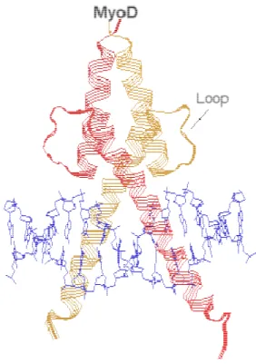
Classification and structure. bHLH transcription factors bear sequence homology,
with overlapping and analogous expression patterns, and highly conserved DNA binding specificity. Members of this family contain two highly conserved domains at either end. N-terminal (AD1 domain) is a basic DNA binding domain that helps the transcription factor to bind to DNA at a consensus hexanucleotide sequence of G(orA)CAXXTGG(orA), namely
34
E-box. At the C-terminal (AD2 domain), there is HLH dimerization domain which is involved in formation of hetero- and homo-dimers with other members of bHLH family.80 Figure-6 shows the structure of MyoD that stands up for the feature structure of many bHLH proteins consisting of two amphipathic long α helices connected by a short loop that mediates homo- and heterodimerization.
Figure 6- Structure of a MyoD basic-helix-loop-helix (bHLH) transcription factor.81
The phylogenetic classification of bHLH proteins as four groups of proteins named A, B, C and D is illustrated in Table-2.82 The bHLH proteins can show ubiquitous (E47, E12, HEB and E2-2) and tissue-restricted (MyoD and neuroD) expression. Class I bHLH proteins (E proteins) are comprised of four members encoded by three distinct genes: E2A (E12 and E47 known collectively as the E2A proteins)VIII encoded by Tcfe2a gene through different splicing, E2-2 and Hela E-box binding protein (HEB). The two former has
VIII
35
different DNA binding affinity with higher affinity for E47. HEB and E2-2 show similar DNA binding activity.83 The variety in tissue distribution, DNA-binding characteristics and the capacity for homo/hetero dimerization provide bHLH with high functional diversity and specificity. Moreover, the transcriptional activity of the bHLH proteins are modulated by IdIX (inhibitor of differentiation) proteins which in turn confers far more functional diversity with respect to the formation of inactive heterodimers that inhibit bHLH proteins from binding to the E-box sites and activating gene transcription.84,85
Table 2- Classification of bHLH proteins.
IX
Id proteins are helix-loop-helix (HLH) proteins consist of four isoforms (Id1, Id2, Id3, and Id4), with homologous HLH domain, that lack the DNA binding basic domain. Id proteins regulate cell type-specific gene expression during cell proliferation, differentiation and commitment, cell cycle, and apoptosis. Acting as negative regulators of bHLH transcription factors, Id proteins are considered as dominant negative regulators of differentiation pathways.
36
E proteins and hematopiesis. Multiple basic helix-loop-helix (bHLH) genes play a
critical role in regulation of hematopietic cell proliferation and differentiation. The function of E proteins in lymphocyte development and differentiation, and their role in lymphoma development is addressed on a large scale, but very little is known yet about the mechanisms through which E proteins may contribute to myeloid lineage specification, and particularly erythropoiesis. Functional heterodimers between the E proteins and other tissue-specific bHLH regulators have been observed. E proteins bind to E-box motif (NNCANNTGNN) on their target genes as either heterodimers with Class II bHLH proteins in non lymphoid cells or homo/hetero dimmers in lymphoid cells.86
E proteins in lymphoid-lineage. Research on E proteins has identified that the E
proteins contribute to B lineage- and T lineage-specific gene expression programs by which they regulate lymphocyte survival and cellular proliferation. The expression of several lymphoid lineage-specific genes is regulated through different types of hetero/homodimers, of which the presence of E2A is indispensable.87 E2A drives HSCs to the establishment and generation of early B cells by activating B cell-specific gene expression programs and immunoglobulin heavy chain (Igh) gene rearrangement. The function of E2A proteins as transcriptional activators is imperative for the completion of immunoglobulin gene rearrangement and normal lymphoid cell development.88 In E2A knockout mice, B-cell development between the pre-B and pro-B-cell stages, at the pro-pre-B stage before expression of the IL-7Ralpha, and the rearrangement of the immunoglobulin genes are severely disrupted.89 Other studies have revealed complete B cell development block in E2A-deficient mouse models.90
In B-cells, E proteins mainly exist as E47 homodimers, or as E47/E12 heterodimers associated with HEB. HEB and E2-2 are necessary for formation of pro-B cells while interacting with E2A. That is to say E2A is necessary for B lymphoid commitment while for further proliferation and differentiation HEB, particularly, and E2-2 are required at later
37
stages.91 Latest data from generation of double-deficient pre-B cell lines for E2A and HEB proteins have confirmed these findings.92
Directly relevant to above enquiries, the role of the E proteins E2A and HEB during T lymphocyte development have been well established. Different combinations of bHLH proteins; SCL, E2A and HEB, control the regulation of T-lineage, non-T-lineage and cell cycle genes at each stage of T-cell development by activating or repressing receptor-dependent signals.93 E proteins are expressed all over the stages of T cell development and their effects are counteracted by Id proteins to adjust the stepwise transcriptional control required for lymphopiesis.94,95 E47 and E12 proteins are important for early thymocyte development and similar to the role they perform in B-lineage development, the loss of E2A gene activity, in E2A gene-null mice, gives rise to a partial block at the earliest stage of T-lineage development and consequently to development of T-cell lymphoma.96 Similarly, any disruption in HEB expression results in partial block in T-cell development.97
E proteins and ETO family. Transcriptional repression plays a critical role in
development and homeostasis likewise. Besides the formation of heterodimers with inhibitory Id proteins, E proteins can show repressive effects on gene expression through the recruitment of different co-regulatory factors to the AD domains. For instance, in mammalian cells, AD1 can either activate transcription through recruitment of the histone acetyltransferases CBP and p300 or repress transcription by direct recruitment of ETO family.98,99,100 E2A activity is inhibited by Id2 through its interaction with E47 DNA binding domain and also by repression E47 target gene through the interaction of its N-terminal domain (AD1 domain) with ETO-2.101,102 Id proteins show inhibitory effects on both positive and negative regulatory properties of E proteins and consequently end in repression or activation respectively, whereas ETO members might only drive transcriptional repression.
38
The ETO family is comprised of three highly conserved and ubiquitously expressed transcriptional regulatory proteins encoded by three genes in the mammalian genome: eight twenty-one (ETO; MTG8), myeloid transforming gene related protein-1 (MTGR1) and myeloid transforming gene chromosome 16 (MTG16; ETO-2). Three closely-related murine homologues include mETO, cbfa2t3 (murine homologue of MTGR1) and ETO-2. ETO-2 is highly identical to MTG16 suggesting ETO-2 as a murine homologue of MTG16. Mouse ETO is 75% identical to ETO-2 and 99% identical to human ETO. These proteins interact with a number of transcription factors inside the different multiprotein repressor complexes and on the promoters of different target genes.103
ETO gene products can interact with both nuclear corepressors N-CoR and Sin3A, and histone-modifying (chromatin remodeling) proteins histone deacetylase 1 (HDAC-1) and histone deacetylase 3 (HDAC-3).104 N-CoR, mammalian Sin3 (mSin3A and B) work in concert with HDAC-1 to modify the chromatin structure in favor of transcriptional repression.105 ETO has received a large attention because the hybrid gene product (AML/ETO), resulting from the translocation of AML (CBF2) gene on chromosome 21 and the ETO (MTG8/CDR) gene on chromosome 8, forms a fusion oncoprotein which is associated with leukomogenesis and is found in French-American-British (FAB) type M2 acute myeloid leukemia (AML).106 The proposed explanation for underlying mechanism is directly relevant to ETO function as a compelling transcriptional repressor within and in interaction with the N-CoR/mSin3/HDAC-1 complex. AML1/ETO fusion may inhibit expression of AML1 target genes by redirecting aforementioned repressors, hence could inhibit cellular differentiation and disrupt normal hematopoiesis. This HDAC-dependent transcriptional repression ensues in a variety of hematologic lineage-specific gene promoters and turns out to be a common pathway in the development of leukemia.107
Data regarding the role of E proteins in erythroid lineage is still limited. Recently, Goardon N, et al. has shown that, in erythroid cells, E2A, HEB and E2-2 are within the SCL (TAL-1) complex containing ETO-2, HDAC-1 and HDAC-2.108 The function of
TAL-39
1 complex is seminal to the regulation of hematopoietic specific genes from HSCs to differentiated hematopoietic progenitors in both mouse and human and also for differentiation to lymphoid or myeloid progenitors.109 TAL-1/E2A as well as TAL-1/HEB heterodimers play important roles in transformation of T-cell precursors.110
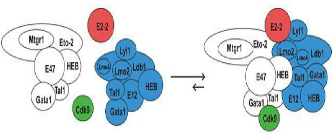
TAL-1 may act as a bifunctional transcriptional regulator (activator and repressor) depending on transcriptional coregulators (coactivators or corepressors). TAL-1 is involved in a complex transcription network bearing either repression or activation capacity for genes whose promoters occupied by TAL-1, E2A, and HEB. E2A, HEB and TAL-1 protein levels and their relative ratio as well as their stage-specific transcriptional partners alter during hematopoiesis and consequently the expression of their target genes are adjusted and lineage-specific gene expression programs are controlled. TAL-1 can either form a heterodimers with E2A and HEB proteins, which prevents E2A to form homodimers necessary for transcription activation, or recruit other transcriptional repressors. This repression effect is proposed as one of the several mechanisms involved in leukomogensis (T-ALLX).111 In hematopoietic progenitor cell lines, TAL-1 interacts with ETO-2 through E2A/HEB where ETO-2 is recruited on its target promoters via this complex. SCL complex gene expression profile is repressed by ETO-2 at the early stage of erythroid differentiation and restores, later in process of cellular maturation, by changing in the ratio of TAL-1. During erythroid differentiation, the relative ratio of TAL-1/ETO-2 rises in favor of TAL-1 that reduces the inhibitory effect of ETO-2 over the expression of TAL-1-dependent erythroid-specific genes.112 A recent study showed that, upon the induction of murine erythroleukemic (MEL) cells and subsequent to detachment of ETO-2 complex from Ldb1 complexXI the level of ETO-2 decrease which leads to induction of differentiation and termination of proliferation (Figure-7).113
X
T cell Acute Lymphoblastic Leukemia
XI
40
Figure 7- Model of Ldb1 complexes in uninduced MEL cells. The horizontal arrows indicate that
the balance of interaction is towards the large complex in proliferating noninduced cells. Upon the induction of differentiation and termination of proliferation the level of Eto-2 drops whereas the level of Lmo4 rises, hence the equilibrium would shift towards the smaller complexes. The presence of several DNA binding proteins in a single complex may explain the role of Ldb1 as a facilitator of long-range interactions.
Taking together, the fluctuation in concentration of transcription factors and/or their cofactors as well as their relative ratio can influence the final composition of protein complexes recruited to gene regulatory regions and therefore play a cause-and-effect relationship in expression of erythroid genes and in making decisions on differentiation versus proliferation.
Further to clear information from prior studies on animal models and considering the insurmountable limitations to the direct experimentation one can perform in human populations, the mouse modeling is most effectual to understand the mechanisms involved in β-globin gene expression. This study is expected to provide insight into epigenetic regulation of β-globin locus during erythropoiesis and help to understand how gene expression is regulated in the context of chromatin. Once DNA regions subject to transcription factor recruitment are identified, changes in transcription factor networks within these sites will be examined in reference to globin gene expression. Herein, we attempt to define the affect of bHLH transcription factors on potentiation and basal level expression of globin gene in hematopoietic progenitor cells and on high level expression in
41
erythroid cells. In an attempt to investigate the whole β-globin locus, the strategy of present project is to focus primarily on HS2, HS3 and promoters of γ-and β-globin genes as these regulatory regions are highly essential for expression of globin genes. By this, we would reemphasize the harmonizing role of LCR and promoter regions in gene expression. Once the components of transcription machinery are identified, we visit knock-out studies, using HEB-/- and E2A-/- mouse models transgenic for β-globin locus, to validate our findings. This study is designed to deliver an answer to address the molecular mechanisms associated with epigenetic modification and transcriptional activation of the β-globin locus and is a continuation of what we have been doing orientated in alternative lineage.
42
Applications of study
Molecular application. Understanding the molecular mechanisms that control
globin gene expression during development provides us with great insights into basis of epigenetic states acquired at multiple levels and shows how they impact globally on gene expression. This project will enhance our knowledge about the mechanism of globin gene expression. Research in this field has an impact on understanding the basic concepts and principles of regulatory mechanisms that coordinately ensure a highly specialized, tissue- and stage-specific gene transcription pattern in other loci. This study could broaden the horizons of eukaryotic gene regulation as new molecular mechanisms will be discovered and existing ones better characterized.
Clinical application. Molecular investigations of spatial and temporal events that
control β-like globin gene expression during erythropoiesis would define many basic mechanisms underlying or relevant to the pathophysiology of hemoglobinopathies, such as sickle cell disease and thalassemia, where the expression of globin genes is disrupted, and would eventually lead to development of molecular therapies and/or cures. For instance, mutations in strategic regions of genes encoding such transcription factors could serve as insults responsible for development and clinical outcome of hemoglobinopathies and become in a position of novel targets for gene therapy in future.
Research Proposal
Rational
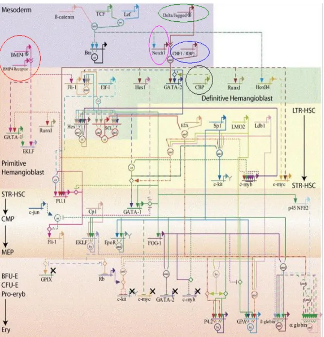
The stepwise establishment of lineage-specific gene expression profiles, during the development of mature blood cells of distinct lineages from HSCs, which progressively restrict the differentiation potential and escalate the proliferation potential, makes hematopiesis an exceptional model system to study TFNs modulating the expression of lineage-specific target genes (Figure-8).114 The human β-globin locus is a highly
43
characterized locus to study the molecular mechanisms of enhancer-promoter interactions and to elucidate cell type-specific and developmental-stage specific regulation of gene expression. E-box sequences, which are potential binding sites for the bHLH proteins, are scattered throughout the β-globin locus within transcriptional regulatory regions at HS core elements of 5’HS2 and 5’HS3 as well as promoters of γ- and β-globin genes.115 For this reason, these elements are theoretically able to recruit various bHLH proteins and their cofactors in the erythroid environment. The animal modeling provides a powerful tool to directly test the effects of bHLH proteins recruitment to LCR and promoters of γ- and β-globin genes on β-globin gene expression and to determine the mechanisms of these effects. Once such mechanisms have been delineated, one can return to human studies to confirm key components.
44
Figure 8- Genetic regulatory networks (GRNs) programming hematopoietic stem cells and erythroid lineage specification. A control logic model of erythroid development describing known
interactions important in the development of the erythroid lineage. The genes have been positioned in the network so that genes expressed at early developmental stages are positioned towards the top of the figure and vice versa. Genes repeated in the network in different positions highlight the different behavior important at different stages.
45
Hypothesis
The prevailing hypothesis is that human β-globin locus potentiation and basal-level of globin gene expression in multipotent hematopoietic progenitor cells (HPCs) and high-level of globin gene expression in erythroid cells are the results of both chromatin modifying activities and the recruitment of general and lineage-specific transcription factors. Informed by key roles of the bHLH transcription factors in lymphoid lineage-specific gene expression, we hypothesized that they could regulate the expression of globin genes in HPC and erythroid cells as well. We hypothesized that E proteins are recruited to both LCR and promoters of γ- and β-genes during the development from HPC to erythroid cells alternating with recruitment of ETO-2 and HDAC-1. We also hypothesize that the absence of E2A and HEB can affect the transcription (a) through the changes in involved transcription factors and/or (b) through the disruption of gene expression. These hypotheses will be tested mainly by performing Chromatin Immunoprecipitation (ChIP) assay on different sorted populations of hematopoietic cells harvested from wild-type, transgenic and knockout mouse models that carry the entire human β-globin locus in their genome.
Objectives
The prime objective is to answer the questions, “Are E2A and HEB are components of lineage-specific transcription factors recruited to LCR and promoter of globin genes?" and "How these transcription factors are important in terms of globin gene silencing in HPCs, high level of globin gene expression in erythroid cells and in alternative lineages and how their recruitment change during erythropoiesis?”, using animal models. The project aims to investigate the major regulatory sites within the β-globin locus that are occupied by E proteins during erythropoiesis in mouse models of human β-globin. With this study we aimed to demonstrate the complex system underlying β-globin gene expression with respect to E2A, HEB, ETO-2 and HDAC-1. Our results would stand as an important step towards gaining a full understanding of processes involved in gene expression.
46
1. To investigate whether E2A, HEB, ETO-2 and HDAC-1 are recruited to LCR and promoters of γ- and β-genes in murine erythroid cells and hematopoietic progenitor cells (HPCs) as well as human erythroid cells. 2. To explore how the arrangement of these factors changes during
erythropoiesis.
3. Following the experiments on normal genetic background, we extend our range of inquiry in knockout mice for E2A and HEB and transgenic for the entire human β-globin locus (E2A-/- β+/+ and HEB-/- β+/+), respectively. A key question is whether removal of E proteins (E2A and HEB) activity might lead to change in the transcription factor complexes at LCR and globin gene promoters, particularly in reference to ETO-2 and HDAC-1, and/or be disruptive to expression of globin genes. This will enable us to take advantage of mouse genetics to further assess the transcriptional changes at the levels of progenitors and mature red cells.
4. Questions persist as to whether the absence of globin gene expression in lymphoid lineage could to some extent be due to different factors engaged at LCR and globin gene promoters, in which all or none of E2A, HEB, ETO-2 and HDAC-1 factors may be present. We extend our findings by similar exploration of LCR and promoters of γ- and β- genes in lymphoid progenitor cells (LPs) in transgenic mice with wild-type genetic background.
47
48
Mouse models
Line 2 mouse. The mouse model we used is called line 2 (ln2) which is a transgenic
mouse for the entire human β-globin locus. This line maintains strong and consistent expression of human globin genes and the mice thereof express β-globin genes normally: the human transgene is expressed in all murine erythroid cells and each cell that contains mouse β-major globin mRNA also contains human β-globin mRNA. This model was previously described by Milot E, et al and Strouboulis J, et al.116,117 ln2 mice were maintained from homozygous breeder line, available in Dr. Eric Milot’s laboratory, born to homozygous mothers crossed with homozygous fathers.
(E2A +/- ln2 +/+) and (HEB +/- ln2 +/+) mice. Most of both E2A knockout and
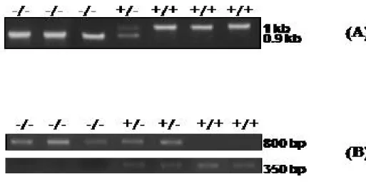
HEB knockout mice die within the first two weeks after birth.XII 118 Homozygous mutant mice for E2A and HEB (E2A +/- and HEB +/-) were kindly provided by Dr. Trang Hoang. These mice were crossed with male or female of ln2 +/+ background to produce the ln2 strains heterozygous for the E2A and HEB allele, respectively. After a long process of crossing and breeding, E2A +/- ln2 +/+ and HEB +/- ln2 +/+ mice were obtained by mating males or females with females or males of same genotype. As discussed above, E2A knockout (E2A-/-) and HEB knockout (HEB -/-) genotypes are almost always lethal;119 therefore, E2A-and HEB-null backgrounds would be best investigated as embryos. For this, E2A +/- ln2 +/+ and HEB +/- ln2 +/+ mice were mated with mice of opposite sex and same genotype, then the embryos were extracted out from the mothers after cervical dislocation on gestational day 13.5 (GD13.5, also called 13.5 dpcXIII). After that, fetal livers were extracted and 13.5 dpc fetal liver-derived hematopoietic cells were harvested by flushing the fetal livers. DNA was prepared from a fraction of these cells and used in polymerase chain reaction (PCR) for genotyping to identify those having E2A -/- ln2 + or HEB -/- ln2 + genotype (see Figure 9). With such a cross, the predicted genotype representation for the
XII
E2A knockout mice are also born at a lower frequency than wild-type mice.
XIII
49
embryos is equivalent for wild-type, homozygous mutant and knockout. The fetal liver erythroid cells (EryC) with E2A -/- ln2 +/+ or HEB -/- ln2 +/+ genotype were utilized in the experiments.
Cell Sorting
Staining with antibodies (Abs) was performed on ice for 30 minutes followed by one wash in phosphate-buffered saline (PBS) 5% fetal bovine serum (FBS). High-speed fluorescence-activated cell sorter (FACS) Vantage Flow Cytometer/Cell Sorter machine with DIgitalized VAntage (DIVA) option (Becton Dickinson, San Jose, CA) was used to analyze and sort the following desired cell populations. For detection of desired hematopoietic surface markers, the commercial antibodies were purchased accordingly. Nonspecific staining was controlled by isotype-matched control antibodies. Sorted populations were always ≥ 90% pure to perform Chromatin Immunoprecipitation assays.
Murine erythroid cells (Ter-119+) and Murine HPCs (Ly-6C-CD31highc-Kit+)
Bone marrow cells were harvested from the long bones (tibiae, femora) of adult ln2 mice. Murine erythroid cells (EryC) were stained with rat anti-mouse Ter119 Abs (TER-119: sc-19592) followed by goat anti-rat phycoerythrin (PE)-conjugated Abs (Santa Cruz Biotechnology, Inc.). For murine HPCs, cells were stained with rat anti-Ly-6C fluorescein isothiocyanate (FITC)-conjugated Abs (ER-MP20: sc-52650) followed by biotinylated rat anti-CD31 (BD Pharmingen: 553371), and then goat anti-rat phycoerythrin (PE)-conjugated Abs (BD Pharmingen), followed by rat anti-mouse c-Kit (CD117) allophycocyanin (APC)-conjugated Abs (BioLegend, Catalog # 105812). The populations of ≥ 90% pure HPCs were separated by FACS.