Université de Montréal
Effect of Mindfulness Meditation on the Neural
Substrates of Emotion Processing and Resting State
in Experienced and Beginner Meditators
par
Véronique Taylor
Département de psychologie Faculté des arts et sciences
Mémoire présenté à la faculté des arts et sciences en vue de l’obtention du grade de maîtrise en sciences (M.Sc.)
en psychologie
Juillet, 2011
Université de Montréal
Faculté des études supérieures et postdoctorales
Ce mémoire intitulé :
Effect of Mindfulness Meditation on the Neural Substrates of Emotion Processing and Resting State in Experienced and Beginner Meditators
Présenté par : Véronique Taylor
a été évalué par un jury composé des personnes suivantes :
Franco Lepore, président-rapporteur
Mario Beauregard, directeur de recherche
Résumé
La méditation par le ‘mindfulness’ favorise la stabilité émotionelle, mais les mécanismes neuroneux qui sous-tendent ces effets sont peu connus. Ce projet investiga l’effet du ‘mindfulness’ sur les réponses cérébrales et subjectives à des images négatives, positives et neutres chez des méditants expérimentés et des débutants au moyen de l’imagerie par résonance magnétique fonctionnelle (IRMf). Le ‘mindfulness’ atténua l’intensité émotionelle via différents mécanismes cérébraux pour chaque groupe. Comparés aux méditants, les débutants manifestèrent une déactivation de l’amygdale en réponse aux stimuli émotifs durant le ‘mindfulness’. Comparés aux débutants, les méditants exhibèrent une déactivation de régions du réseau du mode par défaut (RMD) pendant le ‘mindfulness’ pour tous stimuli (cortex médian préfrontal [CMP], cortex cingulaire postérieur). Le RMD est constitué de régions fonctionnellement connectées, activées au repos et déactivées lors de tâches explicites. Cependant, nous ne connaissons pas les impacts de l’entraînement par la méditation sur la connectivité entre régions du RMD et si ces effets persistent au-delà d’un état méditatif. La connectivité fonctionnelle entre régions du RMD chez les méditants et débutants au repos fut investiguée au moyen de l’IRMf. Comparés aux débutants, les méditants montrèrent une connectivité affaiblie entre subdivisions du CMP, et une connectivité accrue entre le lobule pariétal inférieur et trois regions du RMD. Ces résultats reflètent que les bienfaits immédiats du ‘mindfulness’ sur la psychopathologie pourraient être dûs à une déactivation de régions limbiques impliquées dans la réactivité émotionelle. De plus, les bienfaits à long-terme de la méditation sur la stabilité émotionelle pourrait être dûs à une déactivation de régions corticales et cingulaires impliquées dans l’évaluation de la
Mots clés : Méditation, Pleine conscience (‘mindfulness’), Régulation émotionnelle, Amygdale, Cortex préfrontal, Réseau du mode par défaut, Connectivité fonctionnelle
Abstract
Mindfulness meditation promotes emotional stability, yet little is known of the brain mechanisms through which this is achieved. The impact of mindfulness on the neural and subjective responses to negative, positive, and neutral pictures in experienced meditators and beginners was investigated using functional magnetic resonance imaging (fMRI). Mindfulness attenuated emotional intensity via distinct neural pathways for each group. For beginners, mindfulness induced a deactivation of the amygdala during emotional processing compared to meditators. For meditators (relative to beginners), mindfulness induced deactivations of areas involved in the evaluation of emotional significance and the default mode network (DMN) across all picture categories (medial prefrontal cortex [MPFC], posterior cingulate cortex). The DMN consists of functionally connected brain areas typically activated at rest and deactivated during goal-directed tasks. It remains unknown whether meditation training influences functional connectivity within DMN regions, and if so, whether these effects persist beyond a state of meditation per se. Functional connectivity within DMN regions at rest was examined using fMRI in beginners and meditators. Relative to beginners, meditators exhibited decreased connectivity between MPFC subdivisions, and increased connectivity between the right inferior parietal lobule and three other DMN regions. These findings may reflect that early beneficial effects of mindfulness on psychopathology are due to deactivations of limbic regions involved in emotional reactivity. On the other hand, long-term effects of meditation on emotional stability may occur through a down-regulation of prefrontal and cingulate regions involved in the evaluation of emotional significance, and altered functional connectivity within DMN regions at rest.
Keywords: Meditation, Mindfulness, Emotion Regulation, Amygdala, Prefrontal Cortex, Default Mode Network, Functional Connectivity
Table of Contents
Résumé ... iii
Abstract ... v
Table of Contents ... vii
List of Symbols and Abbreviations ... xi
List of Tables... xv
List of Figures ... xvi
Dedications ... xviii
Acknowledgments ... xix
General Introduction ... 20
Emotions and Emotional Processing ... 21
Affective Behavioral Neuroscience ... 23
Relevance to Psychopathology ... 25
Mindfulness Meditation and the Regulation of Emotion ... 26
Mindfulness, Emotions, and the Brain ... 28
The Default Mode Network ... 31
Principal Objectives ... 33
Hypotheses ... 34
Article 1: Impact of Mindfulness on the Neural Responses to Emotional Pictures in Experienced and Beginner Meditators ... 35
Introduction ... 38
Methods ... 44
Participants ... 44
Stimuli and Experimental Procedure ... 46
fMRI Data Acquisition and Analysis ... 49
Results ... 52
Self-Report Data... 52
fMRI Data ... 55
Emotional Processing at Baseline ... 55
Brain Activity Related to the Effects of Mindfulness and Group ... 56
Condition x Valence x Group Interactions... 56
Negative Emotional Processing ... 56
Positive Emotional Processing ... 57
Condition x Group Interaction ... 60
Main Effects ... 63
Condition ... 63
Group ... 63
Discussion ... 66
Mindfulness and Emotional Intensity ... 66
Brain Patterns Associated with Mindfulness in Experienced and Beginner Meditators ... 68
Brain Patterns Associated with Mindfulness and Emotional Processing in Experienced and Beginner Meditators... 71
Limitations and Future Directions ... 73
Conclusions ... 74
Acknowledgments ... 75
References ... 76
Article 2: Impact of Mindfulness Training on the Default Mode Network during a Restful State: A Functional Connectivity Study ... 84
Abstract ... 86
Introduction ... 87
Materials and Methods ... 90
Participants ... 90
fMRI Data Acquisition ... 91
Experimental Protocol ... 92
Data Analysis ... 92
Results ... 101
Group Differences in Correlations between DMN Regions ... 101
Group Differences in Partial Correlations between DMN Regions ... 105
Discussion ... 108
Identification of the Default Mode Network using NEDICA ... 108
Increased Connectivity for Experienced Meditators Compared to Beginners ... 109
Decreased Connectivity for Experienced Meditators Relative to Beginners ... 111
Correlations and Partial Correlations between DMN Regions ... 113
Conclusions ... 115
Acknowledgments ... 116
References ... 117
General Discussion ... 126
Mindfulness and Emotional Processing ... 127
Mindfulness and the Default Mode Network ... 128
Limitations and Future Directions ... 131
Conclusions ... 132
List of Symbols and Abbreviations
η2 ... Eta Squared Measure of Effect Size Π ... Partial Correlation Coefficient
ACC ... Anterior Cingulate Cortex
AMY ... Amygdala
BA ... Brodmann Area
BOLD ... Blood-Oxygen Level Dependent
cc ... Correlation Coefficient
CORSICA ... Correction of Structured noise using spatial Independent Component Analysis
d ... Cohen’s D Measure of Effect Size
DMN ... Default Mode Network
DMPFC ... Dorso-Medial Prefrontal Cortex
DR ... Degree of Representativity
DU ... Degree of Unicity
EEG ... Electroencephalography
fMRI ... Functional Magnetic Resonance Imaging
IC ... Independent Component
ICA ... Independent Component Analysis
INS ... Insula
IPL ... Inferior Parietal Lobule
ITC ... Infero-Temporal Cortex
k ... Clsuter Size (Number of Voxels)
LPFC ... Lateral Prefrontal Cortex
MDD ... Major Depressive Disorder
MFG ... Medial Frontal Gyrus
MPFC ... Medial Prefrontal Cortex
MSBR ... Mindfulness-Based Stress Reduction Program
NEDICA ... Network Detection using Independent Component Analysis
OFC ... Orbito-Frontal Cortex
PC ... Precuneus
PCA ... Principal Component Analysis
PCC ... Posterior Cingulate Cortex
PHG ... Parahippocampal Gyrus
PUT ... Putamen
ROI ... Region of Interest
SAD ... Social Anxiety Disorder
T2* ... Transverse Relaxation Time
TE ... Echo Time
TMS ... Transcranial Magnetic Stimulation
TR ... Repetition Time
List of Tables
Article 1: Impact of Mindfulness on the Neural Responses to Emotional Pictures in Experienced and Beginner Meditators
Table 1 Mindfulness-Induced Deactivations: Positive vs. Neutral
Pictures 59
Table 2 Brain activations related to the Main Effect of Group 64
Article 2: Impact of Mindfulness Training on the Default Mode Network during a Restful State: A Functional Connectivity Study
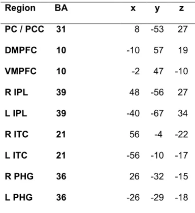
Table 1 Coordinates for Seed Regions within the Default Mode
List of Figures
Article 1: Impact of Mindfulness on the Neural Responses to Emotional Pictures in Experienced and Beginner Meditators
Figure 1. Emotional intensity ratings by Group and Condition for each Valence
category of pictures. 54
Figure 2. Statistical activation maps for the Mindfulness-Induced Deactivations
during Emotional Processing. 58
Figure 3. Statistical activation maps for the Mindfulness-Induced Deactivations
Across all Valence Categories of Pictures. 62
Article 2: Impact of Mindfulness Training on the Default Mode Network during a Restful State: A Functional Connectivity Study
Figure 1. Default mode network t-map identified at the group level using
NEDICA across all participants. 96
Figure 2. Diagram illustrating significant group differences (p < .05)
Figure 3. Correlation values (cc) for all pairwise relationships between
default network regions. Blue triangles represent values for experienced meditators, and orange squares represent values for
beginner meditators. 104
Figure 4. Diagram illustrating significant (p < .05) group differences
in partial correlations (Π) between default mode network regions. 106
Figure 5. Partial Correlation values (Π) for all pairwise relationships between
default network regions. Blue triangles represent values for experienced meditators, and orange squares represent values for
beginner meditators. 107
To Loïc, Who teaches me By example the Value of Hard Work and Perseverance
To my Mother, Who keeps showing me the Importance of Resourcefulness, Strength and Determination And Taught me that a Little Organization goes a Long Way
Acknowledgments
I would like to express my gratitude to Véronique Daneault, for her hard work and dedication in acquiring the data included in this research project. I would also like to thank Joshua Grant, for his invaluable help and feedback on the analyses, interpretations and writing included in this project. I show sincere appreciation to Dr Pierre Rainville, for his insightful comments on the results and rationale included in these studies. I express thanks for the hard work on the part of Geneviève Scavone, Estelle Breton, and Sébastien Roffe-Vidal in the data collection and setup of the experimental design. I also thank Dr Zeffiro (Massachussetts General Hospital, Harvard University), for his help on several questions related to the SPM8 software used for the data analysis in this project. Particularly, I am deeply obliged to the participatants implicated in this research project as they have given us the opportunity to study a very noble discipline. Finally, I express thanks to Dr Mario Beauregard, for his supervision, and, undoubtedly, to NSERC for funding this research project.
General Introduction
Mindfulness meditation is an ancient spiritual practice which attenuates emotional reactivity and is beneficial to the treatment of emotion-related disorders (Baer, 2003). Yet the neural mechanisms through which this is achieved currently remain unknown. In this context, the principal aim of this thesis was to investigate the neural mechanisms through which mindfulness meditation influences the processing of emotional processing and a baseline state of rest. A brief overview of the empirical psychological literature on emotions and emotion regulation is presented, as well as an overview of findings from the neuroscientific literature on brain mechanisms involved in emotional processing (also known as affective behavioural neuroscience). Findings from studies investigating the relationship between meditation, emotional processing and brain function are then presented. Next, the literature on resting states using functional magnetic resonance imaging (fMRI) and findings demonstrating the
involvement of the default-mode network in emotional processing as well as meditation are discussed. The specific objectives and hypotheses are stated, and the results of this thesis are presented within two articles (the first article has been published in the journal
NeuroImage; the second article has been submitted to the journal Social Cognitive and Affective Neuroscience). Finally, the results presented in these articles are discussed,
Emotions and Emotion Regulation
As opposed to moods which are diffuse and long-lasting, emotions are generally short-lived experiences triggered by discrete events (either internal events such as thoughts, or concrete external events; Gross, 2007). Additionally, emotions are thought to depend on the meaning attributed to their triggering events in terms of their relevance to one’s goals, values, and circumstances (Gross, 2007).
Specifically, the sequence of events preceding an emotional response has been described as: 1- the occurrence of a triggering event (external or internal), 2- the allocation of attention to this event, 3- the appraisal or assessment of the event with respect to one’s goals and values, after which 4- the emotional response ensues (Gross, 2007). Thus, the appraisal of the triggering event is thought to play a crucial role in generating emotional responses. This sequence of events is often cyclical, such that one emotional response may constitute a triggering event generating a subsequent emotional response, etc. Additionally, emotional responses occur across several dimensions: subjective experience, behaviour, and physiology (involving central and peripheral nervous system processes). However, an important aspect of emotions is that they are not permanent; rather, emotions are in constant change and are malleable.
Indeed, given that we are primarily social beings, certain emotional responses cannot be expressed within every social context. For example, it would not be considered appropriate to burst out crying in the middle of a professor’s class lecture, but it would be considered appropriate to do so at a funeral. In most cases, when
experiencing an emotion arising in an incongruent context (grief from the death of a relative in the context of a classroom), an attempt is made to regulate, attenuate, or modulate this emotion in some way.
Emotion regulation refers to several types of processes (conscious, unconscious, voluntary, uncontrolled) which are intended to attenuate, maintain, or amplify an emotional response. Some types of emotion regulation strategies are focused on altering aspects of the response itself, such as performing physical exercise to attenuate anxiety before an exam. Other strategies are focused on regulating aspects of the appraisal process. As such, re-appraisal entails that the triggering event is re-interpreted in more neutral terms (Ochsner, Bunge, Gross, and Gabrieli, 2002). Since the regulation of emotion allows individuals to comply with social norms and to function effectively within the environment, failure to effectively regulate emotions can lead to detrimental psychosocial functioning, and is associated with a wide array of psychological disorders including major depression, anxiety disorders, psychopathy, and antisocial personality disorder (Phillips, Drevets, Rauch, and Lane, 2003a). It is therefore crucial to gain a better understanding of emotional experiences and emotion regulation. Specifically, the neural mechanisms underlying these processes have been increasingly studied using a variety of methods assessing brain function.
Affective Behavioural Neuroscience
A wide body of research suggests the involvement of two main cerebral systems in emotional processing: brain regions involved in lower-level subcortical affective appraisal, and brain regions involved in higher order top-down affective appraisal systems (Gross, 2007; Phan, Wager, Taylor, and Liberzon, 2004; Phan, Wager, Taylor, and Liberzon, 2002).
Lower-order affective appraisal brain systems are thought to involve primary innate responses to emotion-triggering events, and consist mostly of subcortical limbic brain structures innervated via a direct pathway from primary sensory areas to the thalamus (Gross, 2007). These structures include the amygdala, the insula, the nucleus accumbens, and the basal ganglia (putamen, caudate nucleus), and play a role in encoding the affective value of environmental stimuli (Calder, Lawrence, and Young, 2001; Phillips, Drevets, Kauch, and Lane, 2003b).
According to evolutionary perspectives, these structures have important survival functions as they allow individuals to react quickly by signalling biologically relevant information, including potential environmental threats. Particularly, the amygdala has been shown to be activated in response to both aversive and fearful information (see, for instance, Ochsner et al., 2002; Ochsner, Knierim, Ludlow, Hanelin, Ramachandran, Glover, and Mackey, 2004; Whalen, Rauch, Etcoff, McInerney, Lee, and Jenike, 1998), as well as apetitve stimuli (Beauregard, Lévesque, and Bourgoin, 2001; Sergerie, Chochol, and Armony, 2008). In contrast, the basal ganglia (caudate nucleus and
putamen) appear to be activated mainly in response to positive, pleasurable and addictive stimuli (Everitt, Parkinson, Olmstead, et al., 1999; Haruno et al., 2004; Naqvi and Bechara, 2010; Phillips and LeDoux, 1992; Prado-Alcala and Wise, 1984). In addition, the anterior portion of the insula has been shown to be involved in the anticipation of aversive events, and is thought to reflect greater awareness of interoceptive states (Craig 2004; Critchley, Mathias, and Dolan, 2001).
Nonetheless, primary affective appraisal systems have been shown to have afferent and efferent connections to higher-order cognitive and control areas of the prefrontal and cingulate cortices (Beauregard et al., 2001; Levesque, Eugene, Joanette, Paquette, Mensour, Beaudoin, Leroux, Bourgouin, and Beauregard, 2003; Ochsner et al., 2002; Ochsner et al., 2004). As such, the amygdala, which plays an important role in fear conditioning, has been shown to be densely connected to the prefrontal cortex, especially the medial prefrontal cortex (MPFC; BA 9 /10) and orbito-frontal cortex (OFC; BA 11 / 47; Amaral, Price, Pitkanen, and Carmichael, 1992; Davidson, 1998). These regions of the prefrontal cortex (PFC) have been shown to play a pivotal role in the extinction of conditioned fear and the dampening of negative affect (Amaral et al., 1992; Davidson, 1998).
Thus, higher-order areas of the PFC and cingulate cortex are thought to be mainly involved in the voluntary regulation of emotion, including the reappraisal of emotional events and the voluntary generation of emotional states (Beauregard et al., 2001; Levesque et al., 2003; Ochsner et al., 2002; Ochsner et al., 2004). In addition, the
rostro-dorsal anterior cingulate cortex (ACC) appears to be involved in monitoring the extent to which regulation strategies actually affect the emotional response (Ochsner et al., 2002; Ochsner et al., 2004).
Finally, subdivisions of higher order PFC regions may also underlie distinct functions. For instance, dorsal regions of the MPFC (8), LPFC (8 / 9), and rostro-dorsal ACC (32) have been associated with description-based appraisal systems, mainly involved in mental descriptions of emotional states as well as the controlled appraisal of emotional states using beliefs and expectations (Gross, 2007). Ventral and orbital portions of the PFC and ACC have been defined as outcome-based systems (Gross, 2007), which are involved in learning basic stimulus-outcome contingencies. Extinction of conditioned learning appears to recruit these areas (Amaral et al., 1992; Davidson et al., 1998). With the use of a variety of brain mapping techniques such as functional magnetic resonance imaging (fMRI), electroencephalography (EEG), and transcranial magnetic stimulation (TMS), the study of the neural substrates underlying emotional processes has revealed considerable insight into clinical affect-related psychopathology.
Relevance to Psychopathology
Different neural processes relevant to emotional processing seem to distinguish clinical from healthy populations (Phillips et al., 2003b). With regard to this issue, hyperactivity of the amygdala has been observed in depressed and anxious patients (Drevets, 2000; Rauch et al, 1997). Depression has also been characterized by hypoactivity of the left PFC, a brain area which has been associated with positive
emotions (Davidson, 2002). In addition, a theory has been put forth that hemispheric differences in parietal cortex activity play a role in arousal: greater left than right parietal activity is associated with low arousal, as observed in depressed individuals without an anxiety disorder; and greater right than left parietal activity is associated with increased arousal, as observed in depressed patients with an anxiety disorder (Bruder; Fong; Tenke; Leite; Towey; Stewart; McGrath, and Quitkin 1997; Davidson 1992; Davidson 2002). Since several mental disorders such as depression and anxiety disorders typically involve some form of emotional dysregulation (Gross, 2007), psychological treatments of clinical populations often focus on implementing and developing various emotion regulation skills.
Mindfulness Meditation and the Regulation of Emotion
One type of mechanism which appears to influence emotional responsiveness and to promote emotional stability (Arch et al., 2006; Baer, 2003; Broderick, 2005) is mindfulness meditation, a practice which is being increasingly studied under experimental psychological and neuroscientific frameworks.
Meditation originates from ancient Eastern traditions, and this spiritual practice dates back from more than two millenaries ago (Nataraja, 2008). Mindfulness is a type of meditation based on cultivating self-observation and present-moment awareness (Kabat-Zinn, 1994), as opposed to living in recollections of the past or in the anticipation of fears concerning the future. As such, Kabat-Zinn (1994) defines
mindfulness as “paying attention in a particular way: on purpose, in the present moment, and non-judgementally”.
Mindfulness meditation is commonly practiced sitting down, and initiated by focusing on a particular object, often physical aspects or sensations related to the breath or posture. In doing so, “each thought, feeling, or sensation that arises in the attentional field is acknowledged and accepted as it is” (Bishop, 2004). After acceptance of these arising events, attention is re-directed to the initial object of focus. Eventually, the frequency of interfering internal thoughts decreases, and ultimately these thoughts cease to arise, at which stage attention is in complete contact with its direct environment. Importantly, during a state of mindfulness, events are not evaluated as good, bad, or assessed in terms of implications for the self (Bishop, 2004). Mindfulness therefore promotes a detached and objective manner of responding to emotional events, as opposed to other maladaptive practices such as avoidance or rumination (Broderick, 2005).
Mindfulness has indeed been shown to reduce ruminative thoughts as well as depressive symptoms (Deyo, Wilson, Ong, and Koopman, 2009). In addition, a focused-breathing manipulation, typically practiced during a state of mindfulness, was shown to reduce emotional volatility to affective slides (Arch and Craske 2006). It is therefore not surprising that this practice has widespread benefits for the treatment of emotion-related psychological disorders, including major depression, which is associated with excessive rumination on negative thoughts (Joormann, 2006), generalized anxiety disorder, which
is associated with excessive worry and avoidance (Dugas, Freeston, Ladouceur, Rheaume, Provencher, and Boisvert, 1998), as well as substance use, chronic pain, and binge eating disorder (Baer, 2003). Nevertheless, the neural mechanisms through which mindfulness influences emotional responses remain unclear.
Mindfulness, Emotions, and the Brain
Few studies have examined the effects of mindfulness, the brain, and emotional processing. Lutz et al. (2008) have investigated the effect of compassion meditation (which involves generating a non-specific, unconditional state of compassion) on brain function in response to emotional aversive, positive and neutral sounds in experienced Buddhist meditators as well as novices. They found that compassion meditation induced increased anterior insula and rostro-dorsal ACC activity during the processing of aversive sounds, and this occurred to a larger extent in experienced meditators. Thus, according to these data, the insula and ACC play a role in generating compassion meditative and empathic states to sounds of distress. In addition, they found that across all valence categories, compassion meditation induced increased activity in the amygdala for the experienced meditators compared to novices. This may reflect general increased emotional arousal during a state of loving-kindness meditation (Lutz, Brefczynski-Lewis, Johnstone, and Davidson 2008).
In contrast, another study conducted by the same group (Brefczynski-Lewis, Lutz, Schaefer, Levinson, and Davidson, 2007) found that for experienced meditators (compared with novices), mindfulness meditation induced decreased activity of the
amygdala during the processing of negative emotional sounds. The magnitude of this effect was also negatively correlated with the extent of meditation experience in the group of experienced meditators. Similarly, Grant et al (2011) found that during aversive painful stimulations, experienced Zen meditators had reduced amygdala activity compared to non-meditators, suggesting an attenuating effect of meditation experience on reactivity limbic systems. These results are consistent with evidence that after completing an eight-week mindfulness-based stress-reduction (MSBR) training, individuals with social anxiety disorder had decreased amygdala activity in response to negative self-beliefs (Goldin and Gross 2010).
There are inconsistencies in the literature with respect to the effect of meditation on lower-level appraisal systems such as the amygdala in response to emotional stimuli. Some reports suggest that meditation attenuates activity in this structure during negative emotional processing (Brefczynski-Lewis et al., 2007), consistent with the idea that meditation attenuates emotional reactivity. On the other hand, other evidence demonstrates a general increase in amygdala activity in response to emotional and non-emotional stimuli during a (compassion) meditative state (Lutz et al., 2008), consistent with the fact that emotions are accepted and unsuppressed during a state of meditation.
With respect to higher-order PFC systems involved in emotional processing, Grant and colleagues (2011) found that experienced meditators exhibited deactivations in the medial PFC (MPFC), a brain region involved in evaluating self-relevance and emotional value to stimuli. In contrast, non-meditators recruited the dorso-lateral PFC, which is
involved in the voluntary regulation of emotion (Ochsner et al., 2004). These findings were interpreted as reflecting that meditation practice attenuates the emotional impact of stimuli by deactivating brain areas involved in the cognitive evaluation of emotional stimul. In other words, mindfulness meditation may be associated with ‘no regulation’ or ‘no appraisal’ due to acceptance of emotional states (Grant et al., 2011). Inherently, as suggested by Grant and colleagues, this may translate into a passive decoupling of PFC-amygdala pathways via deactivation of cortical appraisal systems.
The MPFC deactivations observed in experienced meditators in response to painful stimuli (Grant et al., 2011) are consistent with previous research (Farb, Segal, Mayberg, Bean, McKeon, Fatima, and Anderson, 2007). Indeed, Farb and colleagues (2007) found that an experiential focus condition (evaluating the self in terms of moment-to-moment monitoring of the environment) was associated with decreased activity in the dorsal and ventral MPFC relative to a narrative self condition (evaluating the self in terms of trait-like descriptors). This effect was observed to a larger extent in participants having completed an 8-weeek MSBR program than in novices. Together, the results from Grant et al (2011) and Farb and colleagues (2007) regarding decreased activity in the MPFC associated with meditation - are relevant to the fact that the MPFC is also one of the key structures involved in the so-called ‘default mode network’ (DMN).
The Default Mode Network
The DMN consists of spatially remote brain regions with correlated temporal dynamics. This network is typically actived during rest, and deactivated during specific goal-related tasks (Buckner, Andrews-Hanna, and Schacter, 2008). Though the mental processes related to the DMN remain unclear, it has been proposed that activity in this network during rest (or in the absence of engagement in activities with the external world) reflects mentation about the self (Northoff, Heinzel, de Greck, Bermpohl, Dobrowolny, and Panksepp, 2006), simulations of past and future scenarios, and the planification of future actions (Buckner, Andrews-Hanna, and Schacter, 2008). The core brain regions in the DMN are located within posterior portions of the brain (inferior parietal lobule [IPL], precuneus, posterior cingulate cortex [PCC]), the temporal lobe (lateral and mid-temporal cortex, and the parahippocampal formation), and the prefrontal cortex (dorso-medial PFC [DMPFC] and ventro-medial PFC [VMPFC]).
Specifically, DMN function has been widely studied using fMRI, by recording brain activity during ‘resting states’ (typically, sessions of several minutes during which participants are instructed to rest and not to engage in any explicit goal-related task). These studies have provided valuable insight into clinical disorders, such as marked differences in DMN functioning in patients with Alzheimer’s Disease (who show decreased connectivity between DMN structures and the hippocampus; Greicius; Srivastava; Reiss, and Menon 2004) and major depression (who exhibit increased connectivity between DMN structures and the subgenual ACC; Greicius, Flores, Menon, Glover, Solvason; Kenna, Reiss, and Schatzberg 2007).
The DMN has also been shown to be related to emotional processing (Sheline, Barch, Price, Rundle, Vaishnavi, Snyder, Mintun, Wang, Coalson, and Raichle, 2009). Indeed, Sheline and colleagues (2009) demonstrated that depressed patients failed to deactivate DMN regions (IPL [BA 39], VMPFC [BA 10], and lateral temporal cortex [BA 21]) during the passive viewing and re-appraisal of negative pictures. This was interpreted as reflecting greater interference from internal emotional states. In parallel with these findings, there is also evidence that compassion meditation is associated with increased activity in some regions of the DMN, such as the right IPL, precuneus, and parahippocampal gyrus (Lutz et al., 2008). Enhanced activity in these areas has been interpreted as reflecting greater compassion and understanding of others’ intentions. Other reports have demonstrated increased DMN function during a focused-breath induction after viewing negative self-beliefs in social anxiety disorder (SAD) patients in precuneus, IPL and parahippocampal gyrus (Goldin et al, 2010). Finally, Brefczynski-Lewis and colleagues (2007) investigated brain function during concentration meditation, which is similar to mindfulness as it involves focusing attention on one particular object and gently bringing it back to this object when distracted. They found that during a state of concentration meditation, there was a negative correlation between activity in DMN regions (MPFC and PCC) and the number of hours of meditation experience. Therefore, it appears that there is a relationship between the DMN, meditation, and emotional processing. However, the DMN is thought to operate as a coherent network during rest, since the spatially remote regions within this network are functionally connected (i.e. the time-courses between regions are correlated). Yet, to
date, the impact of meditation training on functional connectivity between DMN regions during rest remains completely unknown.
Principal Objectives
The central objectives of this research project were twofold. First, given the premise that mindfulness meditation promotes affective balance (Kabat-Zinn 1994; Bishop, 2004; Arche et al., 2006; Broderick, 2005) and benefits the treatment of affective and anxiety disorders (Baer, 2003), a central goal of this thesis was to determine the effects of mindfulness on the neural responses to emotional information. This question was addressed in Study #1, in which brain activity using fMRI was recorded while experienced and beginner meditators viewed emotional (positive and negative) as well as neutral pictures. Subjective emotional intensity ratings were also recorded immediately following the presentation of each picture. There were two conditions, one in which pictures were viewed in a meditative state, and the other condition involved viewing pictures in a normal (non-meditative) state.
Second, given evidence of altered DMN function associated with emotional processing (Sheline et al., 2009) and meditation (Brefczynski-Lewis et al., 2007), another main objective of this research project was to investigate the relationship between mindfulness training and functional connectivity within DMN regions during a restful state. This question was addressed in Study #2, in which brain activity was recorded during a state of rest (6-minute fMRI sessions in the absence of any goal-related or specific mental activity) in experienced and beginner meditators. This was
investigated using data-driven connectivity analysis approaches (spatial independent component analysis) to identify a DMN group map, after which pair-wise correlations and partial correlations between DMN regions were examined and compared between groups.
Hypotheses
In Study #1, it was hypothesized that the mindful condition would attenuate subjective emotional intensity perceived from emotional pictures. From a neural circuitry perspective, it was hypothesized that mindfulness would induce reduced amygdala activity in response to emotional stimuli, and more so in experienced vs. beginners (Brefczynski-Lewis et al., 2007; Grant et al., 2011). Finally, based on the MPFC deactivations associated with meditation (Farb et al., 2007; Grant et al., 2011), it was hypothesized that mindfulness would induce reduced activity in prefrontal areas involved in controlled regulation of emotion, and to a larger extent in experienced than in beginner meditators.
In Study #2, it was hypothesized that mindfulness training would be associated with weakened connectivity between MPFC and other DMN structures at rest. This hypothesis was also based on MPFC deactivations associated with mindfulness meditation (Farb et al., 2007; Grant et al., 2011), and on the premise that mindfulness is associated with reduced appraisal of emotional events (Bishop, 2004).
Article 1
Title: Impact of Mindfulness on the Neural Responses to Emotional Pictures in
Experienced and Beginner Meditators
Authors : Véronique Taylor, Joshua Grant, Véronique Daneault, Geneviève Scavone,
Estelle Breton, Sébastien Roffe-Vidal, Jérôme Courtemanche, Anaïs S. Lavarenne, Mario Beauregard
Article Status
A revised version of this article was published in NeuroImage, 57, Taylor, V. A. Grant, J. A., Daneault, V., Scavone, G., Breton, E., Roffe-Vidal, S., Courtemanche, J.,
Lavarenne, A. S., and Beauregard, M. Impact of Mindfulness on the Neural Responses to Emotional Pictures in Experienced and Beginner Meditators, 1524-33, Copyright Elsevier (2011).
Author Contributions
V.T. wrote the manuscript, and conducted all of the statistical analyses on the data presented in this article. J.G. actively participated in the writing process, as well as giving feedback on the experimental design, analyses, and interpretations. V.D. was responsible for coordinating and collecting the data included in this research project. G.S. assisted V.D. in the data collection. S. R.-V. and E.B. contributed to the setup of experimental design and tasks. J.C. contributed to the experimental design as well as the recruitment of participants. A.S.L. helped in some of the data acquisition. M.B.
Impact of Mindfulness on the Neural Responses to Emotional Pictures in
Experienced and Beginner Meditators
Véronique Taylor1 Joshua Grant1,2 Véronique Daneault1 Geneviève Scavone1 Estelle Breton1 Sébastien Roffe-Vidal1 Jérôme Courtemanche1 Anaïs S. Lavarenne1 Mario Beauregard1,2,3,4 * 1
Centre de Recherche en Neuropsychologie et Cognition (CERNEC), Département de Psychologie, Université de Montréal, Montréal, Québec, Canada
2
Centre de Recherche en Sciences Neurologiques, Département de Physiologie, Université de Montréal, Montréal, Québec, Canada
3
Département de Radiologie, Université de Montréal, Montréal, Québec, Canada 4
Centre de recherche du Centre hospitalier de l’Université de Montréal (CRCHUM), Montréal, Québec, Canada
*Correspondence: Mario Beauregard Ph.D., Mind/Brain Research Lab (MBRL), Centre de Recherche en Neuropsychologie et Cognition (CERNEC), Département de Psychologie, Université de Montréal, C.P. 6128, succursale Centre-Ville, Montréal, Québec, Canada H3C 3J7
Abstract
There is mounting evidence that mindfulness meditation is beneficial for the treatment of mood and anxiety disorders. Little is known regarding the neural mechanisms through which mindfulness modulates emotional responses. A central objective of this functional magnetic resonance imaging study was to investigate the effects of mindfulness on the neural responses to emotionally laden stimuli. Another major goal of this study was to examine the impact of the extent of mindfulness training on the brain mechanisms supporting the processing of emotional stimuli. Twelve experienced (with over 1000 hours of practice) and 10 beginner meditators were scanned as they viewed negative, positive, and neutral pictures in a mindful state and a non-mindful state of awareness. For both groups, pictures viewed in a mindful state were subjectively perceived as less intense than when viewed in a non-mindful state. Compared with experienced meditators, mindfulness led to a down-regulation of the left amygdala during emotional (negative and positive) processing in beginners. Compared with beginners, mindfulness was associated with a down-regulation of activity in brain areas involved in responsiveness to emotional stimuli, self-referential processing, and the default mode network (medial prefrontal and posterior cingulate cortex) in experienced meditators across all valence categories. These results are consistent with the view that mindfulness attenuates emotional reactivity and influences neural mechanisms involved in emotion processing. These findings have implications for emotion-related psychological disorders.
Originating from ancient Eastern traditions (Nataraja, 2008), meditation is an increasingly prominent object of study within the fields of clinical psychology and behavioral neuroscience. Mindfulness is a form of meditation which has been operationally defined as “a kind of nonelaborative, nonjudgmental, present-centered awareness in which each thought, feeling, or sensation that arises in the attentional field is acknowledged and accepted as it is.” (Bishop, 2004). The detached observation state which is adopted during mindfulness is thought to promote an objective and adaptive manner of responding to emotional triggers in contrast to a habitual pattern of emotional reactivation typically driven by past experiences, fears and preconceptions (Bishop, 2004). Consistent with this view, it has been demonstrated that mindfulness-based clinical interventions have beneficial outcomes on the treatment of affect-related psychopathology, including major depression (Bondolfi et al., 2010; Teasdale et al., 2000) and anxiety disorders (Bondolfi, Jermann, der Linden, Gex-Fabry, Bizzini, Rouget, Myers-Arrazola, Gonzalez, Segal, Aubry, and Bertschy, 2010; Goldin and Gross, 2010; Kabat-Zinn;, Massion, Kristeller; Peterson, Fletcher; Pbert, Lenderking, and Santorelli, 1992; Kim, Lee, Choi, Suh, Kim, Kim, Cho, Kim, Yook, Ryu, Song, and Yook, 2009).
To date, a few studies have examined the effects of meditation on the processing of emotional stimuli. Focused-breathing typically practiced during mindfulness has been shown to promote emotional self-regulation by reducing reactivity to emotionally laden pictures (positive and negative) (Arch et al., 2006). Mindfulness meditation practiced following a sad mood induction was also shown to decrease dysphoria (Broderick,
2005). Additionally, Brefczynski-Lewis and colleagues (2007) found a negative correlation between the number of hours of meditation training and right amygdala activation during concentration meditation in a group of experienced meditators while processing negative emotional sounds. In another study (Farb, Anderson, Mayberg, Bean, McKeon, and Segal, 2010), the MBSR program led to reduced activation in regions associated with autobiographical memory and increased activity in the right insula (Farb et al, 2010). Finally, Lutz and colleagues (2008) investigated the effects of compassion meditation on the processing of emotional auditory stimuli. They found increased right insula activation for experienced meditators, relative to beginner meditators, during a state of compassion meditation while listening to negative emotional sounds (compared to emotionally positive sounds). This brain region plays an important role in awareness of interoceptive states (Craig, 2004; Critchley, Wiens, Rotshtein, Ohman, and Dolan, 2004). Compassion meditation also induced increased amygdala activation while listening to emotional and neutral sounds for experienced meditators relative to beginners. It is noteworthy that since compassion meditation involves maintaining a non-referential state of compassion, different brain processes may be involved in mindfulness meditation during emotional processing as opposed to compassion meditation.
Emotional self-regulation strategies, such as cognitive distancing and reappraisal, have commonly been associated with the recruitment of prefrontal cortical areas critically involved in executive control functions (e.g., lateral prefrontal cortex [LPFC], medial prefrontal cortex [MPFC], and anterior cingulate cortex [ACC]), and decreased
activation in subcortical structures implicated in emotional processing (e.g., amygdala) (Beauregard et al., 2001; Levesque et al., 2003; Ochsner et al., 2002; Ochsner et al., 2004). Convergent with neuroimaging studies of emotional self-regulation, there are some reports that meditative states are associated with increased activation in various prefrontal cortical areas. Such heightened prefrontal activation has been interpreted as reflecting increased recruitment of attentional resources during meditation (Newberg, Alavi, Baime, Pourdehnad, Santanna, and d'Aquili, 2001). Interestingly, mindfulness was shown to be associated with increased MPFC/LPFC activation and reduced amygdala activation during affect labelling, which is commonly employed during meditation in order to identify emotional states (Creswell, Way, Eisenberger, and Lieberman, 2007). In this study, Creswell and collaborators (2007) found that participants high in trait mindfulness also had negative correlations between areas of the PFC (MPFC and LPFC) and the right amygdala, but not those low in trait mindfulness. These findings are consistent with evidence that the MPFC, through its inhibitory downstream connections to the amygdala, is implicated in the extinction of conditioned fear as well as the dampening of negative affect (Amaral et al., 1992; Davidson, 1998). Similar results were found in clinical populations. For example, Goldin and Gross (2010) reported that patients with social anxiety disorder (SAD) exhibited decreased right amygdalar activation after viewing phrases of negative self-beliefs, and increased activation in regions relevant to the control of attention (including regions of the parietal cortex) after having completed an 8-week mindfulness-based stress reduction program (MBSR). Goldin and Gross (2010) interpreted these results as indicating that mindfulness meditation helps to reduce avoidance-related behavior to threatening
stimuli in SAD patients by enhancing the recruitment of brain regions involved in attentional control.
From a phenomenological perspective, it should be noted that cognitive distancing and reappraisal differ to a large extent from mindfulness. Indeed, these emotional self-regulation strategies aim at altering emotional states, whereas mindfulness is based on accepting emotional states as they are (Kabat-Zinn, 1994).
Consistent with this view, some neuroimaging studies suggest that long-term mindfulness meditation training can lead to decreased activation in prefrontal cortical areas, particularly the MPFC (Farb et al., 2007; Grant et al., 2011). In one of these studies, Farb and colleagues (2007) found that an experiential focus condition (monitoring the self in terms of present-moment circumstances) was associated with deactivations of the ventral and dorsal MPFC compared with a narrative self-focus task (monitoring the self in terms of self-descriptive traits). This effect was more pronounced and widespread in individuals having completed an 8-week MSBR program compared to a control group. A similar pattern of results has recently been reported in Zen meditators experiencing emotionally salient painful stimuli (Grant et al., 2011). In this study, when normally attending to painful stimulations, experienced meditators exhibited decreased activation in several brain regions involved in executive control and emotional appraisal, including the MPFC, LPFC, and amygdala compared to non-meditators. These decreased activations found in executive control areas have been
proposed to reflect that meditation training is associated with a reduction in cognitive elaboration of aversive stimuli (Grant et al., 2011).
Clearly, however, methodological discrepancies in meditative and control tasks, as well as the different strategies or cognitive processes employed and the level or type of expertise in meditators may account for the inconsistencies in the literature. Nevertheless, the premise that meditation stabilizes emotion by not evaluating or appraising salient stimuli is in line with the prefrontal deactivations reported during mindfulness (Farb et al., 2007; Grant et al., 2011). Yet to date, little is known regarding the brain mechanisms through which mindfulness meditation modulates the processing of emotional stimuli.
In this context, a central goal of the present functional magnetic resonance imaging (fMRI) study was to investigate the neural mechanisms mediating the effect of this form of meditation on the processing of emotionally laden stimuli. Another major goal of this study was to examine the impact of the duration of mindfulness meditation training on the brain mechanisms supporting the processing of emotional stimuli. Thus, the effect of mindfulness was measured in a group of experienced meditators and a group of beginner meditators. Participants were scanned while they viewed negative, positive, or neutral pictures in a mindful state and a regular (non-mindful) state of awareness. They also rated stimuli on the emotional intensity they experienced when viewing the pictures. We hypothesized that the emotional stimuli viewed during mindfulness would be perceived as less emotionally intense than those viewed in a non-mindful state of awareness.
Moreover, we hypothesized that mindfulness would lead to decreased activation in cerebral structures involved in emotional processing, such as the amygdala. Based on previous research (Brefczynski-Lewis et al., 2007), this was predicted to occur to a greater extent in experienced meditators relative to beginners, since the former may attain a more sustained meditative state allowing them to more effectively attenuate emotional reactivity. Finally, due to the lack of evaluative processes maintained during mindfulness meditation, we hypothesized that during emotional processing, mindfulness would lead to reduced activation in prefrontal cortical areas. We also hypothesized that beginner meditators would exhibit a smaller reduction of prefrontal activation since they may have a greater need for effortful concentration while meditating.
Methods
Participants
Before being selected for the study, potential participants underwent preliminary telephone screening and were not included if they had any current or previous psychiatric or neurological disorders, consumed any psychotropic drugs, or had any severe medical condition. The group of experienced meditators consisted of 12 individuals with more than 1000 hours of experience in Zen meditation (7 females, 5 males; 11 right-handed, 1 left-handed; 25 - 60 yrs of age, M = 46, SD = 11), and were recruited from meditation centers located in Montreal (Quebec, Canada). One participant from this group had 45 000 hours of meditation practice experience, and deviated in the number of hours of experience from the rest of the group (which ranged from 1000 – 3000 hours, M = 1709, SD = 694). Therefore, the analyses reported below were also conducted without the inclusion of this participant, and the results remained essentially unchanged. Given this, to avoid losing statistical power, this participant was kept in the statistical analyses.
The group of beginner meditators consisted of 10 individuals (4 females, 6 males; 10 right-handed, 1 ambidextrous, 1 left-handed; 22 - 54 years of age, M = 34, SD = 12) with no prior experience in meditation or similar practices such as yoga. There were no significant between-group differences with respect to age (p > 0.05) or to the ratio of male/female participants (p > 0.05). In addition, to ensure that age and sex did not interact with any other experimental factors, the analyses reported below were also
unchanged. These factors were therefore dropped from the analyses and are not discussed further. The level of education was similar across groups, with all participants having completed a minimum of undergraduate university studies (except for one participant in the group of experienced meditators who had not studied beyond a highschool education, and two participants in the group of beginners whose highest level of education completed were CEGEP studies).
The beginner meditators were recruited from Université de Montréal and the Centre de recherche de l’Institut Universitaire de Gériatrie de Montréal (CRIUGM) through advertisement posters. They were given detailed instructions on how to practice mindfulness meditation based on various sources (Ricard, 2008; Kabat-Zinn, 1994; Thich Nhat Hanh, 1994). They were also given a written record of these instructions as well as a compact disc on which a guided mindfulness meditation session was recorded by the experimenters. Based on this guided session, beginners were instructed to practice mindfulness meditation 20 minutes per day, for 7 days before the fMRI experiment. During this week of training, the experimenters followed up with participants to ensure that they understood how to practice meditation and to verify that they had been completing their practice daily. Compliance was followed up upon by a telephone interview, once in the middle of the week of training, during which participants were asked if they had questions. Before beginning their training period, participants were given the experimenters’ contact information and were welcomed to ask any questions. Only one individual had two days of meditation practice prior to testing (due to scheduling restraints), but reported compensating accordingly by practicing additional time each day. In all other participants, 100% compliance was
observed. All participants reported having successfully understood the mindfulness exercises. Participants were remunerated 50$ for their participation in this study. They all gave written informed consent and the study was approved by the ethics research committee of the CRIUGM.
Stimuli and Experimental Procedure
Before the start of the experiment, in order for participants to be comfortable to practice mindfulness within a scanner environment, participants were given some practice trials in a mock scanner, which simulated the physical environment. Participants were given approximately 20 practice trials in the Baseline condition, and 20 practice trials in the Mindful condition.
Blood oxygen level dependent (BOLD) signal changes were measured while participants viewed pictures in a ‘normal’ state (i.e., without attempting to modulate attention; this condition is referred to as the ‘Baseline’ condition) and in a mindful state (during which they were instructed to mindfully attend to the stimuli; this condition is referred to as the ‘Mindful’ condition). Two runs were acquired per condition. In order to prepare themselves for the runs in the Mindful Condition, participants were instructed to relax and focus on their breath (for approximately 1-2 min) until they felt they had attained a mindful state of awareness, and were instructed to maintain this state throughout the run. Before the start of the runs in the Baseline condition, participants were told that they were not required to meditate during the upcoming session. The Baseline and Mindful conditions alternated between runs, but the order in which the conditions were completed was counter-balanced across participants (Mindful, Baseline,
Mindful, Baseline vs. Baseline, Mindful, Baseline, Mindful). The task design was based on a previous neuroimaging study of emotional self-regulation (Ochsner and Gross, 2005). Within each run, each trial consisted of the following events: a cue was first presented for 2 s in the centre of a screen (‘Baseline’ or ‘Mindful’ – depending on the condition), simply to serve as an indicator of the upcoming stimulus presentation. Next, a positive, negative or neutral picture was presented for 6 s, after which a rating scale appeared for participants to rate the strength of their emotional state induced by the stimuli (0 = no emotion, 4 = very strong emotion) using a five-button response box. Finally, there was a brief rest period until the beginning of the next trial during which the word ‘Relax’ appeared for a variable time (1 - 3 s) in the center of the screen. The duration of the rest period between stimuli was randomized throughout each scan, in order to prevent participants from anticipating the onset of subsequent stimuli. Each run lasted approximately 10 minutes. Participants were explicitly instructed to watch the screen at all times.
The stimuli consisted of a total of 216 pictures selected from the International Affective Picture System ([IAPS] Lang et al 1998). An event-related design was implemented. Each run consisted of 54 trials (18 positive pictures, valence = 7.20 + 0.48, arousal = 5.01 + 1.04; 18 negative pictures, valence = 3.01 + 0.78, arousal = 5.74 + 0.89, 18 neutral pictures, valence = 4.93 + 0.27, arousal = 2.88 + 0.47). Within each run and for each participant, the pictures were presented in a randomized manner. Valence and arousal ratings (Lang et al, 1998) for positive, negative, and neutral pictures were equated across conditions (negative pictures: valence = 2.97 + 0.76, arousal = 5.78 + 0.86 for the Baseline condition, valence = 3.00 + 0.79, arousal = 5.79 + 0.78 for the
Mindful condition; neutral pictures: valence = 4.94 + 0.32, arousal = 2.90 + 0.45 for the Baseline condition, valence = 4.89 + 0.26, arousal = 2.86 + 0.48 for the Mindful condition; positive pictures: valence = 7.20 + 0.45, arousal = 4.96 + 1.10 for the Baseline condition, valence = 7.21 + 0.52, arousal = 5.06 + 0.98 for the Mindful condition).
Stimulus presentation and response selection were controlled by the program E-Prime (version 1.0, Psychology Software Tools, Inc.) running on a separate portable computer. Stimuli were projected via a projector, through a lense onto a rear-projection screen that was attached in the magnet bore at the level of the neck. Participants viewed the stimuli on a slanted mirror placed inside the head coil.
To examine the effects of mindfulness on the magnitude of the subjective emotional responses experienced while viewing the stimuli, a Condition x Valence x Group mixed measures ANOVA was performed. The responses from two beginner participants were excluded, one because responses had been rated according to valence, and the other due to response box recording failure during scanning. Thus, a total n = 20 (12 experienced, 8 beginners) cases were included in these analyses. For each condition, the mean response for pictures in each emotional category was computed. The assumptions for conducting a mixed-measure ANOVA, such as homogeneity of variance between groups and linearity, were met. All variables were normally distributed, with skewness and kurtosis values ranging from -1.5 to 1.5. No univariate or multivariate outliers were detected. Due to response box failure on certain trials, some stimuli were missing emotional intensity ratings (with no more than 25% of participant ratings missing for
any given stimulus), which were distributed at random across groups. These missing values were replaced with the group’s corresponding mean value to avoid losing statistical power. The Greenhouse-Geisser correction was used to correct for sphericity of repeated measures. Statistical significance was set at an alpha level of .05. Self-report data were analyzed using the SPSS package (Version 17.0).
fMRI Data Acquisition and Analysis
MRI was performed using a whole-body 3.0 Tesla MRI system (Magnetom Trio, Siemens Electric, Erlangen, Germany) located at the Unité de Neuroimagerie Fonctionnelle (UNF) of the CRIUGM. Thirty-five contiguous slices (3.5 mm thick, voxel size = 3 mm x 3 mm x 3.5 mm) were acquired in an inclined axial plane. These T2* weighted functional pictures were acquired using a two-dimensional echo-planar-imaging pulse sequence (repetition time [TR] = 2500 ms, echo time [TE] = 40 ms, flip angle = 90o, matrix size = 64 x 64 voxels). For each participant, a high resolution anatomical scan was also performed (three-dimensional, spoiled gradient echo sequence; 176 slices, slice thickness = 1 mm, TR = 19 ms, TE = 4.92 ms, flip angle = 25o; matrix size = 256 x 256 voxels). The anatomical scan was acquired after the first two functional scans were completed.
fMRI data were analyzed using Statistical Parametric Mapping software (SPM8, Wellcome Department of Cognitive Neurology, London, UK). Two participants in each group only had data for two runs (one in each condition) due to testing constraints or excessive movement; however, the number of degrees of freedom was adjusted
accordingly in the group analyses. The first three volumes of each run was excluded from the analyses to eliminate any T2*-equilibrium effects. For each participant, images were motion corrected and spatially normalized into an MRI stereotactic space (Talairach and Tournoux, 1988). Volumes deviating in translation by more than 3 mm, which only occurred at the very beginning or very end of a given session, were considered outliers and were excluded from the analyses. Images were spatially smoothed with a gaussian kernel of 8 mm at full-width half maximum to improve the signal-to-noise ratio, and to accommodate for residual variations in functional neuroanatomy that usually persist between participants after spatial normalization. For the statistical analyses, the time series of the images were convolved with a canonical hemodynamic response function, and effects at each voxel were estimated using the general linear model. For each run, the onsets for the three categories of pictures (negative, neutral, positive) were included into the model. In addition, the six motion parameters (x, y, z, pitch, roll, yaw) were entered as regressors of no interest in order to remove unspecific residual activation patterns related to movement (Friston, Holmes, Poline, Price, and Frith, 1996). For each participant, a fixed-effect model was used to compute specific contrast pictures. At the group-level, a random-effect full factorial Condition x Valence x Group ANOVA was performed by entering the contrast images for each condition and emotional category. Global normalization was carried out by including the global value (i.e. the average BOLD signal intensity for all within-brain voxels) from each participant as a nuisance variable. Unless otherwise specified, statistical parametric maps were first thresholded at p < .001 uncorrected for multiple comparisons. Then, an a priori search strategy was conducted to detect activation loci by
performing small volume corrections in the regions of interest (ROIs). The ROIs encompassed the amygdala, insula (Brodmann area [BA] 13 and 14), putamen, caudate nucleus, hippocampus, dorsal (BA 24 and 32) and rostro-ventral (BA 24 and 25) ACC, MPFC (BA 9 and 10), LPFC (BA 9 and 10), and OFC (BA 11 and 47). These brain regions have been found to be activated on a more or less consistent basis in previous functional neuroimaging studies of emotional processing and self-regulation (Phan et al., 2002). For this a priori search, a cluster-level threshold of p < 0.05 corrected for multiple comparisons at the family-wise error rate (FWE) was used. Clusters exceeding a threshold of p < 0.05 FWE corrected at the whole –brain level are also reported. The predefined ROIs were located using masks created with the program MARINA (© Bertram Walter, 2002, Bender Institute of Neuroimaging, University of Glessen, Germany). A separate mask was created for each ROI, and small volume searches were performed for each region separately, within their own mask. Only clusters showing a spatial extent of at least 5 contiguous voxels were kept for image analysis. To aid in the clarity of the presentation of the results, “Mindfulness-Induced Activations” refer to increased activity observed in the Mindful relative to Baseline condition, and “Mindfulness-Induced Deactivations” refer to decreased activity observed in the Mindful compared to the Baseline condition. Cohen’s D (d) effect sizes were examined for all significant loci of activation reported (by transforming t-maps into effect size maps using the Volumes toolbox implemented in SPM8).
Results
Self-Report Data
The ANOVA revealed a significant main effect of Valence (F(2, 18) = 122.39, p < .001, η2 = .87). Negative pictures (p < .0001, M = 2.08; SD = 0.65) and positive pictures (p < .001, M = 1.64; SD = 0.67) were rated as more emotionally intense than neutral pictures (M = 0.52; SD = 0.41). A significant main effect of Condition (F(1, 18) = 7.23, p < .05, η2 = .29) was also found. This effect was due to the fact that pictures viewed in the Mindful condition (M = 1.29; SD = 0.57) were rated as less emotionally intense than those viewed at Baseline (M = 1.53; SD = 0.59).
No significant effect of Group (F(1, 18) = 0.63, p = .438) was found. In addition, the Group x Condition (F(1, 18) = 0.72, p = .382), Group x Valence x Condition (F(1, 18) = 0.45, p = .747), or Valence x Condition (F(1, 18) = 1.94, p = .17, η2 = .170) interactions were not significant. Nonetheless, to examine whether mindfulness had the same impact within each emotional category, simple effects of Condition (one-way repeated measures ANOVAs) were computed within each emotional picture category (collapsing across groups since no main effect or interactions with this factor were found). These analyses revealed a significant main effect of mindfulness on the processing of negative pictures (F(1, 18) = 6.23, p = .022, η2 = .25), such that negative pictures viewed in a state of mindfulness were rated as less intense (M = 2.16, SD = 0.71) than negative pictures viewed in a normal state (M = 1.93, SD = 0.65). A similar effect was found for positive pictures (F(1, 18) = 6.23, p = .015, η2 = .25; M = 2.79, SD = 0.64 for the Baseline condition; M = 2.43, SD = 0.71, for the Mindful condition). For