HAL Id: tel-02180581
https://tel.archives-ouvertes.fr/tel-02180581
Submitted on 11 Jul 2019HAL is a multi-disciplinary open access archive for the deposit and dissemination of sci-entific research documents, whether they are pub-lished or not. The documents may come from teaching and research institutions in France or abroad, or from public or private research centers.
L’archive ouverte pluridisciplinaire HAL, est destinée au dépôt et à la diffusion de documents scientifiques de niveau recherche, publiés ou non, émanant des établissements d’enseignement et de recherche français ou étrangers, des laboratoires publics ou privés.
Arianna Fiorentino
To cite this version:
Arianna Fiorentino. Glomerulogenesis and renal tubular differentiation : role of HNF1β. Human health and pathology. Université Sorbonne Paris Cité, 2016. English. �NNT : 2016USPCB121�. �tel-02180581�
L’UNIVERSITE PARIS DESCARTES Ecole doctorale « Bio Sorbonne Paris Cité », ED 562
Département Développement, Génétique, Reproduction, Neurobiologie et Vieillissement (DGRNV)
Spécialité : Développement
Présentée pour obtenir le titre de DOCTEUR de l’Université Paris Descartes
« Glomerulogenesis and renal tubular differentiation : Role of HNF1β »
ParMlle Arianna FIORENTINO
Soutenance le 13 décembre 2016 Composition du jury :
Mme. Evelyne Fischer Directrice de thèse M. Marco Pontoglio Examinateur M. Jean-Jacques Boffa Rapporteur M. Yves Allory Rapporteur M Rémi Salomon Examinateur M Jean-Claude Dussaule Examinateur
Equipe "Expression Génique, Développement et Maladies" (EGDM) INSERM U1016/ CNRS UMR 8104 / Université Paris-Descartes Institut Cochin, Dpt. Développement, Reproduction et Cancer 24, Rue du Faubourg Saint Jacques, 75014 Paris, France
2
“Connaître ce n'est pas démontrer, ni expliquer.
C'est accéder à la vision.”
(Le Petit Prince- Antoine de Saint-Exupéry)
3 Aknowledgments - Remerciements – Ringraziamenti
During this long adventure of the PhD, I was surrounded by many people that I will try to thank to in these pages.
In first place, I would like to thank the members of the jury that have kindly accepted to evaluate my work: Jean-Jacques Boffa, Yves Allory, Jean-Claude Dussaule and Rémi Salomon.
For the supervision and the precious advices, I would like to thank Evelyne Fischer and Marco Pontoglio that overviewed all my work. I thank Evelyne, my thesis director, for the scientific exchanges of ideas, for the guidance to complete my project and for her help in difficult moments. I thank Marco for the discussions, even for the heated ones, because the pressure in the environment not only helped me to work harder on science but more importantly on my character, to face the problems and solve them.
I would like to thank all members of Pontoglio’s Lab. I am glad to meet them in my adventure of the PhD. Each one has a special place in my heart. Cecile that was technician in the lab when I arrived was the closest person to me, giving me all the information and support for the first days when I still did not understand and speak French. She was my first friend in Paris and I will never forget her. Serge always helped me with informatic problems, was patience with my bad French especially at the beginning and never complained about the thousands of stitching he was obliged to do with my immunofluorescence pictures. I thank Armelle for the help with the experiments and her understanding during all the period that we shared at the bench. Thanks to Magali for the help in the lab, especially with mice, but more for always finding a positive side of things, except on Mondays. I want to thank Alessia for the Italian help translating everything in English when I did not have the words, for the time spent together during the lunch break and for always being close to me. I thank John for the funny moments in the lab, but also for his advices about troubles of PhD life. I want to thanks Parla for the help with the English and for sharing her experience in work, at last for correcting the English of my thesis, but more for the faith that she always demonstrate to me as a friend. Finally, I thank Salvina for her presence on my side in the office, supporting all my good and bad moments and for her “comic” side that really help during working days. Last but not last, I want to thank Michel because of his presence and help in any situation. You are someone to lean on, always on my side. I discovered you later but when we really met each other, we built a
4 deeper friendship and work alliance. I thank all of you for all the help, for all the love and good/bad moments that we shared and for the support in all this time. You are really a wonderful group and my family lab. My PhD could be not the same without you.
During my thesis at the Cochin, I met a lot of kind people: I would like to thank Pierre Billuart for his support in this last period, not only to make me use is office during this long summer, but more for his friendship and empathy. Thanks to all the people of the histology, microscopy and animal house platforms for the technical support during all the work and all the students of the Jecco, in particular Rozen and Mariangela. A special thank goes to the best secretary ever, Maryse.
The experience of the PhD gave me another gift; I had the luck to be part of a Marie Curie Program in which I found wonderful people that shared with me both the science and the life. For this reason, I want to thank Chiara, Zoraide, Laura V, Aylin, Kanishka, Laura R, Martin, Alkaly and Tareq and all the PI that followed us during this journey. This kind of program are great for PhD to be in contact with other groups, for networking and exchange of experience.
During this three year far from home, I was lucky because I found very important people for my life. When I arrived in Paris, I found back my Italian friends Alessandra and Giuseppe that helped me from the first moment not to feel lost, to find a home and to be always close to me every time I need. I want to thank the first family that I had here “Casa Leonardo”, a group of Italian students that became like a family in short time. I thank them for the nice time that we spent together in Paris and to be still present in my life as good friends. Two people of this group became my roommates and my second family, Anna and Claudia that I thank for their support in all my bad moments, to share lab problems and crisis of a PhD student, but also to be present in my life, for the fun and the love that we shared until now. I want to thanks all the friends that I met in Paris and with whom I spent good moments: Jean, Isabella, Lulù, Fabrizio, Luca, Lorenzo and Daria.
However, what really surprises me is that I was so lucky to find another big family, “Pariggini” group! Our friendship started like a joke when Chiara and Sara Zum invited me to sing in a gospel choir, I was so excited and happy for this experience, but I could not imagine that so quickly this could become more than a choir group. Thanks guys for all the fun, for the days and night spent together, between serious and stupid arguments, for your presence when I was happy but more when I was sad. I would like to thank you one by one
5 but probably these pages will be not sufficient to do it. Thanks to Matteo, Lorenzo, Amedeo, Chiara and Veronica for this crazy summer trying to move me out of the lab; Sara Cigna to share the anxiety of writing a thesis; Chiara (Sarto) and Marco to save me in hard moments and just to be present; Anthony my little French friend that I love so much, and Samantha my crazy side, supporter and motivator that always believed in me even when I felt lost. I thank all the people of this fantastic group: Araldo, Marchesini, Michele, Zum e Silvio, Fiask, Filippo e Luciana, Enrico, Leo, Sara Biz, Daniela, Diana e Giovanni, Debora, Elisa, Eugenio, Giancarlo, Giorgia and Angelo. I think this friendship will never finish even if part of us are already around the world.
All these wonderful people will always have a special place in my heart and my memories. A special thanks go to friends in Italy that are still waiting for me to come back, and to the people that believed more than me in my capacity and pushed me to accept this challenge of the thesis: Daniele, Isabella and Silvia and all the other MBC friends.
In fine, voglio ringraziare la mia famiglia che per tutto questo tempo mi ha seguita a distanza dall’ Italia, per il supporto, la fede nelle mie capacità e l’amore che mi dimostra ogni giorno. Il più grande ringraziamento va ai miei genitori e a mia sorella Ylenia, che mi hanno sempre spinto a seguire i miei sogni e ad affrontare questa avventura con forza, sebbene io sappia quanto gli costi stare lontani. La mia più grande soddisfazione è vederli orgogliosi di ogni mio passo, loro sono la mia forza e la mia vita.
These last three years were full of experiences, good vibes and life. I am glad to have this opportunity. PhD is not only an experience of work in science; it is an experience of life and a challenge with one’s self. This experience makes me feel more confident and proud of my-self and I think it will be forever a part of me.
6
TABLE OF CONTENTS
INTRODUCTION ... 8
CHAPTER 1: KIDNEY MORPHOLOGY AND FUNCTION ... 9
I. Anatomy of Kidney ... 10
II. Anatomical description of the different nephron components ... 12
III. Renal tubular transporters ... 15
CHAPTER 2: KIDNEY DEVELOPMENT ... 20
I. UB outgrowth and collecting duct system development. ... 23
II. UB branching morphogenesis ... 25
III. Nephron formation. ... 27
i. Comma and S-shaped body development. ... 28
ii. Precapillary-loop precursor. ... 31
iii. The development of the glomerulus. ... 32
- Podocytes. ... 32
- Capillaries and mesangial cells ... 33
- Glomerular filtration barrier ... 34
- Bowman capsule ... 35
iv. Tubular development ... 40
- Tubular expansion. ... 40
- Tubular elongation. ... 40
- Apicobasal cell polarity. ... 45
CHAPTER 3: HNF1 TRANSCRIPTION FAMILY AND MODY SYNDROMES ... 49
I. HNF1 Transcription Factor Family ... 50
II. HNF1 Mutations in Human ... 52
i. MODY syndrome ... 52
II. MODY3 and MODY5 syndrome ... 54
III. Renal defects correlated to MODY3 and MODY5/HNF1B mutations ... 55
i. Tubular dysfunction in MODY3 patients ... 55
ii. Renal manifestations in patients carrying HNF1B mutations. ... 55
- HNF1B mutations ... 56
- Clinical presentation ... 56
iii. Other clinical manifestations observed in MODY3 and MODY5 patients ... 58
CHAPTER 4: HNF1alpha AND HNF1beta EXPRESSION PATTERN ... 60
I. HNF1alpha and HNF1beta during embryogenesis ... 61
7
III. HNF1alpha and HNF1beta in kidney ... 65
i. HNF1beta in kidney early development ... 66
ii. HNF1beta in tubular and glomerular specification ... 66
iii. HNF1beta in tubular expansion ... 68
CHAPTER 5: TUBULAR DIFFERENTIATION AND FIBROSIS... 73
I. Phenotypic modifications of tubular cells in renal fibrosis ... 74
i. Partial Epithelial to Mesenchymal Transition (EMT) ... 74
ii. Cell cycle arrest ... 76
iii. Metabolic alterations ... 77
II. Molecular pathways involved in tubular cells during fibrosis and their link with HNF1beta ... 79
PROJECT DESIGN ... 82
MATERIAL AND METHODS ... 86
RESULTS PART I ... 93
Role of HNF1beta in glomerular cyst formation... 94
Self-strangling renal glomeruli inHNF1B-glomerulocystic disease ... 99
FIGURES PART I ... 120
RESULTS PART II ... 126
Role of HNF1beta in renal tubules ... 127
Partial epithelial to mesenchymal transition in the absence of HNF1beta in mature renal tubules………. ... 131
FIGURES PART II ... 156
DISCUSSION ... 170
I. The role of HNF1beta during glomerulogenesis ... 171
II. The role of HNF1beta during tubular maturation ... 173
PERSPECTIVES ... 177
I. GLOMERULAR MATURATION ... 179
Perspective 1: Origin of the intercalating parietal cells... 179
Perspective 2: Identification of the signaling pathways and the molecular cascades directly or indirectly controlled by HNF1beta during glomerulogenesis ... 180
II. ROLE OF HNF1Beta IN TUBULAR MATURATION ... 183
Perspective 3: Absence of homeostatic growth in Hnf1b deficiency ... 183
Perspective 4: Tubular dilation in the absence of HNF1beta in the post weaning period ... 185
Perspective 5: Identification of direct target genes bind of HNF1beta in proliferative versus quiescent status. ... 186
BIBLIOGRAPHY ... 187
8
INTRODUCTION
9
CHAPTER 1:
10
KIDNEY MORPHOLOGY AND FUNCTION
I.
Anatomy of Kidney
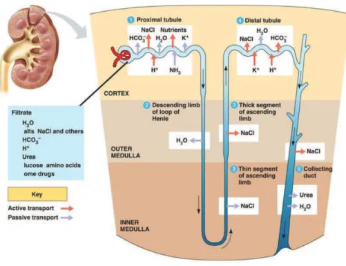
Kidneys are retroperitoneal paired organs located on each side of the vertebral column (Fig 1A). Macroscopically, kidneys are surrounded by a fibrous capsule, and from the outside towards the inside, are composed of the cortex and the medulla. In human, a mature and functional kidney is composed of 500000 to one million structural and functional units called nephrons. Each nephron consists of a glomerulus, the blood filtration unit, and a tubular component. The epithelial tubular portion of the nephron is composed of more than 20 subtypes of highly specialized cells, organized in successive subsegments. Starting from the glomerulus, the tubule of the nephron is composed of the Proximal Tubule (subdivided in S1, S2 and S3 segments), the Henle’s Loop and the Distal Convoluted Tubule which is connected to the Collecting Duct system (Fig1C). Morphologically, S1 and S2 convoluted segments of proximal tubules, the glomerular globular structures and the convoluted distal tubules are localized in the cortical part of the kidney, whereas the medulla contains the straight S3 segment of proximal tubule, the Henle’s loop branches and the collecting duct system. In the deeper part of the medulla, the collecting duct system will progressively form multipyramidal structures, renal calyces, pelvis and finally the ureter (Fig1B).
In the organism, kidneys have different important functions. They play a major role in the maintenance of whole body homeostasis by regulating the acid-base balance, electrolyte concentrations and extracellular fluid volume. They also participate to the elimination of wastes derived from the metabolism. Additionally, kidneys have endocrine functions and produce important hormones: erythropoietin which stimulates the production of red blood cells, and renin, an active actor in the blood pressure regulation via the renin-angiotensin system. Finally, through conversion of 25-hydroxycholecalciferol to 1,25-dihydroxycholecalciferol, kidneys play a key role in the synthesis of the active form of vitamin D that participates to the homeostasis of calcium, important for bone morphogenesis and maintenance.
To maintain whole body homeostasis, the primary urine produced by the glomerular filtration is modified through the reabsorption or secretion of water and small molecules by the renal tubules. These functions are accomplished through the cellular complexity and peculiar spatial organization of the kidney. I will briefly describe the different characteristics and specific functions of the sub segments of the nephron.
11
Figure 1. Anatomical structure of the kidney.
(A) Macroscopic representation of the kidney with its external connection to the urinary system and to the vascular network (adapted from https://media1.britannica.com ). (B) Kidney is composed of two main parts: the cortex, containing the glomeruli and the convoluted portion of the proximal and distal tubules, and the medulla, composed of the straight portion of the proximal tubule, the Henle’s loop and the collecting duct (C) Macroscopic representation of the nephron segments (adapted from https://www.cnx.org )
A
12
II.
Anatomical description of the different nephron components
The mature renal glomerulus is a globular structure responsible for the filtration of blood. Its inner part is a cup-like structure composed of a layer of podocytes (visceral epithelial cells) that surround a network of capillaries supported by the mesangial matrix and contractile mesangial cells. The Bowman’s capsule with its inner monolayer of Bowman’s capsule cells (parietal epithelial cells) surrounds this capillary tuft. Glomerular capillaries arise from a single afferent arteriole that divides into capillaries and finally converges into a single efferent arteriole. The site where blood vessels enter the glomerulus is called vascular pole. The blood is filtered through a filtration barrier composed of three layers: a fenestrated endothelial layer, a basal membrane and the filtration slits created by the foot-processes of podocytes. This barrier is highly selective and allows only small molecules (low molecular weight proteins or ions) to pass. On the contrary, cells and high molecular weight proteins (for example, albumin) are normally retained in the circulation. The Bowman’s capsule, surrounding the vascular tuft and delimiting the urinary space, collects the primary urine derived from blood filtration. Bowman capsular cells (or parietal cells) are flattened cells with a cytoplasm barely visible by light microscopy. They are in continuity with tubular cells at the urinary pole that represents the connection between the glomerulus and the proximal tubule (Fig 2) (Brenner and Rector’s The Kidney 2012).Figure 2. Structure of the glomerulus. The glomerulus is composed of a complex net of anastomosed
capillaries surrounded by podocytes. The glomerular tuft is surrounded by the Bowman capsule, delimiting the urinary space. The macula densa cells are located between the afferent and efferent arterioles, associated to juxtaglomerular cells (adapted from (Human Anatomy and Physiology, 5th Edition by Marieb: Benjamin-Cummings, San Francisco, CA 9780805349894 Hardcover, 5th Edition. - a2zbooks n.d.))
Podocytes
13 The proximal tubule (PT) is subdivided into three segments. The S1 segment represents the initial portion starting from the urinary pole of the glomerulus and constitutes approximately two thirds of the pars convolute. The S2 segment consists of the last portion of the pars convoluta and the initial portion of the pars recta. The S3 segment represents the straight part of the proximal tubule located in the deep cortex and the outer stripe of the outer medulla. The function of each subsegment depends on the specific set of genes expressed by these tubular cells. The proximal tubule is lined by a cuboidal epithelium whose cells have several characteristic features: the presence of a brush border of microvilli at the apical side, which increases the surface area to reabsorbsmall molecules from the filtrate to the interstitium, and a high number of mitochondria (Brenner and Rector’s The Kidney 2012).
The Henle’s loop has a peculiar spatial organization, with a characteristic U shape. This tubular segment enters the medulla of the kidney and returns back to the cortex where it contacts its own glomerulus. Schematically, two functionally distinct limbs form the Henle’s loop segment: the thin descending (TDL) limb and the thick ascending (TAL) limb of Henle. The descending thin segment is lined by simple squamous epithelium, whereas the ascending thick segment is lined by a cuboidal epithelium with scattered microvilli, high density of mitochondria and an interdigitated basal membrane.
The Macula Densa (MD) plaque is a group of epithelial cells located in the cortical thick ascending limb in close proximity with the vascular pole of each glomerulus (Fig 2). In association with the Juxtaglomerular cells ("J-G cells") in the wall of the afferent arteriole, they form the juxtaglomerular apparatus. This complex structure constitutes a unique anatomic arrangement where the vascular and the epithelial networks of the kidney come into contact. The recognized role of MD cells is to detect the increase of NaCl concentration in tubular luminal fluid and to transmit signals that modulate both the vascular tone of the afferent arteriole and the renin secretion from granular cells of the juxtaglomerular apparatus. These cells represent the critical link between renal salt and water excretion and glomerular hemodynamics, thus playing a key role in regulation of body fluid volume (Bell et al., 2003; Lapointe et al., 2003).
The distal convoluted tubule (DCT) and the short connecting tubule (CNT) are the portion of the nephron immediately downstream of the macula densa. These segments are
14 composed of a cuboidal epithelium that is structurally similar to the one of the thick ascending limb. This portion of the renal tubule continues to reabsorb useful solutes from the filtrate to the peritubular capillaries, actively pumping small molecules out of the tubule lumen into the interstitial space.
The DCT is then connected via the CNT to the Collecting Duct (CD) system. The collecting ducts are formed in the renal cortex by the connection of several nephrons. They go down within the medullary rays of the cortex, penetrate the outer and the inner medulla where they successively fuse together to bring the urine produced by the nephrons into the ureter and finally in the bladder. Based on their location within the kidney, the collecting ducts can be subdivided into cortical collecting duct (CCD), outer medullary collecting duct (OMCD), and inner medullary collecting duct (IMCD). The cells lining the CD can be divided into at least two cell types: principal cells and intercalated (IC) cells. The principal cells represent 70–75% of all cells, whereas IC cells represent the remaining 25–30% of cells.
Figure 3. Schematic representation of nephron components. Primary urine produced by the glomerulus
pass through the different tubular portions of the kidney that reabsorb water and solutes and define the final urine composition. (Figures 44.14 and 44.19, page 879 and 884, Campbell'sBiology, 5thEdition)
15
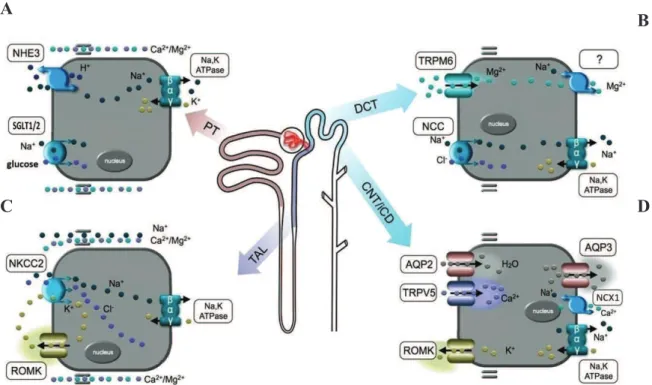
III. Renal tubular transporters
A complex set of tubular reabsorption and secretion mechanisms is activated in the kidney to maintain the whole body homeostasis. Approximately 2/3 of the water and Na+ and most of the electrolytes and amino acids filtered by the glomerulus are reabsorbed during their passage through the proximal tubule. Additional 25% of the filtered Na+ and water are reabsorbed in the Henle’s loop. Final adjustments in the composition and volume of urine are made in the distal part of the nephron (distal and collecting duct) mainly under the influence of specific hormones (aldosterone and vasopressin, respectively).
In the next part, I will briefly describe the general mechanisms leading to tubular reabsorption in each tubular segment, and give more details concerning the molecular mechanisms involved in glucose, calcium and uric acid reabsorption.
The proximal tubule is responsible of the reabsorption of the 70% of the ultrafiltrate produced in the glomerulus. The driving force for this massive reabsorption is represented by the active sodium transport carried by the Na+-K+-ATPase present in the basolateral pole of the cells. The efflux of Na+ out of the cell creates a sodium gradient between the apical urinary medium and the intracellular environment. This gradient is favorable to an entry of sodium in the tubular cell. The transport of sodium is coupled to the transport of several dissolved substances and performed by specific transporters, which act as co-transporters or counter-co-transporters (Brenner and Rector’s The Kidney 2012). For example, the reabsorption of Na+ can be coupled with glucose (Na+/glucose cotransporter) or exchange with H+ (Na+/H+ exchanger). Glucose is an important metabolite that is reabsorbed entirely in the proximal tubule via the glucose cotransporters SLC5A1 (SGLT1) and SLC5A2 (SGLT2), which present different affinity and stoichiometry for glucose. The low affinity Na+/glucose co-transporter SLC5A2, that reabsorbs one molecule of Na+ per molecule of glucose, is located in the S1 segment, where glucose concentration in the urine is high. The Na+/glucose high affinity transporter SLC5A1 is located in the straight S3 segment where only few molecules of glucose have still to be reabsorbed (Kanai et al., 1994) (Kamiyama et al., 2012). The reabsorption of Na+ via Na+/H+ exchangers (NHEs) leads to the secretion of an H+ in the tubular lumen. This process is important for the acid-base homeostasis, since NHEs are responsible for 70% of the reabsorption of HCO3- (bicarbonate) in the PT, with the help of a luminal and intracellular carbonic anhydrase. Once the HCO3− has been reabsorbed, it is exported in the bloodstream
16 through a basal sodium/bicarbonate cotransporter, whose defect causes proximal renal tubular acidosis (Hamm et al., 2015).
The gradient generated by the reabsorption of solutes allows the passive reabsorption of the water, whereas the reabsorption of small molecules physiologically filtered by the glomerulus requires a receptor-mediated endocytosis system (Christensen et al., 2009; Nielsen, 1994).
In the convoluted part of the proximal tubule, 60% of the Ca2+ filtered by the glomerulus is reabsorbed in a passive and paracellular way. The passage of the calcium from the proximal tubular lumen into the blood occurs through the tight junction, via the membrane proteins Claudins which form the channels for the passage of the ions. 2, Claudin-1, Claudin-10a and -12 are involved in cations reabsorption in proximal tubule. As an example, it has been shown that inactivation of Claudin2 in mice leads to hypercalciuria (Alexander et al., 2013) (Fig 4A).
Finally, uric acid is mainly reabsorbed in the PT through the apical URAT1 transporter (encoded by SLC22A12) and exchanged with anions that return into the lumen. On the other hand, the anion transporters, OAT1 and OAT3, present on the basolateral membrane of the proximal tubule are involved in the secretion of urate in the urine. Mutations in SLC22A12 gene result in hypouricemia, with high levels of urate in the urine (Enomoto et al., 2002) (reviewed by Hediger et al., 2005).
The ability of the kidney to concentrate or dilute urine depends mostly on the generation of a cortico-medullary gradient. The active Na+ transport in the thick ascending limb of Henle’s loop, from the lumen to the surrounding interstitium, contributes to the formation of a hypertonic medullary interstitium. Thanks to the closely associated capillary network, reabsorbed osmoles are gradually accumulated to the deepest part of the kidney, via a countercurrent multiplier system. Thick and thin limbs of Henle’s loop are characterized by different properties concerning solutes and water permeability. In the TDL portion, the reabsorption is limited to water, whereas the solutes are retained in the urine (Kokko, 1970). This process increases the concentration of the urine that reaches its maximum around the tip of the loop in the medulla. On the opposite the TAL is almost impermeable to water, but permeable to solutes. The energy for the reabsorption of Na+ in the TAL is again provided by the basolateral Na+-K+-ATPase. In these tubular cells, Na+ reabsorption is provided by SLC12A1 (NKCC2) and K+ is recycled back to the lumen through the apical ROMK channel. Mutations in ROMK channel result in defective fluid and salt
17 reabsorption, with a secondary excretion of potassium in the distal portion and a consequent hypokalemia (Lorenz et al., 2002; Brenner and Rector’s The Kidney 2012) (Reviewed in Zacchia et al., 2016). Uromodulin, a protein expressed in the TAL, regulates the activity of the SLC12A1 (NKCC2) transporter, through its amino-terminal phosphorylation (Mutig et al., 2011). Mutations in UMOD reduced urinary concentrating ability, probably due to a defective sodium reabsorption in the TAL (Bernascone et al., 2010; Hart et al., 2002).
The TAL is also involved in HCO3– reabsorption, that is Na+ dependent (Na+-HCO3- co-transporter, NBCN1), and in the paracellular transport of divalent cations such as Ca2+ and Mg2+. Similarly to the situation in the proximal tubule, the passage of Ca2+ and Mg2+ is permitted by the presence of claudins in the tight junction between the epithelial tubular cells. In this subsegment, Claudin-16 and Claudin-19 form a cationic pore for the passage of Ca2+ and Mg2+. In human mutations in the genes encoding for these two proteins are associated to familial hypomagnesemia with hypercalciuria and nephrocalcinosis (FHHNC), characterized by progressive renal Mg2+ and Ca2+ waste. In this pathology, the nephrocalcinosis is caused by the increased waste of calcium in the urine, resulting finally in a chronic kidney disease (Alexander et al., 2013; Hou et al., 2008; Brenner and Rector's The Kidney 2012) (Fig 4B).
The reabsorption of sodium, chloride and calcium in the DCT and CNT is driven by the high Na+-K+-ATPase activity which generates the electrochemical gradient at the basis of ions transport. Mutations in FXYD2, encoding for the γ-subunit of the Na+-K+-ATPase pump, are involved in calcium-magnesium reabsorption defects and patients with FXYD2 mutation have autosomal dominant renal hypomagnesaemia with hypocalciuria (Meij et al., 2000) (reviewed in (Hoenderop and Bindels, 2005)) (Fig 4C). The SLC12A3 (NCC), a sodium-chloride symporter, is located at the apical membrane of the DCT and benefits from the sodium gradient to transport Na+ and Cl− from the tubular fluid into these cells. Defects in this transporter are associated with Gitelman syndrome, characterized by hypokalemia, hypomagnesaemia and hypocalciuria (Simon et al., 1996). The apical side of DCT and CNT cells carries also the active trans-cellular transporters for Mg+ (TRPM6) and Ca2+ (TRPV5). In the epithelial cells, Ca2+ is extruded into the blood through the Na+ -Ca2+-exchanger (NCX1). In a similar way, Mg+ passes from the cytoplasm of cells into the bloodstream. Patients carrying TRPM6 mutations suffer from hypomagnesaemia associated to Mg+ reabsorption defect (Walder et al., 2002), whereas animal model defective for Ca2+
18 transporter is characterized by calcium waste associated to polyuria (Hoenderop et al., 2003) (Zacchia et al., 2016). In addition, the presence of a K+ channels at the basolateral side (KCNJ in the DCT) mediated the passage of the potassium in the interstitum (Woda et al., 2003). Defects of these channels lead to salt waste and hyperkalemia (Lorenz et al., 2002; Yang et al., 2010)(reviewed in Zacchia M et al 2016).
In the collecting duct, several transporters and channels are present at the apical membrane of principal cells, and are mainly regulate by the action of hormones, such as aldosterone and vasopressin. These transporters and channels play an important role in the final regulation of urine volume and concentration (Muto, 2001). In particular, water reabsorption is directly regulated by the vasopressin via the water channel Aquaporin2 (AQP2). Vasopressin induces the activation of the protein kinase A (PKA) responsible for the phosphorylation of AQP2 in intracellular vesicles (Katsura et al., 1997; Nishimoto et al., 1999). When it is phosphorylated, AQP2 is exocytosed to the apical side of the tubular cells (Fushimi et al., 1997). PKA is also important for the phosphorylation of transcription factors that control AQP2 gene expression, such as CREB-P and c-Jun/c-Fos. The up or down regulation of AQP2 expression in the CD determines the amount of water reabsorption and the urine concentration. For example, in experimental rat models of bilateral ureteral obstruction (BUO), the expression of aquaporin channels, sodium and urea transporters were strongly decreased, leading to a marked polyuria (Frøkiaer et al., 1996; Li et al., 2003). On the contrary, in unilateral obstruction, even if the expression of the transporters was altered, secretion of sodium and water was not changed probably due to compensatory mechanisms by the controlateral kidney (Frøkiaer et al. 1996)(reviewed by Nielsen et al 2007) (Figure 4D).
19
Figure 4. Schematic representation of the principal transporters/channels and their distribution in the different nephron tubular segments. A Na+-K+-ATPase pump is present all along the tubular compartments on the basolateral side of the epithelial cells, in order to provide the energy for electrolyte transport. (A) In the PT, there is a massive reabsorption of sodium, due for example to an exchange with H+ or to a cotransport with the glucose by SGLUT1 (in the S3) and SGLT2 (in the S1). (B) In the TAL, the reabsorption of sodium, chloride and potassium is achievd via the NKCC2. Potassium is then recycled in the lumen by the ROMK channel. (A,B) In both PT and TAL, Ca2+ and Mg2+ are reabsorbed in a passive way. (C) In the DCT, NCC transporter is responsible of the reabsorption of the sodium and chloride, and the active transport of the Mg2+ is mediated via TRPM6 (D) In the CNT, the active transport of the Ca2+ is mediated through TRPV5 whereas the K+ is excreted via the ROMK channel. In the CD, aquaporin channels reabsorb water and determine urine concentration (Adapted from (Dimke et al., 2010)).
B A
20
CHAPTER 2:
21
Kidney development
In the adult kidney, the functional unit, the nephron, is composed of highly differentiated glomerular and tubular cells, whose spatial organization is very complex. During renal morphogenesis, tubular and glomerular progenitor cells activate progressively the expression of a peculiar subset of genes to perform their specific functions in adulthood. Such high level of complexity is achieved through the action of several signaling pathways and crosstalk controlled mostly at the transcriptional level during kidney development. During development, embryonic kidneys derive from the intermediate mesoderm (IM), one of the three mesodermal layers, and form continuum along the rostro-caudal axis. In mammals, three successive structures will develop in a distinct temporal sequence. In the anterior part, the two first primitive renal structures are represented by the pronephros and the mesonephros, which are transient structures. In mouse and human, the pronephros is represented by a group of primitive renal tubules, barely detectable, whereas the mesonephros is formed by well developed tubules with a glomerulus that is connected to the nephric duct. The mesonephros is a functional filtration unit during early embryonic life but starts to degenerate in concomitance with the metanephros development (reviewed in (Dressler, 2006)). The formation of the definitive kidney, the metanephros, starts at embryonic day 10.5 (E10.5) in mouse and around 5weeks in human, and is initiated by an outgrowth of the Wollfian duct, the ureteric bud (UB), which invades the mass of the metanephric mesenchyme (MM) (Figure 5).
Metanephros development depends on the reciprocal heterotypical interactions between the UB and the MM. Signals from the MM induce UB growth and branching that will ultimately form the urinary collective system. In parallel, the UB secretes factors to induce mesenchyme aggregation around the UB tips and mesenchymal to epithelial transition (MET) to generate the renal epithelial compartment of the nephron. This process of reciprocal induction is repeated many times during development in order to form the complex structure of the mature kidney (reviewed in (Dressler, 1999)).
22
Figure 5. Schematic representation of the development of renal structures. Two primitive structures, the
pronephros and the mesonephros represent transient structures in mammals, degenerate and are replaced by the definitive renal structure, the metanephros (adapted from(Dressler, 2009))
23
I.
UB outgrowth and collecting duct system development.
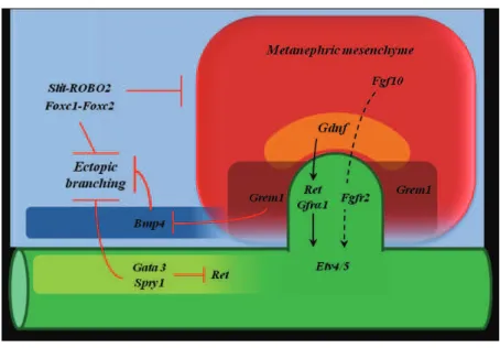
The outgrowth of the UB from the nephric duct (ND) is initiated by the interaction between the Glial-cell-line Derived Neurotrophic Factor (GDNF) peptide secreted by MM and its receptor Ret, a member of the receptor tyrosine kinase super-family, expressed by the UB (reviewed by (Krause et al., 2015)). The activation of the GDNF-Ret signaling modulates the expression of different genes in the UB. As an example, this signaling induces Wnt11 secretion by the UB which is crucial to maintain Gdnf expression in the MM, in a positive feedback loop (Pepicelli et al., 1997). In addition, two transcription factors Etv4 and Etv5 localized in the UB tip are also activated by GDNF-Ret signaling. These two factors induce the expression of target genes involved in proliferation and elongation of the UB, such as CxCr4, Myb and Mmp14 (Lu et al., 2009). Any dysfunctions in the pathways regulating the emergence of the UB may lead to kidney agenesis, due to the absence of UB outgrowth.
On the other hand, the emergence of a unique UB is controlled by specific crosstalk between genes expressed in the nephric duct and in the MM. Misregulations in these crosstalk may lead to ectopic UB outgrowth and surnumerary kidneys. During morphogenesis, Pax2/8 genes have several important roles in metanephric kidney. In addition to their involvement in the maintenance of Gdnf in the MM, they regulate the expression of the Gata3 gene in the ND, that inhibits the expression of Ret (reviewed by (Sharma et al., 2015)). Gata3 inactivation in the nephric duct leads to the formation of ectopic buds. In this model, the emergence of ectopic buds is due to presence of Ret positive cells all along the nephric ducts that response to the mesenchymal GDNF (Grote et al., 2008). In addition to Gata3, different genes are involved in the emergence of a unique UB and the inhibition of the ectopic branching, such as Sprouty1, Gremlin1, Bmp4 and Slit-ROBO2. Activated by the GDNF-Ret signaling, Sprouty1 (Spry1), a receptor tyrosine kinase antagonist, inhibits in a negative feed-back loop the expression of Ret in the nephric duct and in the UB. This inhibition of Ret is necessary to induce normal branching morphogenesis. When Spry1 is inactivated, mutant kidneys present supernumerary buds and defective branching (Basson et al., 2006). As another example, the interaction between the ligand SLIT2, expressed in the nephric duct, and its receptor ROBO2, expressed in the MM, has an essential role in the unique emergence of the UB, via the definition of the boundary of Gdnf expression in the mesenchyme (Figure 6). Mutant mice for the either
24 Slit2 or Robo2 developed ectopic buds along the stalk of the nephric duct, due to the persistent expression of the Gndf in the anterior nephrogenic mesenchyme (Grieshammer et al., 2004).
The transcription factor HNF1beta plays a crucial role in the definition of a unique emergence of the UB. Unpublished results obtained in our laboratory have shown that embryos deficient for this transcription factor display numerous and abortive ectopic UB outgrowth, due to the misregulation of the expression of Spry1 and a decrease in ROBO-Slit signaling. In addition the expression of Pax2 in the ND and UB epithelium has been shown to be directly regulated by HNF1beta. The cooperation of these two genes is necessary for the normal morphogenesis of the collecting duct system. Double heterozygous mice Hnf1b-/+ Pax2-/+ are characterized by the formation of ectopic UBs in the caudal portion of the nephric duct and a reduced number of branching events (Paces-Fessy et al., 2012).
Figure 6. Principal signaling pathways involved in the UB outgrowth. The UB outgrowth is the results of
the interplay between inductive and inhibiting signals in the MM and nephric duct. Gata3/Spry1, Bmp4 and
Slit-ROBO2 inhibit the ectopic branching and define the correct position of the UB, whereas Grem1 inhibits Bmp4 and promote UB outgrowth. This set of signals defines the correct position of the UB formation and its
25
II.
UB branching morphogenesis
GDNF/Ret signaling is important to maintain UB branching during kidney morphogenesis and to define the identity and fate of UB tip cells. During development of the UB tree, Ret-expressing cells continue to be part of the tips and participate to each round of new branching, while cells that do not express Ret will form the UB trunk. The elongation of the UB trunk via proliferation will form collecting duct, renal medulla and papilla (reviewed by (Costantini and Shakya, 2006)). In addition, other signaling pathways involved in the outgrowth of the UB participate also to the formation of each new tip during the branching process. In particular, Slit-ROBO2 signaling is important to determine the interaction between the tip and the surrounding mesenchyme. In metanephroi culture, over expression of ROBO2 inhibits normal branching due to a reduction in the number of condensing cells around each UB tip (Ji et al., 2012).
Intricate signaling pathways and molecular cascades are involved in the branching morphogenesis. As an example, the transcription factors Sox8/9 control the expression of crucial genes downstream of Ret, such as Ert4 and Spry1. Deletion of one copy of Sox9 in Sox8 deficient mice inhibits the branching, probably due to a defective GDNF/Ret signaling (Reginensi et al., 2011). The transcription factor Emx2 (Empty Spiracles2) is involved in the branching morphogenesis but do not control the emergence of the UB. In fact, in Emx2 mutant mice the UB emerges from the nephric duct, invades the MM but does not branch (Miyamoto et al., 1997). An important role in the branching morphogenesis is played also by the Fgf (Fibroblast growth factor) signaling, whose components are detected in both the UB and MM. In particular Fgf10 and Fgf7 ligands are expressed by the MM and interact during branching morphogenesis with the Fgfr2IIIb receptor expressed in the UB tips. Inactivation of either ligands or receptors cause defective branching and renal hypoplasia (reviewed by (Blake and Rosenblum, 2014)) (Figure7).
26
Figure 7. Schematic representation of the pathways involved in the UB branching. During UB branching the GDNF/Ret signaling is activated in the UB tip (marked in red), whereas the expression of
Spry1 in the ureteric bud stalk inhibits Ret signaling to avoid ectopic branching. In the surrounding MM,
Grem1 inhibits Bmp4 and promotes UB branching and invasion of the MM. At the UB tip the receptor Fgfr2 interacts with its ligands Fgf7 and Fgf10 express in the MM. Emx2 and Sox8/9 control branching morphogenesis. Emx2 maintains the expression of Ret in the UB tip (adapted from (Massa, 2012)).
27
III. Nephron formation
Around each UB tip, the metanephric mensenchyme starts to condensate and to form the cap mesenchyme. These condensed cells express Six2 and Cited, and represent the nephrogenic progenitor pool that will give rise to all epithelial component of the nephron. In the next step of nephrogenesis, cap cells can either maintain the expression of both Cited1 and Six2, forming the self-renewing progenitor population, or lose the expression of Cited1, and become engaged in the epithelial differentiation (Self et al., 2006; Kobayashi et al., 2008).
The initial and crucial event in nephron formation is represented by the mesenchymal to epithelial transition in the MM. This transition is initiated by the UB via the secretion of Wnt9b, a ligand involved in Wnt signaling (Carroll et al., 2005). In response to the Wnt9 signal, condensed cells will start an epithelial transition, form pre-aggregates and stop to express Cited1. The induced preaggregates start to express Wnt4, another Wnt ligand, whose binding to its receptor will activate the canonical Wnt signaling. In this context, the stabilization of the b-catenin will induce the activation of crucial genes involved in epithelial differentiation, including Lhx1 and Fgf8 (Dressler, 1997) (Figure 8).
Figure 8. Mesenchymal to epithelial transition. Cap mesenchyme is composed of two populations of cells:
self renewing progenitor Six2+Cited1+, which remain in the mesenchyme around the UB tip, and the
Six2+Cited1-, which are committed in the epithelial differentiation. These cells form the pre-aggregate in which the expression of Wnt4 drives nephron morphogenesis ( adapted from (Massa, 2012))
28 The next step of epithelial nephron precursors is the renal vesicle. This precursor is a small structure composed of approximately 70 epithelial cells. These vesicle cells surround a lumen that will fuse with the lumen of the UB, forming a continuity between the lumen of the collecting duct system and the lumen of the future nephron. The renal vesicle is a polarized structure, characterized by the expression of different set of genes along a proximal-distal axis (Figure 10A). For example, cells close to the UB will start to express Lhx1, Brn1 and molecules involved in the Notch signaling (the receptor Notch 2 and the ligands Dll1 and Jag1), which are part of a genetic program involved in the formation of the future tubular compartment of the nephron. Complementary, cells forming the proximal pole of the vesicle are characterized by the expression of Wt1, a crucial gene for glomerular development (Georgas et al., 2009). In the proximal portion of the vesicle the transcription factors Pax2 and Wt1 are co-expressed until the comma-shaped body, collaborating in the same regulatory pathway to allow a correct glomerular development (Ryan et al., 1995).
i. Comma and S-shaped body development. The polarized expression of specific markers of the different nephron segments, tubules and glomerulus, in the nephron precursors is maintained along a proximal-distal axis during the successive steps of nephrogenesis. Through a complex set of cell proliferation/cell rearrangement, the vesicle will give rise to comma-shaped body and then to S-shaped body (Figure 9). During morphogenesis these nephron precursors are characterized by the formation of two invagination processes: the first one takes place during the transition between the vesicle and comma-shaped body and a second one during the transition between the comma and S-shaped body. The first slit, the vascular cleft will separate the proximal part from the distal part of the vesicle. This process is characterized by a deep morphological cellular rearrangement. Few cells, at the junction of these two parts, reorganize their cytoskeleton and move the nucleus on the apical side, creating a notch on the vesicle surface. This notch will be invaded by vessels that will give rise to the future glomerular tuft. The second slit appears on the opposite side, close to the UB, and is formed by the expansion of the future tubular compartment. These two invagination processes will give rise to the typical S-shaped body, a nephron precursor characterized by the presence of three distinct subsegments, i.e. the distal, mid and proximal limbs, that will form different portion of the mature nephron (Saxén and Sariola, 1987). The distal portion will give rise to the distal tubule; the mid limb corresponds to the future Henle’s loop and the proximal tubule and
29 the proximal limb will form the glomerular cup. At the S-shaped body stage, podocytes plate starts to invaginate and to form a cup that wraps the developing capillary network. At this stage, the position of the prospective urinary pole is contiguous to the prospective vascular pole, represented here by the rim of the cup (Figure 9).
Figure 9. Schematic representation of the transition between vesicle and S-shaped body nephron precursor stages. During nephrogenesis, the nephron precursors undergo two invagination processes. The
first process occurs during the transition between the vesicle and comma-shaped body to form the vascular cleft. The second invagination process occurs during the transition between the comma and the S-shaped body. At the S-shaped body the prospective urinary pole is contiguous to the prospective vascular pole, represented by the rim of the podocytes cup. (Adapted from chapter 277 “Cellular and Molecular Biology of the Kidney”, Harrison's Principles of Internal Medicine, 18e, 2012)
Recent analyses have characterized the molecular program expressed in the distinct segments of the S-shaped body and a set of genes that are selectively expressed in each limb of this nephron precursor (Figure 10C) (Brunskill et al., 2008). For example, the distal and medial portions express Lhx1 (Karavanov et al., 1998; Kobayashi et al., 2005) and Brn1 (Nakai et al., 2003). On the other hand, the transcription factors Irx1-3 (Nichane et al., 2006; Reggiani et al., 2007), important regulators of the intermediate tubule fate (Nichane et al., 2006), and ligands of the Notch pathway, required for the proximal tubule development (Cheng et al., 2007), are restricted to the mid limb. Interestingly, the expression of all these molecules is regulated by the transcription factor HNF1beta, a crucial player in the differentiation of the future tubular component. Its inactivation in the Six2+ cap mesenchyme cells or in Wnt4 expressing cells in the pretubular aggregate leads
30 to defective morphology of the S-shaped body probably due to a concomitant down-regulation of Irx1-2 and the Notch ligands Dll1 and Jagged1 (Heliot et al., 2013; Massa et al., 2013). During normal development, the transcription factor Pax2 continues to be express in the distal portion of the S-shaped body, but its expression is reduced in the proximal portion, where podocytes will be formed (Kreidberg et al., 1993) (Figure 9). A complex reciprocal control between these two genes depending on their spatial and temporal expression pattern has been shown: Pax2 activates the Wt1 promoter, whereas Wt1 represses Pax2 expression. As the renal differentiation proceeds, the expression of Pax2 is progressively downregulated in the glomerular structures.
Figure 10. Schematic representation of nephron precursors and gene pathways involved in nephrogenesis. During early nephrogenesis, the expression pattern of several genes is polarized along a
proximal-distal axis (A) In the vesicle, the proximal part expresses Wt1 that starts to inhibit the expression of
Pax2 whereas, the distal portion expresses Lhx1 and Notch ligands, represented by Dll1 and Jag1. (B) In the
comma-shaped body, the proximal portion is committed to form the glomerular portion of the nephron. On the other side, in the distal portion the expression of genes specific for the diverse tubular segment starts to be delineated. (C) In the S-shaped body there are three different limbs: the distal limb that will form the distal tubule, the mid limb corresponding to Henle’s loop and proximal tubule, and the proximal limb that will form the glomerulus. Each limb expresses already a specific set of genes (Adapted from (Desgrange and Cereghini, 2015)).
31 ii. Precapillary-loop precursor. The transition from the S-shaped body to the typical configuration of the precapillary-loop precursor is characterized by the expansion of the tubular compartment. In addition, the podocyte plate undergoes a progressive invagination process followed by a complex reorganization that will position the urinary pole on the external side of the glomerular cup (Potter, 1965; Saxén and Sariola, 1987). The mechanism at the basis of these latter structural changes is still unclear. I will describe in details the morphological processes occurring during the formation of the glomerulus, in the first part of my project.
At precapillary-loop stage, the elongating tubules give rise to the mature distal and proximal tubules connected by the Henle's loop. In parallel, cells that will differentiate into podocytes and Bowman's capsular cells will form the mature blood filtration unit, the glomerulus (Rak-Raszewska et al., 2015) (Pietilä and Vainio, 2014) (Boyle et al., 2014) (Figure 11). During this final step of nephron maturation, some embryonic pathways are downregulated, being progressively replaced by the genetic program leading to mature epithelial cells. As an example, the expression of Pax2 is gradually reduced in the maturing nephron precursors, until becoming restricted to the collecting duct. Down regulation of Pax2 is important to allow the terminal differentiation of the nephron. Indeed, its aberrant expression has been reported in several renal tumors in both human and rat, suggesting a state of undifferentiation or dedifferentiation of the epithelial cells (Davies et al., 2004).
Figure 11. Schematic representation of the transition between the S-shaped body and the mature glomerulus. At the precapillary loop stage the prospective urinary pole (UP) is close to the prospective
vascular pole (VP). The transition between the capillary loop stage and the mature glomerulus is represented by the relative position of the urinary pole on the external side of the glomerular cup. (Adapted from chapter 277 “Cellular and Molecular Biology of the Kidney”, Harrison's Principles of Internal Medicine, 18e, 2012)
32 iii. The development of the glomerulus. During the transition of the S-shaped body to mature nephron, important morphological processes will participate to the formation of the glomerulus. The early glomerulus in the more proximal portion of the S-shaped body is composed by the podocyte precursor plate (a thick columnar epithelium), the layer of Bowman capsule cells and the glomerular tuft, itself composed of glomerular capillaries and mesangial cells.
- Podocytes. During glomerulogenesis, podocyte precursors change their morphology. These changes are characterized by the expansion of the apical surface and the progressive movement of the initially apical junction belt towards the basal side. In the meantime, once the prospective vascular pole is colonized by capillaries, podocytes migrate and surround the developing capillary loops. At the end of this process, podocytes have developed large cytoplasmic projections, called foot processes that extend completely around the developing capillary loops (Schell et al., 2014). The normal podocyte development requires the expression of Wt1, a transcription four zinc fingers factor that can bind DNA and RNA. The high expression of Wt1 in mature podocytes that this factor could also play a role in the maintenance of normal podocyte functions throughout life. Wt1 controls the expression of key genes for podocyte function and specification, in particular:
- expression of Nephrin and Podocalyxin, involved in the organization of the specialized junctions of the slit diaphragm and podocyte foot processes;
- expression of actin cytoskeleton proteins, as Synpo, involved in podocyte polarity and cytoskeleton arrangement;
- expression of proteins involved in the cell-matrix adhesion of podocytes to the glomerular basement membrane (GBM) (reviewed by (Dong et al., 2015)).
Moreover, Wt1 is also necessary to maintain the paracrine and intercellular signaling between podocytes and mesangial cells and to regulate the expression of Vascular Endothelial Growth Factor A (VegfA) during glomerular development (Schell et al., 2014) (Gao et al., 2005) (Figure 12A). In mouse, podocyte-specific Wt1 knock-out leads to defects in podocyte differentiation, whereas its inactivation later in already developed podocytes promotes foot processes effacement, proteinuria and glomerulosclerosis. In human, WT1 mutations lead to glomerulopathies characterized by alteration in podocyte development or maintenance (reviewed by (Dong et al., 2015)). Mutations of WT1 are also associated with diffuse mesangial sclerosis (DMS), characterized by increased deposition of extracellular matrix (ECM) in the vascular side of the GBM. In this pathology, the
33 defective expression of WT1 and its target genes leads to an aberrant communication between podocytes, mesangial and endothelial cells that may control the deposition of ECM and the correct assembly of the glomerular tuft (reviewed by (Quaggin and Kreidberg, 2008)).
Another important transcription factor involved in the development and maintenance of mature podocytes is Tcf21 (Pod1). This transcription factor controls the expression of several key angiogenic factors, including Vegfa, necessary for the formation of the glomerular tuft. Mouse model of Tcf21-specific knock-out in podocyte showed retardation in glomerular maturation that results in simplified glomeruli with a decreased number of endothelial and mesangial cells. Around 50% of the mutant mice developed proteinuria and lesions similar to focal segmental glomerlosclerosis (FSGS) (Maezawa et al., 2014).
- Capillaries and mesangial cells. During glomerulogenesis, the secretion of VEGFA by podocytes precursors attracts angioblasts from the interstitium. This induces the formation of the capillaries by endothelial cells that invade the vascular cleft. Later on, VEGFA induces also the remodeling of the endothelial cells in capillary loop that becomes fenestrated (Eremina and Quaggin, 2004) (Figure 12B). Model of inactivation of Vegfa results in defective development of the filtration barrier and reduction of endothelial cells in the glomeruli (Eremina et al., 2003). Another gene involved in capillary loop formation is Crim1, a transmembrane cystein-rich repeat-containing protein. This gene is expressed in podocytes, mesangial cells, parietal cells and vascular smooth muscle, and regulates the delivery of VEGFA from podocytes to endothelial cells. This protein sequesters growth factors on the surface of the cells once they are secreted, in order to control the rate of maturation and the effective concentration gradient. Its mutation during kidney development leads to an abnormal VEGFA delivery to glomerular endothelial cells and results in increased activation of the VEGFA signaling pathway in these cells. This conditions leads to enlarged capillary loop, podocyte effacement and mesangiolysis (Wilkinson et al., 2007).
The second important event during glomerular maturation is the secretion of the PDGFB by podocytes that recruits mesangial cells via the activation of PDGFB/Pdgfrb pathway. In the mature glomerulus, mesangial cells are in contact with the endothelial cells in the glomerular tuft, and play a crucial role to maintain the capillary loop structure (Figure 12C). During glomerulogenesis, these cells derived from the stromal mesenchyme as
34 specialized vascular smooth muscle cells. They are activated by PDGFB and invade the developing glomerulus where they attach to the forming blood vessels. Normal structure and function of glomerulus depend on the interactions between endothelial cells, podocytes and mesangial cells (Lindahl et al., 1998) (Krause et al., 2015). The mature glomerulus is finally composed by mesangial cells at the basis of the capillary tuft that is enveloped by the GBM and the podocytes (Figure 12D).
Figure 12. Development of the glomerular endothelial and mesangial cells. (A) Podocytes (in blue)
produce VEGF-A to attract endothelial cells (EC, in red), and PDGF-B to recruit mesangial progenitor cells (MC, in violet). (B) The endothelial cells organize the structure of capillaries whereas (C) the mesangial cells support endothelial cells to form the capillary loop. (D) Mature glomerulus: mature podocytes and basal membrane surrounded the fenestrated endothelium of capillaries, which are supported by mesangial cells (Adapted from (Schell et al., 2014))
- Glomerular filtration barrier. The filtration barrier is a multilayered structure that includes the fenestrated endothelium of the capillaries, the GBM and the slit-diaphragm, between podocyte foot processes. During development, capillaries and podocytes fuse their basal lamina in order to form a unique GBM. At the same time, specialized junctions
35 between the foot processes composed the slit diaphragm that connects podocytes one to each other. (Quaggin and Kreidberg, 2008) (Schell et al., 2014).
- Bowman capsule. The glomerular tuft is enveloped by the parietal epithelial cells (PECs) lying on the Bowman capsule. The PECs are characterized by a small and flat cell body that lines the urinary space of the glomerulus. PECs and podocytes derived from the same sub-compartment in nephron precursors and share a common phenotype until their mature differentiation at the precapillary loop stage. At this stage, in concomitance with the migration of endothelial and mesangial cells in the vascular cleft, podocytes start to express Nephrin and Podocalyxin, whereas the parietal cells are characterized by the expression of Pax2 and Claudin-1.
In the adult kidney different subpopulations of PECs can be distinguished on the basis of the markers they express and on their localization in the glomerulus (Figure 13). For example, at the vascular stalk, a few cells called transitional cells are localized between podocytes and PECs. These transitional cells have phenotypical characteristics and marker expression common to both cell types. On the other hand, rare cells lining the Bowman’s capsule express podocyte markers and are able to form foot processes. These cells are called ectopic or parietal podocytes (reviewed by (Shankland et al., 2014)).
In human, during glomerular disease, PECs can acquire an activated phenotype, characterized by increased proliferation, migration and matrix deposition. They are identified by the expression of CD44, a specific marker of aPECs, which is absent in other glomerular resident cells. CD44 is a typical marker of mononuclear cells, such as macrophages and monocytes (Fatima et al., 2012). As an example, during crescentic glomerulopathy, some PECs acquire a proliferating capacity and produce cellular crescents in Bowman’s space. Activated PECs represent the predominant cellular type in this pathological multilayered neoformation, even if podocytes and macrophages have been identified as possible players (Bariéty et al., 2001, 2005). In glomerulosclerosis, the presence of adhesion between the Bowman capsule and the sclerotic lesions in the tuft facilitate the migration of aPECs into the capillary tuft, where they continue to deposit extracellular matrix leading to a global endocapillary glomerulosclerosis.
Seen the common genetic program shared by podocytes and PEC during renal development before their terminal differentiation, it has been hypothesized that PEC could trans-differentiate into podocytes under pathological situations and play a role as adult
36 podocyte stem or progenitor cells. In human adult glomeruli, subpopulations of cells expressing renal progenitor markers (CD24 and glycCD133) and podocyte markers have been identified at the urinary pole and/or close to the vascular pole. Moreover, in human pathologies characterized by a reduction of the number of podocytes, there is an increase of cells expressing both podocyte and PEC markers, supporting the hypothesis of a regenerative role played by this last subpopulation. However, several observations do not support this paradigm. First, there is no evidence that cells expressing podocyte markers during glomerular diseases acquire also the morphological and functional characteristics of mature podocytes. Second, cell-fate tracking studies have demonstrated that some parietal cells can move into the glomerular tuft to form functional podocytes, suggesting the presence of a pool of cells lining the Bowman capsule still committed to form podocytes. Nevertheless, this phenomenon is present only during glomerular development in new born mice. Turnover of podocytes or recruitment of PECs in the capillary tuft in adult mice, in ageing or after induction of compensatory glomerular hypertrophy have never being observed (reviewed by (Shankland et al., 2014)). Altogether, these studies suggest that PEC form a heterogeneous population, with some characteristics of podocyte progenitors, but their functional relevance in pathological conditions remains to be demonstrated.
37
Figure 13. Representation of the different cell populations in glomerulus. In mature glomerulus different
PEC population are distinguished on the basis of expression of specific markers: podocyte committed
progenitors expressing CD133+ CD24+PODXL+ line the Bowman capsule close to vascular pole, whereas
transitional cells with an intermediate phenotype are present at the vascular pole between Bowman capsule
and podocytes. However some podocytes could be observed lining Bowman capsule close to the vascular pole and are indicated as parietal/ectopic podocytes. Resident multipotent cells, which could give rise to parietal or tubular cells, line the Bowman capsule close to the urinary pole, express CD133+ CD24+, but are PODXL-, and are indicated as adult parietal epithelial multipotent progenitors (APEMP). aPEC (in red) are PEC activated during glomerular disease with a different expression pattern and an increased capacity to proliferate and migrate (adapted from (Shankland et al., 2014))
38 During aberrant renal morphogenesis, some malformations include the formation of the glomerular cysts, characterized by a dilated Bowman capsule (Figure 14). This phenotypic trait is usually associated with other hereditary cystic disease, such as medullary cystic kidney disease, polycystic kidney disease, tuberous sclerosis complex, and have also been observed in patients carrying HNF1B mutations (Table 1) (Lennerz et al., 2010). The mechanisms at the basis of the glomerular cyst formation are not still clear. Some studies have suggested a potential role of the primary cilium in the pathogenesis of this cystic phenotype. This proposition was essentially based on the fact that most of the proteins encoded by the different genes mutated in glomerular cyst are normally part of this organelle. Interestingly, most of the glomerular cystic phenotypes develop during embryonic life. This observation suggests that an alteration of the morphogenetic process of the glomerular development could be at the basis of cyst formation (Bissler, 2010)
Figure 14. Schematic morphologic spectrum of glomerular cysts. Illustration of progressive changes in
glomerular cysts development from left to right. Glomerular cysts can be spherical, oval or polygonal. The tuft is present on one side of the Bowman capsule either as separated dysplastic tuft (a,b,c,d) or as a flatted tuft-remnant (e,g) (Adapted from (Lennerz et al., 2010)).
39 Table 1. Cystic kidney disease associated to glomerular cysts
Human disease
Mutated gene
Protein
Autosomal dominant PKD PKD1-2 Polycystin-1 Polycystin-2 GCK inPKD Autosomal recessive PKD PKHD1 FibrocystinMODY5 (RCAD) HNF1B Hepatocyte
nuclear factor-1β
Hereditary GCK Autosomal dominant
GKCD UMOD Uromodulin
PKDTS (Polycystic kidney disease and Tuberous sclerosis)
TSC2 Tuberin
RHPD (renal hepatic
pancreatic dysplasia) NPHP3 Nephrocystin 3
Urinary tract abnormalities ___ ___ Obstructive GCK Ischemic GCK Or Drug-induced GCK ___ ___ Sporadic GCK