HAL Id: hal-03004592
https://hal-cnrs.archives-ouvertes.fr/hal-03004592
Submitted on 20 Nov 2020
HAL is a multi-disciplinary open access
archive for the deposit and dissemination of
sci-entific research documents, whether they are
pub-lished or not. The documents may come from
teaching and research institutions in France or
abroad, or from public or private research centers.
L’archive ouverte pluridisciplinaire HAL, est
destinée au dépôt et à la diffusion de documents
scientifiques de niveau recherche, publiés ou non,
émanant des établissements d’enseignement et de
recherche français ou étrangers, des laboratoires
publics ou privés.
Structural Basis for Clinical Longevity of Carbapenem
Antibiotics in the Face of Challenge by the Common
Class A β-Lactamases from the Antibiotic-Resistant
Bacteria
Laurent Maveyraud, Lionel Mourey, Lakshmi Kotra, Jean-Denis Pedelacq,
Valérie Guillet, Shahriar Mobashery, Jean-Pierre Samama
To cite this version:
Laurent Maveyraud, Lionel Mourey, Lakshmi Kotra, Jean-Denis Pedelacq, Valérie Guillet, et al..
Structural Basis for Clinical Longevity of Carbapenem Antibiotics in the Face of Challenge by the
Common Class A β-Lactamases from the Antibiotic-Resistant Bacteria. Journal of the American
Chemical Society, American Chemical Society, 1998, 120 (38), pp.9748-9752. �10.1021/ja9818001�.
�hal-03004592�
Structural Basis for Clinical Longevity of Carbapenem Antibiotics in
the Face of Challenge by the Common Class A
β-Lactamases from
the Antibiotic-Resistant Bacteria
Laurent Maveyraud,†Lionel Mourey,† Lakshmi P. Kotra,‡ Jean-Denis Pedelacq,† Vale´rie Guillet,†Shahriar Mobashery,*,‡and Jean-Pierre Samama*,†
Groupe de Cristallographie Biologique, Institut de Pharmacologie et de Biologie Structurale du CNRS, 205 route de Narbonne, 31077-Toulouse Cedex, France, and Department of Chemistry, Wayne State UniVersity, Detroit, Michigan 48202-3489
ReceiVed May 26, 1998
Abstract: Bacteria resistant to antibiotics are being selected in a relatively short time, and cases of infections
resistant to treatment by all known antibiotics are being identified at alarming rates. The primary mechanism for resistance toβ-lactam antibiotics is the catalytic function of β-lactamases. However, imipenem (a β-lactam) resists the action of mostβ-lactamases and is virtually the last effective agent against the vancomycin-resistant Gram-positive bacteria, as well as against multiple antibiotic-resistant Gram-negative organisms. Here, we report the crystal structure, to 1.8 Å resolution, of an acyl-enzyme intermediate for imipenem bound to the TEM-1β-lactamase from Escherichia coli, the parent enzyme of 67 clinical variants. The structure indicates an unprecedented conformational change for the complex which accounts for the ability of this antibiotic to resist hydrolytic deactivation byβ-lactamases. Computational molecular dynamics underscored the importance of the motion of the acyl-enzyme intermediate, which may be a general feature for catalysis by these enzymes.
Assuming that the current efforts in the pharmaceutical companies toward discovery and development of novel antibiot-ics are successful, the use of these new agents would inevitably select for resistance in microbial populations in a relatively short time.1-3 It is critical that clinical selection of resistance be
delayed as long as possible and that the principles for such resistance mechanisms be elucidated to help prolong the utility of existing drugs.4 One class ofβ-lactam antibiotics which has
largely been resistant to the deleterious effects of the bacterial resistance enzymes is carbapenems,5-8 a member of which,
imipenem (1), has found clinical utility.9 Imipenem is often
considered an antibiotic of last resort against the vancomycin-resistant Gram-positive bacteria, as well as against multiple antibiotic-resistant Gram-negative organisms. The structural reason for this resistance of carbapenems to the action of
β-lactamases is unknown, and it represents a subject which has
yet to diminish in its biomedical importance.
Despite the high affinity of the enzyme for the drug, the poor turnover of imipenem (1) by class Aβ-lactamases stems only from a slow deacylation rate. Furthermore, the kinetics of
turnover are biphasic, suggestive of two distinct acyl-enzyme species.10 We report herein the crystal structure for one
acyl-enzyme intermediate for imipenem with the TEM-1β-lactamase, a common class Aβ-lactamase from Escherichia coli. Imi-penem is found covalently bound to Ser-70 Oγ (Figure 1A) as the∆2tautomer (Scheme 1).
The backbone atoms of the protein in the native enzyme and in the TEM-1-imipenem complex are identical within experi-mental errors (rmsd of 0.24 Å), except for residues 129-131,
†Institut de Pharmacologie et de Biologie Structurale du CNRS. ‡Wayne State University.
(1) Swartz, M. N. Proc. Natl. Acad. Sci. U.S.A. 1994, 91, 2420. (2) Jones, R. N. Am. J. Med. 1996, 100 (suppl. 6A), 3S.
(3) Davies, J. E. Antibiotic resistance: origins, eVolution, selection and
spread; Ciba Foundation Symposium 207; Chadwick, D. J., Goode, J., Eds.;
John-Wiley: Chichester, 1997; pp 15-35. (4) Neu, H. C. Science 1992, 257, 1064.
(5) Bush, K.; Jacoby, G. J. Antimicrob. Chemother. 1997, 39, 1. (6) Kropp, H.; Gerckens, L.; Sundelof, J. G.; Kahan, F. M. ReV. Infect.
Dis. 1985, 7 (suppl. 3), S389.
(7) Rasmussen, B. A.; Bush, K. Antimicrob. Agents Chemother. 1997,
41, 223.
(8) Matagne, A.; Lamotte-Brasseur, J.; Fre`re, J.-M. Biochem. J. 1998,
330, 581.
(9) Balfour, J. A.; Bryson, H. M.; Brogden, R. N. Drugs 1996, 51, 99.
(10) (a) Taibi, P.; Mobashery, S. J. Am. Chem. Soc. 1995, 117, 7600. (b) The value for kcatfor the fast phase of the turnover of imipenem by the
TEM-1β-lactamase has been reported at 0.04 s-1(ref 10a). Considering
that the kcatvalues for the favorable substrates, such as certain penicillins,
approach 2000 s-1, this difference amounts to 50000-fold. The effect of this rate attenuation has been determined to be due to the 6R-1R-hydroxyethyl group of imipenem. Consistent with these determinations, studies with judiciously designed penicillanate derivatives have documented that the 6R-1R-hydroxyethyl group can lower the value of kcat/Kmfor the
substrate of the TEM-1β-lactamase by as much as 10000-fold (Miyashita, K.; Massora, I.; Mobashery, S. Bioorg. Med. Chem. Lett. 1996, 6, 319). Scheme 1
S0002-7863(98)01800-9 CCC: $15.00 © 1998 American Chemical Society Published on Web 09/09/1998
Downloaded via INIST-CNRS on November 13, 2020 at 15:44:15 (UTC).
237-244, and the N- and C-terminal helices. The rms deviation on these main-chain regions are 1.5 Å, 0.5, 0.4, and 0.5 Å, respectively. The conformation adopted by the 129-131 region seems necessary to accommodate the 6R-1R-hydroxyethyl substituent of imipenem in the acyl-enzyme intermediate. The peptide bond between residues 129-130 rotates by nearly 180°,
and the carbonyl oxygen atom of residue 129 is hydrogen bonded to the Nζ atom of Lys-234 (3.1 Å), which shifted by 1.4 Å compared to the native enzyme (Figure 1A). Lys-234 is believed to provide a strong electrostatic pull in anchoring the carboxylate of the substrate(s).11 This role is substantially
diminished in importance for the interaction of the enzyme with
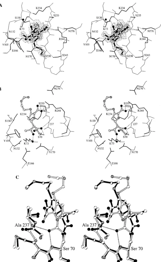
Figure 1. (A) Stereoview of the X-ray structure for the acyl-enzyme intermediate of imipenem with the TEM-1β-lactamase shown in the final electron density map. Hydrogen bonds are represented as dotted lines. Water molecules are depicted as black spheres. (B) The energy-minimized structure for the canonical acyl-enzyme intermediate of imipenem with the TEM-1β-lactamase. (C) The structures in (A) and (B) are superimposed here to demonstrate the motion of the acyl-enzyme species. The ester carbonyl of the canonical acyl-enzyme species (in gray) is held in the oxyanion hole by two hydrogen bonds (dotted lines) to the main-chain amines of Ser-70 and Ala-237. The acyl-enzyme species seen in the crystal structure is depicted in black.
imipenem, since the distance between the Nζ and the closest carboxylate oxygen of imipenem is 6.4 Å. The displacement of the loop 129-131 generated a cavity filled by a water molecule, which provides the single polar interaction to the essential Lys-73 (Figure 1A).
The main-chain atoms of residues 237-244 shifted toward the substrate binding cavity. A salt bridge interaction was observed between the side chain of Arg-244 and the carboxylate of imipenem (2.61 Å). In this interaction, the carboxylate displaced the Arg-244 side chain away from the active site, a movement which propagated to helices H1 (residues 26-40) and H11 (residues 272-288), through the hydrogen bond between Arg-244 and Asn-276 (Figure 1A).
The conformation of the acyl-enzyme species of imipenem is unprecedented because the ester carbonyl oxygen was not located in the oxyanion hole (Figure 1A). It points toward Ser-130 Oγ, at a distance of 3.3 Å. The hydroxyl group of the 6R-1R-hydroxyethyl substituent formed a hydrogen bond to Asn-132 Oδ1 (2.7 Å). The methyl group of the 6R-1R-hydroxyethyl substituent was found at van der Waals distance to the Cβ atom of Ser-130 and to the Cδ1 atom of Tyr-105. This side chain is pointing toward the carbapenem and has rotated by 110 degrees away from its position seen in the several structures of this enzyme which have been determined.12-16
The hydrolytic water is present in the TEM-1-imipenem complex, and was merely displaced by 0.3 Å from its position in the native enzyme. It formed a hydrogen bond to the hydroxyl group of the 6R-1R-hydroxyethyl substituent (2.5 Å), an interaction which likely would decrease its nucleophilicity. In class Aβ-lactamases, activation of the carbonyl oxygen atom of theβ-lactam ring for acylation of Ser-70 Oγ involves displacement of a water molecule from the oxyanion hole. This
water molecule is not found in the oxyanion hole even though the ester carbonyl oxygen is not present in that position. The absence of water molecule in the oxyanion hole of the TEM-1-imipenem acyl-enzyme complex implies that this water was excluded by imipenem in the acylation step, before the con-formational rearrangement leading to the observed complex.
We generated a computational model for the canonical acyl-enzyme intermediate, the immediate product of Ser-70 acylation (Figure 1B), in which the ester carbonyl oxygen is positioned in the oxyanion hole (species 2A in Scheme 1). We suggest that this immediate product of enzyme acylation is the species that undergoes the relatively more rapid initial hydrolysis, before transition to the second acyl-enzyme intermediate observed in the X-ray structure (species 2B in Scheme 1; Figure 1A). The 6R-1R-hydroxyethyl moiety in this model presents a steric and electrostatic barrier to the approach of the hydrolytic water to the ester carbonyl and would explain, consistent with the kinetic findings, that even in the more rapid phase of imipenem turnover, the rate of deacylation is slow compared to that for good substrates.10 The second acyl-enzyme intermediate, that
revealed by the X-ray structure, undergoes deacylation even more slowly than the first species. This species has to revert back to the canonical acyl-enzyme intermediate prior to deacylation. Figure 1C depicts the superimposition of the immediate acyl-enzyme intermediate (Figure 1B) and that seen in the X-ray crystal structure (Figure 1A).
Molecular dynamics simulations were carried out to gain an understanding of the rearrangement process. These simulations were performed on two acyl-enzyme species, (1) the compu-tational structure for the canonical acyl-enzyme species of imipenem (Figure 1B) and (2) the same complex without the 6R-1R-hydroxyethyl group. In the complex of the enzyme with the variant of imipenem, the distances between the ester carbonyl oxygen and the backbone nitrogens of Ser-70 and Ala-237, which constitute the “oxyanion hole” (Figure 2), remain within good hydrogen-bonding range (<3 Å) for the entire period of simulation (Figure 3A). On the other hand, in the canonical TEM-imipenem complex the distance between the ester carbonyl and the Ser-70 amine increased to approximately 4 Å between 37 and 38 ps of simulation, indicating that the hydrogen bond was lost (Figure 3B). The other hydrogen bond to the amine of Ala-237 also increased in length, going beyond 3 Å
(11) Ellerby, L. M.; Escobar, W. A.; Fink, A. L.; Mitchinson, C.; Wells, J. A. Biochemistry 1990, 29, 5797.
(12) Jelsch, C.; Mourey, L.; Masson, J. M.; Samama. J. P. Proteins
Struct. Funct. 1993, 16, 364.
(13) Strynadka, N. C. J.; Adachi, H.; Jensen, S. E.; Johns, K.; Sielecki, A.; Betzel, C.; Sutoh, K.; James, M. N. Nature 1992, 359, 700.
(14) Fonze´, E.; Charlier, P.; Toth, Y.; Vermeire, M.; Raquet, X.; Dubus, A.; Fre`re, J.-M. Acta Crystallogr., Sect. D 1995, 51, 682.
(15) Strynadka, N. C. J.; Martin, R.; Jensen, S. E.; Gold, M.; Jones, J. B. Nature Struct. Biol. 1996, 3, 688.
(16) Maveyraud, L.; Massova, I.; Birck, C.; Miyashita, K.; Samama, J. P.; Mobashery, S. J. Am. Chem. Soc. 1996, 118, 7435.
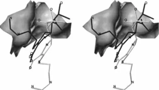
Figure 2. A different perspective for the two superimposed acyl-enzyme intermediates. A portion of the active site which encompasses the oxyanion hole is shown as a surface. The ester carbonyl of the canonical acyl-enzyme species (in gray) is shown housed in the oxyanion hole, and that for the species from the crystal structure is shown pointing out of it (in dark gray). The heteroatoms in imipenem are labeled.
several times during the simulation after the initial 38 ps. After the initial 125 ps, these movements became more pronounced, and the ester moiety moved more freely for the remainder of the simulation. The simulation illustrated that the acyl-enzyme intermediate was able to move in and out of the oxyanion hole and, in essence, partition between the two intermediary acyl-enzyme species, as it should, on the basis of the known kinetic behavior for turnover of imipenem.10
We suggest that the torsional strain caused by the 6R-1R-hydroxyethyl substituent accounts for this molecular motion. The torsion angle of CR-Cβ-Oγ-C is 95.5°in the model of the canonical acyl-enzyme intermediate for imipenem (species 2A in Scheme 1). In the X-ray structure, this torsion angle is -173.8°, which is effectively unstrained (species 2B in Scheme 1). The driving force for this rearrangement is the presence of the methyl group of the 6R-1R-hydroxyethyl. Indeed, formation of the canonical acyl-enzyme intermediate for
6R-hydroxym-ethylpenicillanate with TEM-1 induced no conformational
change of the protein under the reported experimental conditions, and this crystal structure16indicates that the decoration with
the additional methyl group in the 6R-1R-hydroxyethyl sub-stituent would induce steric encumbrance with protein atoms. This methyl group forces the structural adaptation observed in the TEM-1-imipenem complex and facilitates the transition to the second acyl-enzyme intermediate. This process is favored by the relative stability (slow rate of hydrolysis) of the canonical acyl-enzyme species, which likely provides the opportunity for this species to explore a different low-energy state.
The motion in and out of the oxyanion hole is quite intriguing
and occurs in the simulations at the picosecond time scale, while the deacylation chemistry even for the more favorable substrates occurs at the millisecond time scale.17 It argues for the fact
that the motion may be of a general nature, pertinent for discussion of the catalytic process, when considering the turnover of good and poorβ-lactamase substrates, particularly in the deacylation step.
The flexibility in the structure of the active site that we have documented for the first time here with this crystal structure, in conjunction with the findings of the simulations, presents a dynamic picture for the turnover chemistry of β-lactams by
β-lactamases, which heretofore was not appreciated in view of
the structural aspects of the function of these enzymes.
Experimental Section
Methods. Crystals18were soaked, at 4 °C, in a saturated
imipenem solution in 20 mM Na,K-phosphate, pH 7.8, contain-ing 50% of saturated ammonium sulfate. After 20 min, half of the solution was replaced by a fresh saturated imipenem solution, and the soaking was pursued for another 20 min. The crystal was mounted in a cryoloop, transferred in a 50% (v/v) PEG 20 K solution for a few seconds and frozen in a 100 K nitrogen flux. Diffraction data were collected on a MarResearch imaging plate at the×31 synchrotron beamline of the EMBL outstation at DESY (Hamburg, Germany), with detector distance 120 mm, wavelength 1.1271 Å and frame size 1°. Data were processed using MOSFLM 5.4019and CCP4.20 A rigid body refinement,
using the TEM-1 structure (PDB entry 1btl), was performed to account for the cell parameters variation between the native and derivatized crystals. All reflections between 18 and 1.8 Å were used in refinement with REFMAC.21 An external bulk solvent
contribution was computed with X-PLOR22and updated after
each major refinement cycle. The electron density map after the first refinement cycle allowed identification of all the substrate atoms. Water molecules were automatically introduced/ removed and refined using a procedure that alternates the use of REFMAC21and ARP.23 The final crystallographic R and (17) Christensen, H.; Martin, M. T.; Waley, S. G. Biochem. J. 1990,
266, 853.
(18) Jelsch, C.; Lenfant, F.; Masson, J.-M.; Samama, J. P. J. Mol. Biol. 1992, 223, 377.
(19) Leslie, A. G. W. In Proceedings of the CCP4 Study Weekend; J. R. Heliwell, P. A. Machin, M. Z. Papiz, Eds.; Daresbury, Warrington WA4 4AD, UK, Daresbury Laboratory, 1987; pp 39-50.
(20) Collaborative Computational Project, Number 4. Acta Crystallogr. 1994, D50, 760.
(21) Murshudov, G. N.; Vagin, A. A.; Dodson, E. J. Acta Crystallogr. 1997, D53, 240.
(22) Bru¨nger, A. T. X-PLOR Version 3.1: A system for X-ray
crystal-lography and NMR; Yale University Press: New Haven, CT, 1992.
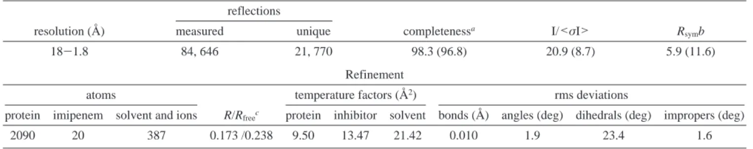
(23) Lamzin, V. S.; Wilson, K. S. Acta Crystallogr. 1993, D49, 129. Table 1. Summary of Crystallographic Analysis
Data Collection reflections
resolution (Å) measured unique completenessa I/<σI> R
symb
18-1.8 84, 646 21, 770 98.3 (96.8) 20.9 (8.7) 5.9 (11.6)
Refinement
atoms temperature factors (Å2) rms deviations
protein imipenem solvent and ions R/Rfreec protein inhibitor solvent bonds (Å) angles (deg) dihedrals (deg) impropers (deg)
2090 20 387 0.173 /0.238 9.50 13.47 21.42 0.010 1.9 23.4 1.6
aNumbers in parentheses refer to the highest resolution shell (1.89-1.80 Å).bR
sym)∑∑|〈I〉- Ii|/∑∑Ii.cR )∑||Fo| - |Fc||/∑|Fo|. The Rfree
value was calculated from a random set of reflections (10%) which were omitted from structure refinement. The R value was calculated from the remaining reflections.
Figure 3. The distance versus time profiles for the dynamics simulations on (A) the immediate acyl-enzyme intermediates for the TEM-imipenem complex and (B) the TEM-imipenem analogue without the 6R-1R-hydroxyethyl side chain. Black and gray traces represent the profile of the distances between the main-chain amine of Ser-70 and the acyl oxygen and that of Ala-237 and the acyl oxygen, respectively.
Rfree24values are 0.173 and 0.238, respectively. The final model
comprises all proteins and substrate atoms, 8 sulfate ions, and 347 water molecules. Alternate conformations were assigned to 13 side chains (Table 1).
The energy minimization procedures and molecular dynamics simulations were performed using the AMBER 4.1 package.25
In all starting conformations, the ester oxygen in the canonical acyl-enzyme species was in the oxyanion hole, making hydrogen bonds to the backbone nitrogens of Ser-70 and Ala-237. The crystallographic water molecules were retained and the complexes were immersed in a shell of water of 12 Å thickness from the surface of the enzyme complex. The energy-minimized (10 000 cycles) complexes were used as the starting
conformations for the molecular dynamics simulations. The structures were warmed from 0 to 300 K in steps of 20 K per 1 ps. Simulations were carried out for 165 ps for each of the two complexes at 300 K. The coordinates were collected for every 0.05 ps during the first 20 ps, and for every 0.2 ps thereafter. They were measured using the CARNAL module in AMBER package and plotted as a function of time.
Acknowledgment. We thank Ste´phanie Cabantous for excellent technical assistance. The work in France was funded in part by INSERM (CRE contract 930612; J.P.S.) and the Regional Midi-Pyrenees (contract 9200843; J.P.S.). The work in the U.S.A. was supported by grants from the National Institutes of Health and the National Science Foundation (S.M.). The crystallographic coordinates are deposited in the Brookhaven Protein Data Bank.
JA9818001
(24) Bru¨nger, A. T. Nature 1992, 355, 472.
(25) Weiner, S. J.; Kollman, P. A.; Case, D. A.; Singh, U. C.; Ghio, C.; Alagona, G.; Profeta, S., Jr.; Weiner, P. A. J. Am. Chem. Soc. 1984, 106, 765.