tf,3//ô.
Université de Montréal
Morphology of soleus and EDL
Neuromuscular Junctions
in
the Transected
Rat Model Following Fetal Tissue Transplant
par
Rérny
J. Pilon
Département de Kinésiologie
Mémoire présenté à la Faculté des études supérieures
en
vue
de l’obtention du grade de
Maîtrise en sciences (M.Sc.)
en sciences de l’activité physique
Août 2003
oI
Université
de Montréal
Direction des bibliothèques
AVIS
L’auteur a autorisé l’Université de Montréal à reproduire et diffuser, en totalité ou en partie, par quelque moyen que ce soit et sur quelque support que ce soit, et exclusivement à des fins non lucratives d’enseignement et de recherche, des copies de ce mémoire ou de cette thèse.
L’auteur et les coauteurs le cas échéant conservent la propriété du droit d’auteur et des droits moraux qui protègent ce document. Ni la thèse ou le mémoire, ni des extraits substantiels de ce document, ne doivent être imprimés ou autrement reproduits sans l’autorisation de l’auteur.
Afin de se conformer à la Loi canadienne sur la protection des
renseignements personnels, quelques formulaires secondaires, coordonnées ou signatures intégrées au texte ont pu être enlevés de ce document. Bien que cela ait pu affecter la pagination, il n’y a aucun contenu manquant.
NOTICE
The author of this thesis or dissertation has granted a nonexclusive license allowing Université de Montréal to reproduce and publish the document, in part or in whole, and in any format, solely for noncommercial educational and research purposes.
The author and co-authors if applicable retain copyright ownership and moral
rights in this document. Neither the whole thesis or dissertation, nor
substantial extracts from it, may be printed or otherwise reproduced without the author’s permission.
In compliance with the Canadian Privacy Act some supporting forms, contact information or signatures may have been removed from the document. While this may affect the document page count, it does not represent any loss of content from the document.
111
Université de Montréal
Faculté des études supérieures
Morphology of soleus and EDL
Neurornuscular Junctions in the Transected
Rat Model Following Fetal Tissue Transplant
présenté par:
Rérny Pilon
a été évalué par un jury composé des personnes suivantes
Dr. Phillip Gardiner
-Directeur de recherche
Dr. Louise Béliveau
—Membre de jury
Dr. Martin Brochu
—Membre de jury
Mémoire présenté à la Faculté des études supérieures
en
vue
de l’obtention du grade de
Maîtrise en sciences (M.Sc.)
en sciences de l’activité physique
Août 2003
iv
ABSTRACT
Recently rnany studies have tried to add to the ever-growing body ofknowledge with regards to spinal cord transection by introducing ernbryonic stem celis at the site of injury with the goal of providing an environment that could prornote the restoration of physical ftmction in the transected model.
The present study exarnined the effect of transection of the spinal cord on
neuromuscular junction (NMJ) parameters in rat hind lirnb muscles (SOL and EDL) 4 weeks post-lesion at the lower thoracic level and the influence oftransplanted embryonic stem celis into the transection site. A histological technique modified from that previously described by Pestronk and Dracirnan (1978) was used for endplate staining. This method allowed for the quantification ofthe cholinesterase-containing end plate as a demarcated transparent blue area, against which the sHver-stained nerve-terminals stood out. This study was composed of three groups of rats: an unoperated control group (group CNTRL, n=3), a spinal transection group (group TRANS, n=3) and a spinal transection plus fetal transplant group (TRNPL, n3). The NMJ pararneters quantified included end-plate area, end-plate longitudinal length, number of nerve terminal branch points and muscle fiber width.
Post transection, the soleus had significant increases in end-plate length, number of branch points and a significant decrease in muscle fiber width. The end-plate length was the only SOL pararneter that was attenuated with the transplant post-transection.
V
Sirnilar to the soleus, the EDL underwent a significant decrease in muscle fiber width afler transection. With regards to end-plate (E?) length, EP area and branch points, the parameters were not significantly different from CNTRL values (p> 0.05). The transplant procedure did flot influence any of these properties. This could be attributed to the more fast twitch nature of the EDL when cornpared to the SOL, thus taking longer to undergo end-plate morphological changes.
Though the reproducibility and relevance ofthese findings have yet to be determined, embryonic stem celi transplants and their influence on neuromuscularjunctions hold much interest and promise for future research in the restoration ofphysical function to those who suffer from spinal cord injuries.
vi
RÉSUMÉ
Récemment, plusieurs études ont utilisé des cellules embryonnaires pour fournir un environnement permettant le rétablissement de fonction physique au modèle qui ont subit une transection expérimentale.
Cette étude a examiné l’effet d’une transplantation des cellules embryonnaires sur des paramètres de jonctions neuromusculaires des muscles SOL et EDL qui ont subi une
transection expérimentale au niveau du thorax inférieur.
Une méthode histologique a été modifiée de celle décrite par Pestronk et Drachman (1978) pour teindre les plaques motrices. Cette méthode permet la quantification de la cholinestérase de la plaque motrice comme une région bleue en mettant en contraste les terminales des nerfs rendus argentés par la teinture.
Cette étude est composée de trois groupes de rats: un groupe contrôle (non opéré) (CNTRL, n=3), un groupe transecté (TRANS, n=3) et un groupe transecté avec
transplantation foetale (TRNPL, n=3). Les paramètres de la jonction neuromusculaire (NMJ) comprennent l’aire de la plaque motrice, la longueur longitudinale de la plaque motrice, le nombre de branches neuronales (« branch points ») et la largeur des fibres musculaires.
Après la transection, une augmentation de la longueur de la plaque motrice a été observée pour le soléaire ainsi qu’une augmentation du nombre de branches neuronales et une diminution de largeur des fibres musculaires. Avec la transplantation, la longueur de la plaque motrice a été le seul paramètre atténué.
vii
Comme le soléaire, FEDL a subi une diminution en largeur de la fibre musculaire. Eu égard à la longueur de la plaque motrice, à l’aire et aux points de branche, les paramètres n’étaient pas significativement différents des valeurs contrôles (p> 0,05).
La procédure de transplantation n’a eu aucune influence sur ces propriétés. Ceci peut être attribué au caractère rapide du EDL en comparaison avec le soléaire, d’où une durée plus longue pour le changement morphologique des plaques motrices.
Même si le pertinance ou l’abilité de reproduire ces résultats sont ndeterminé jusqu’à maintenant, l’effet d’une transplantation des cellules embryonnaires surdes jonctions
VIII
TABLE 0F CONTENTS
ABSTRACT iv
RÉSUMÉ
TABLE 0F CONTENTS viii
LIST 0F DIAGRAMS x
LIST 0F FIGURES x
LIST 0F TABLES xi
LIST 0F APPENDICES xii
LIST 0F ABBREVIATIONS xiii
ACKNOWLEDGEMENTS xiv
REVIEW 0F LITERATURE 1
Part I Spinal Cord Injury 2
Part II Restoration of Function Following Transection 4
Part III Spinal Cord Transection (Experimental Model) 6
Part 1V Regeneration In Peripheral Vs Central Nervous System 7
PartV Tissue Transplant: A Medium To Encourage Axonal Regeneration 10
PartVI Morphology ofThe Neuromuscular Junction 12
PartVII Sprouting ofMotoneurones 14
PartVIII FiberType Differences With Regards To The NMJ 1$
Part IX SummaryofLiterature 20
Part X Purpose0fThe $tudy 21
ix
MATERIALS AND METHODS
I. Subjects 23
2. Surgical Procedures 24
2.1 Spinal Cord Transection 24
2.2 Transplanted Embryonic Tissue 25
3. Harvesting of Tissues 26 4. Histochemical Analysis 27 5. Statistical Analysis 31 RESULTS EP Longitudinal Length 32 EPArea 37
Muscle Fiber Width 41
Branch Points 46
SUMMARY PRINCIPAL RESULTS 49
DISCUSSION 50 STUDY LIMITATIONS 56 CONCLUSION 57 REFERENCES 5$ APPENDIX A 64 APPENDIXB 65
X
LIST 0F DIAGRAMS
Diagram 1 - Cholinesterase Staining of End-Plate Areas 2$
Diagram 2 - Measurement of End-Plate Longitudinal Length 28
Diagram 3 - Calculation ofEnd-Plate Brandi Points 29
Diagram 4 - Measurement offiber Width 29
LIST 0F FIGURES
Figure 1 - Muscle Main Effect on End-Plate Length 32
Figure 2 - Treatment Main Effect on End-Plate length 33
Figure 3 - SOL EP length distribution according to percentile 35
Figure 4 -EDL EP length distribution according to percentile 36
Figure 5 -Muscle Main Effect 0f End-Plate Area 37
Figure 6 -Treatment Main Effect on end-plate area 38
Figure 7 - SOL EP area distribution according to percentile 39
Figure $- EDL EP area distribution according to percentile 40
Figure 9- Muscle Main Effect On Muscle Fiber Width 41
Figure 10- Treatment Main Effect on Fiber Width 42
Figure 11 - SOL fiber width distribution according to percentile 44
Figure 12 - EDL fiber width distribution according to percentile 45
Figure 13 - Muscle Main Effect ofBranch Pôints 46
xi
LIST 0f TABLES
Table I - End-Plate lengths for SOL and EDL (jim) ± S.D. 35
Table 2 - End-Plate areas for SOL and EDL (im2) ± S.D. 39
Table 3 - Fiber width for SOL and EDL (tm) ± S.D. 43
xii
LIST 0f APPENDICES
APPENDIX A: Staining for Quantitative Measurement ofNeuromuscular Junction 64
• Tissue Processing
• Cholinesterase-Staining Procedure
XIII
LIST 0F ABBREVIATONS
CNS Central Nervous System
CTRL Control group
EP End-Plate
EDL Extensor Digitorum Longus
ESC Embryonic Stem Celis
IP Intraperitoneal
MyHC Myosin Heavy Chain
NMJ Neuromuscular junction
SC Stem celis
SCI Spinal Cord Injury
SOL Soleus TRANS Transection
xiv
ACKNOWLEDGEMENTS
first I would like to thank Dr, Phullip Gardiner for lis inexhaustible enthusiasm and encouragement througbout my studies at the Université de Montréal. Ris dedication lias made my time spent with him a most enriching and rewarding experience, and for this I am truly grateful.
I would also like to dedicate this prolonged work to my parents, Lillian and René Pilon, for ail their immeasurable support throughout the years. From a young age, they instilled within me the desire and the ‘justification for higlier education.” Thanks to their guidance and tutelage, they have motivated me in every facet ofmy life, including my recent move to Ottawa, to pursue a career in the pharmaceutical industry. Words simply cannot express how thankful I am to be their son.
I gratefully acknowledge the rest ofmy family and friends for providing support in their own special ways, and of course my fiancée Catherine Chan, who has inspired me in her own right. So a special thank you to Peter Tzavaris for convincing me to apply and for Dr. Phil Gardiner for pairing Catherine and I together. Sometimes life’s greatest surprises can be right before your very eyes.
Lastly, I would also like to thank Dr. Houle, from the University ofArkansas, for generously providing the transected and transplant subjects used in this study.
REVIEW 0F LITERATURE
Every sensation, action and thouglit revolves around the complicated processes ofthe central nervous system (CNS), which consists ofthe spinal cord and the brain. Despite over a century of exhaustive research into spinal cord injury (SCI), we stili can offer no cure. The Christopher Reeves Paralysis Foundation estirnates 250,000 spinal cord injured individuals in the United States, with an additional 11,000 new injuries occurring every year. The majority of these cases occur between the ages of 16 to 30, with the average age of injury being 31. Adaptation to skeletal muscle fibers to increased use and decreased use has been extensively investigated.
Littie is known about the adaptations that occur to other components of the neuromuscular system. This review ofliterature will summarize the current literature regarding spinal cord injury, the transected experimental model and the morphology ofthe neuromuscularjunction. The neuromuscularjunction is specifically defined as the synaptic site between a motoneuron and muscle libers belonging to a motor unit. The NMJ is a dynamic structure that undergoes both morphological and fiinctional changes throughout the course of a lifetime (Sieck & Prakash, 1997).
Though extremely small and measured in micrometers, the NMJ plays a critical role in ail neuromuscular functions and its morphology post-transection with fetal tissue
z
SPINAL CORD INJURY
The Christopher Reeves Paralysis Foundation estimates 250,000 spinal cord injured (SCI) individuals in the United States, with an additional 11,000 new injuries occurring every year. The rnajority of these cases occur between the ages of 16 to 30, witli the average age of injury being 31.
The spine supplies the main defense for the spinal cord, providing a protective barrier against injury. A fluid fflled area, known as the syrinx, offers additional protection by absorbing shocks (Christopher Reeve Paralysis Foundation, 1999).
Though generally extremely beneficial, both these defenses cause complications upon injury. Swelling induces additional damage to the spinal cord as pressure builds in the confined space between the cord and vertebrae. Though the cord is flot usually completely severed during injury, the swelling impedes vital blood supply to neurons, resulting in cdl death. The syrinx contributes to the scar tissue that builds up around the area of injury, blocking the neurons from reconnecting once the cord lias been severed (Brown and Tronton,
197$).
The complex interactions between the brain and neurons, in combination with the enon-nous number of individual neurons and synapses, make reconnection of the nerve celis post-transection extremely difficult (Christopher Reeve Paralysis foundation, 1999).
The types of disabilities associated with spinal cord injury vary greatly depending on the severity of the injury, the segment ofthe spinal cord at which the injury occurs, and the particular nerve fibers that are damaged. The destruction ofnerve fibers that carry motor signais from the brain to the torso and limbs leads to muscle paralysis, while damage to afferent fibers leads to loss of sensation sucli as touch, pressure, and temperature. There are
3
numerous other serious consequences which include exaggerated reflexes; loss ofbladder and bowel control; sexual dysfunction; lost or decreased breathing capacity; impaired cough reflexes; and muscular spasticity (Chnstopher Reeve Paralysis foundation, 1 999).
A severe spinal cord injury disconnects the major conduits through which sensoiy and motor signais pass from the body to the brain and vice versa. It was beiieved this condition was ineversible because it was thought that the environment ofthe central nervous system was inhibitory to neurite growth. The potential for regeneration and extension ofthe CNS axons was recognized by the tum ofthe century by Ramon y Cajai (1928). Cajal and Camiilo Golgi received the Nobel Prize in 1906 for introducing the silver-chromate stain. With his reduced siiver nitrate technique he was able to demonstrate neurons and their connections quite easiiy. His introduction ofhis goid chloride-mercury bichioride technique to demonstrate astrocytes was a monumental contribution as was his work on degeneration and regeneration of the nervous system. Cajal and Golgi (1928) hypothesized that in a permissive environment, axons would be able to grow for iong distances following a CNS lesion.
This is a hypothesis, though nearly a century old, stili raises questions ami encourages researchers to further investigate the morphology of the neuromuscular junction.
4
RESTORATION 0F FUNCTION FOLLOWING TRANSECTION
The inability ofthe axons ofthe central nervous system to regenerate across lesions in the spinal cord is an extremely puzzling phenomenon. The sprouting nerves will simply flot traverse the lesion and consequently cannot reach their targets to form cormections and restore function. Contrastingly, a lesion of a penpheral nerve need flot be followed by permanent disability. Peripheral axons have been shown to reiimervate skeletal muscles (Bregman et al., 1993; Kunkel-Bagden & Bregman, 1989), allowing for controlled
movements post lesion. This discrepancy between central and peripheral nerves that exists in the mammalian species, does flot exist in lower vertebrates or invertebrates. The leech for example, afier complete transection ofthe CNS, can grow and reconnect nerves and targets with astonishing precision (Nicholls, 1987).
McDonald and colleagues (1999) from Washington University $chool of Medicine successfully implanted embryonic stem ceils in laboratory rats. They induced mid-thoracic spical cord injury in the rat using a 2.5 mm diameter metal rod in diameter resulting in paralysis. Nine days afier the injury, McDonald et al. transplanted roughly I million ES celis in the cavity. Two weeks afler the transplantation ES cells fihled the area normally occupied by guai scarring. Aller five weeks the stem ceils had migrated further away from the
implantation site. Although a number ofthem had died, there was stili enough raw material to have a growing supply ofneurons and glial celis. Most ofthe surviving celis were oligodendrocytes and astrocytes, but some neurons were found in the middle ofthe cord. The rats had actually regained limited use of their legs. Paralysis had apparently been cured at least partially.
5
Though McDonald’s work represented new successes in stem celI technology, there is much work ahead of us before we might test any ofthis technology in humans. Scientists are achieving results, though they do not fully understand the mechanisms or factors behind controlled nerve regeneration. McDonald et al. (1999) hypothesized that the regaining of function was attnbutable to the few differentiated neurons. Perhaps function was regained due to embryonic stem ceils producing growth factors. Complications could also arise from such technology. One example pertains to the rejection offoreign bodies by the host’s immune system. Cyclosporin is most commonly used in rats, but the human body promises to be much more complicated. The brain and spinal cord are complex and mysterious, and until science can predict the exact effects ofour evolving teclmology, no testing on humans can occur.
But before these questions can be answered, a successful restoration ofphysical fiinction post-transection must be achieved on animal models prior to any ethical research on humans may be attempted.
6
SPINAL CORD TRANSECTION (EXPERIMENTAL MODEL)
Transection is a preferred experimental method by which to study neuromuscular inactivity since there is no direct damage to the muscle and the innervation is flot physically disrupted. Spinal cord transection is a transverse duttmg ofthe spinal cord resulting in loss of ail sensations and voluntary movements inferior to the lesion (Dupont-Versteegden et al., 1998). Except for reflex movement, transection produces unmistakable lesions that result in qualitative and quantifiable motor deficits (Houle, 198$).
Typically, following a laminectomy in an anesthetized animal, the pia mater is cut and a small glass micropipette is used to aspirate spinal cord tissue until a cavity about 2 mm long is created. Motoneurons caudal to the transection lose neural input, affecting both their tonic and phasic firing pattems, and influencing their electrophysiological and metabolic activities (Houle and Reier, 198$).
Characteristic changes in muscles afier spinal cord injury include decrease in gross muscle size, atrophy ofindividual myofibers, and altered expression ofmyosin heavy chain proteins, such that the type II isoforms become prevalent (Lieber et al., 1986). The rapid transformation from a slow to a fast phenotype, afier a spinal cord transection, supports the observation that appropriate neural activity is an important component in maintaining the expression of slow characteristics ofthe muscle (Lieber et al., 1986). Neural input or muscular activity (active or passive) are not the sole regulatory mechanism ofmuscular phenotypic expression. Humoral signals, particularly thyroid hormones, can positïvely influence fast fiber transformation, independent ofthe state ofinnnervation (lannuzo et al., 1977).
I
REGENERATION IN PERIPHERAL VS CENTRAL NERVOUS SYSTEM
The CNS environrnent does flot favour axonal growth. However in the PNS, severed axons sprout to restore normal function, partly due to the assistance ofSchwann ceils. These ceils have been associated with assisting extending nerve processes in rcsponse to
denervation and were found to be associated with most nerve sprouts in partially denervated
muscle. Schwann ceils have been shown to both induce and guide the process of axonal
sprouting (Son and Thompson, 1995).
Schwann ceils that make up the myehn do not die post-transection, but in fact stay
where they are allowing for a new sprouting axon to use the previously existing pathway
(Son and Thompson, 1995). When the axons start to grow, they can travel through the same “tunnel” that an axon previously occupied, and eventually reach the same location ofthe skin or muscle.
Contrastingly, in the CNS, the most axons can grow is a fcw mm. Part of the problem
is that the Schwann ceil homologue in the CNS is the oligodendrite, which does not remain to assist in the regrowth of sevcred axons. To make matters worse, oligodendrites possess
molecules on their surface that are inhibitory to axonal growth cones (Schwab, 1988). This inhibitory effect can be neutralized by monoclonal antibodies. Schwab used these antibodies
in the transected cat and axons were seen to regenerate past the lesion for distances over 1 centimeter, which was significantly greater than in the control groups. Thcugh the number of
regenerated axons was deemed too few to have any significant functional effect, Schwab
(1990) concluded that the antibodies were clearly making the CNS environment lcss
In an attempt to promote favorable conditions for axonal growth around the site of an injury in the CNS, numerous experimental approaches have been utilized (Guth et aI., 1993). There are two main strategies cunently being used to overcome this inability ofthe neurons in the CNS to regenerate new axons thus reinnervating the appropriate post-synaptic neurons.
Thefirst consists ofmanipulating the molecular environment to facilitate
regeneration. It bas been found that there are a number ofmolecules that wifl either promote or inhibit axonal regeneration (Guth et al., 1993). These generally include a number ofnerve growth factors that work primarily by increasing the expression of certain genes necessary for axon elongation.
The second, of great interest to this report, centers around the bridging of the lesion with the utilization of transplant tissue that is conducive to growth into the lesioned cord. In this strategy, the transplant acts as a “bridge” for the central neurons to grow on. Transplants have resulted in axonal growth, a reduction of muscle atrophy and even reinnervation (Reier et al., 1983; Howiand et al., 1995(a); Miya et. al., 1997; Houle et al., 1999).
It has been reported that spinal lesions sparing even small amounts of tissue permit considerable function in the cat (Blight and DeCrescito, 1986), therefore one would assume that increased fiinction might be achieved when the spinal cord is repaired by a transplant, even without complete restitution of alI the original pathways (Miya et al., 1997). Despite having a significantly diminished number of intact ascending and descending axons, patients with subtotal spinal cord transection generally have limited ffinction. Fawcett (1992)
hypothesizes that perhaps what might seem an insignificant number of axons, should they be oftheright kind and reconnecting the right areas, just might 5e of “considerable functional benefit.” He goes on to add that this might be arranged by the addition of Schwann celis or peripheral nerve grafis. “The limitations of sucli grafis pertain to the fact that the axons that
9
grow tlirough the grafi cannot penetrate far enough into spinal cord tissue distal to the graftto make useful connections (Kulilengel et al., 1990).
Younger subjects generally possess the ability to accept transplanted fetal tissue and regrow axons better than senescent individuals. Tlierefore, it lias been liypothesized that fetal donor tissue would hold more promise than aduit tissue transplant since it possesses a greater ability for growth and sprouting (Kunkel-Bagden & Bregman, 1989).
A fetal transplant into the site of spinal transection eau influence both regeneration of axons and recovery of motor functions in tlic rat. Howiand et al. (1995b) used embryonic spinal transplants that were placed into the site of a complete midthoracic spinal transection in kittens, and observed that it permitted the development of locomotion that exceeded that of littermates with transections alone.
Fawcett (1992) postulated that this problem may be somewhat improved by
employing a combination oftreatments, including but flot limited to: nerve growth factors to neuronal cdl bodies and axons to enhance outgrowth, blockers ofoligodendrites inhibition and astrocyte active factors for regeneration of axons leaving the grafi to penetrate the spinal cord thus making the appropriate neural connections.
‘U
TISSUE TRANSPLANT:
A MEDIUM TO ENCOURAGE AXONAL REGENERATION
Since the early studies of Ramon y Cajal (1928), there lias been much speculation that immature mammalian CNS might show regeneration after spinal injury. A trend in spinal cord research centers around transpianting pieces offetal spinal cord tissue into the site ofthe lesion of transected recipients (Houle al., 1999; Kunkel-Bagden & Bregman 1989; Miya et al. 1997). The strategy behind transpianting grafis involves establishing a “bridge” which might assist axon elongation through and beyond the injury. It lias been proposed that the transplants provide an environment that supports growth of axotomized and late developing axons and a rescue of neurons destined to undergo retrograde ceil death (Miya et al., 1997). Transplants are thought to rescue severed neurons by serving as a surrogate source oftarget derived neurotrophic factors (Bregman and Reier, 1986).
Researchers have been in agreement that transplantation of adult nerve tissues does not work, while embryonic or fetal transplantation can be quite successful (Houle, 1991). In other words, transplants may restore some elements of spinal circuitry or otherwise affect a reorganization ofhost tissues.
There is evidence that axonal integration between the transplant and host spinal cord occurs, either directly by growth ofhost axons through the transplant or indirectly by a relay ofmechanisms involving synaptic contacts between host and transplant neurons (Bregman et al., 1997; Houleand Reier, 1988).
11
In many cases, the growth ofdescending pathways into the fetal transplants was substantially greater afier lesions al birth than at rnaturity, in both rats and cats (Bregman et al., 1993; Bregman and Goldberger, 1982). The capacity for the CNS neurons for axonal regrowth afier injury decreases as the age ofthe animal at the tirne of injury increases. In the adult, Bregman et aI. (1993) demonstrated that the growth is restncted to within 200 um of the host/transplant border. Studies utilizing neonatal spinal rats given transplants seemed to produce optimism for the transplant theory in rats (Iwashita et al., 1 994). When a transplant was inserted into a lesioned cord of a newbom, the host axons descend into the transplant and exhibit extensive regeneration growth into and beyond the transplant.
Houle et al. (1999) compared the effects of a fetal transplant grafi versus passive hindlimb cycling in the SOL. They found that the grafi had no effect on myosin heavy chain (MyHC) expression, though it did limit muscle atrophy afier complete transection ofthe spinal cord. Muscle fiber cross sectional areas (CSA) ofthe transection (TRANS) group was
50% that ofthe control (CNTRL), while the transplant (TRNPL) and exercise groups were 77% and 74% when compared to CNTRL. It was suggested that these results support the notion that a different regulatory mechanism for the control of muscle size and for the expression ofproteins that are considered important for muscle contractility.
Studies have shown that spinal lesion sparing small amounts of tissue permits
considerable function (Blïght and DeCrescito, 1986). One might postulate that function may be possible with a transplant, even without full restoration of normal pathways. Intraspinal transplants offetal spinal cord have been shown to enhance the development oflocomotor performance afier complete transection in the newbom rat (Diener and Bregman, 1994; Miya et al., 1997).
12
MORPDOLOGY 0F THE NEUROMUSCULAR JUNCTION
It has long been dernonstrated that activity directly affects the morphology ofthe
mammalian motoneuron (Manu, 1894). The neuromuscularjunction is specifically deflned as the synaptic site between a motoneuron and muscle fibers belonging to a motor unit. This dynamic structure undergoes both morphological and functional changes throughout the course ofa lifetime (Sieck & Prakash, 1997).
Various studies have demonstrated that, in some ways, the structure ofthe NMJ is specific to muscle fiber type (Brown et al., 1980). The literature bas shown that both the
morphology and the physiology of the NMJ have been shown to adapt to both increased and
decreased levels of activity (see review: Panenic and Gardiner, 1998). Increased activity for
instance aerobic or anaerobic types of exercise stimulates adaptations that enhance neuromuscular transmission, thus improving muscular performance. On the contrary,
decreased activity of this myoneural synapse, due for instance to limb immobilization or spinal cord transection initiates morphological changes that can impair transmission and function.
Both complete and incomplete muscle disuse appears to bring about similar signs of
NMJ degeneration and regeneration (fahim, 1989; fahim and Robbins, 1986). Adaptations
to disuse include the number and size of structural components (Deschens et aI., 1993), as well as the functional characteristics (Robbins and fischbach, 1971; Brenner and Rudin, 1989) oftheNMJ.
Some ofthe structural adaptations from muscle disuse include motor end-plate expansion (Eldridge et al.1981), enlarged presynaptic nerve terminal area, amplified brandi
J-5
number, increases in sprouting and complexity ofnerve terminais (Tomas et al., 1989). Motor units seem to exist in a dynamic state and display a high degree ofplasticity (Mendeli,
1994). This piasticity is inherent in the motor unit and lias been attnbuted to the
developmental process and to changes in the aduit, which are under continuaI influence by extemal stimuli (MenUe!!, 1994; Navarrette and Vrbova, 1993).
14
SPROUTING 0f MOTONEURONES
Total disuse rapidly affects both pre- and post-synaptic charactenstics ofthe mammalian NMJ. The most prevaient changes inciude increased sprouting at nerve
terminais, enhanced synaptic transmission, and atrophy ofpost-synaptic folds. Sprouting is the production ofnew processes (outgrowths) by nervecelis. During the embryonic stages sprouting is referred to as undergoing primary differention, and redifferentiating when it occurs in the aduit neurons in response to nervous tissue damage or in dissociated neurons.
Motor nerve sprouting was first observed in response to partial denervation , and the possible consequences that follow partial denervation have ail been postulated at one time or another as the sprouting stimulus. Brown et ai. (1980) also observed sprouting upon
inducing deneiwation-like changes in muscle by simply blocking nerve-induced activity. Motor nerve terminais have the ability to sprout under a variety of normal and
pathologicai conditions. Continuai sprouting occurs at he normal NMJ providing renewal of nerve terminais. Partial denervation of muscle tends to evoke growth of the nerve terminais. Accidentai or experimental nerve injury resuit in sprouting of the intact motor nerves (Pestronk & Drachman, 197$).
Muscle activity exerts a trophic influence on motoneurones. It may be a factor in the regulation of sprouting (Snider & Hanis, 1979). Brown and Ironton (1977) found fine, ultra terminal sprouts emanating from the endplates of muscles rendered inactive by chronic conduction biock ofthe muscle nerve. Pestronk and Drachman (1978) observed increased motor neiwe terminal branching and a consequent increase in endplate size in similar conditions.
I)
Total disuse of the mammalian NMJ rapidly affects both pre- and post-synaptic characteristics, producing sprouting oftemiinals, enhanced synaptic transmission, and atrophy ofpost-synaptic folds ofthe perijunctional ‘raised area within 3-7 days (Fahim & Robbins, 1986). A well-documented fonu ofneuromuscular disuse which lasts 2-3 weeks, produces about 90% disuse and does not entail use ofdrugs or nerve injury, is obtained by limb immobilization. This procedure causes changes in synaptic transmission within 3-5 days. A rapid morphologicai response to subtotal disuse occurs within 5 days and consists of sprouting and longitudinal distortion ofnerve terminais, flattening ofprimary grooves and partial loss ofperi-junctional surface features. A briefperiod of partial disuse induces considerable and rapid synaptic plasticity in the aduit nervous system.
It has been suggested that that this atrophy is initiated by the dismption ofthe NMJ that leads to progressive end-plate degeneration and graduai withdrawal ofthe terminal axon (Stebbins et al., 1985). The rate at which the process ofatrophy develops can vary both within and among muscles. With this in mmd, muscle-specific preferential fiber-type atrophy (Tuffery, 1971) and liber-type conversion have been observed, suggesting that the fiber-type composition of muscle may influence the process and development of atrophy.
The extent and nature of the motoneuron sprouting response has been shown to differ between slow and fast muscles (Brown et al., 1980).
Axonal withdrawal has been impiicated as a causation factor for liber atrophy associated with aging muscles. The peripheral nervous system is also active in the process that reduces fiber atrophy. This process known as terminal sprouting, occuring throughout the life-span, is believed to be a mechanism involved in end-piate growth and reconstruction. Outgrowths (sprouts) on the motor axons migrate toward and eventuaiiy innervating the parent end-piate.
1C
The process of terminal sprouting and withdrawal may act to sustain and reorganize motor end-plate morphology in response to changes in functional demand during growth, aging or disease (Cardasis & Padykula, 1981). Increases in neuromuscular contact through terminal sprouting helps to maintain integnty of the NMJ.
Sprouting is evident at the microscopic level by formation ofnew nerve growth either along the axon or endplate. Motoneuron sprouting was documented in the late eighteen hundreds and was thought to be a possib]e explanation for the lack of degenerating fibers observed in partially denervated muscle (Exner, I $84).
following partial denervation, fast muscles motoneurons showed more sprouting than their slow muscle counterparts, though the production of sprouts was similar subsequent to complete paralysis with botulinum toxin (Brown et al., 1980). Brown & Ironton (1977) hypothesized that the signal for terminal sprouting originated ftom the denervated muscle but also proposed that the origins for nodal sprouting may be linked to nerve degeneration.
The only common denominator for the sprouting stimulus has been reported as either an inactive muscle or a muscle state showing characteristic of denervated muscle (Brown et al., 1980). Brown & Lunn (1988) later revised their conclusions to include central changes such as chromatolysis in the cell body, loss of afferent input and in addition to peripheral changes, altered neuronal interactions.
Van Mier and Lichtman (1994) have published convincing evidence for the presence of a growth signal emanating from inactive muscles that attracts nearby axons to form new connections. Their results showed that damaged muscle fibers provided a directional
sprouting signal that was detected by nearby axons. It was also noted that in cases where the original endplate areas were left intact, new sprouts avoided the areas but nonetheless made contact with the regenerating fibers. The sprouts were subsequently displaced by
1 7
reinnervation oftlie original axon at the original site. However, if both the fiber and its original terminal site were destroyed, there was more extensive reinnervation ofthe fiber and sorne evidence for a protracted competition between newly amved sprouts, until one contact became the permanent and sole contact.
During the mid-nineteen liundreds, new nerve growtli was observed to be “sprouting” from the Nodes ofRanvier and from the terminais of intact nerves ofpartially denervated muscles (Hoffluan, 1950). In addition, Hoffman observed these new growths made contact with the denervated muscle fibers and underwent remyelination, suggesting that these wcre newfunctionaÏ motor units. This reorganization by the residual systems is seen as an attempt to restore flrnctions to damaged areas and lias been shown to restore functions in the partially denervated model.
Sprouting occurs relatively rapidly and its extent is quantifiable by the measuring of branch points. The literature re-iterates that it is important to note that these are probably overestimates ofthe time required for the sprouting response to occur because the laboratory techniques utilized may not be sensitive enough to pick up early morphological changes.
1Ô
FIBER TYPE DIFFERENCES WITH REGARDS TO THE NMJ
Differences between type I and type Il muscle fibers have long been documented. Granit et al. (1957) discovered that the nerves innervating white muscle (fast/type Il) were larger than those ilmervating red muscle (slow) and suggested a functional implication for the differcnce in diameter.
Postulations for these differences between the types offibers pertained to the intrinsic speed differences ofthe muscle that are at least partially regulated by neural mechanisms (Buller et al., 1960). Wigston (1989) suggested that the degree ofremodeling post
transection ofthe NMJ may vary among fast and slow twitch muscles, perhaps attributable to the pattem of use.
Duchen (1970) reported that this distinction in sprouting, between fast and slow muscles, persisted up to 4 weeks afler transection. The deep portion ofthe fast muscles (plantaris and gastrodnemius) resembled the soleus in terms ofthe number ofsprouts, terminal morphology and cholinesterase distribution 28 days post-transection. The superficial regions ofthe fast mixed muscles began to show some sprouting but stili contained a predominance ofnon-sproutednerve terminals.
Dramatic differences were observed in the sprouting responses ofmotor units from paralysed (botulinum toxin) soleus and gastrocnemius muscles and even amongst motor imits from different regions ofthe same muscle (gastrocnemius and plantaris). The data showed nearly immediate extensive sprouting in slow soleus, somewhat less extensive in the deep regions ofthe gastrocnemius and plantaris, and absence in the highly concentrated type IIb superficial regions (Duchen, 1970).
19
In addition, fast and slow muscles responded differently to penods ofparalysis induced by injection oftetanus toxin, a presynaptic blocking agent. The injection oftetanus toxin elicited terniinal sprouting in the soleus, though these new growths were not observed in the extensor digitorum longus muscle (Duchen and Tonge, 1973).
It lias becn suggested by Brown et al. (1980) that the relatively fast muscles have less sprouting upon denervation than when compared to their slow muscle counterparts. This reduction ofnerve morphology can be attributed to their higher resistance from a reduced neuronal input.
Traditionally, the sprouting ofthe soleus is seen as a direct resuit of changes in muscular activity and/or neural input as in transection or limb-immobilization. Whereas the traditional sprouting for the EDL is generally associated with the natural process ofaging. This has been attnbuted to the fact that slow-twitch liber is mainly responsible for postural and everyday functions, whereas the utilization ofthe fast-twitch muscles generally decline with senescence. for a more detailed analysis ofmorphological adaptation differences between liber types, including sprouting, sec Sieck and Prakash (1997).
Lu
SUMMARY 0F THE LITERATURE
Damage to central nervous system axons is devastating. There has been rnuch effort to increase regenerative capacity experimentally. The utilization of transplant tissue to enhance reparation lias been most promising in recently. Specifically, transplant tissue inserted into tlie transected spinal cord lias spared muscle tissue innervated by axons caudal to the transection. Though this is an extremely promising step for spinal cord repair, the meclianisms remain unknown.
The effect of transplant tissue on transected nerve terminal morphology remains complctely unknown.
PURPOSE 0F THE STUDY
Nurnerous studies have looked at the potential offetal transplants for CNS restoration in the spinal cord transection model. However, in comparison, only modest research bas studied the morphological effects ofthese transplants on the NMJ.
The purpose ofthis study was to analyze morphological parameters ofthe
neuromuscular junctions in hindlimb muscles (soleus and EDL) of rats following spinal cord
transection, with and without a fetal transplant procedure performed at the time of
transection. Due to the diffeHng nature of fast and slow muscle fibers (Sieck and Prakash, 1997), one would expect the embryonic transplant to elicit different responses in the SOL and EDL end-plates. Due to its slow-twich properties, it is hypothesized that the transplant would attenuate the EP morphological changes generally seen post-transection in the SOL more so than in the EDL.
The parameters quantified were endplate area, end-plate longitudinal length, branch points within the endplate and muscle fiber width.
22
WHY WERE THE EDL AND SOLEUS SELECTED?
The present investigation assessed different morphological parameters ofnerve terminais from control (CNTRL), transected (TRANS) and transection-transplanted groups (TRNP). The slow-twitch soleus and the phasic fast-twitch EDL muscles were selected for several specific reasons:
a) The soleus and EDL perform fiinctionally different actions (flexion and extension, respectively);
b) There are differences in theirfiringpattems during rest (Fischbach and Robbins, 1969; Navarrette and Vrbova, 1980);
c) The EDL and soleus are composed primarily oftypes II and I fibers, respectively (f ahim et al., 1984; Panenicand Gardiner, 1991).
MATERIALS AND METHODS
1. SUBJECTS
Ail experiments were performed in accordance with the guidelines ofthe Canadian Council on Animai Care and were approved by the animal ethics committee ofthe University ofMontreal. A total of 9 aduit (16 to 24 weeks old) female Sprague-Dawley rats (6 from University ofArkansas, 3 from Charles River, St-Constant, Quebec) were studied. The animais received a standard rat diet (ProLab RMH 401$) and water ad libitum. They wcre housed in an environmentally controÏied facility (12 hours light- 12 hours dark cycle, 22°C).
Female rats were chosen because they tend to gain weight less rapidly than males whcn eating ad libitum, and therefore there is less of a difference in body weight between controls and treated rats when the treatment influences food intake (Harpur 1980).
The study was composed ofthree groups: an unoperated control group (group
CNTRL, n=3), a spinal transection group (group TRANS, n=3) and a spinal transection plus fetal transplant group (TRNPL, n=3).
24
2. SURGICAL PROCEDURES
The rats from the University ofArkansas underwent the following surgical procedures.
2.1 Spinal Cord Transection
The rats were ancsthetized using an intraperitoneal injection ofketamine/xylazine and the vertebral column at the thoracic level T9-T1 O was exposed by a dorsal laminectomy of vertebral bone. Afler opening the dura mater the dorsal spinal vein was cauterized at two places, separated by about 6 mm, limiting extensive bleeding. A sut through the pia mater was made and a small glass micropipette was used to aspirate spinal cord tissue until a cavity about 2 mm long was created. The cavity extended completely to the lateral and ventral borders ofthe meninges. Bleeding in the cavity was controlled with gelfoam, which was lefi in place while the tissue for transplantation was being prepared (in the case of the TRNPL rats). The dura mater was sutured closed with 10-O silk thread. Animais received antibiotics for 1 week post-op and an IP injection of glucose-saline immediately post-op. The urinary bladder required manual expression 2-3 times per day for about 2 weeks. Rats were
monitored daily for signs ofurinary tract infection or self-mutilation ofparalyzed hind limbs (Houle, 198$).
2.2 Transplanted embryonic tissue
A pregnant dam was anesthetized with chlorai hydrate (4% solution, I mJ/1 00 gram body weight). I-2 embryos were removed from the uterus and immersed in a Hanks balanced sait solution. The embryo was pinned to a wax plate, dorsal side up, and the spinal cord was carefully dissected from the embryo with microforceps and tungsten wires that had been sharpened. Spinal cord tissue was eut into small segments and passed through a graded series ofsmaller hypodermic needles, up to about 2$ gauge. This created a coarse slurry of tissue that can be transferred by micropipette to the lesion cavity in the aduit rat spinal cord. The cavity was kept fairly dry at the time of transplantation and it was critical that no air bubbles be present in the tissue afler the transplantation procedure. The dura mater was sutured closed with 10-0 siik thread. As with TRANS rats, the TRNPL rats received antibiotics for I week post-op and an IP injection of glucose-saline immediately post-op. The urinary bladder required manual expression 2-3 times per day for about 2 weeks. Rats were monitored daily for signs ofurinary tract infection or self-mutilation ofparalyzed hind iimbs. Ail operated animais were either transected, or had a transplant for 4
3. HARVESTING 0F TISSUES
following the treatrnent penod (4 weeks, +1- 3 days), rats underwent a I 2-hour termina] expcnment under ketamine/xylazine anesthesia whicli involved electrophysiological recording from spinal motoneurones. These experiments were not part ofthe present work but were performed off site by Dr. Houle (Arkansas). At the end ofthe experiments, the rat was killed with an overdose of anesthetic. Hindlimb muscles were quickly removed, laid on 1/4-inch cork material such that ah visible slack was removed, covered with paraffin
embedding compound, and frozen in melting isopentane. Muscles frozen in this manner included soleus (SOL) and the extensor digitomm longus (EDL). Tissue samples were subsequently transferred to a storage freezer kept at a temperature of—80 °C, where they were kept until later cutting and histochemical processing.
27
4. HISTOCHEMICAL ANALYSIS
Excised muscles were removed from the freezer (-80 °C) and were piaced into the cryostat (-14°C) 30 minutes prior to cutting. Longitudinal sections, 5Oum thick, were cut in the cryostat, and placed on plastic siides. They were submerged in cooled 3 % EDTA solution for at lcast I minute to prevent contraction, and were subsequently allowed to air-dry at room temperature.
Histochemical processing ofthe end plates used a technique modified from that previously described by Pestronk and Drachman (1978). Cholinesterase staining was performed by a bromoindoxylacetate technique detailed in Appendix A. Nerve terminais were demonstrated by the technique detailed in Appendix B. This method allowed for the quantification ofthe cholinesterase-containing end plate as a demarcated transparent blue area, against which the silver-stained nerve-terminals stood out.
The following measurements were taken:
• Area ofthe end-plate, as outlined by the blue cholinesterase stain (Figure I); • The longitudinal length of each endplate, as outlined by the cholinesterase stain,
parallel to the length of the muscle fiber (figure II);
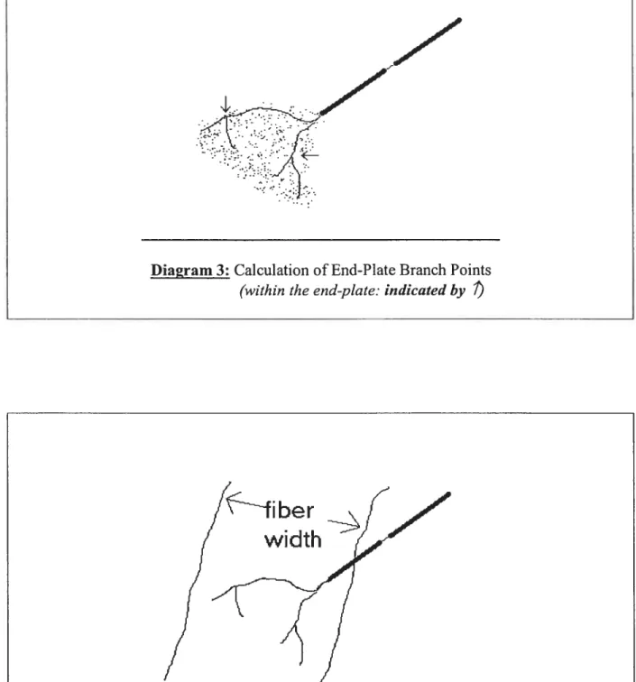
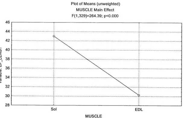
• The number ofnerve terminal brandi points witbin each end plate (Figure III); • Muscle width, as a measure of atrophy of the muscle in question (figure IV).
28
Dia%ram 1: Cholinesterase Staining of End-Plate Areas
longitudinal
29
Diagram3: Calculation ofEnd-Plate Branch Points
(within the end-plate: indicated by
width
iiber
30
Data were collected and recorded until the pool of data ofeach parameter (ie: EP area, EP length, brandi points and fiber width) attained a minimum of 145 entries. No more than 4 EPs wcre measured from any one siide. An EP was selected if a least a single
parameter was clearly measurable.
A Nikon Optiphot-2 light microscope, equipped with a JVC TK-5210 video camera (TK-A240 power unit supply) was connected to a video monitor (Javelin). The screen was linked to a PC-based $3386 microcomputer (Jaripel) equipped a digital image processing sofiward (Image Pro II version 2.0). The PhotoShop sofiware permitted the measurement of end-plate area, longitudinal length and nerve terminal branch points. Images were captured on hard drive and disk for later analysis at varying magnifications.
31
5. STATISTICAL ANALYSIS
Data was analyzed using one-way ANOVA, to determine the effects oftransection alone and transection with transplant. When a significant main effect was found, a post hoc test
(Scheffe’s test) was used to determine significance of the difference between specific means. A probability level below .05 was considered significant.
32
RESULTS
EP LONGITUDINAL LENGTH:
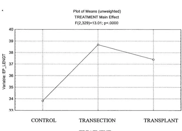
MUSCLE DIFFERENCE
Statistical analysis uncovered a significant main effect
(p<O.O5)
between the endplate lengths of the EDL and SOL (Figure 1). The longitudinal lengths of the soleus endplates were on average 43.3% larger than those ofEDL.
figure 1: Muscle Main Effect on End-Plate Length
46 44 42 H 40 z u — o 36 . 34 (‘3 > 32 30 28 Sot: Soteus
EDL: Extensor Digitorum Longus
Plot of Means (unweighted) MUSCLE Main Effect F(1,329)264.39; p<Q.000
Sol EDL
33
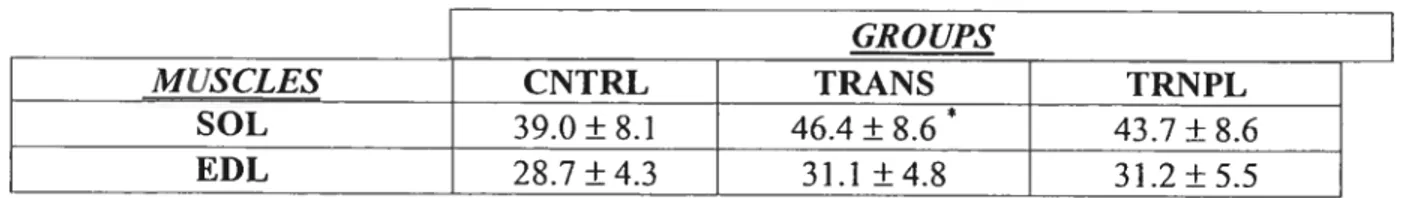
TREATMENTS
Statistical analysis uncovered a significant main effect
(p<O.O5)
between the different treatments (Figure 2). TRANS and TRNPL groups were significantly different (larger) than control. The TRANS and TRNPL groups were flot significantly different from each other.Figure 2: Treatment Main Effect011 End-Plate length
38 j— z o w 36 cJ > 34 40 39
Plot of Means (unweighted) TREATMENT Main Effect
F(2,329)=1 101; p<.0000
CONTROL TRANSECTION TRANSPLANT
34
Soteus:
Table I lists the EP lengths (±SD) for the data presented in the figures 3 and 4. The Scheffe test found significant difference between the SOL CNTRL and SOL TRANS groups (p<O.O5). There was no significant difference between the TRNPL and either the CNTRL or
TRANS groups (p>O.O5; p=O.O69, p=O.43, respectively), indicating that the transplant
procedure tended to attenuate the increase in sol endplate length that occurred with transection alone.
EDL:
35
TABLE 1. EP Iengths for SOL and EUL ([tm) ± S.D. of data from figures 3 & 4.
E L: w -J GROUPS MUSCLES CNTRL TRANS TRNPL SOL 39.0 ± 8.1 46.4 ± 8.6 * 43.7 ± 8.6 EDL 28.7±4.3 31.1 ±4.8 31.2±5.5
End-plate Longitudinal length (Soleus)
*Significantlv different from CNTRL Value (p <0.05)
figures 3 & 4 illustrate the distribution of data for SOL and EDL EP lengths for the three treatments according to their respective percentile.
Figure 3: SOL EP length distribution according to percentile for CNTRL, TRANS arid TRNPL. 70 60 50 40 30 20 -o- CONTROL -o- TRAN$ECTION 10 20 30 40 50 60 70 80 90 100 Percentile -o- TRANSPLANT
Figure 4: EDL EP length distribution according to percentile for CNTRL, TRANS and TRNPL 44 38 32 E :3 -c 26 G) 20 14
EDL END-PLATE LENGTH
CONTROL -n- TRANSECTION -o- TRANSPLANT Q Q Q Q Q CD co Percentile
(%)
37
EP AREA:
MUSCLE DIFFERENCE
Statistical analysis uncovered a significant main effect
(p<O.O5)
between the end plate areas of the EDL and SOL (Figure 5). The area of the SOL endplates was 26.2% greater than that for EDL endplates.Figure 5: Muscle Main Effect 0f End-Plate Area
w 0-I w cl) -D (u (u > Sol: Soleus
EDL: Extensor Digitorum Longus
Plot of Means (unweighted) VAR2 Main Effect F(1,321)=37.71; p<.0000 0uu 780
j
640 620 600 on . Sol MUSCLE EDLTREATMENT
Statistical analysis uncovered a significant main effect
(p<O.O5)
between the end plate areas with regards to the treatments (Figure 6).figure 6: Treatment Main Effect on end-plate area
7D 730 720 710 w <I -0 w U) U) U) > 660
Plot of Means (unweighted) VAR3 MainEffect Ff232 U=3.45; p<.033l
E___
640 WUfl CNTRL TRANS TRNPL TREATMENT CNTRL:CONTROL TRANS:TRANSECTION TRNPL:TRANSPLANT Soteus:Table 2 lists the EP areas (±SD) for the data collected from the SOL & EDL with regards to end-plate area. The Scheffe test found no significant difference between any of the SOL groups (p>0.05).
EDL:
-‘y
TABLE 2. EP areas (ni2)± S.D.
GROUPS
MUSCLES CNTRL TRANS TRNPL
SOL 716.9 ± 278.3 $24.7 ± 268.1 763.4 ±215.7
EUL 571.3 ±239.3 626.9± 176.7 628.0±208.1
Figure 7: SOL EP area distribution according to percentile for CNTRL, TRANS and
TRNPL. (N E D w cl) L w 1800 1400 1000 600 200 -200 Percentile
(%)
-o-CONTROL -u-TRANSECTION -O-TRANSPLANT EP AREA SOLEUS II II Q (N Q QCD Qco QQ40
c’J
E
w
o::
Figure 8: EDL EP area distribution according to percentile for CNTRL, TRANS and
TRNPL. EDL EP AREA 1600 1400 1200 1000 800 600 400 200 O II D D (D percentile
f
¾) -o- CONTROL -o- TRANSECTION -o- TRANSPLANT41
MUSCLE FIBER WIDTH:
Statistical analysis uncovered a significant main effect
(p<O.O5)
between the fiber widths ofthe EDL and SOL (Figure 9). Soleus fiber widths were 25.9% larger than those of EDL.Figure 9: Muscle Main Effect On Muscle Fiber Width
I H D Q) -Q Q) Q) > Sol: Soleus
EDL: Extensor Digitorum Longus
FIBER Means MUSCLE Main Effect F(1,290)=53.14; p<.0000 35 34 33 32 31 30 29 28 27 26
z
Sol EDL MUSCLE42
TREATMENT
Statistical analysis uncovered o significant main effect (p<0.O5) between the fiber widths with regards to the trcatment (Figure 10). The TRANS and TRNPL groups were not significantly different from each other.
Figure 10: Treatment Main Effect on Fiber Width
40 38 36 34 32 > L,
Table VI lists the liber widths (±SD) for the data presented in figures 6 & 7. The Scheffe test found siguificant differences between the SOL CNTRL group and both the SOL
TRANS and TRNPL groups (p<0.05; p=0.00, p=O.00). No significant difference was found
between the TRANS and TRNPL groups (p>005; p=O.153).
TREATMENT Means TREAT Main Effect F(2,290)=53.55; p<0000 CNTRL TRANS TRNPL TREATMENT Soteus: CNTRL:CONTROL 11ANs:TRANSECTION TRNPL:TRANSPLANT
43
EDL:
Significant differences were found between the EDL CNTRL group and both the EDL TRANS and TRNPL groups (p=O.O2,
p=O.02
respectively). No significant difference was found between the TRANS and TRNPL groups (p=O.99).TABLE 3. Fiber width (itm) ± S.D.
GROUPS
MUSCLES CNTRL TRANS TRNPL
SOL 43.4±8.4 26.8±11.4* 31.1 ±6.7*
EDL 31.0±6.1 25.2±6.6* 24.1 ± 5.0*
*Significantly different from CNTRL Value (p <0.05)
44
Figure 11: SOL FIBER WIDTH distribution according to percentile for CNTRL, TRANS and TRNPL. 80 70 60 50 E 40 -c -D • 30 20 10 O
SOL lIBER WIDTH
-o- CONTROL -n- TRANSECTION -o-TRANSPLANT 60% 80% 100% 20% 40% percentile (%)
figure 12: EDL FIBER WIDTH width distribution according to percentie for CNTRL, 35 30 D .z 25 20 15 10 5 -o-CONTROL -o- TRANSECTION -o- TRANSPLANT TRANS and TRNPL. 50 45 40
EDL FIBER WIDTH
D :
—HI
20% 40%
percentile¾
46
BRANCH POINTS
Statistical analysis did flot uncover a significant main effect between the branch points ofthe EDL and SOL (Figure 13).
Figure 13: Muscle Main Effect ofBranch Points
U) o o z ra o w a) n >
Plot 0f Means (unweighted) VAR2 Main Effect Ff1 ,297)=2.78; p<.0963 1.94 1.92 1.90 1._. 1.82 1 .2D lTD .00 SOL D MUSCLE EDL Sol: Soteus
47 (O H Q I o z o:: o w Q) Q) (u >
Table 4 lists the branch points (±SD) for branch point data collected. The Scheffe test found significant differences between the SOL CNTRL group and both the SOL TRANS and TRNPL groups (p=O.000I, p=O.006). No significant difference was found between the
TRANS and TRNPL groups (p=O.978).
TREATMENT
Statistical analysis uncovered a significant main effect (p<O.O5) among the treatment groups with regards to branch points (Figure 14).
Figure 14: Treatment Main Effect on end-plate branch points.
Plot of Means funweighted) VAR3 Main Effect Ff2,297)=15.75; p<.0000 22 2.1 2.0 1.9 1.8 1.7 1.6 1.5 1.4 Soteus: CNTRL TRANS TRNPL TREAIMENT CNTRL: CONTROL TRÀNS: TRAN$ECTION TRI’wL: TRANSPLANT
4
EDL:
The Scheffe test sliowed no significant effect between any ofthe EDL groups.
TABLE 4. Branch points ± S.D.
GROUPS
]
MUSCLES CNTRL TRANS TRNPL
SOL 1.48±0.64 2.19±0.77* 2.07±0.74*
EDL 1.59±0.75 1.94±0.67 1.80±0.67
*
49
SUMMARY PRINCIPLE RESULTS
1) TRANS SOL EP length was larger than in the TRNPL or CNTRL groups, suggesting that the transplant procedure tended to attenuate the increase in SOL EP length that occurs with transection.
2) SOL and EDL EP AREAS had no statistically significant differences between the CNTRL, TRJ’\NS or the TRNPL groups.
3) Both SOL and EDL TRANS and TRNPL groups had significantly larger widths than their respective CNTRL groups, suggesting that the transplant had no effect on attenuating the fiber width atrophy attributable to transection.
4) SOL TRANS and TRNPL groups had significantly more branch points than the CNTRL group, suggesting that the transplant had no effect on attenuating the SOL EP sprouting related to transection.
DISCUSSiON
The goal of this study was to determine what effect, if any, a fetal graft transplant would have on the morphology of fast and slow neuromuscular end-plates of hindlimb muscles (SOL and EDL) following spinal cord transection. The findings indicate that the transection induced some of the expected structural adaptation of motor end-plate expansion (Eldridge et al.,1981), such as an increase in sprouting and complexity of nerve temiinals (Tomas et al., 1989). As expected, the transection of the spinal cord induced muscle fiber with atrophy, end-plate expansion and neuronal sprouting in both the SOL and EDL. The transplant attenuated the increase in SOL end-plate length post-transection when compared to transection alone.
The muscle fiber width, EP area, and number ofbranch point parameters, for both the SOL and EDL, were not attenuated by the transplant post-transection. Therefore it was concluded that the embryonic cdl transplant had no significant change when compared to control subjects, such may be due to the duration of the transplant before muscles were excised. Perhaps a duration of 28 days is flot long enough for the transplant to elucidate any changes within the neuromuscular junction.
Dl
CNTRL EP length values
The CNTRL values for the SOL EP lengths were consistent with the literature. SOL EP length of 39.0 jim ± 8.1 is in the same range as Pestronk and Drachman’s (197$) EP length of 38 tm ± 2 and fahim’s (1989) EP length of 48.4 jim ± 2.0. The EDL EP length of 28.7jim ± 4.3 is comparable with the range of 25-55 tm repeated by Eldridge, Liebhold and Steinbach (1981).
CNTRL EP Area values
The CNTRL values for the rat SOL and EDL EP areas were consistent with the literature. Our SOL EP area of 716.87 ± 27$.33j.im2 is consistent with EP areas reported by Roscnheimer and Smith (1985) (941.20 ± 6.35 (SE)) and by Duchen (1970) (838 ± 297 jim2) (range: 200-1 600tm2).
The EDL EP areas in this work of 571.31 ± 239.26 11m2 is also comparable with Rosenheimer and Smith’s (1985) EP area of 601.24 ± 2.54 (SE), and Duchen (1970) 813 ± 267iim2 (range: 100-2200iim2).
CNTRL fiber widtli values
The CNTRL values for the SOL and EDL fiber widths were consistent with the literature. Our SOL fiber width of 43.36 ± $.3$jim is comparable with f ahim and Adonian’s (1990) 50 ± 2 (SE) and Fahim ami Robbins’s (1986) 44 ± 0.7 SE and Oda’s (1985) fiber diameter range of 30-70 um. Our EDL fiber width of 30.963 ± 6.072 jim is consistent with Fahim and Adonian’s (1990) 39 ± 2 (SE).
Houle et al. (1999) observed that the grafi attenuated the extent of muscle atrophy in the soleus post transection. Their soleus transected cross-sectional area (um2) was found to
-be 50% that ofthe CNTRL (888:1681 um2), while the TRNPL was only 77% that ofthe CNTRL (1155:1681 um2). The present study found the average SOL TRANS width to be 62% ofthe CNTRL while the TRNPL width was determined 72% ofcontrol. The Scheffe test did not reveal a statistical difference between the TRANS and TRNPL groups.
One reason why we did flot observe a significant attenuating effect from the TRNPL group with regards to fiber width could be that Houle (1999) compared another indicator of atrophy, myofiber cross-sectional area, and flot the width ofthe fiber. Another difference in the protocol was that Houle used a duration oftreatment of 90 days, whereas our subjects were sacrificed 4 weeks post-tranplant.
CNTRL EP brandi point values
Our SOL EP brandi points 1.48 ± 0.64 is comparable with Rosenheimer and Smith’s (1985) brandi points of 1.63 ± 0.01 (SE), and Pestronk and Drachman’s (197$) 2.6 ± 0.01
Our EDL EP branch points 1.59 ± 0.75 is consistent with Rosenheimer and Smith’s (1985) branch points of 1.25 ± 0.004 (SE) and Tomas et al. (1989) branch points of 2.99 ± 1.29.
53
WHY WERE THESE 2 MUSCLES INFLUENCED DIFFERENTLY?
The most marked atrophie responses following are typically seen in postural muscles that possess a relatively high percentage of type I fibers, and which experience the greatest change from their normal activity pattem.
The EDL and soleus are composed primarily of types II and I fibers respectively (Fahim et al., 1984), perform different actions (flexion and extension, respectively) and exhibit differences in firing pattems (Fischbach and Robbins, 1969; Navarrette and Vrbova, 1980).
Brown et al. (1980) suggest that the relatively fast muscles generally have less sprouting when denervated when compared to their slower fiber counterparts attributable to their higher resistance to the effects ofnerve degeneration. Slow muscles that are used for postural maintenance, tend to have a more continuous firing pattern than fast muscles and would suffer the greater change in firing pattem post-transection, and undergo greater morphological changes.
The current literature shows that afier transection, the soleus atrophies markedly, while the EDL seems substantially more resistant to the decrease in activity (Dupont Versteegden et al., 1998). This was also evident in the resuits of the present study, where the impact of transection was seen to be greater on SOL parameters when compared with respective control values.
It is hypothesized that transplants may assist the host by providing an environment that supports growth ofaxotomized and the rescue ofneurons destined to undergo retrograde celi death. Transplants are thought to rescue severed neurons by serving as a surrogate source of target-derived neurotrophic factors (Bregman and Reier, 1986). The greater vigor
54
in regenerating axons from embryonic rather than aduit neurons is one of the reasons why rnuch ofthe current research employs embryonic donors in an attempt to regain function. In this study, the transplant seems to have contributed in attenuating the increase in SOL longitudinal length resulting from transection. In other words, the transplant may have restored some elements of spinal circuitry, thus assisting in the reorganization ofthe host tissue.
Studies have shown that spinal lesion sparing small amounts of tissue permits considerable function (Blight and DeCrescito, 1986). Therefore, in this study, it is hypothesized that small amounts of spinal tissue may have been spared, resulting in an attenuated SOL end-plate growth post transection.
Though the transplant had little effect on end-plate morphology post-transection, several mechanisms have been suggested by which transplants placed in transected rats could permit repair of injured spinal cord. These mechanisms include axonal regeneration, the rescue of severed neurons destined to die, and trophic effects on remaining circuitry, thus improving the substrate through which compensatory mechanisms operate and thus permit development improvement motor control.
Presently, trophic molecules are being tested and applied together with antibodies against growth-inhibitory molecules, grafis of Schwann celis are being introduced into lesion sites, and embryonic cells are being implanted. It has been shown that some trophic agents such as NGF, FGF and BDNF are able to boost axon regeneration in vitro (Lindsay, 1988; Babr et al., 1989).
Future research should include introducing various components such as neurotrophins, thus providing an environment that could promote neuronal growth. Offier
aspects that miglit alter NMJ rnorphology would include passive exercise, electrostimulation and changing the underlying gene expression, which alters axonal growth with increased age.
These questions are raised not to be pessimistic or to dampen enthusiasm about prospects for spinal cord repair, but to raise relevant points for future research.
30
STUDY LIMITATIONS
Some ofthe study limitations would include:
1. Small sample sizes might have reduced the statistical power of some tests. Marginal changes could have become statistically significant if the number of rates per groups had been larger.
2. Animais used were female Sprague-Dawley rats.
3. Assumptions were made as to the duration oftransection and transplant periods since part of the protocol was done off-site;
4. Quantification of end-plate parameters relied on human precision, computer screen pixel resolution and microscope magnification standardizations.
CONCLUSIONS
Utilization of spinal tissue for transplantation is an extremely promising area for future research. The transplant tissue is thought to rescue severed neurons by serving as a surrogate source of target-denved neurotrophic factors and by providing “spare parts” thus assisting in the restoration of neuronal pathways.
In this study, the parameters measured included EP area, EP Iength, muscle fiber width and number ofbranch points, ail were seen to undergo the morphological changes expected post-transection. These included an increase in EP area, EP length and brandi point number within the BP, while a decrease in muscle fiber width signified muscle fiber atrophy.
The transplant had no effect on attenuating the transection response ofthe EP area, muscle width and brandi points ofthe EDL or SOL. The SOL longitudinal length response was the only parameter attenuated post-transection by the transplant. Though the relevance and reproducibility ofthese findings has yet to 5e determined, tus finding should propogate furtier studies with regards to the NMJ post-tranplant.