HAL Id: tel-01541523
https://tel.archives-ouvertes.fr/tel-01541523
Submitted on 19 Jun 2017HAL is a multi-disciplinary open access archive for the deposit and dissemination of sci-entific research documents, whether they are pub-lished or not. The documents may come from teaching and research institutions in France or abroad, or from public or private research centers.
L’archive ouverte pluridisciplinaire HAL, est destinée au dépôt et à la diffusion de documents scientifiques de niveau recherche, publiés ou non, émanant des établissements d’enseignement et de recherche français ou étrangers, des laboratoires publics ou privés.
treatment of congestive heart failure
Yohan Farouz
To cite this version:
Yohan Farouz. Designing biomaterials for controlled cardiac stem cell differentiation and enhanced cell therapy in the treatment of congestive heart failure. Biomechanics [physics.med-ph]. Université Sorbonne Paris Cité, 2015. English. �NNT : 2015USPCB114�. �tel-01541523�
Ecole Doctorale ED474 : Frontières du Vivant
Sorbonne Paris Cité – Université Paris Descartes – PARCC – HEGP – Fondation Carpentier INSERM U970 : Regenerative therapies for cardiac and vascular diseases
Paris Sciences et Lettres – Ecole Normale Supérieure de Paris – Institut Pierre-Gilles de Gennes CNRS UMR8640 : Microfluidics, chemical organization and nanotechnologies
Designing biomaterials for controlled
cardiac stem cell differentiation and
enhanced cell therapy in the treatment of
congestive heart failure
[Conception de biomatériaux pour le contrôle de la différenciation cardiaque à partir de cellules souches et pour l’amélioration de la thérapie cellulaire dans le traitement de
l’insuffisance cardiaque sévère]
Par Yohan Farouz
Thèse de doctorat,Spécialité : Biophysics, Biomaterials and Regenerative Medicine (Biophysique, Biomatériaux et Médecine régénératrice)
Dirigée par :
Philippe Menasché
Yong Chen
En vue d’une soutenance publique le 30 septembre 2015 devant un jury composé de :
Cesare Terracciano, MD, PhD
rapporteur
Abdul Barakat, PhD
rapporteur
Matthieu Piel, PhD
examinateur
Onnik Agbulut, PhD
examinateur
Yong Chen, PhD
co-directeur de thèse
Yohan Farouz’ PhD dissertation - 2015 ii © 2015 Yohan Farouz
Yohan Farouz’ PhD dissertation - 2015 iii
Abstract
Cell therapy is a promising strategy to help regenerate the damaged heart. Recent studies have placed a lot of hopes in embryonic stem cells and our lab had previously found a way to differentiate them into cardiac progenitors, cells that can only differentiate into cardiomyocyte, endothelial cells or smooth muscle cells. This early commitment decreases their proliferative capabilities, yet maintains their plasticity for better integration inside the host tissue. However, clinical and pre-clinical injection studies did not really meet the expectations. Even though slight improvements in cardiac function were demonstrated, very low cell viability has been observed, as well as a very low retention of the cells inside the myocardium. To address this problem, my PhD projects not only focus on the design of new biomaterials to act as a vehicle for cell delivery and retention in the infarcted area, but also on the design of biomaterials that control the cellular environment during the differentiation of pluripotent stem cells into cardiomyocytes. Going back and forth between the labs and the clinics, we first developed new techniques for the fabrication and the characterization of a cell-laden fibrin patch that is now undergoing phase I clinical trial. From the approved clinical formulation, we then propose new blends of clinical materials that will eventually improve the maturation of the cardiac progenitors once grafted onto the failing heart. In this perspective, we developed an in vitro model to investigate the combined influence of matrix elasticity and topographical confinement on stem cell differentiation into cardiomyocytes. By using microfabrication techniques to pattern pluripotent stem cells on substrates of controlled stiffness, we demonstrate that even using a widely recognized chemical-based protocol to modulate signaling cascades during differentiation, much heterogeneity emerges depending on the cellular physical environment. We thus extracted the main features that led to controlled and reproducible cardiac differentiation and applied it to the fabrication of next generation of multi-layered anisotropic cardiac patches in compliances with clinical requirements. This work opens new routes to high-scale production of cardiomyocytes and the fabrication of cell-laden or cell-free clinical patches.
Keywords: Cell therapy, regenerative medicine, biomaterials and tissue engineering, cardiac development, mechanotransduction, microfabrication, human pluripotent stem cells.
Résumé
La thérapie cellulaire se positionne comme une stratégie prometteuse pour inciter le cœur infarci à se régénérer. A cet effet, des études récentes placent des espoirs considérables dans l’utilisation des cellules souches embryonnaires et notre laboratoire a déjà démontré comment les différencier en progéniteurs cardiovasculaires, un type de précurseurs cellulaires qui ne peut aboutir qu’à la formation de cardiomyocytes, de cellules endothéliales ou de cellules de muscles lisses. Cet engagement précoce réduit leur capacité de prolifération anarchique et en même temps leur permet de rester suffisamment plastiques pour éventuellement s’intégrer plus facilement avec le tissue hôte. Cependant, les études précliniques et cliniques d’injection de ces cellules s’avérèrent décevantes. Malgré de légères améliorations de la fonction cardiaque, on observa une trop faible survie cellulaire ainsi qu’un taux de rétention des cellules dans le myocarde remarquablement bas. Afin d’étudier ce problème, mes travaux de thèse ont porté non seulement sur la conception de nouveaux biomatériaux pouvant servir de moyen de transport et d’intégration des cellules dans la zone infarcie, mais aussi sur la conception de biomatériaux permettant de contrôler précisément l’environnement cellulaire au cours du processus de différenciation de cellules souches pluripotentes humaines en cardiomyocytes. Grâce aux importantes interactions entre nos laboratoires de recherche fondamentale et de recherche clinique, nous avons tout d’abord développé de nouvelles techniques de fabrication et de caractérisation de patches de fibrine cellularisés qui sont récemment entrés dans un essai clinique de phase I. A partir de cette formulation clinique approuvée par les autorités de régulation, nous avons élaboré toute une gamme de matériaux composites uniquement à base de matières premières pertinentes dans ce cadre clinique, dans le but d’améliorer la maturation des progéniteurs cardiovasculaires une fois greffés sur le cœur défaillant. Dans cette optique, nous avons également développé un modèle in vitro permettant d’étudier précisément l’influence combinée de la rigidité du substrat et du confinement spatial sur la différenciation des cellules souches en cardiomyocytes. Grâce à des techniques de microfabrication sur substrat mou, il a été possible de positionner précisément les cellules souches pluripotentes dans des espaces restreints d’élasticité variable. Ainsi, nous avons pu observer que même en utilisant des protocoles chimiques éprouvés basés sur la modulation de cascades de signalisation impliquées dans le développement cardiaque, une très forte hétérogénéité pouvait apparaître en fonction de l’environnement physique des cellules. Nous avons ainsi pu extraire les caractéristiques principales permettant une différenciation cardiaque efficace, reproductible et standardisée et les avons appliquées à la fabrication d’une nouvelle génération de patches composés de matériaux cliniques et de couches multiples de bandes synchrones de cardiomyocytes. De fait, ces travaux ouvrent de nouvelles voies dans l’utilisation de biomatériaux pour la production industrielle de cardiomyocytes et pour la fabrication de patches cliniques, cellularisés ou non, dans le traitement de l’insuffisance cardiaque. Mots clés : Thérapie cellulaire, médecine régénératrice, biomatériaux et ingénierie tissulaire, développement cardiaque, mécanotransduction, microfabrication, cellules souches pluripotentes humaines.
Yohan Farouz’ PhD dissertation - 2015 v
Acknowledgements - Remerciements
The work presented in this dissertation is not the fruit of a single man, but rather the fortunate consequence of many interactions with people who raised me, guided me, assisted me and encouraged me relentlessly to make this journey instructive and worthwhile. I apologize in advance for any involuntary omission I am about to make.
Before anything, lets acknowledge our many sponsors for their generous funding and support: Ecole Polytechnique Paris Saclay for the 3-year PhD scholarship, Université Paris Descartes – Sorbonne Paris Cité for the 3-year teaching assistantship, Fondation pour la Recherche Médicale for funding the 4th and final year, Fondation Bettencourt-Schueller for travelling stipends, and all the other institutions
that help the various laboratories I have spent time in: Labex Revive, ShapeHeart Leducq Transatlantic Network, ANR, AFM, Fondation de France, the PARCC/HEGP, INSERM, CNRS, the FdV Doctoral School and the Ecole Normale Supérieure de Paris.
Then, I would like to thank my two supervisors, Philippe Menasché and Yong Chen, who could not have been more complementary in their mentoring advice and my training. Philippe Menasché, for his open-mindedness, his trust and positivity, for involving me in the pre-clinical experience and for introducing the clinical world to me. Yong Chen, also for his open-mindedness, trust and positivity, and for brainstorming the most challenging ideas with me.
I am also grateful to Cesare Terracciano and Abdul Barakat for accepting to be my thesis’ “rapporteurs”, for taking the time to review and comment the present work.
Of course, this thesis would not have been possible without Onnik Agbulut and Matthieu Piel, who opened up their lab to me and never refused a scientific discussion. Not to mention members of their laboratories, respectively Pierre Joanne and Solène Boitard, as well as Nicolas Carpi and Emmanuel Terriac. Their technical knowledge was invaluable and they made me see cardiac differentiation and micropatterning (respectively), in ways I would never have imagined otherwise.
I would like to thank Jérôme Larghero, Valérie Vanneaux and all the members of the Cell Therapy team at the Saint-Louis Hospital, who really trained me in experimental stem cell biology, especially Valérie Vanneaux, Hélène Riesterer, Lionel Faivre, Hélène Boucher, Isabelle Cacciapuoti, Alexandre Parouchev, and Manuel Théry for his advice of microfabrication.
To all the members of the INSERM U970 team at the PARCC/HEGP who trained me and assisted me in performing the experiments described in this dissertation: Damelys Calderon, Hadhami Hamdi, Valérie Bellamy, Alain Bel, Mathieu Pieronne, Wardiya Afshar, Elodie Mouloungui, Nisa Renault, Isabelle Hédon, Julie Piquet, Adrien Lalot, Véronique Oberweiss, Marie-Cécile Périer.
To all the members of Ecole Normale Supérieure de Paris who trained me and assisted me in performing the experiments described in this dissertation: Yadong Tang, Diana Molino, Sandrine
Yohan Farouz’ PhD dissertation - 2015 vi
Quignard, Jiangmei (Megan) Lian, Sisi Li, Wang Li, Jian (Olivier) Shi, Zhitao Han, Junjun Li, Naresh Kumar Mani, Anna Venancio Marques, Frédéric Bataille, Dominique Ho-Tin-Noé, Anne Hallopé, Nabil Garroum, Géraldine Hallais, Jacques Fattaccioli and Damien Baigl.
A special thanks to David Smadja at HEGP, Mathieu Pernot at Institut Langevin, as well as to Alba Marcellan at ESPCI for their help and guidance in fibrin-based assays and mechanical characterization.
To André Terzic, Marc Mercola, Karine Vauchez, Gabor Foldes and Michel Pucéat for their scientific and professional inputs.
To François Taddei from the CRI and Sophie Bernard from Université Paris Descartes, who gave me the opportunity to teach and to learn to teach. And, of course, to my students both from the FdV Bachelor Program 13’ and from the BME Master Program 14’ and 15’, who had to patiently experiment my unorthodox and ever changing teaching methods.
To the group of Ecole Polytechnique’s students I mentored during their PSC: Fanny Olivier, Hugo Soulat, Nora Faivre, Clémence Herzog, Gwendoline Tallec, Ricardo Furquim Pereira, Victor Ray. Thank you for your inputs and for your interest in crazy interdisciplinary projects.
I am deeply grateful to Kevin Kit Parker, Molly Stevens, Seung-Wuk Lee and their respective teams who cheerfully welcomed me before my PhD and initiated me to academic research and to the field of tissue engineering. A special mention to Ashutosh Agarwal for his friendship and mentorship and for always encouraging me to ask the right questions and think of the right experiments.
To Xavier Duportet, Camille Delebecque and Philippe Bouaziz, who unconsciously led me to start this PhD journey.
To Kévin Lhoste, for all the good times at the lab, at the CRI and outside.
To Kalthoum Ben M’Barek, friend and best co-PhD-student/colleague, for all the good times up at the mezzanine, down at the lab and outside, for all the support and cheering, and for all the open-minded discussions about Northern African cultures and so on.
To my family in law: Jean-François, Sylvie, Francine, Max, Nicolas and Caroline; and to my Parisian families for always helping me, supporting me and welcoming me so warmly: the Dekels; Gégé; the Masliahs, Soustiels and Sadocks; the Azoulays, Aboulkers, and Nahons.
To Coralie Villa and Juliette Dumoulin, who are always there for me regardless of the distance. Thanks for all the moments we lived since London and high school, respectively. Thanks in advance for all the future ones.
Yohan Farouz’ PhD dissertation - 2015 vii
Thanks to my closest family: my regretted grand-fathers Papi Marc and Papi Soly; Mamie Marcelle, Tonton Joseph, Mamie Mady, Maman, Papa, Nat’ and Jo’, for whom neither my English nor my native French vocabulary will be sufficient to faithfully express my feelings of love, admiration and gratitude. Thanks for your education, for teaching me the values of our roots, traditions and legacy. Thanks for your unconditional and blind support regardless of my decisions, like my choice to pursue an academic career. Thanks for always pushing me beyond my limits. Thanks for our funny relationship to high-tech and for the DIY blood running in parts of the family.
Merci Papa, for teaching me how to balance “la rage de vaincre” with the pleasures of life like gastronomy and the arts.
Merci Maman, for instilling in me both creativity and rigor and for your selfless attentions. Thanks to both for still loving me even if I don’t call you often enough.
And to my beloved Sophie, who has accompanied me since the first year of the PhD, who lived with immense strength the best and worst moments of this journey. Thanks for having done your best to put yourself in my shoes; for having made my life as easy as possible for the sake of this PhD; for dealing with my lack of organization and for dealing with the struggle of our incompatible calendars; for cooking delicious macaroons and fine dishes while I was away feeding stem cells during week-ends; for your organization skills; for keeping me from enslaving myself to my work; for always bringing me new perspectives on the world. And for all the things that would make this paragraph too personal in the context of this dissertation…
Yohan Farouz’ PhD dissertation - 2015 ix
Main abbreviations
AFM: Atomic Force Microscopy
ATMP: Advanced Therapy Medicinal Product BMP: Bone morphogenetic protein
BSA: Bovine Serum Albumine CPC: Cardiac progenitor cells
EB: Embryoid Body (mEB for murine EB, hEB for human EB) ECB: Engineered Cardiac Band
ECM: Extracellular matrix
EMA: European Medicines Agency
ESC: Embryonic stem cells (mESC for murine ESC, hESC for human ESC) ESCd-CM: Embryonic stem cell-derived cardiomyocytes
FBS: Fetal Bovine Serum
FDA: Food and Drug Administration FGF: Fibroblast growth factor GAG: Glycosaminoglycan
GMP: Good Manufacturing Practice HA: Hyaluronic acid
HMDS: Hexamethyldisilazane iPSC: Induced pluripotent stem cells µCP: Micro-contact printing
MSC: Mesenchymal stem cells NRCM: Neonatal rat cardiomyocyte PAA: Polyacrylamide
PBS: Phosphate Buffer Saline PCP: Planar cell polarity PDMS: Polydimethylsiloxane PEG: Poly-ethylene glycol PEG-DA: PEG-diacrylate PFA: Paraformaldehyde
PLL-g-PEG: Poly-L-lysine grafted PEG
PSCd-CM: Pluripotent stem cell-derived cardiomyocytes
RGD: Argininylglycyl aspartic acid. Peptide of sequence L-Arginine (R)- Glycyl (G) – Aspartic acid (D)
RT: Room Temperature SCD: Stem cell differentiation
Yohan Farouz’ PhD dissertation - 2015 x SCD-CM: Stem cell-derived cardiomyocyte
SEM: Scanning Electron Microscopy
SSEA-1: Stage-specific embryonic antigen-1 (also referred to as CD15) SWE: Shear Wave Elastography
TFM: Traction Force Microscopy TMCS: Trimethyl chlorosilane UV: Ultraviolet light
Wnt: (Wint) family of genes related to major developmental pathways. Wnt is a portmanteau word made of int and Wg, for “Wingless-related integration site”.
Y-27632: small molecule that inhibits the ROCK pathway. YAP: Yes activation protein
Yohan Farouz’ PhD dissertation - 2015 xi
Table of Contents
Abstract... iii Résumé ... iii Acknowledgements - Remerciements ... v Main abbreviations ... ixTable of Figures and Movies ... xv
List of Tables ... xviii
Chapter 1 – Introduction ... 1
Cardiovascular diseases ... 2
Rationale of the ESCORT Clinical Trial ... 3
Cell type ... 4
Scaffold type ... 5
Rationale ... 5
Presentation of the thesis project ... 8
References ... 9
Chapter 2 – On the design of biomimetic materials for cardiac stem cell differentiation ... 11
Abstract... 12
Introduction ... 13
Rationale for cardiomyocyte production ... 13
Mechanical forces during cardiac development, systems biology and the main cardiac differentiation protocols ... 13
Force transmission: Cell-Cell Interactions and Cell-ECM interactions ... 18
Interactions in the developing embryo ... 18
Strategies for mimicking cell-cell communication and cell-ECM coupling ... 19
Influence of substrate topography ... 20
General overview ... 20
Influence of topography on SCD into mesodermal progenitors and cardiomyocytes: the importance of colony size ... 22
Influence of topography on sarcomere maturation and force generation of cardiomyocytes: the importance of anisotropy ... 23
Anisotropy in early differentiation and isotropy in ESCd-CM studies ... 25
Influence of Tissue elasticity ... 25
General overview ... 25
Influence of elasticity on mesodermal SCD ... 27
Influence of elasticity on cardiomyocyte sarcomere maturation ... 28
Perspectives ... 30
Yohan Farouz’ PhD dissertation - 2015 xii
Influence of electrical signals on the maturation of cardiomyocytes ... 30
Pharmacological and medical applications ... 32
Conclusion ... 34
References ... 35
Chapter 3 – Safety, Regulatory and Ethical Issue of human studies ... 48
Introduction ... 49 Safety Issues ... 49 Arrhythmias ... 49 Tumor Development ... 50 Rejection ... 52 Stent Restenosis/Thrombosis ... 53 Calcification ... 53 Regulatory Issues ... 53 Preclinical Studies ... 53 Cell Manufacturing ... 54 Release Criteria ... 56
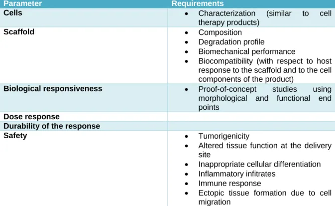
Cell and Scaffold Combined Products ... 58
Ethical Issues ... 59
Conclusion ... 61
References ... 62
Chapter 4 – From the clinics: Design of a Cardiac Patch for the ESCORT clinical trial ... 65
Introduction ... 66
The ESCORT clinical trial ... 66
Design of a cardiac patch for clinical applications ... 69
Material and methods... 73
Determination of fibrinogen and thrombin concentrations ... 73
Preparation of 3D Culture in fibrin gels ... 73
Immunohistology ... 74
Scanning Electron Microscopy (SEM) ... 74
Image analysis ... 75
Materials characterization ... 75
Results ... 76
Concentration of Evicel’s fibrinogen solutions ... 76
Structural characterization of fibrin patches ... 77
Mechanical characterization ... 80
Handling tests ... 82
Yohan Farouz’ PhD dissertation - 2015 xiii
Manipulation during surgery ... 84
Effect of the patch on rats and sheep ... 85
Conclusion and Discussion ... 86
Perspectives ... 87
References ... 89
Chapter 5 – From the clinics to the labs: development of new microfabricated platforms for the standardization of cardiomyocyte differentiation ... 92
Introduction ... 93
Materials and methods ... 94
Stamp fabrication ... 94
Sacrificial coverslip microcontact printing ... 94
Polyacrylamide (PAA) gel fabrication ... 95
Cell Culture ... 95 Immunofluorescence ... 96 RT-qPCR ... 96 Statistical analysis ... 97 Movies ... 97 Results ... 98
Characterization of the substrates ... 98
Morphology of the colonies and cell density ... 100
Beating behavior of the colonies... 102
Transcriptional analysis ... 106
Conclusion on the transcriptional analysis ... 112
Discussion ... 113 Limitations ... 113 Perspectives ... 115 Conclusion ... 117 References ... 118 Supplementary Information ... 121
Chapter 6 – Back to the clinics: new design strategies for the fabrication of Engineering Cardiac Bands ... 131
Introduction ... 132
Materials & Methods ... 134
Cell patterning on clinical fibrin hydrogels ... 134
Preparation of composite 3D scaffolds ... 136
Results I: Soft micropatterning with the ESCORT protocol ... 138
The choice of the substrate: BJ vs. FPBT ... 138
Yohan Farouz’ PhD dissertation - 2015 xiv
Initial Glass patterning success ... 142
Results II: Proof-of-concept patterning strategies using clinical fibrin hydrogels ... 145
First proof of concept for the transfer of patterned cells from PAA to clinical fibrin gels ... 145
Advanced proof of concept using an optimized geometry for single-material differentiation of hiPSCs into cardiomyocyte and subsequent transfer to clinical fibrin ... 148
Alternatives to cell transfer: direct ECM patterning on clinically-relevant hydrogels ... 151
Results III: new blends of clinical materials ... 152
Handling test ... 154
Histological profile ... 155
Mechanical properties ... 156
In vitro experiment ... 157
Conclusion on the new blends ... 159
Conclusion and Perspectives ... 160
References ... 162
Supplementary Information ... 165
Chapter 7 – Conclusions and Perspectives ... 166
Summary ... 167
Perspectives ... 170
Stem cell differentiation, biomaterials and cell-free cell therapy ... 170
References ... 172
Abstract... 174
Yohan Farouz’ PhD dissertation - 2015 xv
Table of Figures and Movies
Figure 1.1 Ischemic heart disease is the major risk factor of heart failure in high income countries
worldwide. ... 2
Figure 1.2 The ideal cardiac patch. ... 3
Figure 1.3 Short summary of the differentiation protocol from hESCs to cardiovascular progenitor cells ... 5
Figure 1.4The importance of scaffold parameters at every step of the fabrication process. ... 6
Figure 2.1 General strategy for differentiating PSCs into cardiomyocytes. ... 14
Figure 2.2 Important players involved in conventional cardiac differentiation protocols, and their interactions with physical factors (not published)... 17
Figure 2.3 Overview of popular techniques for controlling and characterizing materials and cells at the microscale. ... 21
Figure 4.1 Description of the cell production process in GMP conditions ... 69
Figure 4.2 Fibrin polymerization process ... 70
Figure 4.3 Mechanical characterization of fibrin hydrogels ... 75
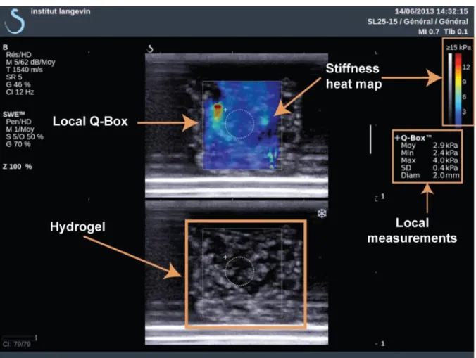
Figure 4.4 Elasticity measurement with Shear Wave Elasticity on Aixplorer® Ultrasound System ... 76
Figure 4.5 Lot-to-lot variability in fibrinogen concentrations ... 77
Figure 4.6 Electron Micrographs of fibrin patches. ... 77
Figure 4.7 Fiber diameter as a function of fibrin composition ... 78
Figure 4.8 Confocal imaging of fluorescently labelled fibrin hydrogels ... 78
Figure 4.9 Photomicrographs of histological sections of fibrin patches, stained with Hematoxylin/Eosin stain. ... 79
Figure 4.10 Variations in pore diameter (μm) as a function of patch concentrations (mg/mL-U/mL). . 79
Figure 4.11 Compressive testing results for fibrin hydrogels of increasing fibrinogen:thrombin concentrations. ... 80
Figure 4.12 SWE measurements of fibrin elasticity. ... 81
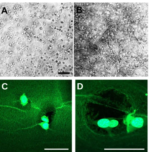
Figure 4.13 Representative images of the behavior of fibroblasts seeded within fibrin patches. ... 83
Figure 4.14 Shape and handling of the fibroblast-laden fibrin gels ... 84
Figure 4.15 Grafting of the patch on a sheep’s heart. ... 85
Figure 4.16Grafting the patch on a rat’s heart ... 85
Figure 5.1 Micropatterning ECM proteins on soft polyacrylamide hydrogels. ... 95
Figure 5.2 Culture conditions and small molecule treatments for purely chemical-based cardiomyocyte differentiation. ... 96
Figure 5.3 Example of beating patterns observed through movie analysis. ... 98
Figure 5.4 Characterization of the substrates. ... 99
Figure 5.5 Morphological analysis of the differentiated clusters. ... 100
Figure 5.6 General morphology of cell colonies at the end of differentiation. ... 101
Figure 5.7 Percentage of beating spots as a function of time and spot diameter (A-i), time and elasticity (A-ii), and spot diameter and elasticity on days 12 (B-i) and 15 (B-ii)... 103
Yohan Farouz’ PhD dissertation - 2015 xvi
Figure 5.8 Beating frequency as a function of time and spot diameter (A-i), time and elasticity (A-ii),
and spot diameter and elasticity at day 12 (B-i) and day 15 (B-ii). ... 104
Figure 5.9 Percentage of spots with circular contraction wave behavior, as a function of time (A), spot diameter (B) and elasticity (C)... 105
Figure 5.10 Percentage of irregularly beating spots as a function of time, spot diameter and elasticity. ... 106
Figure 5.11 Gene expression in change fold versus undifferentiated hiPSC before seeding on micropatterns. ... 107
Figure 5.12 Clustergram of the relative expression profiles in different conditions of elasticity (brown), diameter (pink), and day (blue). ... 110
Figure 5.14 Classification of genetic expression variations depending on spot diameter and elasticity ... 112
Figure 5.15 Design of new platforms for higher-throughput assays. ... 116
Figure 6.1 Representative alginate structure: (A) chain conformation and (B) block distribution. ... 133
Figure 6.2 Representative structure of hyaluronic acid. ... 133
Figure 6.3Comparative BMP-2 induction on BJ feeders or FPBT feeders. ... 139
Figure 6.4Gene expression and purity analysis after specification on FPBT or BJ ... 140
Figure 6.5Immunostaining attempts on patterns with inappropriate pattern cell density. ... 141
Figure 6.6Effect of Y-27632 addition (A) and removal (B) from Nutristem® medium on hESCs seeded on spot patterns. ... 142
Figure 6.7 Immunostaining after BMP2 induction of hESCs on rigid spots. ... 143
Figure 6.8qPCR analysis ... 144
Figure 6.9Example of patterning of BJs + hESCs on 10 kPa PAA substrates patterned with FN spots. ... 144
Figure 6.10PAA to Fibrin patterned cell transfer. ... 145
Figure 6.11Surface of PAA before and after transfer on fibrin patch ... 146
Figure 6.12Surface of fibrin patches after transfer from PAA. ... 147
Figure 6.13Fluorescent fibrinogen observation on 1 kPa PAA patterned with lines and seeded with BJ fibroblasts. (A) Epifluorescence. (B) LSCM of a cluster, (B-i) section, (B-ii) 3D reconstruction. ... 148
Figure 6.14 Beating patterns of engineered cardiac bands. ... 149
Figure 6.15 Proof-of-concept of a two-level fibrin patch made of parallel band of hiPSC-derived cardiomyocytes. ... 150
Figure 1.16 Evaluation of the stiffness of aggregated cardiac bands, ... 151
Figure 6.17 Confocal microscopy of the proof-of-concept fibrin micropatterning with lines of fibronectin + fibrinogen, after transfer from a 10-kPa polyacrylamide layer. ... 152
Figure 6.18 Cardiac muscular thin films constructed from micropatterned alginate substrates. ... 153
Figure 6.19 Histological sections of fibrin patches, stained with Hematoxylin/Eosin. ... 155
Figure 6.20 Histological sections of fibrin/alginate patches, stained with Hematoxylin/Eosin. ... 155
Figure 6.21 Histological sections of fibrin/hyaluronic acid patches, stained with Hematoxylin/Eosin. ... 156
Yohan Farouz’ PhD dissertation - 2015 xvii
Figure 6.22 Compressive test and SWE results for fibrin/alginate hydrogels. ... 156
Figure 6.23 Compressive test and SWE results for fibrin/HA hydrogels. ... 157
Figure 6.24 Microscopic image of F20T4 seeded with hMSCs at Day 7. Scale bar: 200 μm ... 158
Figure 6.25 Microscopic images of fibrin/Alginate seeded with hMSCs at Day 7. ... 158
Figure 6.26 Microscopic images of fibrin/HA seeded with hMSCs at Day 7. ... 158
Figure 6.27 Sheep surgery using a fibrin/HA patch loaded with CD15+ hESC-derived progenitors .. 159
Supplementary Figure 5.1 qPCR’s heat maps of clustered gene expression visualization grouped by diameters (x-axis) and elasticity (y-axis) [106 kPa represents glass coverslips] ... 122
Supplementary Figure 5.2 Web diagram of gene expression for each condition of diameter and elasticity, day 12 ... 123
Supplementary Figure 5.3 Web diagram of gene expression for each condition of diameter and elasticity, day 15 ... 124
Supplementary Figure 5.4 p-value matrices corresponding to Figure 5.6 . Green represents significance. ... 126
Supplementary Figure 5.5 p-value matrices corresponding to Figure 5.7. Green represents significance. ... 127
Supplementary Figure 5.6 p-value matrices corresponding to Figure 5.8. Green represents significance. ... 128
Supplementary Figure 5.7 p-value matrices corresponding to Figure 5.9. Green represents significance. ... 129
Supplementary Figure 5.8 p-value matrices corresponding to Figure 5.10. Green represents significance. ... 130
Supplementary Movie 5.1 Representative movie of the analysis of a cystic cluster beating irregularly ... 121
Supplementary Movie 5.2 Representative movies of beating clusters in each condition ... 121
Supplementary Movie 5.3 Representative movies of circular beating ... 121
Supplementary Movie 6.1 Engineered Cardiac Bands, spontaneously beating on PAA substrates of variable stiffness ... 165
Supplementary Movie 6.2 Ultrasound imaging of a beating fibrin patch with 2 layers of cardiac bands ... 165
Supplementary Movie 6.3 Fibrin patch with 2 layers of cardiac bands, right after fibrin polymerization ... 165
Yohan Farouz’ PhD dissertation - 2015 xviii
List of Tables
Table 3.1 Summary of major steps to be taken into account before submission to the Regulatory
Authorities ... 54
Table 3.2 Main regulatory requirements for autologous and allogeneic cells. ... 56
Table 3.3 Major properties of cells that need to be characterized prior to a clinical use... 57
Table 3.4 Main regulatory requirements for biomaterials alone or associated with cells (combined product) ... 59
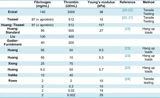
Table 4.1 Literature survey of the use of fibrin for cardiovascular applications. ... 71
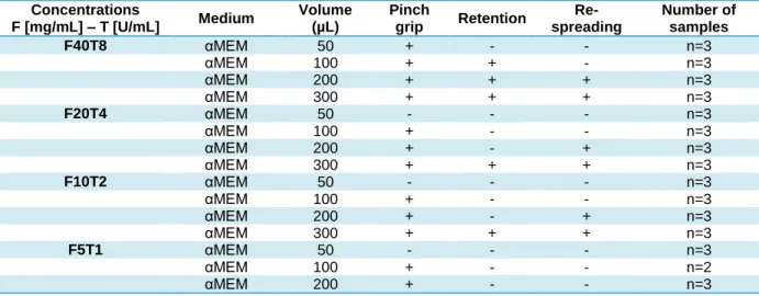
Table 4.2 Handling of patches as a function of fibrinogen and thrombin concentrations. ... 82
Table 5.1 Number of coverslips available for PCR extraction ... 97
Table 5.2 Total number of cluster images for movie analysis ... 97
Table 5.3 Classification of genetic markers depending on developmental stage or major tissue of predilection ... 109
Table 6.1Specification yields after culture on BJs or FPBTs feeders. ... 139
Table 6.2 Specification yields after culture on BJ or FPBT feeders. ... 140
Table 6.3 Handling of patches as a function of fibrinogen, thrombin and alginate concentrations. ... 154
Table 6.4 Handling of patches as a function of fibrinogen, thrombin and hyaluronic acid concentrations. ... 154
Yohan Farouz’ PhD dissertation - 2015 1
Chapter 1 – Introduction
Important Note: The rationale for the PhD studies presented in this chapter is partly described in
French in the following article [1]:
Farouz Y, Chen Y, Menasché P, Ino J, Atlan M, Le Visage C, Letourneur D: Réparer les cœurs brisés et les vaisseaux abîmés. Biofutur 2012:1–4.
Yohan Farouz’ PhD dissertation - 2015 2
Cardiovascular diseases
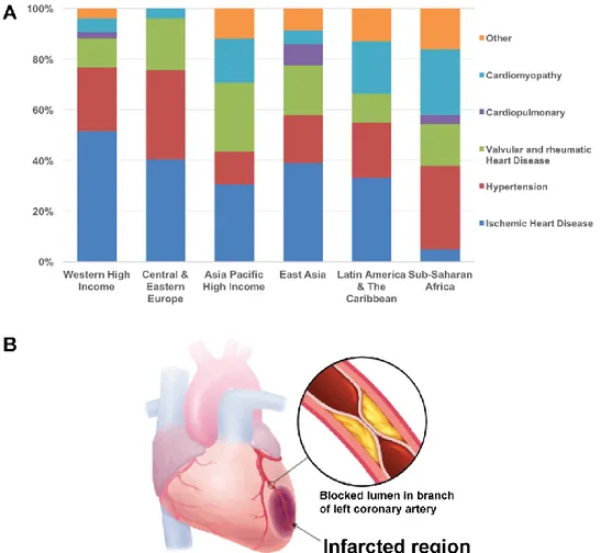
Cardiovascular diseases (CVD) are the main cause of mortality worldwide with more than 17.3 million death per year [2]. Taking the example of the United States (US), there were about 800,000 deaths due to CVD in the year of 2011, compared to less than 600,000 for all cancers together [3]. Cardiovascular diseases are various and include myocardial infarction, dilated cardiomyopathy, valvular heart diseases, hypertension (Figure 1.1). Not only cardiovascular diseases are of prime importance because of their implied mortality, but also because of the cost they represent for the Healthcare system as well as because of the shortage in manpower it generates. In 2011, this cost was estimated to about $320 billion in the US [3] and to about $863 billion worldwide [2].
Figure 1.1Ischemic heart disease is the major risk factor of heart failure in high income countries worldwide. (A) Age- and sex-adjusted relative contributions of different underlying risk factors of heart failure in six world regions (reproduced from [4]). (B) Schematic of the coronary occlusion leading to myocardial ischemia (adapted from NursingCrib.com).
One of the recurring consequences of such diseases is cardiac failure. Cardiac failure is defined as the inability of the heart to pump enough blood to the rest of the body, resulting in tiredness, coughing, water retention in lower limbs and dyspnea (shortness of breath). Once diagnosed, the 5-year survival rate from cardiac failure is only 50% [3]. There are many causes of cardiac failure, and their incidence strongly varies between countries. However, it appears
Yohan Farouz’ PhD dissertation - 2015 3 that cardiac failures usually results from ischemic heart diseases [4] (Figure 1.1-A), a problem caused by the obstruction of the coronary artery, leading to the death of the downstream cardiomyocytes that are no longer provided with nutrients and oxygen (Figure 1.1-B). Once these cells are dead, this part of the tissue cannot regenerate nor can it contract. Therefore, there is a tremendous loss in efficiency during heart contraction. In addition, scar tissue comprised mainly of collagen is formed in the infarcted area, resulting in increased stiffness and therefore, the healthy surrounding cells need to generate considerably more power to supply the body with blood. This increased activity leads to accelerated tiredness of the healthy cardiomyocyte, which eventually acquire an arrhythmogenic phenotype or even start to die.
Rationale of the ESCORT Clinical Trial
Many approaches have been considered in order to improve the function of the infarcted heart. Assisting devices can be implanted to stimulate cardiac pumping, but the best solution is still heart transplant, which is problematic due to the shortage of available hearts. Nowadays, cell therapy is considered as one of the most promising strategies to help regenerate the damaged heart. Indeed, replacing the dead cells with new cells would be a sustainable solution that corresponds best with physiological needs. Figure 1.2 shows a naïve representation of what could an ideal patch could look like in the close future, as imagine at the beginning of my PhD studies.
Figure 1.2 The ideal cardiac patch.
Global view and sections of a normal heart (A) and an infarcted heart (B) with the strategy for positioning the patch. The “ideal” patch (C) contains cardiac and endothelial progenitor cells. It delivers angiogenic factors and mechanically supports the dilating heart to ensure improved contractility, while waiting for grafted cells to have secreted their own extracellular matrix in the hope that they would replace the scar tissue made of stiff collagen and residual fibroblasts. Figure adapted from Th. Winslow’s artwork and Y. Farouz’ schematic published in [1].
Yohan Farouz’ PhD dissertation - 2015 4
Cell type
Many cell types have been considered as good candidates for the repopulation of the damaged heart. One would think that the best cells would be the exact same type as the ones that have died. This would include mainly cardiomyocytes, but also blood vessel cell types like endothelial cells or vascular smooth muscle cells. However, cardiomyocytes are very difficult to obtain because they can hardly be harvested from the patient’s own body (and there would be a risk to get some damaged cells) and because they are already mature (meaning that they already established a well organized network of sarcomeres and formed gap junctions with their neighbors). Hence, it would be almost impossible to expand them and there would be a very low probability that they would be able to integrate in between the healthy cardiomyocytes of the patient’s heart [5].
Another option is the use of myoblasts that can proliferate and then differentiate to form myotubes. These cells are interesting because, like cardiomyocytes, they become striated muscle cells. However, they lack of the ability to contract spontaneously and need an electrical stimulus in order to contract. This is not a problem a priori, but again their integration inside the already existing tissue is source of a lot of interrogations.
Therefore, recent studies have placed a lot of hopes in stem cells. Cells like mesenchymal stem cells or adipocyte-derived stem cells are not expected to replace the dead cells, thing that they can hardly do (although some laboratories claim they can) but instead would support the revascularization of the infarcted region by generating paracrine factors like VEGF, SDF-1 or PDGF. These growth factors will help the recruitment of new endothelial cells, smooth muscle cells and reduce apoptosis in the infarcted area [6, 7].
If adult stem cells can only act by paracrine stimulations, embryonic stem cells have a lot more potential, as they can actually differentiate into the three cardiac lineages: cardiomyocytes, endothelial cells and smooth muscle cells. The only concern when using embryonic stem cells would be their anarchical proliferation and their large panel of available commitments. To avoid these problems, we think the best solution is to differentiate these embryonic stem cells into cardiovascular progenitors (Figure 1.3), a type of cell that can only differentiate into one of the three cardiac cell types [8]. This early commitment decreases their proliferative capabilities, yet maintains their plasticity. It is this characteristic that we are looking for when considering the integration of the cells inside the host’s tissue.
Yohan Farouz’ PhD dissertation - 2015 5 Figure 1.3Short summary of the differentiation protocol from hESCs to cardiovascular progenitor cells
as patented and published by Blin et al. [9]
Scaffold type
Clinical trials and other pre-clinical studies did not really meet the expectations mentioned above. Indeed, even though slight improvements in cardiac function were observed in terms of wall thickness, ejection fraction during the cardiac cycle, or in term of revascularization of the infarcted area, very low cell viability has been observed, as well as a very low retention of the cells inside the myocardium.
To address this problem, several teams including ours are designing new biomaterials to act as a vehicle for cell delivery and retention in the infarcted area [10, 11]. The ideal scaffold will be the one that would allow the cells to attach and maybe proliferate inside the patch, allowing them to generate a new extracellular matrix around them while the biomaterial is degrading or resorbing.
Rationale
Different cell types require different environments in order to develop appropriately and form a functional tissue. We want to start from human embryonic stem cells (Figure 1.4, step 1), to drive them to differentiate into cardiac progenitors (Figure 1.4, step 2), then we want to incorporate them in a scaffold (Figure 1.4, step 3) that would be grafted onto the myocardium (Figure 1.4, step 4) in order for them to integrate with the patient’s tissue (Figure 1.4, step 5), forming new vessels as well as new contractile units. In each step of the project, different mechanical properties of materials are important, such as the elasticity for the stem cell differentiation, or the rate of degradability once the patch is inside the body. Also, mechanical stability has to be taken into consideration for surgical reasons, as a good patch will not only
Yohan Farouz’ PhD dissertation - 2015 6 be a patch that provides a physiological environment, but it will be a patch that would be easily manipulated inside the operating theater.
Furthermore, as cells change their phenotype about twice (from embryonic stem cells to cardiac progenitors to cardiac cells, see Figure 1.3), different environments need to be provided for each of these phenotypes. Defining more clearly these two environments is the global aim of my PhD projects.
Figure 1.4The importance of scaffold parameters at every step of the fabrication process. [Main image adapted from Julian George’s PhD thesis]
Although differentiation has been deeply investigated by the use of developmental factors to induce cell maturation, it has been recently demonstrated that other properties like matrix elasticity, composition and structuration are of prime importance in the differentiation process. Culturing mesenchymal stem cells on surfaces with low stiffness (<1 kPa) led to their differentiation into neuron like cells, while increasing the stiffness led to their differentiation into skin cells (1 kPa), muscle cells (10 kPa) or bone cells (~100 kPa) [12]. Other independent studies showed that culturing embryonic cells on surfaces that were patterned with lines led to the formation of mature cardiomyocytes (see next chapter). Indeed, these cardiomyocytes formed anisotropic sarcomeres and formed gap junctions in order to maintain electrical coupling along the tissue [13-15]. Some studies also report variations in phenotypes
Yohan Farouz’ PhD dissertation - 2015 7 depending on the type of extracellular matrix protein the cells were seeded on. For instance, mimicking peptides like RGD (present in fibronectin or collagen, for instance) or YISGR (present in laminin) do not allow cells to form mature sarcomeric structures [16, 17].
As shown in Figure 1.4, the next step toward the fabrication of a cardiac patch is the sorting of the cells. Sorting is a step that usually takes advantage of the differential affinity of cells for different adhesion proteins or substrates. The current technique is magnet-assisted cell sorting where antibodies are attached to magnetic beads. These antibodies are selective to the cell type we want to select or exclude. The current issues lie in the fact that we currently need to do positive sorting, meaning that the cells linked to the beads are the cells of interest. These beads, if not removed, can interfere with the good behavior of cells once inside the patch and then in the body.
The next step is the incorporation of the cells inside a patch. Again, many approaches have been considered. Patches can be made in two dimensions, where cells are seeded on top of the film. This method is very attractive because cells have been extensively studied in two-dimension and we know very well how to control their organization [18-20]. Also, it has the advantage of putting the cells directly in contact first with the culture medium and second with the host tissue. However, although we do not exclude such a strategy in the future, there are currently some clinical hurdles that would need to be overcome. First, having cells on a surface makes the patch handling much more complicated: the surgeon has to be very careful not to scratch the cells away, making sure that the right side is put in contact with the tissue. Second, the quantity of cells that we can seed on a surface is much lower than in a 3D gels, even though stacking 2D layers of cells is still an option to keep in mind.
In order to compensate for the cell mortality during the surgery, it is interesting to incorporate the cells in a three-dimensional matrix. It mimics better the natural cell environment, ease the manipulation of such a patch and increase the number of cells that can be delivered. However, it can be difficult to control the anisotropy and the porosity of the matrix and ensure its mechanical stability at the same time.
We plan on varying the composition of the fibrin precursors to alter the porosity of the fibrin gel, as well as to ensure good cell viability and manipulability. An important point will be the thickness of the construct, which will have to allow nutrients and oxygen to diffuse inside the patch.
Yohan Farouz’ PhD dissertation - 2015 8
Presentation of the thesis project
As described here, the physical characteristics of the biomaterial, like its elasticity, porosity, degradability, the biochemical characteristics of the biomaterial, like its growth factor composition, its adhesion proteins and their distribution, have a dramatic influence on the cell behavior, and this influence depends strongly on the cell type. The two main steps in this big project starting from hES culture and ending in the operating theater will therefore be investigated by taking into accounts these many parameters, but with very distinct points of view. First, we describe what is known regarding differentiation of stem cell into cardiomyocytes and how physical constraints play a role in it (Chapter 2). Second, we draw a picture of the practical challenges that arise when trying to translate biomaterials based stem cell therapies to the clinics (Chapter 3). From these observations, we came to the conclusion that the most urgent would be to make sure that hESC are a viable choice and thus, we focused on the fabrication of a patch made out of clinical components only. The cardiac patch had to be optimized with regards to clinical constraints in order to quickly receive an approval from the regulatory agencies to start clinical trials and get a first feedback from human patients (Chapter 4). Next, we take a step back and investigate how we could improve cardiac differentiation and finely control their final phenotype. Pluripotent stem cells will be studied with a rigorous set of conditions to determine the best microenvironment for their differentiation into cardiac progenitors (Chapter 5). Before taking another step back to look at the big picture of what we have achieved and what remains to do (Chapter 7), we propose strategies to design new ways to bridge the gap between cell culture and patch fabrication by taking advantage of the preferential adhesion of the cells onto ECM proteins, and we propose new blends of clinical materials that would allow modulating their mechanical properties while keeping their potential for fast clinical translation (Chapter 6).
Yohan Farouz’ PhD dissertation - 2015 9
References
1. Farouz Y, Chen Y, Menasché P, Ino J, Atlan M, Le Visage C, Letourneur D: [Mending broken hearts
and repairing damaged vessels] (French title: Réparer les cœurs brisés et les vaisseaux abîmés). Biofutur 2012:1–4.
2. Alwan A, Armstrong T, Bettcher D, Branca F: Global Status Report on Noncommunicable
Diseases 2010: Description of the Global Burden of NCDs, Their Risk Factors and Determinants.
World Health Organization; 2011.
3. Mozaffarian D, Benjamin EJ, Go AS, Arnett DK, Blaha MJ, Cushman M, de Ferranti S, Després J-P, Fullerton HJ, Howard VJ, Huffman MD, Judd SE, Kissela BM, Lackland DT, Lichtman JH, Lisabeth LD, Liu S, Mackey RH, Matchar DB, McGuire DK, Mohler ER, Moy CS, Muntner P, Mussolino ME, Nasir K, Neumar RW, Nichol G, Palaniappan L, Pandey DK, Reeves MJ, et al.: Heart disease and stroke
statistics--2015 update: a report from the American Heart Association. Circulation 2015, 131:e29–
322.
4. Khatibzadeh S, Farzadfar F, Oliver J, Ezzati M, Moran A: Worldwide risk factors for heart failure:
A systematic review and pooled analysis. International Journal of Cardiology 2013, 168:1186–1194.
5. Ghafar-Zadeh E, Waldeisen JR, Lee LP: Engineered approaches to the stem cell
microenvironment for cardiac tissue regeneration. Lab Chip 2011, 11:3031–3048.
6. HAO X, Silva EA, MANSSONBROBERG A, GRINNEMO K, SIDDIQUI A, DELLGREN G, WARDELL E, BRODIN L, Mooney DJ, SYLVEN C: Angiogenic effects of sequential release of VEGF-A165 and
PDGF-BB with alginate hydrogels after myocardial infarction. Cardiovascular Research 2007, 75:178–185.
7. Yu J, Du KT, Fang Q, Gu Y, Mihardja SS: The use of human mesenchymal stem cells
encapsulated in RGD modified alginate microspheres in the repair of myocardial infarction in the rat. Biomaterials 2010.
8. Blin G, Nury D, Stefanovic S, Neri T, Guillevic O, Brinon B, Bellamy V, Rücker-Martin C, Barbry P, Bel A, Bruneval P, Cowan C, Pouly J, Mitalipov S, Gouadon E, Binder P, Hagège A, Desnos M, Renaud J-F, Menasché P, Pucéat M: A purified population of multipotent cardiovascular progenitors
derived from primate pluripotent stem cells engrafts in postmyocardial infarcted nonhuman primates. J Clin Invest 2010, 120:1125–1139.
9. Blin G, Nury D, Stefanovic S, Neri T, Guillevic O, Brinon B, Bellamy V, Rücker-Martin C, Barbry P, Bel A, Bruneval P, Cowan C, Pouly J, Mitalipov S, Gouadon E, Binder P, Hagège A, Desnos M, Renaud J-F, Menasché P, Pucéat M: A purified population of multipotent cardiovascular progenitors
derived from primate pluripotent stem cells engrafts in postmyocardial infarcted nonhuman primates. J Clin Invest 2010, 120:1125–1139.
10. Hamdi H, Planat-Benard V, Bel A, Puymirat E, Geha R, Pidial L, Nematalla H, Bellamy V, Bouaziz P, Peyrard S, Casteilla L, Bruneval P, Hagège AA, Agbulut O, Menasché P: Epicardial adipose stem
cell sheets results in greater post-infarction survival than intramyocardial injections.
Cardiovascular Research 2011, 91:483–491.
11. Le Visage C, Gournay O, Benguirat N, Hamidi S, Chaussumier L, Mougenot N, Flanders JA, Isnard R, Michel J-B, Hatem S: Mesenchymal stem cell delivery into rat infarcted myocardium using a
porous polysaccharide-based scaffold: a quantitative comparison with endocardial injection.
Tissue Engineering : Part A 2011, 18:35–44.
12. Engler AJ, Sen S, Sweeney HL, Discher DE: Matrix elasticity directs stem cell lineage
specification. Cell 2006, 126:677–689.
13. Parker KK, Tan J, Chen CS, Tung L: Myofibrillar architecture in engineered cardiac myocytes. Circulation Research 2008, 103:340–342.
Yohan Farouz’ PhD dissertation - 2015 10
14. Alford PW, Feinberg AW, Sheehy SP, Parker KK: Biohybrid thin films for measuring contractility
in engineered cardiovascular muscle. Biomaterials 2010, 31:3613–3621.
15. Kim D-H, Lipke EA, Kim P, Cheong R, Thompson S, Delannoy M, Suh K-Y, Tung L, Levchenko A:
Nanoscale cues regulate the structure and function of macroscopic cardiac tissue constructs.
PNAS 2010, 107:565–570.
16. Shachar M, Tsur-Gang O, Dvir T, Leor J, Cohen S: The effect of immobilized RGD peptide in
alginate scaffolds on cardiac tissue engineering. Acta Biomaterialia 2011, 7:152–162.
17. Boateng SY: RGD and YIGSR synthetic peptides facilitate identical cellular adhesion as
laminin and fibronectin but alter the physiology of neonatal cardiac myocytes. AJP: Cell
Physiology 2004.
18. Takahashi H, Nakayama M, Shimizu T, Yamato M, Okano T: Anisotropic cell sheets for
constructing three-dimensional tissue with well-organized cell orientation. Biomaterials 2011, 32:8830–8838.
19. Williams C, Tsuda Y, Isenberg BC, Yamato M, Shimizu T, Okano T, Wong JY: Aligned Cell Sheets
Grown on Thermo-Responsive Substrates with Microcontact Printed Protein Patterns. Adv Mater
2009, 21:2161–2164.
20. Masuda S, Shimizu T, Yamato M, Okano T: Cell sheet engineering for heart tissue repair. Advanced Drug Delivery Reviews 2007, 60:1–9.
Yohan Farouz’ PhD dissertation - 2015 11
Chapter 2 – On the design of biomimetic materials for
cardiac stem cell differentiation
Important Note: Most of the content of this chapter has been published in Stem Cells in 2015 [1]: Farouz Y, Chen Y, Terzic A, Menasché P: Concise review: growing hearts in the right place: on the design of biomimetic materials for cardiac stem cell differentiation. Stem Cells 2015,
Yohan Farouz’ PhD dissertation - 2015 12
Abstract
Tissue engineering aims at recapitulating permissive conditions that enable cells to collaborate and form functional tissues. Applications range from human tissue modeling for diagnostic purposes to therapeutic solutions in regenerative medicine and surgery. Across this spectrum, human stem cells are the active ingredient, expandable virtually indefinitely and with the propensity to generate new tissue. Engaging lineage-specific differentiation requires a precise concerto of key spatial and temporal factors, such as soluble molecules and growth factors, but also physical and mechanical stimuli. These stimuli compete to modulate distinct developmental signaling pathways and ultimately affect the differentiation efficiency. The heart is a chemo-mechano-electrical biological system that behaves as both a sensor and an actuator. It can transduce electrical inputs to generate mechanical contraction and electrical wave propagation. Such a complex organ arises from multipart developmental events that interact with one another to self-regulate. Here, we overview the main events of heart development and the role of mechanical forces in modifying the microenvironment of the progenitor cells. We analyze the cascades regulating cardiac gene activation to illustrate how mechanotransduction is already involved in the most popular protocols for stem cell differentiation (SCD) into cardiomyocytes. We then review how forces are transmitted to embryonic stem cells (ESC) by cell-substrate or cell-cell communications, and how biomaterials can be designed to mimic these interactions and help reproduce key features of the developmental milieu. Putting this back in a clinical perspective, many challenges needs to be overcome before biomaterials-based SCD protocols can be scaled up and marketed.
Yohan Farouz’ PhD dissertation - 2015 13
Introduction
Rationale for cardiomyocyte production
Beyond chemical signaling, mediated by soluble growth factors, providing the foundation for SCD protocols [2], there is growing evidence that environmental cues are also of prime importance in guiding differentiation events [3]. Indeed, it appears that the whole stem cell niche is important in determining cell fate. In addition to chemical factors (including transcription factors and other proteins), oxygenation [4], extracellular matrix (ECM) proteins [5-8], innervation, support cells [9, 10] and mechanical loading [11] are some of the key parameters that have been identified. In the heart, cardiac cells form anisotropic layers able to contract in response to electrical signals. Therefore, mechanical properties are thought to contribute to differentiation and further maturation during embryogenesis. Here we focus on the impact of the mechanical and topographical properties of materials used for cell culture on the differentiation of stem cells into cardiomyocytes. Adequate differentiation of stem cells into cardiomyocytes has significant medical applications offering the aptitude to recreate cardiac-like tissue for patient-specific in vitro drug toxicity assays [12, 13] as well as designing cell-based therapies for treatment of cardiac diseases [14-16]. Indeed, hopes were reinforced after historical observations on the benefits of human embryonic stem cell-derived cardiomyocytes (hESC-CM) on infarcted rat hearts[17] have been recently extended to non-human primate hearts [18, 19].
Mechanical forces during cardiac development, systems biology
and the main cardiac differentiation protocols
With the formation of the four-chambered heart, the cellular arrangement of cells highly evolves from a cardiac crescent to a cardiac tube, followed by two looping events, the formation of the four chambers and finally septation (Figure 2.1-A). During these steps, differential growth occurs as well as increased blood flow and the initiation of electrical signals. Hence, cells are stretched, sheared, thereby resulting in different cell phenotypes at different stages of development [20, 21].
However, in vitro SCD is often realized only by activating signaling cascades mimicking the way they are activated during embryogenesis. This is achieved by using transcription factors [2], small molecules [2], or miRNAs [22, 23], often identified from high-throughput screenings [24-27].
The most used protocols involve modulation of developmental signaling pathway such as the canonical Wnt pathway [26, 28-32], the Wnt/PCP (non-canonical) pathway [33-36], TGF-β and
Yohan Farouz’ PhD dissertation - 2015 14 BMP pathways [37-40], and all their combinations [41]. These protocols are increasingly efficient and simpler than the original ones [42], and while the first versions relied on reagents that were either difficult to translate to the clinics or simply too expensive, a lot of effort is now made in order to create the simplest cocktails possible [43].
Figure 2.1General strategy for differentiating PSCs into cardiomyocytes.
A. Observation of the heart’s developmental stages. After gastrulation and somite formation (i), the foregut arise and folding brings the two branches of splanchnic mesoderm together (ii-iv) before eventually merging (v) and forming the heart tube (vi, vi’). Cardiomyocytes are already beating and further differentiating but some keep proliferating (vii), cause the tube to start its C-loop (vii’). The S-loop (viii’) causes the cardiomyocytes to stretch even more (viii) and when the four chambers are finally formed (ix’), they almost completed their elongation and differentiation (ix)
B. Analysis of the relationships between external mechanical forces and gene expression. Although the main principles are
similar, ESCs (top) and cardiomyocytes (bottom) have different mechanosensing architectures. ESCs connect together using E-cadherins and form round shaped colonies (cortical actomyosin network), while cardiomyocytes connect through N-cadherins, establish gap junctions for electrical signal transmission and express an important set of proteins for crystallization of actomyosin into highly anisotropic sarcomeric structures. Connections to the extracellular matrix are regulated via chemical (angiotensin II) or mechanical stimuli. Along with differentiation, tissues secrete different kind of ECM protein. For instance, the heart as a higher fibronectin:laminin ratio than the blastocyst.
C. Basic biochemical mimicry can be achieved by culturing ESCs on petri dishes coated with ECM mixtures or mimics.
Using sequential addition of cocktails of growth factors and/or small molecules, developmental signaling pathways are triggered and ESCs are directed toward cardiomyocyte differentiation.
D. Advanced biophysical mimicry emerges through microfabrication techniques. 2D or 3D patterning of circular shapes
allow for mimicking of the blastocyst structure (top) while patterning of lanes allow for mimicking aligned cardiac tissues (bottom).
Yohan Farouz’ PhD dissertation - 2015 16 While the two main pathways used in in vitro cardiac differentiation are the Wnt/β-catenin pathway and the BMP pathway, it is becoming clearer that the common denominators are mechanosensing and calcium signaling (Figure 2.2). The Wnt/PCP pathway is responsible for convergence/extension of the gastrulating embryo [44-46] while the BMP pathway and smad genes activation is promoted by membrane mechanosensors [47] as well as calcium signaling [48]. Calcium is also responsible for stabilization of cell-cell interactions through N-cadherin [49]. As N-cadherins are ultimately coupled to β-catenin [50-52] and α-catenin, overpresence of N-cadherin contacts at the membrane can trigger overactivation of N-cadherin as well as inhibition of β-catenin translocation in the nucleus, which is equivalent to Wnt/β-catenin inhibition or GSK3Β overactivation [35, 53]. Another example of the importance of forces and calcium signaling is the establishment of the Left/Right axis, which precedes cardiac looping. Combining two models, it is thought that a right-to-left shear flow induces the generation of a gradient of growth factors, but also induces the bending of the primary cilia. Bending of the primary cilia will lead to the increase in calcium on the left side, cooperating with Nodal and BMP signaling to activate Pitx2 and trigger cardiac looping [21, 54, 55]. Finally, calcium is also one of the main actors of cardiac contraction, reestablishing the link between mechanical forces and calcium signaling (Figure 2.1-B).
Of importance, the Hippo pathway has also been found to be involved in cardiogenesis through the YAP/TAZ molecules [56], already known to act as mechanical transducers in tissue-growth servo-regulation pathways [57, 58]. Similarly, myocardial differentiation was observed by GSK3β activation (i.e. Wnt/β-Catenin inhibition) in a signaling cascade involving the insulin-like growth factor pathways, under control of the YAP/TAZ pathway [59]. This is no longer surprising under the light of a study by Azzolin et al., where Wnt inhibition is reinforced by the presence of YAP/TAZ in the beta-catenin destruction complex, while Wnt activation triggers both YAP/TAZ and beta-catenin release and nuclear translocation, a phenomenon responsible for the inhibition of mesendodermal differentiation[60]. More details on the relationship between Hippo, Wnt and stem cell differentiation can be found in recent reviews by Hao et al. and Varelas et al. [61, 62]. As further discussed later in this review, both the topography and the elasticity of the substrate influenced the fate of adult cardiac progenitor cells (CPC) through a YAP/TAZ-dependent mechanism [63]. Signaling studies on the relationship between the Hippo pathway, mechanotransduction and cardiogenesis have not been translated to efficient SCD protocols yet, but increased evidence of their intricate relationships can be found in other models, like for MSC differentiation [64] or neuronal differentiation from iPSCs [65].
Yohan Farouz’ PhD dissertation - 2015 17 In this review, we highlight the mechanotransduction effects in a hierarchical fashion, first by defining briefly how cells can sense forces, and then, zooming out on the physical forces generated by the cell-matrix interactions, the cell-cell interactions, aggregate mechanics, tissue mechanics and finally the heart development itself. At each scale, we will summarize current knowledge regarding the biophysics of development and maturation of cardiomyocytes, and which techniques are available to recapitulate these environmental properties in a modular bottom-up fashion.
Figure 2.2Important players involved in conventional cardiac differentiation protocols, and their interactions with physical factors (not published).
Debate 1: Murine vs. Human cells
Differentiation studies using murine models are very important in the field of developmental biology. Like other model species (the fruit fly, the xenopus or the zebra fish), they allowed identifying the main regulatory pathways that control embryonic development, with regard to both biological and mechanical behaviors. However, major differences exist between murine and human pluripotent cells. While murine ESCs (mESCs) can be cultivated on gelatin-coated petri dishes with the only addition of LIF (Leukemia Inhibitory Factor), human ESCs (hESCs) need a far more complex ECM coating (fibroblast feeder layer, Matrigel or vitronectin for non cellular materials). Another distinction is the timing of development and of expression of