THESE
THESE
En vue de l'obtention du
DOCTORAT DE L’UNIVERSITÉ DE TOULOUSE
DOCTORAT DE L’UNIVERSITÉ DE TOULOUSE
Délivré par l'Université Toulouse III - Paul Sabatier Discipline ou spécialité : Biochimie et Biologie moléculaire
JURY
Pr. Bernard SALLES - Professeur (UPS - Toulouse) - Président du Jury . Pr. Cristina CARDOSO - Professeur (Technische Universität - Darmstadt) - Rapporteur
Dr. Mario COSTA - Researcher (Institue of Neuroscience - Pisa) - Rapporteur Dr. Frédéric COIN -Directeur de Reherche (IGBMC - Strasbourg) - Examinateur
Dr. Wim Vermeulen - Directeur de Recherche - (Erasmus MC - Rotterdam) - Examinateur Dr. Giuseppina GIGLIA-MARI - Chargé de Recherche (IPBS - Toulouse) - Directrice de thèse Dr. Etienne JOLY - Chargé de Recherche (IPBS - Toulouse) - Directeur de thèse
Dr. Bernard SALLES - Professeur (UPS - Toulouse) - Président du Jury
Dr. Cristina CARDOSO - Professeur (Technische Universität - Darmstadt) - Rapporteur Dr. Mario COSTA - Researcher (Institue of Neuroscience - Pisa) - Rapporteur
Dr. Frédéric COIN -Directeur de Recherche (IGBMC - Strasbourg) - Examinateur
Dr. Wim Vermeulen - Directeur de Recherche - (Erasmus MC - Rotterdam) - Examinateur Dr. Giuseppina GIGLIA-MARI - Chargé de Recherche (IPBS - Toulouse) - Directrice de thèse Dr. Etienne JOLY - Chargé de Recherche (IPBS - Toulouse) Directeur de thèse
Ecole doctorale : Biologie-Santé-Biotechnologies Unité de recherche : IPBS CNRS - UMR5089
Directeur(s) de Thèse : Dr. Giuseppina GIGLIA-MARI et Dr. Etienne JOLY Rapporteurs : Pr. Cristina CARDOSO et Dr. Mario COSTA
Présentée et soutenue par Lara KADDOUM Le 17 Décembre 2010
Titre : La protéine MeCP2: Etude de son implication dans la réponse aux dommages à
Résumé
Le syndrome de Rett est une maladie neurodéveloppementale progressive conduisant à un polyhandicap lourd associé à des retards mentaux sévères. Cette pathologie a pour principale origine des mutations du gène codant pour la protéine Methyl-CpG Binding Pro-tein 2 (MeCP2), localisé sur le chromosome X. Elle touche essentiellement les filles avec une fréquence d’environ 1/10 000 naissances. Différentes fonctions ont été attribuées à MeCP2 : modulateur transcriptionnel (active ou réprime la transcription), épissage alternatif de cer-tains ARN, maintien de l’état de méthylation des gènes au cours de la réplication de l’ADN et modification de la structure tridimensionnelle et/ou du niveau de compaction de la chro-matine.
Initialement, mes travaux de thèse ont consisté à explorer l’hypothèse que MeCP2 aurait la capacité de passer d’une cellule à l’autre. Les résultats obtenus suggèrent fortement que le transfert intercellulaire de MeCP2 ne se produit pas in vivo mais serait dû à une diffusion inter-cellulaire de la protéine suite à l’étape de fixation cellulaire à l’acétone nécessaire à la suite de l’expérimentation. Cependant, ces travaux nous ont permis de mettre au point une nouvelle méthode pour la détection des protéines dans les cellules de mammifères basée sur le système de split GFP.
Dans le cadre de mon projet de thèse, j’ai également produit et caractérisé des anticorps dirigés spécifiquement contre chacune des 2 isoformes de MeCP2. Ces anticorps origin-aux vont permettre d’étudier les niveorigin-aux d’expression et le rôle de chaque isoforme dans divers types cellulaires de l’organisme. Cela va pouvoir améliorer notre compréhension de la pathologie du syndrome de Rett.
Plus récemment, mes travaux se sont focalisés sur la relation entre MeCP2 et les mé-canismes de réparation de l’ADN, et nous ont permis de mettre en évidence la capacité de MeCP2 de s’accumuler sur l’ADN endommagé. Cette accumulation est indépendante de la transcription et des voies GG-NER et TC-NER. Mais, elle dépend de la région C-terminal de la protéine MeCP2. Les futurs projets de l’équipe viseront à élucider les mécanismes impliqués dans cette nouvelle fonction de MeCP2.
Abstract
Rett syndrome is a severe and progressive X-linked neurodevelopmental disorder that affects 1/10000 female birth. RTT is caused by mutations in the mecp2 gene, encoding the Methyl CpG binding Protein 2. MeCP2 binds to methylated DNA and has several roles in: transcription activation or repression, chromatin remodeling, alternative splicing of mRNA, etc.
Initially, my thesis project was to explore the hypothesis that MeCP2 may be able to transfer between cells. My results suggest that this phenomenon appears after cell fixation with acetone and doesn’t occur in vivo. This work, however, allowed us to develop a new staining method to detect and localize proteins in mammalian cells using the split GFP sys-tem.
Within the frame of this project, I have also produced antibodies specific for each of the two MeCP2 isoforms. These novel antibodies should prove to be interesting tools to understand the role of each isoform in the pathology of Rett syndrome.
More recently, my work was focalized on the relationship between MeCP2 and DNA damage. I was able to show that MeCP2 accumulates on DNA damage. This accumulation is independent from transcription, GG-NER and TNER pathways but depend on the C-terminal region of MeCP2. Future work will be aimed at understanding the mechanisms involved in this newly uncovered function of MeCP2, and will hopefully improve our un-derstanding of Rett syndrome pathogenesis.
Many Thanks
. . .
M
A thèse a été réalisée au sein de l’Institut de Pharmacologie et de Biologie Structurale dirigée par Dr Jean-Philippe Girard. Je tiens à vous remercier pour votre accueil au sein du laboratoire.I would like to thank Pr. Bernard Salles who accepted to be the president of my thesis jury.
I want also to thank Dr. Mario Costa, Dr. Frédéric Coin and Dr. Wim Vermeulen who accepted to evaluate my thesis work. I hope that you appreciated your journey in Toulouse besides all the weather and the airport problems. Dr. Cristina Cardoso, thank you for your careful evaluation of my thesis report. I regret that I haven’t got the occasion to meet you in person but it was a pleasure to have a skype discussion with you during my thesis defence. Ma thèse a été financée par l’Association Française du syndrome de Rett (AFSR) et la fondation pour la recherche médicale (FRM). Je vous exprime ma profonde gratitude pour les soutiens que vous m’avez accordés.
Je voudrais remercier mes deux directeurs de thèse pour leur soutien au quotidien du-rant ces quatre années de thèse.
Merci à Dr. Etienne JOLY pour m’avoir accueilli dans l’équipe dès mon DEA. Merci de m’avoir fait confiance et de m’avoir proposé de continuer en thèse sur le syndrome de Rett. Merci aussi de m’avoir laissé la liberté dans mon travail et de m’avoir poussé à aller toujours vers l’avant. Je souhaite aussi remercier Dr. Ambra Giglia-Mari pour son soutien et ses encouragements constants. Merci de m’avoir accepté dans ton équipe en milieu du chemin et d’avoir accepté de me guider sur ce nouveau projet. Merci pour ta patience et d’avoir pris le temps de m’expliquer les voies du DNA repair.
Merci à vous deux pour les discussions scientifiques si enrichissantes qu’on a pu avoir. Je remercie chaleureusement toutes les personnes que j’ai côtoyé pendant ces quatre an-nées et qui ont fait que les jouran-nées de travail ont été si agréables: Denis (Merci pour tes
conseils, ta disponibilité et ta gentillesse et pour toutes les discussions qu’on a pu partagées. En travaillant à côté de toi, j’ai appris comment on peut concilier enseignements, recherche et les moments en famille. Les mots d’ordre sont " Organisation et Efficacité!!! " Merci de m’avoir encourager à faire des enseignements et d’avoir été là jusqu’à la fin de ma thèse), Julie (Tu as été ma maman " MeCP2 ". Merci pour ton sourire, ta douceur et ta patience et pour tout ce que j’ai pu apprendre à tes côtés), Anne (Merci pour ton dynamisme et ta joie de vivre. Je retiendrais de toi ta philosophie sur la vie surtout concernant les enfants), Sandrine (Merci pour ton sourire et tes blagues parfois un peu à côté), Eddy (Merci pour ta gentillesse, ton calme, ta disponibilité et pour tout ce que tu m’as appris à l’animalerie. Merci pour ton amitié), Christine (Merci pour ton humour, ton sourire et ton dynamisme. Tu es la joie de vivre au labo au quotidien). Pierre-Olivier (Merci pour ta disponibilité et tout le temps que tu as passé à m’apprendre à utiliser le multiphoton), Magali (Ma voisine de bureau, Merci pour les discussions qu’on a pu avoir surtout sur les souris, tu m’a ap-pris énormément), Sophie (Merci pour ta douceur, ta getillesse et ton sourire. J’espère que MeCP2 te sera de grande utilité dans tes mises au point de ChIP), Camille (ou Madame Godon pour les proches !!! Merci pour ta bonne humeur, ton humour décalé et ton efficacité à organiser un pot de dernières minutes. Je garderai un bon souvenir de tes expressions si étranges et de tes cours d’initiation au verlan), Julie et Joris (Merci d’avoir accepté mon anglais " si parfait ". Grâce à vous je m’améliore de jour en jour). Merci pour votre présence à tous, votre gentillesse et votre joie de vivre.
Merci à toutes les personnes de passage ou celles avec qui j’ai pu discuté dans un couloir ou autour d’un café, d’un repas ou une soirée. Ceux qui m’ont prêté un produit ou partagé un protocole ou des astuces: Florence, Roxane, Ludivine, Marielle, Aline, Luc, Cécile, Béa-trice, Marlène. Je tiens à remercier mes voisins d’étage avec qui j’ai partagé de bons mo-ments au cours de ces années : à celles qui sont déjà partie Audrey, Elisabeth, Claire, Dania, Claire, ceux qui sont encore là Isabelle (Merci pour tes encouragements constants), Leyre (Merci pour ta disponibilité pour nous tous, merci pour les nombreuses fois où tu as été ma motivation pour le travail tard le soir ou le WE, cela m’a évité le PTI), Pierre (Merci pour ta gentillesse et ta bonne humeur et tes encouragements au moment de la rédaction en plein été. Je te souhaite les meilleurs Western au monde !!!) et les derniers arrivés Thomas et Guillaume. Merci à tous pour vos aides et surtout le jour de la soutenance.
Many Thanks. . .
ont été les premiers à m’accueillir en master 1ère année. Merci de m’avoir donné l’envie de continuer dans la voie de la recherche. D’autre part je remercie l’équipe du Dr. Isabelle Maridonneau-Parini et surtout Arnaud pour avoir contribué à ma formation en DEA. Je garde un très bon souvenir de mon passage chez vous, merci pour tout ce que vous m’avez appris.
J’ai eu l’occasion de collaborer avec l’équipe du Dr Laurent Villard. Je tiens à les re-mercier pour leur accueil à Marseille et je remercie Dr. Jean-Christophe Roux et Nicolas Panayotis pour leur aide à réaliser les expériences d’immunohistochimie sur les tissus de souris.
Je remercie l’équipe pédagogique de biochimie de l’université Paul Sabatier à Toulouse. Merci pour m’avoir fait confiance et pour tous vos conseils qui m’ont été utiles pour mener à bien les enseignements. C’était une très belle expérience.
Grand merci à " mes copains du DEA " et " mes copains du Vendredi midi ". Aurel, Mitch et Caro, merci d’avoir été là dans les moments difficiles mais aussi pour tous les moments de joie qu’on a pu partager (surtout les kirs, pêches Melba. . .. et les chips !!! du Hoegarden les Vendredi soirs) . Aurel tu as été la plus courageuse et tu m’as supporté jusqu’au bout, merci pour toutes les discussions qu’on a pu avoir au biphoton pour faire passer le temps entre 2 acquisitions et les pauses cafés histoire de râler un peu. Mitch ou Mister Biblio, je ne sais toujours pas comment tu fais pour retenir le 5ème auteur d’une publi qui date de 1980 mais je te remercie pour toutes les discussions scientifiques qu’on a pu avoir autour de la pause café de 7h30 le matin. Caro, merci pour ta gentillesse et ton courage. Tu m’a appris que quand on veut quelque chose, il faut foncer et ne pas se laisser décourager. Gaelle, merci pour ta gentillesse et pour toutes les fois où tu m’as aidé à préparer mes oraux ou mes dossiers, pour les pauses de 16h agrémentées de quelques discussions protocoles, manips ou vacances. Le dernier " copain du Vendredi midi " Thomas merci pour tous les goûters et les pauses " expérimentations scientifiques " étranges mais intéressantes qu’on a pu partager. . .. Une thèse, ça fait aussi gagner des amis!!!
Toutes ces années, en France, n’auraient jamais été si agréables sans la présence des amis. Fred, Joel, Marion, Georgette, Joseph, Liliane, Cindy et Diana, merci pour tout ce que vous avez fait pour nous. Laurence, Claudia, Elodie, et bien sûr les loustics (Nath, Stéph fille et Stéph garçon, JB et Damien). Vous êtes des gens formidables. Merci pour les magnifiques soirées et les moments de délire qu’on a passé ensemble. Merci pour votre présence et
votre soutien pendant la rédaction de cette thèse. Le meilleur souvenir pour moi restera quand même le cochon et la chasse aux œufs sous la pluie. Avouez c’était la classe !!!! Et je n’oublierai pas bien sûr " Que font les rats sans Lara ?? " c’était génial !!!
Je ne serais pas arrivée là sans l’aide et l’encouragement de ma famille. à mes parents, merci de m’avoir fait confiance et d’avoir dit " oui " à mes projets : venir en France seule et me lancer dans une thèse. J’espère que vous ne m’en voudrez pas trop d’avoir encouragé Elsy et Achaya de venir continuer leurs études à mes côtés. Sœurette, qu’est ce que j’aurais fait sans toi. Pour moi tu as été ma sœur, mon amie, ma confidente, ma colocataire, mon défouloir. . . Merci pour ton sourire au quotidien, pour ton grand cœur et pour tous les moments qu’on a partagé et qu’on partagera ensemble. Merci pour toutes les aides informatiques que tu m’as apportées : sans toi je serai encore en train d’installer ma free et attendre des nuits et des nuits ce " chenillard " qui ne veut pas s’arrêter, surtout sans toi mon manuscrit n’aura jamais été aussi " Classe " !!!! Je te souhaite plein de courage pour que tu arrives toi aussi au bout de ta thèse et que tu réalises tes rêves. Frero, ces deux dernières années ont été superbes. Je suis contente que Toulouse t’ait plu. Je suis aussi très contente d’avoir pu partager ces deux années avec toi. Merci pour tout ce qu’on a fait ensemble. Rugby, Stade, pizza, bière, london town, fléchette. . .. Qui dit mieux !!!! Je te souhaite beaucoup de réussite pour tes projets futurs. Pense à moi quand tu seras riche, je suis toujours d’accord pour faire le tour du monde et " visiter la pierre ".
Enfin je remercie tous ceux que j’ai oubliés de citer et qui m’ont apporté leur soutien et leurs conseils à un moment donné de ma thèse.
≪Somewhere, something incredible is waiting to be known. ≫ Dr. Carl Sagan
Résumé général des travaux de
thèse et perspectives
L
E syndrome de Rett est une maladie neuro-développementale progressive conduisant à un polyhandicap lourd associé à des retards mentaux sévères. Cette maladie touche principalement les filles avec une fréquence d’environ 1/10 000 naissances et représente la première cause de polyhandicap dans les sociétés occidentales. Après une première année asymptomatique, ce syndrome est caractérisé, entre autres, par l’apparition progressive de troubles moteurs, de mouvements stéréotypés, de dysfonctionnements massifs du système nerveux autonome, avec épilepsie, autisme et retard mental.En 1999, l’équipe du Dr Huda Y. Zoghbi a découvert que le syndrome de Rett (RTT) est dû à des mutations dominantes du gène mecp2, localisé sur le chromosome X. La pro-téine MeCP2 appartient à la famille des MBP (Methyl-CpG-Binding Protein), qui sont des répresseurs de transcription se liant à l’ADN au niveau des dinucléotides CpG méthylés. Il existe 2 isoformes de MeCP2 (e1 et e2) issues de l’épissage alternatif d’un même ARNm ; la forme MeCP2e1 (ou MeCP2B) est majoritaire dans le cerveau. Différentes fonctions ont été attribuées à MeCP2 : modulateur transcriptionnel (active/réprime la transcription), épis-sage alternatif de certains ARN mesépis-sagers, maintien de l’état de méthylation des gènes au cours de la réplication de l’ADN et modification de la structure tridimensionnelle et/ou du niveau de compaction de la chromatine. Ces différentes fonctions peuvent expliquer les larges variétés de symptômes observés chez les patientes atteintes du syndrome de Rett.
Au cours d’une étude, menée dans l’équipe de Dr Joly, visant à étudier la régulation, par MeCP2, de l’expression des gènes codant pour le complexe majeur d’histocompatibilité CMH I (cf. Appendix), l’équipe a observé que, dans des co-cultures de cellules neuronales transfectées par une forme étiquetée de MeCP2 et des cellules non transfectées, MeCP2 fai-sait l’objet d’un transfert intercellulaire.
Des données préliminaires indiquent que le passage de MeCP2 est rapide: un marquage du noyau des cellules receveuses adjacentes aux cellules donneuses est détectable après quelques dizaines de minutes. Ce passage est également spécifique car le transfert inter-cellulaire de MeCP2 s’opère vers d’autres cellules neuronales murines, mais pas vers des fibroblastes murins ou humains, ni vers des cellules neuronales humaines. De plus, dans les mêmes conditions, le répresseur transcriptionnel MBD2, qui appartient à la même famille, n’est pas transféré.
La première partie de ma thèse (cf. Appendix et Chapitre 5)a consisté à caractériser les mécanismes moléculaires impliqués dans ce phénomène et de caractériser la région de la protéine qui est nécessaire à ce transfert intercellulaire.
Les résultats préliminaires de ce projet ont été obtenus en réalisant des expériences d’immunofluorescences sur des cellules fixées. La première étape a donc été de confirmer ces résultats sur des cellules vivantes. Pour cela, MeCP2 a été fusionnée à la protéine fluores-cente GFP. Après des expériences de co-cultures entre des cellules exprimant MeCP2-GFP et des cellules ne l’exprimant pas, nous n’avons pas pu observé le passage intercellulaire de MeCP2. Ces résultats peuvent être dus à la grande taille de la GFP qui peut gêner le transfert. Nous avons donc décidé d’utiliser le système de la split-GFP.
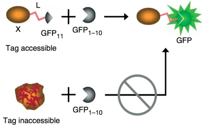
Le système « split-GFP » est basé sur l’auto-complémentation de deux fragments de la GFP : un fragment de 15 acides aminés appelé GFP 11, correspondant au dernier brin β de la GFP et le fragment complémentaire GFP 1-10 correspondant au reste de la molécule GFP. Ces deux fragments exprimés séparément ne sont pas fluorescents ; ce n’est que lorsqu’ils sont mis en commun qu’ils s’associent spontanément, permettant la reconstitution de la molécule GFP et la formation du chromophore.
Il a été montré que lorsque le fragment GFP 11 est fusionné avec une autre protéine, il est toujours capable de s’associer à GFP 1-10, et de restaurer la fluorescence GFP de façon quantitative au nombre de molécules étiquetées avec la GFP 11.
Pour notre projet, nous avons fusionné MeCP2 au petit fragment GFP 11. Cette con-struction a été exprimée d’une façon stable dans des cellules neuronales donneuses. Le fragment complémentaire de la GFP (GFP 1-10) a été exprimé dans d’autres cellules neu-ronales receveuses. Après co-culture, nous n’avons pas pu détecter l’apparition d’un signal fluorescent dans les cellules donneuses.
Résumé général des travaux de thèse et perspectives
Malgré l’absence de contrôle positif lors des expériences de co-cultures cellulaires (par exemple les protéines Engrailed -1 ou 2, fusionnées à la GFP 11, connues pour trans-férer du noyau d’une cellule à l’autre), nous suspectons que le transfert inter-cellulaire de MeCP2 soit dû aux étapes de fixation et de perméabilisation instantanées avec l’acétone lors des expériences d’immunofluorescence. En effet, quand les cellules sont fixées avec le paraformaldéhyde et perméabilisées avec le Triton X100, nous n’observons plus le transfert de MeCP2.
Ce travail avec la split-GFP nous a permis de développer une nouvelle application à ce système. Nous avons montré que la protéine GFP1-10 recombinante peut être utilisée comme réactif en microscopie ou en cytométrie de flux pour détecter la présence de pro-téines fusionnées à la GFP 11 quelque soit leur localisation dans les cellules de mammifères. En comparaison avec les marquages avec des anticorps, cette technique s’avère être plus rapide et plus spécifique avec un ratio signal/bruit très élevé.
La deuxième partie de ma thèse (cf. Chapitre 6)a permis de produire et de caractériser, pour la première fois, de nouveaux anticorps capables de détecter séparément et spécifique-ment chacune des deux isoformes de MeCP2.
Au laboratoire, nous avons montré que ces anticorps peuvent être utilisés pour des expériences de Western blot, d’immunofluorescence, d’immunohistochime et d’immunoprécipitation de la chromatine (ChIP).
Des expériences de western blot sur des extraits nucléaires issus de différents tissus (cerveau, poumon, coeur, foie, thymus, rate et rein) d’une souris âgée de 3 mois ont montré que l’isoforme MeCP2e1 est fortement exprimée dans le système nerveux central et moyennement exprimée dans les poumons et le rein. D’autre part, des expériences d’immunohistochimie sur des coupes de cerveau issues de cerveaux de souris âgées de 24 et de 55 jours, sauvages ou KO pour le gène mecp2, ont montré que l’isoforme MeCP2e1 est exprimée dans différentes structures du cerveau comme par exemple l’hippocampe, le noyau paraventriculaire et le noyau arque. Dans ces deux types d’experiences, nous n’avons pas pu détecter la présence de l’isoforme MeCP2e2 suggérant que cette isoforme n’est pas exprimée dans l’organisme ou bien elle est exprimée à un très faible niveau non détectable dans nos conditions expérimentales.
La troisième partie de ma thèse (cf. Chapitre 7)a consisté à étudier le rôle de MeCP2 dans la réponse aux dommages à l’ADN. En effet, quand nous avons induit des lésions à l’ADN
avec une micro-irradiation laser à 800 nm, nous avons observé une accumulation de MeCP2 dans la région du dommage. Nous avons aussi démontré que le recrutement de MeCP2 sur les lésions est indépendant de la transcription, et il est indépendant des 2 voies de réparation de l’ADN par excision des nucléotides : GG-NER et TC-NER. En revanche, nous avons observé que la partie C-terminale de MeCP2 (après l’acide aminé 308) est nécessaire pour ce recrutement. Cette partie de la protéine, qui est absente dans le modèle de souris de Dr. Zoghbi présentant des symptômes du syndrome de Rett, est importante pour la liaison de MeCP2 à l’ADN non méthylé et aux interactions protéines/ protéines.
En parallèle à ce travail nous avons pu montrer qu’en absence de la protéine CSB, l’interaction de MeCP2 avec la chromatine est diminuée.
La protéine CSB est une protéine de 168 kDa membre de la famille des hélicases SWI/SNF. Elle joue un rôle dans la réparation de l’ADN couplée à la transcription (TC-NER) en interagissant avec l’ARN polymérase II bloquée sur les lésions d’ADN. De plus, il a été montré que CSB joue un rôle dans le remodelage de la chromatine. Des mutations dans cette protéine ont été retrouvées chez des patients atteints du syndrome de Cockayne. Ces patients présentent un tableau de symptômes neurologiques très similaire à celui observé pour les patientes atteintes du syndrome de Rett. Ces symptômes neurologiques pour-raient être causés par l’impossibilité pour MeCP2 de se lier correctement à la chromatine et d’effectuer sa fonction correctement dans le cerveau.
Il est maintenant important de comprendre le rôle de MeCP2 dans la réponse aux dom-mages à l’ADN. Est ce que MeCP2 agit directement au niveau d’une voie de réparation ? Est-elle capable de détecter une lésion et de recruter les protéines nécessaires pour la répa-ration du dommage ? Peut-elle jouer un rôle dans l’architecture de la chromatine autour de l’ADN endommagé ?
Ensuite, il est important de comprendre la relation entre MeCP2 et CSB. MeCP2 intéragit-elle directement avec CSB ? Sinon, se trouvent-intéragit-elles dans le même complexe protéique ?
La découverte de ce nouveau rôle potentiel de MeCP2 dans la réponse aux dommages à l’ADN augmente la complexité des études réalisées sur cette protéine. Le fait que la partie C-terminal de la protéine soit impliquée dans le recrutement de MeCP2 sur les lésions de l’ADN et que des mutations dans cette région soient identifiées chez des patientes atteintes du syndrome de Rett implique que ce nouveau rôle pour la protéine doit être pris en compte dans la pathologie de Rett.
Résumé général des travaux de thèse et perspectives
Les résultats de notre étude sont donc importants pour le syndrome de Rett mais aussi pour la maladie de Cockayne. À la fois ils pourraient permettre d’avancer dans la com-préhension de ces deux maladies mais aussi de pouvoir envisager des thérapies communes, comme la supplémentation en anti-oxidants pour les patientes Rett.
Contents
Many Thanks. . . v
Résumé général des travaux de thèse et perspectives ix Abreviation 1 I Introduction 5 1 Rett Syndrome 7 1.1 Definition . . . 7
1.2 Clinical features . . . 7
1.2.1 The classical form . . . 7
1.2.2 The atypical form . . . 8
1.3 Genetic origin of Rett Syndrome . . . 9
1.4 RTT causing mutations . . . 10
1.4.1 Mutations in MeCP2 gene . . . 10
1.4.2 Mutations in other genes . . . 11
1.5 Genotype/Phenotype correlations . . . 11
1.5.1 Type of mutations . . . 11
1.5.2 Effect of XCI . . . 11
1.5.3 MeCP2 Mutations in boys . . . 12
1.6 Mouse Models of Rett syndrome . . . 13
1.6.1 MeCP2-null mouse model . . . 13
1.6.2 MeCP2 conditional mutants . . . 14
1.6.3 MeCP2 truncation . . . 14
1.6.4 MeCP2 transgenic mice . . . 15
1.6.5 MeCP2-eGFP knock-in mice . . . 15
Bibliography . . . 17
2 Methyl CpG Binding Protein 2 (MeCP2) 21 2.1 MeCP2 gene and protein . . . 21
2.2 MeCP2 function domain . . . 22
2.4 MeCP2 biological function . . . 25
2.4.1 Transcription repression . . . 25
2.4.1.1 Histone deactylation . . . 25
2.4.1.2 Histone methylation . . . 26
2.4.1.3 Chromatin Compaction and architecture . . . 26
2.4.1.4 Direct interaction with the transcriptional machinery . . . 26
2.4.2 Transcription activator . . . 27
2.4.3 Alternative splicing of mRNA . . . 27
2.4.4 Maintaining of DNA methylation . . . 27
2.5 MeCP2 mutations and other diseases . . . 28
2.5.1 The Angelman syndrome (AS) . . . 28
2.5.2 The severe neonatal encephalopathy (SNE) . . . 28
2.5.3 The X-linked Mental retardation (XLMR) . . . 28
2.5.4 Autism . . . 29
2.6 Conclusions . . . 29
Bibliography . . . 30
3 Green Fluorescent protein and Microscopy 35 3.1 Introduction . . . 35
3.2 Green Fluorescent Protein (GFP) . . . 35
3.3 Split GFP system . . . 37
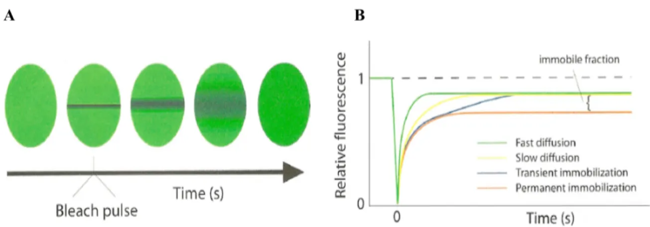
3.4 Fluorescence Recovery After Photobleaching (FRAP) . . . 38
Bibliography . . . 41
4 DNA Damage Repair 43 4.1 Introduction . . . 43
4.2 DNA repair pathways . . . 44
4.2.1 DNA Double Strand Breaks (DSB) repair . . . 44
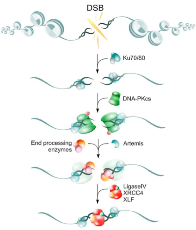
4.2.1.1 Non Homologous End Joining (NHEJ) . . . 45
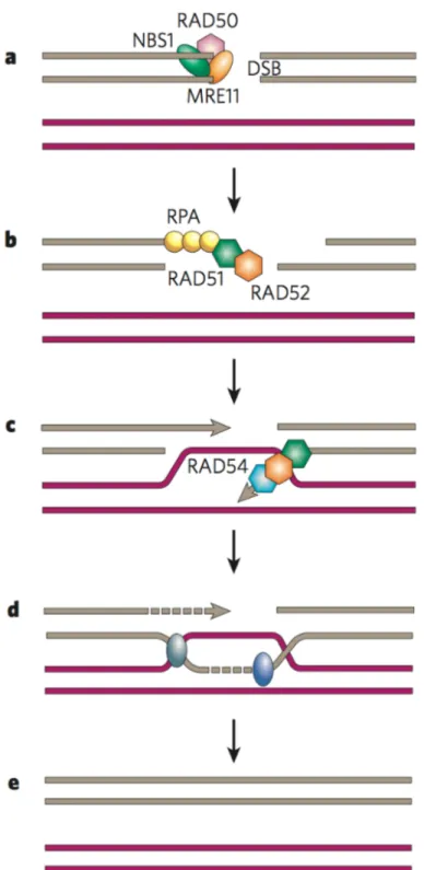
4.2.1.2 Homologous Recombination (HR) . . . 47
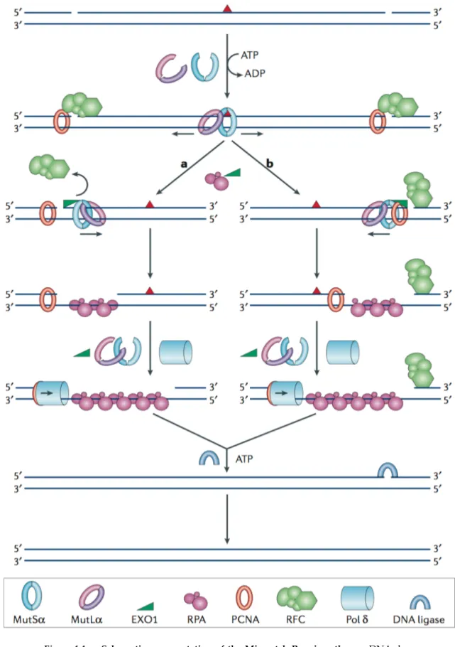
4.2.2 Mismatch repair (MMR) . . . 49
4.2.3 Base Excision Repair (BER) . . . 51
4.2.4 Nucleotide Excision Repair (NER) . . . 53
4.2.4.1 Xeroderma pigmentosum (XP) . . . 55 4.2.4.2 Trichothiodystrophy (TTD) . . . 55 4.2.4.3 Cockayne Syndrome (CS) . . . 55 Bibliography . . . 57 Working Context 61 Bibliography . . . 65
Contents
II Results 67
5 One-step split GFP staining for sensitive protein detection and localization in
mammalian cells 69
5.1 Abstract . . . 70
5.2 Introduction . . . 70
5.3 Materials and methods . . . 71
5.3.1 Plasmids . . . 71
5.3.2 DNA sequence of the GFP11m vector cassette . . . 71
5.3.2.1 GFP11m N-terminal fusion . . . 71 5.3.2.2 GFP11m C-terminal fusion . . . 71 5.3.2.3 Recombinant GFP 1-10 . . . 72 5.3.3 Cell culture . . . 72 5.3.4 Transfection . . . 72 5.3.5 Antibodies . . . 72 5.3.5.1 Primary antibodies . . . 72 5.3.5.2 Secondary antibodies . . . 73
5.3.6 Flow cytometry analysis . . . 73
5.3.7 Immunofluorescence staining and microscopy . . . 73
5.3.8 Microplate assay . . . 74
5.4 Results and discussion . . . 75
5.4.1 Characterization of the GFP 1-10 staining assay for FACS . . . 75
5.4.2 Sensitivity of the GFP 1-10 staining in microplate assay . . . 75
5.4.3 Comparison with MAb staining . . . 77
5.4.4 Validation of the GFP 1-10 staining assay for FACS measurements . . . 77
5.4.5 GFP 1-10 staining for microscopy . . . 79
5.5 Acknowledgments . . . 81
5.6 Competing interests . . . 81
5.7 Correspondence . . . 81
Bibliography . . . 88
6 Generation and characterization of isoform-specific anti-MeCP2 antibodies 91 6.1 Abstract . . . 91
6.2 Introduction . . . 91
6.3 Material and methods . . . 93
6.3.1 Rabbit immunization . . . 93
6.3.2 Cell culture and transfections . . . 94
6.3.3 Immunofluorescence staining of tissue culture cells and microscopy . 94 6.3.4 Western Blot . . . 95
6.3.5 Brain tissue immunostaining . . . 95
6.4 Results and discussion . . . 96
6.4.1 Peptide design and Rabbit immunization . . . 96
6.4.2 Antibodies characterization . . . 97
6.4.2.1 Immunofluorescence and western blot . . . 97
6.4.2.2 Immunostaining on brain sections of wildtype and MeCP2 KO mice . . . 98
6.5 Conclusion . . . 100
Bibliography . . . 102
7 MeCP2 Involvement in the DNA Damage Response 103 7.1 Introduction . . . 103
7.2 Results . . . 104
7.2.1 Expression and detection of MeCP2-GFP and mutants derivatives . . . 104
7.2.2 MeCP2 localization on locally damaged DNA. . . 106
7.2.3 MeCP2 recruitment on local damage is transcription and CSB inde-pendent . . . 106
7.2.4 MeCP2 recruitment on Local Damage is dependant on pro-tein/protein interaction but is DNA methylation independent. . . 109
7.2.5 MeCP2 Binding to chromatin is altered in neuron from CSB-/- mice . 110 7.3 Discussion . . . 112
7.4 Materials and Methods . . . 115
7.4.1 Cell lines and cell culture . . . 115
7.4.2 DNA constructs and cell transfection . . . 115
7.4.3 Transfection and Generation of stable cell lines . . . 115
7.4.4 Immunofluorescence staining . . . 116
7.4.5 Western blot analysis . . . 116
7.4.6 MeCP2 extraction from Brain . . . 117
7.4.7 Strip-FRAP experiments . . . 118 7.4.8 UV treatment . . . 118 7.4.9 Laser micro-irradiation . . . 118 Bibliography . . . 119 Conclusion 123 III Appendix 127 A Appendix 129 A.1 Abstract . . . 130 A.2 Introduction . . . 130 A.3 Results . . . 132
A.3.1 Overexpression of MeCP2 downregulates basal MHC class I . . . 132
A.3.2 Overexpression of MeCP2 markedly reduces MHC class I upregula-tion by IFN-γ . . . 135
A.3.3 MeCP2 mutants retain their repressive effect on MHC class I expression 137 A.3.4 Expression of MHC class I in cells from MeCP2-knockout mice is no different to that in cells from wild-type mice . . . 138
A.4 Discussion . . . 140
A.5 Materials and Methods . . . 145
A.5.1 Mice . . . 145
A.5.2 Cell lines . . . 146
A.5.3 Antibodies . . . 146
Contents
A.5.5 Mutagenesis . . . 146
A.5.6 Transfection . . . 148
A.5.7 Quantitative Western Blot . . . 148
A.5.8 Cells staining and cytometry . . . 148
A.5.9 Immunohistochemistry of brain slices . . . 149
A.5.10 Immunofluorescence staining . . . 149
A.5.11 Primary cultures . . . 150
A.6 Acknowledgments . . . 150
A.7 Author Contributions . . . 150
Bibliography . . . 151
Abreviation
- Aa : Amino acid
- APE1 : APurinic/APyrimidinic Endonuclease1
- AS : Angelman Syndrome
- ATM : Ataxia Telangiectasia Mutated
- BER : Base Excision Repair
- BFP : Blue Fluorescent Protein
- BRCA : BReast CAncer
- CDKL5 : Cyclin-Dependent Kinase-Like 5
- CFP : Cyan Fluorescent Protein
- CPD : Cyclobutane Pyrimidine Dimer
- CpG : Cytosine-phosphate-Guanine
- cREB : cAMP Response Element-Binding
- CS : Cockayne Syndrome
- DNA : DesoxyRibonucleic Acid
- DNMT1 : DNA methyltransferase 1
- DSB : Double Strand Break
- eGFP : enhanced Green Fluorescent Protein
- ES : Embryonic Stem cells
- Exol : Exonuclease 1
- FBP11 : Formin Binding Protein 11
- FEN1 : Flap endonuclease 1
- FRAP : Fluorescence Recovery After Photobleaching
- GG-NER : Global Genome-Nucleotide Excision Repair
- GGR : Global Genome Repair
- HDAC : Histone Deacetylase Complex
- HMGA1 : High Mobility Group A 1
- HR : Homologous Recombination
- ID : Insertion / Deletion
- MBD : Methyl Binding Domain
- MeCP2 : Methyl CpG Binding Protein
- MLH : MutL Homolog
- MMR : Mismatch Repair
- MSH : MutS Homolog
- N-COR : Nuclear receptor corepressor
- NER : Nucleotide Excision Repair
- NHEJ : Non Homologous End Joining
- Paf : Patchy fur
- PCNA : Proliferating Cell Cuclear Antigen
- 6-4PP : Pyrimidine-Pyrimidone (6-4) products
- RFC : Replication Factor C
- RPA : Replication Protein A
- RTT : Rett syndrome
- RNA : Ribonucleic Acid
- STK9 : Serine/Threonine Kinase 9
- SNE : Severe neonatal encephalopathy
- SWI/SNF : SWItch/Sucrose NonFermentable
- TC-NER : Transcription Coupled- Nucleotide Excision Repair
Abreviation
- TFIIB : Trancription Factor II B
- TFIIH : Trancription Factor II H
- TTD : TrichoThioDystrophy
- TRD : Transcription Repression Domain
- 3’-UTR : 3’-Untranslated Region
- XCI : X Chromosome Inactivation
- XLF : XRCC4-like factor
- XLMR : X-linked Mental retardation
- XP : Xeroderma Pigmentosum
- XRCC : X-ray repair cross-complementing
- Yb-1 : Y Box protein 1
Studying the MeCP2 protein:
Exploring its involvement in the DNA Damage
Response, and developing new tools for its detection.
1
Rett Syndrome
1.1
Definition
Rett syndrome (RTT, MIM 312750) is a severe postnatal progressive neurodevelopmen-tal disorder that manifests mainly in girls during early childhood. Rett syndrome is one of the most common causes of mental retardation in females with an incidence of 1 birth in 10000 to 20000.
This syndrome was described for the first time in 1966 by Andreas Rett, an Austrian neurologist in Vienna. But the syndrome became internationally recognised 17 years later when Dr. B. Hagberg, a Swedish neurologist, reported 35 cases of RTT [1, 2].
1.2
Clinical features
There is a large variability in the progression and the severity of Rett syndrome. We can distinguish between the classical and the atypical forms of RTT.
1.2.1 The classical form
At birth, girls with the classic form of the disease have a normal head circumference, ap-pear to develop normally until 6-18 months and achieve the expected motor skills, language and social milestones. Nevertheless, some studies revealed subtle behavioural abnormal-ities soon after birth. After this period, the neurological development is arrested and the regression phase begins. This regression is commonly divided into four stages [2, 3, 4] and (Figure 1.1):
1
is stopped and the head growth undergoes deceleration that usually leads to micro-cephaly. Babies may show less eye contact and start to lose interest in toys. They may also show signs of hypotonia and have delays in the acquisition of sitting, crawling or walking.
⪧ Stage II: The regressive stage (1-4 years): Children with Rett syndrome gradually lose the ability to speak and to use their hands purposefully. They show the classic "hand-washing" stereotypic activity. Some children with Rett syndrome hold their breath or hyperventilate and may scream or cry for no apparent reason. It’s often difficult for them to move on their own. Girls show autistic features and half of them develop seizures.
⪧ Stage III: The relative stabilization period (2-10 years): Although problems with move-ment continue, behavior may improve. Children in this stage often cry less and become less irritable. During this stage, the use of eye contact and hands for communication generally improve. Many people affected by Rett syndrome remain in stage III for the rest of their lives.
⪧ Stage IV: The late motor impairment period (10-15 years): The last stage is marked by reduced mobility, muscle weakness and scoliosis. Understanding, communication and hand skills generally don’t decline during this stage but repetitive hand movements may decrease.
1.2.2 The atypical form
Five distinct categories of atypical RTT have been described on the basis of clinical crite-ria. These variants can be either milder or more severe than the classic RTT phenotype. We can distinguish 3 types of milder variants [2, 3, 4] and (Figure 1.1):
⪧ the ”forme fruste” where regression begins later than classic RTT (between 1 and 3 years old). In this form the hand use is sometimes preserved with minimal stereotypic movement.
⪧ the preserved speech variant where girls can speak a few words. Patients with this variant have a normal head size.
1.3. Genetic origin of Rett Syndrome
1
Figure 1.1 — Revised diagnostic criteria for classical and variant RTT. (Figure adapted
from [2]).
⪧ the late regression variantis characterized by normal head circumference and gradual loss of acquired speech and fine motor skills in late childhood.
And 2 types of more severe forms:
⪧ the congenital formwhere patients show RTT features straight from birth. In addition to lacking the typical early normal period, they are floppy and very retarded.
⪧ the early seizure-onset variant: In this variant, the normal perinatal period is soon followed by the appearance of seizures preceding the regression period.
1.3
Genetic origin of Rett Syndrome
Early reports suggested that RTT is an X-linked dominant disorder because of the almost exclusive occurrence in females, the high concordance rate among monozygotic twins and the rare familial cases with maternal inheritance.
The causes of RTT were difficult to determine because only 1 % of the RTT cases are fa-milial (99% of RTT cases are sporadic). Genetic mapping studies on the X chromosome in familial cases identified the Xq28 locus as the origin of RTT [5, 6]. In 1999, Huda Zoghbi’s
1
group carried out systematic mutational analysis of genes located in Xq28 in patients af-fected by the sporadic or the familial forms of RTT. They identified mutations in the MeCP2 gene encoding for the X-linked methyl CpG binding protein 2 as the causes of some cases of RTT [7]. As many other genes located on the X chromosome, this gene undergoes X inactivation.
1.4
RTT causing mutations
1.4.1 Mutations in MeCP2 gene
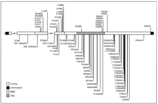
MeCP2 mutations account for up to 95% of classic RTT cases [7, 8]. Almost all muta-tions occur de novo and they include missense, frameshift, nonsense and intragenic delemuta-tions ([9, 10] and Figure 1.2). About 70% of MeCP2 mutations are caused by a C to T transition at 8 CpG nucleotides located in the third and the fourth exons, whereas carboxy-terminal deletions occur in 10 to 15% of RTT patients [11].
Figure 1.2 — Summary of mutations found in coding region from exon 2 to exon 4
of MeCP2 gene: Missense mutations are shown above the genomic structure of MeCP2
whereas nonsense or frameshift mutations are shown below. Asterisks represent muta-tions causing non-syndromic mental retardation. (Figure adapted from [10]).
1.5. Genotype/Phenotype correlations
1
1.4.2 Mutations in other genes
Some RTT patients don’t show mutations in MeCP2 gene. It was proposed that there is at least one other gene responsible for RTT syndrome. Two groups showed that some cases of RTT-like phenotypes (with severe mental retardation and early seizure onset) are caused by truncating frameshift and missense mutations in the gene coding for the cyclin-dependant kinase-like 5 (CDKL5/ STK9) [12].
Another group reported that some mutations in the FOXG1 gene are responsible for the congenital form of Rett syndrome, a severe variant where girls are retarded from the first months of life. The first studies revealed deletions and nonsense mutations in this gene as the origin of variant Rett syndrome [13, 14]. Recently a study revealed a frameshift point mutation in a boy with the congenital variant form of Rett syndrome [15].
1.5
Genotype/Phenotype correlations
RTT patients present a large phenotypic variability associated with different MeCP2 mu-tations. Recent genotype-phenotype studies showed that severity of RTT phenotype de-pends on the type of the mutation, the genetic background and the X-chromosome inactiva-tion balance.
1.5.1 Type of mutations
Patients carrying mutations that truncate the protein in the C-terminal domain (late trun-cating mutations) present milder phenotype and are less typical of classical Rett Syndrome than those carrying missense or early truncating mutations. Jian et al. reported in 2005 that R270X mutation (X representing here a stop codon) is associated with elevated mortality. Wan et al. showed that girls carrying the same mutations could sometimes present different phenotypes. This observation is consistent with an important role for the X-chromosome inactivation (XCI) balance [16, 11].
1.5.2 Effect of XCI
The purpose of the inactivation of one of the X chromosome is to equalize X-linked gene products between XX females and XY males. This process occurs randomly in
differenti-1
ating embryonic cells in females, resulting in cells that are mosaic with respect to which chromosome is active.
MeCP2 gene is located on the X-chromosome and is subjected to XCI. Girls with Rett syndrome are thus mosaic for MeCP2 mutations, meaning that, on average, half of their cells express the wild-type MeCP2 allele and the other half express the mutated one. Some studies reported that healthy-carrier mothers or females with less severe RTT phenotype have a skewed XCI with inactivation of the mutated allele. This was illustrated, for ex-ample, with a study showing monozygotic twins with R294X mutation with very different phenotypes. While one of the twins with a severe phenotype presented a balanced XCI, the other one showed a less severe phenotype caused by a skewed XCI towards the maternal wild-type allele [17]. These results are, however, controversial because, in these studies, the XCI profile was determined on peripheral lymphocytes. In fact, it was demonstrated that the lymphocyte clone carrying the wild-type allele developed and proliferated faster than the clones carrying the mutated allele, thus biasing the XCI skewing [18]. Studies performed on RTT brain tissues suggest that balanced XCI patterns are prevalent [4].
1.5.3 MeCP2 Mutations in boys
At the beginning, RTT was considered to be an X-linked dominant disorder with male lethality. In 1999, Jan et al. identified males with Rett syndrome [19]. Screening of MeCP2 mutations in males with neurological pathologies revealed that these mutations can cause variable phenotypes such as:
⪧ Classical RTT phenotype due to somatic mosaicism for mutations in MeCP2 or in cases of Klinefelter syndrome (XXY).
⪧ Mild to severe mental retardation: These patients carry mutations different from the ones found in girls.
⪧ Severe neonatal encephalopathy: These patients carry the same mutations identified in girls but the phenotype is more severe due to the presence of just one X chromosome in males. Male patients usually die in the first two years of life [20].
1.6. Mouse Models of Rett syndrome
1
1.6
Mouse Models of Rett syndrome
To understand the molecular mechanisms implicated in RTT, different mouse models have been generated. These mouse models mimic the clinical features observed in RTT patients.
1.6.1 MeCP2-null mouse model
The first attempts to create MeCP2-null mice using male mouse embryonic stem (ES) cells lacking MeCP2 were unsuccessful. Chimeric embryos exhibited high lethality. In 2001, two groups directed by Dr Adrian Bird and Dr. Rudolph Jaenisch decided to use condi-tional knock-out approaches based on Cre-Lox technologies. Dr. Jaenisch generated the MeCP21lox mouse model with MeCP2 with conditionally deleted exon 3 [21] and Dr. Bird the MeCP2tl1−1Birdwith MeCP2 with conditionally deleted exons 3 and 4 [22].
MeCP2-null male mice are apparently healthy at birth until 3 to 8 weeks of age. After this period, mice begin to show neurological symptoms like those observed in RTT patients: stiff and uncoordinated gait, hindlimb clasping, and irregular breathing. Uneven wearing of the teeth and misalignment of the jaws are also observed. Testes of MeCP2-null males were always internal. Symptoms progression leads to weight loss and early death around 54 days. Brain architecture in null mice is grossly normal, although a slight decrease in the size and weight can be noticed in comparison with wild-type littermates. This is due to neurons compaction in hippocampus, cerebral cortex and cerebellum.
MeCP2+/− females mice are viable, fertile and grow normally until adulthood. At 3 months, they start showing hind limb clasping and by 9 months they show RTT phenotypes such as breathing irregularity and inertia.
These results show that MeCP2-null mice can be a good model to study RTT because of delayed onset of neurological symptoms affecting posture, gait, breathing and spontaneous movements.
In 2006, Dr Patrick Tam generated mice with a targeted deletion of the methyl binding domain (MBD) resulting in complete loss of MeCP2 protein. MeCP2tm1Tam phenotype is comparable with that of the Jaenisch and Bird’s mice phenotype. In addition, at 8 to 10 weeks after birth, they display reduced level of anxiety, locomotors activity and cerebellar
1
learning. In this study, authors wanted to verify if the Y chromosome has an effect on the mouse phenotype. For this heterozygous female, called MeCP2tm1neoTam , were mated with Paf males on a C3H/HeSNJ background. Paf (or Patchy fur) is a mutation that provokes default in X and Y chromosome segregation during meiosis leading to a frequent loss of the Y chromosome. Comparison between MeCP2tm1Tam 39XO females and MeCP2tm1Tam 40XY males carrying mutated X alleles and differing only by expression of Y chromosome show similar profiles of postnatal viability and growth suggesting that Y chromosome has no impact on MeCP2-null phenotype [23].
1.6.2 MeCP2 conditional mutants
The group of Dr Jaenisch wanted to study the effect of specific deletion of MeCP2 in the central nervous system. MeCP2-null mice were hence crossed with transgenic mice carrying Nestin-Cre transgene. This crossing gave birth to mice in which MeCP2 was deleted specif-ically in neurons and glial cells. These mice displayed a phenotype similar to that observed in MeCP2-null mice suggesting that the primary site where the lack of MeCP2 is causing the Rett pathology is indeed in the brain. Chen et al also investigated the role of MeCP2 in post-mitotic neurons. In order to do this, they introduced the Cam kinase Cre transgene which is active in the excitatory neurons in the postnatal forebrain, hippocampus and brainstem. For these conditional mutants the phenotype seemed to be less severe than null mice or Nestin-Cre conditional mutants. CamK-Nestin-Cre conditional mutants were healthy until 3 months. After this period, they began to show symptoms including gain of body wait, ataxic gait and re-duced nocturnal activity. The brain weight of these mice was rere-duced with smaller neuronal cell bodies in cortex and hippocampus [21].
1.6.3 MeCP2 truncation
In 2002, Dr Huda Zoghbi’s group generated a mouse carrying a truncation at amino acid 308. This truncation eliminates the C-terminal region of MeCP2.
In these mice the phenotype is milder than those seen previously. MeCP2308/y are nor-mal until 6 weeks and then they develop symptoms like tremors, kyphosis, learning and memory deficits, social behaviour abnormalities, etc. Heterozygote females MeCP2308/+ have impaired motor features starting at 35-39 weeks after birth. As in the case of human female patients, these mice show phenotypic variability due to the XCI. In this study,
au-1.6. Mouse Models of Rett syndrome
1 thors show that the truncated protein has a normal nuclear localization on heterochromatic
regions but histone H3 is hyperacetylated in the brain. In contrast, the acetylation level of histone H4 was the same as the one observed in their wild-type animals [24].
1.6.4 MeCP2 transgenic mice
In 2004, Luikenhuis et al. overexpressed a MeCP2 transgene, coding for MeCP2e2, in postmitotic neurons under the control of the Tau promoter [25]. Expression of tau-MeCP2 transgene in neurons is sufficient to rescue RTT phenotype as shown by the fact that when this transgene is expressed in MeCP2 deficient mice, the latter show a phenotype similar to wild-type animals. In 2007, Dr. Bird’s group created a mouse model in which MeCP2 is silenced by the insertion of a lox-stop cassette [26]. The expression of MeCP2 can be re-activated by the addition of a transgene expressing a fusion between the Cre recombi-nase and a modified estrogen receptor (Cre-ER). The Cre-ER fusion protein remains in the cytoplasm until treatement with the estrogen analog tamoxifen. After treatement with ta-moxifen, authors were able to rescue symptomatic animals suggesting that in the absence of MeCP2, neurons do not die and the phenotype can be reversible by delayed restoration of MeCP2 gene [26]. However, transgenic mice overexpressing MeCP2 show severe motor dysfunction: Collins et al. generated transgenic mice overexpressing the wild-type human MeCP2e2 protein at 2 fold the wild-type level. Early in the development, these mice show increased learning ability and synaptic plasticity. However, after the age of 10 weeks they developed seizures, hypoactivity and spasticity, dying by one year of age [27]
It was demonstrated that overexpression of MeCP2 in human males can cause autis-tic features and profound mental retardation [28, 29]. All these observations indicate that deficiency or elevated expression of MeCP2 cause neurodevelopmental disorders with a variability of phenotypes. These observations should be considered when attempting to develop therapies against Rett syndrome.
1.6.5 MeCP2-eGFP knock-in mice
This mouse was generated by Schmid et al. in 2007 [30]. In these mice, MeCP2 was tagged by eGFP in frame into exon 4 just before the stop-codon. These mice are viable, fertile and without any detectable abnormalities suggesting that MeCP2-GFP fusion is functional. Immunofluorescence colocalisation and confocal microscopy analysis revealed that
MeCP2-1
eGFP expression is restricted to the nucleus and targeted to heterochromatic regions. Results revealed that the fusion protein is expressed in all neurons, interneuron and astrocytes at dif-ferent levels as described in other earlier studies. Authors didn’t detect any MeCP2 expres-sion in proliferative cells. In concluexpres-sion, this study revealed that MeCP2-eGFP expresexpres-sion matches endogenous MeCP2 expression suggesting that these mice can be an important tool to study MeCP2 expression or MeCP2 dynamics in vivo.
Bibliography
1
Bibliography
[1] B. Hagberg, J. Aicardi, K. Dias, and O. Ramos. A progressive syndrome of autism, dementia, ataxia, and loss of purposeful hand use in girls: Rett’s Syndrome: report of 35 cases. Ann Neurol, 14(4):471–9, 1983.
[2] S. L. Williamson and J. Christodoulou. Rett Syndrome: new clinical and molecular insights. Eur J Hum Genet, 14(8):896–903, 2006.
[3] K. A. Jellinger. Rett Syndrome – an update. J Neural Transm, 110(6):681–701, 2003. [4] M. D. Shahbazian and H. Y. Zoghbi. Rett Syndrome and MeCP2: Linking epigenetics
and neuronal function. Am J Hum Genet, 71(6):1259–72, 2002.
[5] N. C. Schanen, E. J. Dahle, F. Capozzoli, V. A. Holm, H. Y. Zoghbi, and U. Francke. A new Rett Syndrome family consistent with X-linked inheritance expands the X chro-mosome exclusion map. Am J Hum Genet, 61(3):634–41, 1997.
[6] N. Sirianni, S. Naidu, J. Pereira, R. F. Pillotto, and E. P. Hoffman. Rett Syndrome: con-firmation of X-linked dominant inheritance, and localization of the gene to Xq28. Am J Hum Genet, 63(5):1552–8, 1998.
[7] R. E. Amir, I. B. Van den Veyver, M. Wan, C. Q. Tran, U. Francke, and H. Y. Zoghbi. Rett Syndrome is caused by mutations in X-linked MECP2, encoding methyl-CpG-binding protein 2. Nat Genet, 23(2):185–8, 1999.
[8] T. Bienvenu, A. Carrie, N. de Roux, M. C. Vinet, P. Jonveaux, P. Couvert, L. Villard, A. Arzimanoglou, C. Beldjord, M. Fontes, M. Tardieu, and J. Chelly. MeCP2 mutations account for most cases of typical forms of Rett Syndrome. Hum Mol Genet, 9(9):1377–84, 2000.
[9] J. Christodoulou, A. Grimm, T. Maher, and B. Bennetts. RettBASE: The IRSA MeCP2 variation database-a new mutation database in evolution. Hum Mutat, 21(5):466–72, 2003.
[10] M. D. Shahbazian and H. Y. Zoghbi. Molecular genetics of Rett Syndrome and clinical spectrum of MeCP2 mutations. Curr Opin Neurol, 14(2):171–6, 2001.
[11] M. Wan, S. S. Lee, X. Zhang, I. Houwink-Manville, H. R. Song, R. E. Amir, S. Budden, S. Naidu, J. L. Pereira, I. F. Lo, H. Y. Zoghbi, N. C. Schanen, and U. Francke. Rett Syndrome and beyond: recurrent spontaneous and familial MeCP2 mutations at CpG hotspots. Am J Hum Genet, 65(6):1520–9, 1999.
[12] L. S. Weaving, J. Christodoulou, S. L. Williamson, K. L. Friend, O. L. McKenzie, H. Archer, J. Evans, A. Clarke, G. J. Pelka, P. P. Tam, C. Watson, H. Lahooti, C. J. Ellaway, B. Bennetts, H. Leonard, and J. Gecz. Mutations of CDKL5 cause a severe neurodevel-opmental disorder with infantile spasms and mental retardation. Am J Hum Genet, 75(6):1079–93, 2004.
[13] F. Ariani, G. Hayek, D. Rondinella, R. Artuso, M. A. Mencarelli, A. Spanhol-Rosseto, M. Pollazzon, S. Buoni, O. Spiga, S. Ricciardi, I. Meloni, I. Longo, F. Mari, V. Broccoli, M. Zappella, and A. Renieri. FOXG1 is responsible for the congenital variant of Rett Syndrome. Am J Hum Genet, 83(1):89–93, 2008.
[14] F. T. Papa, M. A. Mencarelli, R. Caselli, E. Katzaki, K. Sampieri, I. Meloni, F. Ariani, I. Longo, A. Maggio, P. Balestri, S. Grosso, M. A. Farnetani, R. Berardi, F. Mari, and A. Renieri. A 3 Mb deletion in 14q12 causes severe mental retardation, mild facial dysmorphisms and Rett-like features. Am J Med Genet A, 146A(15):1994–8, 2008.
1
[15] T. Le Guen, N. Bahi-Buisson, J. Nectoux, N. Boddaert, Y. Fichou, B. Diebold, I. Des-guerre, F. Raqbi, V. C. Daire, J. Chelly, and T. Bienvenu. A FOXG1 mutation in a boy with congenital variant of Rett Syndrome. Neurogenetics, 2010.
[16] L. Jian, H. L. Archer, D. Ravine, A. Kerr, N. de Klerk, J. Christodoulou, M. E. Bailey, C. Laurvick, and H. Leonard. p.R270X MeCP2 mutation and mortality in Rett Syn-drome. Eur J Hum Genet, 13(11):1235–8, 2005.
[17] T. Ishii, Y. Makita, A. Ogawa, S. Amamiya, M. Yamamoto, A. Miyamoto, and J. Oki. The role of different X-inactivation pattern on the variable clinical phenotype with Rett Syndrome. Brain Dev, 23 Suppl 1:S161–4, 2001.
[18] D. Balmer, J. Arredondo, R. C. Samaco, and J. M. LaSalle. MeCP2 mutations in Rett Syndrome adversely affect lymphocyte growth, but do not affect imprinted gene ex-pression in blood or brain. Hum Genet, 110(6):545–52, 2002.
[19] M. M. Jan, J. M. Dooley, and K. E. Gordon. Male Rett Syndrome variant: application of diagnostic criteria. Pediatr Neurol, 20(3):238–40, 1999.
[20] K. Ravn, J. B. Nielsen, P. Uldall, F. J. Hansen, and M. Schwartz. No correlation between phenotype and genotype in boys with a truncating MeCP2 mutation. J Med Genet, 40(1):e5, 2003.
[21] R. Z. Chen, S. Akbarian, M. Tudor, and R. Jaenisch. Deficiency of methyl-CpG binding protein-2 in CNS neurons results in a Rett-like phenotype in mice. Nat Genet, 27(3):327– 31, 2001.
[22] J. Guy, B. Hendrich, M. Holmes, J. E. Martin, and A. Bird. A mouse MeCP2-null muta-tion causes neurological symptoms that mimic Rett Syndrome. Nat Genet, 27(3):322–6, 2001.
[23] G. J. Pelka, C. M. Watson, T. Radziewic, M. Hayward, H. Lahooti, J. Christodoulou, and P. P. Tam. MeCP2 deficiency is associated with learning and cognitive deficits and altered gene activity in the hippocampal region of mice. Brain, 129(Pt 4):887–98, 2006. [24] M. Shahbazian, J. Young, L. Yuva-Paylor, C. Spencer, B. Antalffy, J. Noebels, D.
Arm-strong, R. Paylor, and H. Zoghbi. Mice with truncated MeCP2 recapitulate many Rett Syndrome features and display hyperacetylation of histone H3. Neuron, 35(2):243–54, 2002.
[25] S. Luikenhuis, E. Giacometti, C. F. Beard, and R. Jaenisch. Expression of MeCP2 in post-mitotic neurons rescues Rett Syndrome in mice. Proc Natl Acad Sci U S A, 101(16):6033– 8, 2004.
[26] J. Guy, J. Gan, J. Selfridge, S. Cobb, and A. Bird. Reversal of neurological defects in a mouse model of Rett Syndrome. Science, 315(5815):1143–7, 2007.
[27] A. L. Collins, J. M. Levenson, A. P. Vilaythong, R. Richman, D. L. Armstrong, J. L. Noebels, J. David Sweatt, and H. Y. Zoghbi. Mild overexpression of MeCP2 causes a progressive neurological disorder in mice. Hum Mol Genet, 13(21):2679–89, 2004. [28] M. Meins, J. Lehmann, F. Gerresheim, J. Herchenbach, M. Hagedorn, K. Hameister, and
J. T. Epplen. Submicroscopic duplication in Xq28 causes increased expression of the MeCP2 gene in a boy with severe mental retardation and features of Rett Syndrome. J Med Genet, 42(2):e12, 2005.
[29] H. Van Esch, M. Bauters, J. Ignatius, M. Jansen, M. Raynaud, K. Hollanders, D. Lugten-berg, T. Bienvenu, L. R. Jensen, J. Gecz, C. Moraine, P. Marynen, J. P. Fryns, and G. Froyen. Duplication of the MeCP2 region is a frequent cause of severe mental retar-dation and progressive neurological symptoms in males. Am J Hum Genet, 77(3):442–53, 2005.
Bibliography
1 [30] R. S. Schmid, N. Tsujimoto, Q. Qu, H. Lei, E. Li, T. Chen, and C. S. Blaustein. A
methyl-CpG-binding protein 2-enhanced green fluorescent protein reporter mouse model pro-vides a new tool for studying the neuronal basis of Rett Syndrome. Neuroreport, 19(4):393–8, 2008.
2
Methyl CpG Binding Protein 2
(MeCP2)
2.1
MeCP2 gene and protein
The mecp2 protein was identified and purified for the first time in 1992 in Dr Adrian Bird’s group based on its capacity to bind methylated DNA [1].
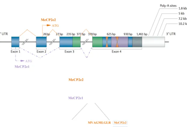
The MeCP2 gene consists of four exons encoding for different variants of transcripts due to differential splicing or alternative usage of the polyadenylation sites in the highly con-served 3’-untranslated region (3’-UTR) [2, 3]. Northern blot analysis revealed the presence of 3 transcripts of 1.8, 7.5 and 10 kb. 1.8 and 10kb are the major transcripts. They have simi-lar half-lives but differ by their translation efficiency [2, 4, 5]. The functional significance of these different transcripts is unknown (Figure 2.1).
In 2004, two studies revealed the presence of two isoforms of MeCP2 that differ in their N-terminal region [6, 7]. This is due to an alternative splicing of exon 2. The first transcript (MeCP2e1 or MeCP2α in mice or MeCP2B in human) lacks exon 2 and the initiation site is in exon 1. The second transcript (MeCP2e2 or MeCP2β in mouse or MeCP2A in human) contains the 4 exons and the initiation site is in exon 2 (Figure 2.1). The two splice variants differ in translation efficiency and are expressed at different relative amounts in different tissues. MeCP2e1 is more abundant in the brain, thymus and lung and during neuronal differentiation.
MeCP2e1 protein (498 aa) is 12 amino acids longer than MeCP2e2 (486 aa). It contains 21 amino acids encoded by exon 1 and lacks the 9 amino acids encoded by exon 2. The major difference between the two isoforms is the presence of a poly-alanine and a poly-glycine repeats in the N-terminal region of the longer isoform, MeCP2e1. Both MeCP2 isoforms are
2
nuclear and colocalize with methylated heterochromatic foci in mouse cells [6, 7].
MeCP2e2 MeCP2e2 ATG ATG MeCP2e1 MeCP2e1 MeCP2e2 MeCP2e2 MeCP2e1 MeCP2e1 MVAGMLGLR MAAAAATAAAAAAPSGGGGGGEEER MeCP2e2 MeCP2e1
Figure 2.1 — The MeCP2 gene and its splicing isoforms: Structure of the MeCP2 gene
with its four exons and the different polyadenylation sites generating transcripts of 1.8, 5, 7.2 and 10.2 Kb. The presence of the 5Kb transcript remains controversial (8). The two main protein isoforms, MeCP2e2 (486 amino acids) and MeCP2e1 (498 amino acids) are produced by alternative splicing of the MeCP2 transcript and differ in their N-terminal regions which are encoded by exon 1 in the case of MeCP2e1 and exon 2 for MeP2e2. (Figure adapted from [8]).
2.2
MeCP2 function domain
MeCP2 belongs to a family of proteins that contain a MBD domain which binds to methy-lated DNA (Figure 2.2).
Three domains compose MeCP2: the methyl binding domain (MBD), the transcription repression domain (TRD) and the C-terminal domain (Figure 2.1).
The MBD, an 85 amino acids domain (from amino acid 78 to 162 in MeCP2) [10], specif-ically binds to symmetrspecif-ically methylated CpG dinucleotides, with a preference for CpG sequences with adjacent A/T-rich motifs [11]. The MBD can also bind to four-way DNA junctions (also called Holliday junctions formed during homologous recombination) on un-methylated DNA with the same affinity as on un-methylated CpG [12].
2.2. MeCP2 function domain
2
Figure 2.2 — Characteristic domains of the methyl CpG binding (MBD) protein fam-ily: MBD proteins display homology within their MBD domain. The family contains five members: MBD1 and MBD2 are transcriptional repressors, MeCP2, the founding mem-ber, plays different roles as transcription modulator or mRNA splicing factor. MBD3 is a member of the Mi2/NuRD co-repressor complex and MBD4 is a G-T mismatch glycosy-lase playing a role in DNA repair. (Figure adapted from [9])
The TRD, a 104 amino acids domain (from amino acid 207 to 310), is very basic containing 26% lysine and arginine. It also contains non-polar amino acids (12.5% alanine, 10.5% valine and 8% proline) [13]. TRD is required to repress transcription by recruiting transcriptional co-repressors such as Sin3a, c-ski, N-COR and histone deactylase complex HDAC 1 and 2 [14, 15, 16]. The TRD can repress transcription at long-range distance ranging from 500 to 1500 bp away from the promoter [13]. The TRD contains also the MeCP2 nuclear localisation signal between amino acids 255 and 271 [17].
Although the C-terminal region is not well characterized, this region is very important for MeCP2 function. In fact, the mouse model carrying the R308X mutation (X is a stop codon) reproduces many features of RTT phenotype [18]. MeCP2 C-terminus presents a WDR domain (amino acids 325 to 486) containing a poly-proline rich sequence (amino acids 376 to 405). This region can bind to splicing factors containing a group II WW domain like the FBP11 (Formin binding protein 11) and HYPC proteins [19]. Furthermore, the C-terminal region allows MeCP2 fixation on naked DNA and on nucleosomal cores [20, 21].
Amino acids 188-194, between the MBD and the TRD, are identical to those found within the AT hook DNA binding domains of HMGA1 [22]. This region can bind to the xenopus
2
protein p20, this interaction reduces the turnover and stabilize MeCP2 [23].
2.3
MeCP2 expression profile
Several studies, carried out in rodents [24, 25, 5], monkeys [26] and human [5], aimed to analyse the expression profile of MeCP2. From these studies, it appears that MeCP2 is ex-pressed in many tissues. Western blot analysis on mice tissue samples revealed that the MeCP2 protein is not abundant in liver, stomach and small intestine, moderately expressed in kidney and heart and highly abundant in brain, lung and spleen [5]. Within the cerebral tissue, MeCP2 is not abundant in astrocytes [27] and immature neurons. However its ex-pression increases to be highest in mature neurons and it is maintained high in these cells throughout life. LaSalle et al. showed that MeCP2 expression varies between neuronal pop-ulations from different regions and different structures within the central nervous system (CNS) [28]. In this study, they characterised two populations of cells: The MeCP2lo cells, expressing low levels of MeCP2, were found in the brain (glial and neuronal cells) and in the periphery whereas the MeCP2hi cells, expressing high levels of MeCP2, were found in a higher proportion in the layer IV of the cerebral cortex and in the molecular layer of the cerebellum.
MeCP2 expression is regulated in a developmental stage and cell type specific manner. Few things are known about molecular mechanisms implicated in this regulation. It was shown that the mecp2 gene contains multiple transcription starting sites embedded in a re-gion that is GC rich and contains CpG islands. In this study, they showed that the mouse mecp2 promoter does not contain any canonical boxes like TATA or CAAT. They also iden-tified a promoter region (-677/+56) that is responsible for the expression of MeCP2 in neu-ronal cells. In this region, there is a positive regulatory element of 19 bp (-64 to +46) that controls the major activity of the promoter region [29].
In 2006, a study focused on mapping the regulatory regions of the locus of human MeCP2 showed that the locus is characterized by a very large intron 2 (60 Kb), a 3’- UTR of 8,5 kb with highly conserved domains and different polyadenylation sites and a 40 kb intergenic region separating MeCP2 from the nearest upstream gene [30]. It also identi-fied a region supposed to be the core promoter (-179 to -309 bp upstream the initiation site) supporting basal gene expression. Two positive regulatory elements were characterized
2.4. MeCP2 biological function
2 (-681/-847) and (-847/-1071) as well as two negative regulatory elements (-309/-370) and
(-553/-681). In order to identify potential Cis-regulatory elements able to control spatio-temporal expression of MeCP2, authors performed an interspecies sequence comparison and identified four enhancers and two silencer elements in the 210 kb surrounding MeCP2 locus [30].
2.4
MeCP2 biological function
2.4.1 Transcription repression
As previously mentioned, MeCP2 contains two domains: The MBD domain that binds to methylated DNA and a TRD domain that represses transcription by interacting with differ-ent partners. It was shown that MeCP2 could repress transcription by differdiffer-ent mechanisms:
2.4.1.1 Histone deactylation
Transcription can be regulated by histone modifications: histone acetylation contributes to gene activation whereas histone deacetylation is accompanied by transcription repression. It was shown that MeCP2 can repress transcription by deacetylating histones. In fact, the transcription repression domain of MeCP2 interacts with the co-repressor Sin3a complex. This complex contains the histone deacetylase complex (HDAC 1 and HDAC 2). This inter-action leads to the chromatin remodelling and compinter-action causing transcription repression [14, 16]. MeCP2 interacts also with c-ski and NCOR that are components of the HDAC complex [15].
Uta Francke’s group analysed the histone acetylation profile in clonal lymphoblastoid cell culture from RTT female carrying the R168X and in cells from hemizygous male carrying the V288X mutation. Western blot analysis showed that histone H4 was hyperacetylated on residue 16, whilst histone H3 acetylation profile was not modified [31]. In contrast, another study done in the MeCP2308/y mice showed an elevated level of histone H3 acetylation [18]. Finally, Thatcher and Lasalle demonstrated that H3K9 acetylation (H3K9ac) shows a dynamic developmental localization pattern coincident with MeCP2 increase. Nuclei with mature neuronal phenotype characterized by a large euchromatic nucleus and a single large nucleolus presented concomitantly a high level of MeCP2 and a reduced level of H3K9ac