RESEARCH
Variability in total serum IgE over 1 year
in severe asthmatics
Renaud Louis
1*, Charles Pilette
2, Olivier Michel
3, Alain Michils
4, Guy Brusselle
5, Antoine Poskin
6,
Jan Van Schoor
6, Kris Denhaerynck
7,8, Stefaan Vancayzeele
6, Ivo Abraham
7and Sandra Gurdain
6Abstract
Background: Immunoglobulin E (IgE) is the treatment target of omalizumab, a monoclonal antibody indicated in
the treatment of severe allergic asthma. Long-term variability of serum total IgE (sIgEtot) in asthmatics remains poorly documented.
Methods: In this prospective study, sIgEtot levels were measured over 1 year at 7 time points in 41 severe asthmatics treated with high-dose of inhaled corticosteroids and long-acting β2 agonists. 33 patients were atopic based on at least one positive RAST to common aeroallergens. Patients were divided into three groups according to their baseline sIgEtot level: low (< 76 IU/mL; n = 10), intermediate (76–700 IU/mL; n = 20) or high (> 700 IU/mL; n = 11). Patients also completed the six-item Juniper Asthma Control Questionnaire (ACQ6). The sIgEtot variability and factors predictive for this variability were studied, as well as ACQ6 outcomes.
Results: The variation in sIgEtot level was mostly the consequence of between patient-variability, which represented 96%, 71% and 96% of the total variability in the low, intermediate and high sIgEtot subgroups, respectively. The residual within-patient variability was therefore limited. In 10/41 patients, sIgEtot levels increased or decreased, for at least one visit, beyond the predefined range of the subgroups to which they were assigned (< 76 IU/mL; 76–700 IU/ mL; > 700 IU/mL). There was a significant but weak correlation between sIgEtot and ACQ6 score over all time points (r = 0.15, p = 0.02), but sIgEtot failed to associate with severe exacerbation. sIgEtot decreased by 3% with any additional year of age for the whole group (p = 0.01) and increased by 5% per one unit of allergen exposure score in atopic patients (p = 0.002).
Conclusion: In severe asthmatics, limited within-patient variability of sIgEtot levels was observed over 1 year as opposed to marked between-subject variability. sIgEtot decreases with age. Variation in sIgEtot weakly associates with asthma control but not with exacerbation.
Keywords: Asthma, IgE, Variability
© The Author(s) 2019. This article is distributed under the terms of the Creative Commons Attribution 4.0 International License (http://creat iveco mmons .org/licen ses/by/4.0/), which permits unrestricted use, distribution, and reproduction in any medium, provided you give appropriate credit to the original author(s) and the source, provide a link to the Creative Commons license, and indicate if changes were made. The Creative Commons Public Domain Dedication waiver (http://creat iveco mmons .org/ publi cdoma in/zero/1.0/) applies to the data made available in this article, unless otherwise stated.
Introduction
Immunoglobulin E (IgE) is an antibody associated with hypersensitivity and allergic reactions. IgE mainly binds on the high-affinity IgE receptor (FcεRI) on mast cells and basophils. Upon FcεRI cross-linking following allergen exposure, inflammatory mediators including histamine, leukotrienes and pro-inflammatory cytokines are released [1]. However, in murine models, the binding
of IgE itself on mast cell surface may already favour cell survival and cytokine release irrespective of the presence of any allergen [2, 3]. Although direct evidence for this is still lacking in humans, it has been reported that asthmatics with high sputum IgE levels have also raised sputum levels of TNFα and Interleukin-6, two cytokines released from mast cells [4]. Therefore, IgE might possibly be considered as an inflammatory mediator independent of atopic status and deserves to be monitored.
In a study of a community population with measurements at two time points over an 8-year period, the level of serum total IgE (sIgEtot) tended to be stable in
Open Access
*Correspondence: R.Louis@chuliege.be
1 Service de Pneumologie-Allergologie, CHU Sart Tilman B35, 4000 Liege,
Belgium
subjects above the age of 35 years while levels decreased in children and young adults. Atopic subjects tended to show a decrease with age even after the age of 35 years [5]. Further, a longitudinal study spanning a period of up to 20 years also showed that smoking opposed the natural decline in IgE and synergized with atopy to result in increased levels in subjects over the age of 50 years [6]. However, variation in sIgEtot beyond two or three
time points in the same subject has not been studied extensively.
Omalizumab, a monoclonal antibody that binds serum IgE, has become part of asthma treatment for the severe spectrum of the disease in which patients remain uncontrolled despite a combination of high dose inhaled corticosteroids (ICS) with long-acting β2 agonist (LABA).
The drug has been validated in asthmatics whose serum IgE ranges from 30 to 700 IU/mL but most of the effect in terms of reduction of exacerbation was confined to patients with serum IgE ranging from 76 to 700 IU/mL [7]. A retrospective chart review of severe asthmatics with two to four measurements of sIgEtot over an average
period of 2 years showed some clinically significant variability in sIgEtot affecting candidacy and dosing of
omalizumab [8]. A recent prospective study conducted with 17 moderate-to-severe asthma patients with 6 serial measurements confirmed this variability in sIgEtot over
1 year [9].
To better understand sIgEtot variation and factors
influencing this variation, we conducted a 12-month prospective study of sIgEtot levels in severe asthmatics
treated with high-dose ICS and LABA. Furthermore, we also examined how fluctuation in IgE may relate to day-to-day environmental exposure, asthma control and exacerbations.
Methods
Patient characteristics
Patients enrolled in the study had an established diagnosis of severe asthma as defined by the ERS/ATS consensus statement [10], aged ≥ 18 years, and currently treated with high-dose ICS (daily dose of fluticasone propionate > 500 µg or equivalent) and a LABA. Patients were excluded from the study if: (i) they were being or had been treated with omalizumab (Xolair®, Novartis, Basel, Switzerland), unless treatment was stopped at least 1 year before inclusion; (ii) they were undergoing immunotherapy; and/or (iii) used an investigational drug at the time of enrollment or within 30 days prior to enrollment in the study.
Study design
This was a prospective, multicenter study to assess variability of sIgEtot levels in severe asthmatics conducted
in 5 Belgian centers. The 12-month variability in sIgEtot
levels in the total population was assessed and the proportion of the total variability explained by within- and between-patient variability was calculated (primary objective). In accordance with reimbursement criteria in Belgium, patients were stratified post hoc into three groups on the basis of baseline sIgEtot: < 76 IU/mL (low
IgE), 76–700 IU/mL (intermediate IgE), and > 700 IU/ mL (high IgE). These three groups were selected based on the study of Bousquet et al., which demonstrated that omalizumab reduces the annualized rate of asthma exacerbations more in patients with sIgEtot ≥ 76 IU/mL
[7]; and based on the upper limit of serum IgE inclusion criteria of the INNOVATE registration study (< 700 IU/ mL) [11]. These two studies are currently used as the basis for the reimbursement of omalizumab in Belgium. An additional objective of the study was to evaluate demographic, environmental, and treatment factors that may influence sIgEtot variation and whether sIgEtot relates
to asthma control and asthma exacerbation.
At baseline patients completed the six-item Juniper Asthma Control Questionnaire (ACQ6) [12]; blood was
sampled to measure sIgEtot levels; and atopic status of
the patient was evaluated with a radio allergosorbent test (RAST) directed towards common allergens: grass mix, birch, mould mix, house dust mite (Dermatophagoides pteronyssinus), and cat and dog dander. Patients were considered atopic if RAST levels to any allergen exceeded 0.35 kU/L. Additionally, an environmental questionnaire was completed which enquired about the frequency of patients’ exposure (1: “once, more than 1 month ago”; 2: “once, more than 1 week ago”; 3: “once, within the last week”; 4: “occasionally”; 5: “frequently” or 6: “constantly”) to house dusts, pollens, molds, cats and dogs, smoke, humidifier/vaporizer, and carpeting.
Subsequently, 6 visits were planned every 2 months for a follow-up period of 12 months. At each visit, sIgEtot was
measured, the ACQ6 and environmental questionnaires
were completed, and the treatment prescribed by the physician was registered. Treatment prescription, continuation, or discontinuation was per the physician’s best clinical judgment and in accordance with the Belgian reimbursement criteria. Also, to allow for the monitoring of exacerbations during the study, patients had to report if and how many times they had an exacerbation since the last visit. The presence of an exacerbation in the week preceding the visit was recorded specifically. A severe exacerbation was defined as an exacerbation requiring a treatment containing systemic corticosteroids for at least three consecutive days and/or an emergency room visit and/or a hospitalization.
This study was approved by the ethical committee of the CHU Liege under approval number B70720096731.
All patients had to provide an informed consent before entering the study.
Statistical analysis
Descriptive statistics were calculated using frequencies, percentages, means, standard deviations, medians and ranges as appropriate. Comparisons in baseline characteristics between the three subgroups (low, intermediate, high sIgEtot) were tested using ANOVA
with Tukey post hoc test comparison for age and a general linear model with contrasts for other (categorical) variables. Random-intercepts regression analysis was used to model log-transformed sIgEtot, with
‘patient’ as the random variable. The model allowed us to calculate intra-cluster correlations to determine how much of the variability observed in sIgEtot levels over
time could be attributed to variability between individual patients, compared to variability within each patient. It also allowed us to test predictors of sIgEtot (exposure
to allergens, active/passive exposure to tobacco smoke, change in asthma treatment, treatment with maintenance systemic corticosteroids, and ACQ6 score). Modeling
of exacerbations (absent/present) was performed by logistic regression analysis, using generalized estimating equations to account for repeated measurements over time. This analysis was used to assess influence of treatment changes and time of year. The correlation between sIgEtot and ACQ6 score at baseline was evaluated
with a Spearman correlation. As this was a real-life study, data were collected from routine clinical practice and there may be missing data. Missing values were not imputed in the descriptive and statistical analyses. p values < 0.05 were considered statistically significant.
Results
Patient characteristics
In total, 41 patients were recruited. The median sIgEtot
level at baseline in the entire study group was 248 IU/ mL (range 17–7620 IU/mL). Mean ACQ6 score was
1.5 ± 0.87. Only 14/39 patients (35%; 2 responses unknown) had asthma GINA-defined as controlled at baseline. Of these 41 patients, 10 (27%) were included in the low sIgEtot subgroup (≤ 76 IU/mL; median baseline
sIgEtot = 43.5 IU/mL, range 17–71 IU/mL); 20 patients
(49%) were included in the intermediate sIgEtot subgroup
(76–700 IU/mL; median baseline sIgEtot level = 247.0 IU/
mL (range 86–667 IU/mL); and 11 patients (24%) were included in the high sIgEtot subgroup (> 700 IU/mL;
median baseline sIgEtot level = 1332.0 IU/mL, range
729–7620 IU/mL). Atopy, defined as a positive RAST to at least one common aeroallergen, was present in 33/41 subjects (80%). The baseline characteristics by subgroup are given in Table 1.
Variability in sIgEtot over 12 months
The between-patient variability represented 93.4% of the total variability for the entire sample, and 96%, 71% and 96% in the low, intermediate, and high sIgEtot
subgroups, respectively. Therefore, within-patient variability represented 4%, 29% and 4% of the total variability in the low, intermediate and high sIgEtot
subgroups, respectively. The within-patient variability was significantly higher in the intermediate sIgEtot group
than in the other groups (p < 0.0001) (Table 2).
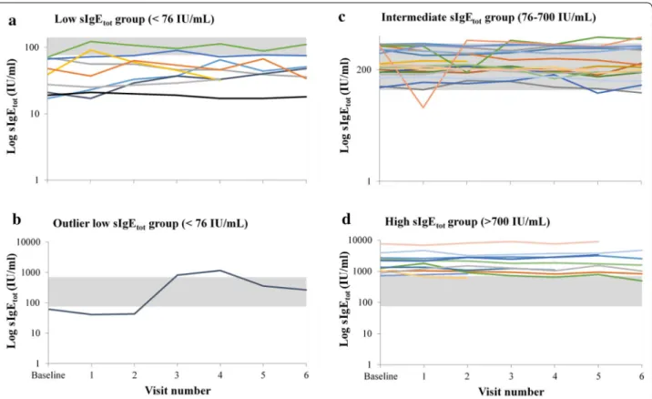
Detailed individual trajectories over time by baseline IgE strata are shown in Fig. 1, panels a, c, and d. Ten of the 41 patients (24%) reached values beyond the limit of their own baseline sIgEtot subgroup during at least one
of the follow-up visits. Of these 10 patients, 4 were in the low, 4 in the intermediate, and 2 in the high sIgEtot
subgroup, therefore representing 40% of the patients in the low sIgEtot subgroup, 20% of the patients in the
intermediate sIgEtot subgroup and 18% of the patients in
the high sIgEtot, respectively.
Two patients had sIgEtot levels in the three categories
over the 1 year period of observation. One patient initially in the low sIgE group had a rise in IgE above 700 IU/mL for at least 2 consecutive measurements before returning to intermediate levels at the two last visits (Fig. 1b). This patient was subsequently found to have developed a parasitic infection.
The relevant clinical data for this patient are presented in Table 3. The patient did not have any positive RAST results at baseline and was therefore considered non-atopic.
Relationship between variation in sIgEtot and demographics, environmental exposure and treatment features
Age was a significant predictor of sIgEtot levels over
12 months. sIgEtot decreased by 3% with each additional
year of age (p = 0.01). Allergen exposure for patients with a history of a positive RAST test was associated with change in sIgEtot. Exposure scores ranged from 0 to
12 and reflected the sum of the frequencies of exposure for the different allergens for which the patient had shown a positive RAST. A unit increase in the exposure score was associated with a 5% rise sIgEtot (p = 0.002).
No relationship was found between sIgEtot and gender,
smoke exposure, and asthma treatment.
Relationship between sIgEtot and lung function, asthma control, and exacerbation
There was no relationship between lung function (% predicted FEV1) and sIgEtot variation at baseline or
Table 1 Baseline characteristics stratified by sIgEtot group (low, intermediate, high)
BMI, Body Mass Index; D. pteronyssinus, Dermatophagoides pteronyssinus; FEV1, forced expiratory volume in 1 s; GERD, gastroesophageal reflux disease; ICS, inhaled corticosteroids; ICS/LABA, inhaled corticosteroids/long-acting β2 agonists; LABA, long-acting β2 agonists; LTRA, leukotriene receptor antagonist; OCS, oral
corticosteroids; RAST, radio-allergo sorbent test; SD, standard deviation; SABA, short-acting β2 agonists; SABA/SAAC, short-acting anticholinergic
* p-value < 0.05 were considered statistically significant; ** Regression analysis for age/Fisher’s exact for other (categorical) variables; *** Tukey test for age/general linear model with contrasts for other (categorical) variables, the column indicates between which groups were observed the statistical differences: 1 = low sIgEtot
subgroup, 2 = intermediate sIgEtot subgroup and 3 = high sIgEtot subgroup
Baseline characteristics (N = 41) Low IgE group (< 76 IU/mL) (N = 10)
Intermediate IgE group
(76–700 IU/mL) (N = 20) High IgE group (> 700 IU/mL) (N = 11)
p value** Difference specification***
Age (years) Mean (± SD) 57.7 ± 15.7 49.5 ± 14.7 40.4 ± 13.4 0.03* 1 vs 3 Gender Male 5 (29%) 8 (47%) 4 (24%) 0.84 Female 5 (21%) 12 (50%) 7 (29%) Race Caucasian 10 (28%) 19 (53%) 7 (19%) 0.03* 1 vs 3; 2 vs 3 Other 0 (0%) 1 (20%) 4 (80%) Smoking status Never smoked 6 (26%) 12 (52%) 5 (22%) 0.27 Former smoker 3 (25%) 7 (58%) 2 (17%) Current smoker 1 (17%) 1 (17%) 4 (66%) BMI (kg/m2) < 30 9 (25%) 17 (47%) 10 (28%) 0.79 ≥ 30 1 (25%) 3 (75%) 0 (0%) Medical history Allergic rhinitis 2 (9%) 13 (56%) 8 (35%) 0.02* 1 vs 3; 1 vs 2 Atopic dermatitis 3 (17%) 11 (61%) 4 (22%) 0.33 GERD 4 (44%) 4 (44%) 1 (11%) 0.23 Nasal polyps 2 (25%) 3 (38%) 3 (38%) 0.59 Residence City 4 (18%) 10 (45%) 8 (36%) 0.51 Suburbs 4 (40%) 5 (50%) 1 (10%) Rural/country 2 (22%) 5 (56%) 2 (22%)
GINA level of asthma control
Controlled 3 (21%) 8 (57%) 3 (21%) 0.39
Partly controlled 2 (18%) 7 (64%) 2 (18%)
Uncontrolled 5 (33%) 4 (27%) 6 (40%)
Positive RAST testing
D. pteronyssinus 4 (18%) 11 (50%) 7 (32%) 0.65 Grass mix 4 (22%) 6 (33%) 8 (44%) 0.07 2 vs 3 Cat dander 2 (12%) 10 (59%) 5 (29%) 0.41 Dog dander 2 (12%) 7 (44%) 7 (44%) 0.16 Birch 0 (0%) 5 (42%) 7 (58%) 0.005* 1 vs 3; 2 vs 3; 2 vs 3 Mold 1 (8%) 5 (42%) 6 (50%) 0.1 Treatment ICS/LABA 10 (24%) 20 (49%) 11 (27%) – SABA 5 (18%) 18 (64%) 5 (18%) 0.02* 1 vs 2; 2 vs 3 LTRA 2 (10%) 12 (60%) 6 (30%) 0.11 OCS 1 (13%) 3 (37%) 4 (50%) 0.34 SABA/SAAC 1 (33%) 1 (33%) 1 (33%) 1 LABA 0 (0%) 1 (100%) 0 (0%) 1 Asthma status % Predicted FEV1—mean (SD) 72.3 ± 28.6 86.6 ± 18.6 80.2 ± 12.2 0.20 ACQ total score—median (range) 12.0 (2.0–14.0) 8.0 (5.0–13.0) 8.0 (4.0–13.0) 0.77 Exhaled nitric oxide—median (range) 15.6 (7.1–46.7) 20.8 (9.6–154.0) 120.9 (5.7–236.0) 0.82
exacerbations the year prior to enrollment were not correlated significantly with baseline sIgEtot in the
total study group (OR = 0.95; p = 0.21), nor in the low (OR = 0.85; p = 0.95), intermediate (OR = 0.82; p = 0.42) or high subgroup (OR = 0.96; p = 0.31). Over the entire study period, there was no correlation between sIgEtot
and severe exacerbations since the previous visit nor with
exacerbation that occurred the week before the visits. At baseline there was no relationship between sIgE and ACQ6 in the whole group (Spearman’s ρ = 0.01, p = 0.96)
nor in any of the three groups. There was a significant but weak correlation between sIgEtot and ACQ6 score across
all time points (r = 0.15, p = 0.02).
Influence of seasons on sIgEtot asthma control
Figure 2 shows the variation in sIgEtot (panel a) and
asthma control (panel b) according to the seasons. In the total study group, there was no association between seasons (March–May, June–August; September– November; December–February) and sIgEtot (p = 0.47) or
ACQ6 score (p = 0.43).
Discussion
There are limited data in the literature on the stability of IgE measurements over time in asthmatics. In the current study, we investigated variations in sIgEtot in
severe asthmatics over a 1 year period with repeated measurements every 2 months. The principal finding is
Table 2 Intra-cluster correlation (ICC) of serum total IgE (sIgEtot) evaluated over the 12-month study period in three strata of asthmatic patients based on sIgEtot at baseline: < 76 IU/mL (n = 10); 76–700 IU/mL (n = 20) and > 700 IU/mL (n = 11)
CI, confidence interval
* Between-patient variability/(between- + within-patient variability)** likelihood ratio test comparing within-patient variabilities
ICC (95% CI) (*) 1-ICC (95% CI) p-value (**)
< 76 IU/mL 96 (93–98) % 4 (2–7) %
76–700 IU/mL 71 (62–82) % 29 (18–38) % < 0.0001 > 700 IU/mL 96 (94–99) % 4 (1–6) %
Fig. 1 The 12-month variability in sIgEtot in severe asthmatic patients stratified by sIgEtot at baseline (low, intermediate and high). The 12-month
variability in sIgEtot was evaluated in 41 patients divided post hoc in three strata based on sIgEtot at baseline: < 76 IU/mL (n = 10) (a); 76–700 IU/
mL (n = 20) (c) and > 700 IU/mL (n = 11) (d). One outliner patient from the low sIgEtot group is shown separately (b). The gray zone indicates the
76–700 IU/mL region (intermediate group) to allow for the identification of patients whose sIgEtot reached the level of a different group during the
that most of the variability sIgEtot was due to
between-patient variability whereas within-between-patient variability of sIgEtot levels was rather limited. sIgEtot was found to
decrease with age.
The relative stability in sIgEtot suggests that the
mechanisms regulating IgE levels are sustained over time in most patients. Nonetheless, 24% of patients showed sIgEtot level scores beyond the range of their own group
for at least one visit. This was particularly the case in patients with baseline sIgEtot < 76 IU/mL as 4 out of 10
patients had a measurement above 76 IU/mL in at least one subsequent blood sample.
Omalizumab is a biologic with proven efficacy to improve asthma quality of life and reduce asthma exacerbations in severe allergic asthmatics. The findings from our study suggest that regular monitoring of sIgEtot
in routine clinical practice may assist in optimizing treatment by aligning it with observed variations in sIgEtot and thus avoid under-treatment with omalizumab.
As the 40% of patients with sIgEtot < 76 IU/mL at baseline
but subsequent serum levels in the 76–700 IU/mL range underscore, without repeated blood sampling these patients might not have been initiated on omalizumab, which may have translated into a subsequent preventable need for corticosteroid therapy or into preventable exacerbations and hospitalizations. Likewise, the 20% of patients with sIgEtot between 700 and 1000 IU/
mL who subsequently showed serum levels below the 700 IU/mL threshold might not have been initiated on omalizumab without repeated testing. However, patients with very high serum IgE levels (> 2000 IU/mL) remain fairly stable in this “high” zone. Although between-patient variability was the major trend identified in our
study, limited within-patient variability was still present. Therefore, regular monitoring of sIgE over 1 year results in an increase from 50 to 66% of patients falling into the range between 76 and 700 IU/mL. The limited patient variability resulted in the fact that two-third (27/41) of our patients treated with high doses ICS/LABA had a serum IgE values between 76 and 700 IU/mL after a 1 year period of observation (including 7 measurements for most of them), a figure clearly higher than 50% of patients as observed at baseline.
Despite the limited within–patient variability, one patient with baseline sIgEtot < 76 IU/mL showed
significant changes in serum levels over the observation period. This patient who was non-atopic, maintained low IgE levels (< 76 IU/mL) at the first two visits, after which there was a marked increase to 818 IU/mL at the third and 1159 IU/mL at the fourth visit, before declining at subsequent visits without, however, returning to low serum levels. This patient was a severe corticosteroid-dependent asthmatic in whom a parasitic infection was detected and treated accordingly. Remarkably, when the sIgEtot increased sharply at visit 3, the patient’s
ACQ6 scores rose from 2.3 to 4.2 without any change
in FEV1 but with an augmentation of the OCS dose as
treatment adjustment. This case points at the importance of considering possible etiologies when a sharp rise in IgE is detected. IgE levels are known to increase in the case of parasitic infection, namely helminth infections [13]; yet other causes of elevated IgE outside an atopic status should also be considered including infections by mycobacterium tuberculosis, Epstein–Barr virus, cytomegalovirus, malignancies, or chronic inflammatory/ dysimmune disorders [14].
Table 3 Clinical data for the outliner patient belonging to the low serum total IgE (sIgEtot) subgroup (see Fig. 1b)
FEV1, forced expiratory volume in 1 s; FENO, fractional exhaled nitric oxide; OCS, oral corticosteroids
Visit Baseline 1 2 3 4 5 6
sIgEtot (IU/mL) 61.3 41 43 818 1159 357 266
FEV1 predicted (%) 49 61 64 52 59 65
Total ACQ score 13 19 14 25 19 15 16
FENO (ppm) 7.1 4.8 18.6 28.1
Asthma exacerbation x x x x x
Asthma exacerbation requiring OCS (if
yes dosis mg) 32 16
Parasitic infection x
Budesonide/formoterol (µg/days) 1200/54 1200/54 800/36 1200/54 1200/54 1200/54 1200/54
OCS dosis (mg/days) 16 16 8 16 16 16 16
Beclamethasone/formoterol 200/18 200/18 200/18 200/18
Salbutamol x x x
Fenoterol/ipratropium x
Theophylline x x x x x
Though not powered for that purpose, our study also showed an inverse relationship between age and serum IgE, a finding we have recently reported in a
large cross-sectional study in asthmatics [15] and which was previously established in longitudinal population studies [5, 6]. A decrease in sIgEtot by 3% with every year Fig. 2 Variation in sIgEtot and asthma control in severe asthmatic patients (n = 41) according to the seasons. Data from the 6 follow-up visits
are shown according to the month of the visit (the baseline data are not included). In these graphics, the geometric mean sIgEtot with standard
increase in age was observed in the present study. Not unexpectedly, allergen exposure score was associated with a rise of sIgEtot in sensitized subjects, supporting a
role for allergen exposure in IgE synthesis in addition to its role in mast cell activation thereby confirming a recent study [9].
In contrast to what is known for eosinophils [16, 17], there are limited data on the relationship between sIgEtot and asthma control and severity. A higher sIgEtot
level in severe asthmatics as compared to that in mild to moderate asthmatics has recently been reported in a large real-life cross-sectional study even if there was a considerable overlap between the groups [15]. In the same study, there was however no correlation between ACQ7
and serum IgE. In the study reported here, though not present at baseline, a weak but significant correlation was observed between sIgEtot and ACQ6 score throughout
the study period, which suggests that fluctuation in IgE over time in a subject may relate to day-to-day asthma symptoms. By contrast, no relationship was found between sIgEtot and severe exacerbation rates. Recent
data from SARP (Severe Asthma Research Program) have shown an inverse and surprising relationship between serum IgE and propensity to severe exacerbation in asthmatics [18].
Omalizumab convincingly reduces asthma exacerbation in clinical trial and routine practice in patients with serum IgE ranging from 30 to 700 IU/ mL. The efficacy of omalizumab was shown when free circulating IgE was reduced by more than 95% and maintained at lower than 30 IU/mL [19]; however, sIgEtot itself prior to treatment initiation does not
predict the ability of the drug to prevent exacerbation [20]. Rather than considering the baseline IgE level, the IgE level at which omalizumab is able to maintain free circulating IgE may be more relevant in terms of reduction in exacerbations. It is also likely that airway (and not serum) IgE, which can be assessed in sputum [4, 21] and bronchial biopsies [22, 23], plays a more important role in the disease than circulating IgE though a convincing correlation was reported between serum and sputum IgE levels [4]. We might have found a stronger relationship between IgE and exacerbation if we had measured airway IgE rather than serum IgE in the present study.
The main limitation of our study is the limited number of patients included and followed up throughout the protocol. This may have resulted in a lack of association between sIgE and seasons in contrast to what was reported by others [9]. Moreover, we did not found a relationship between smoke exposure and serum IgE. Our negative finding does not preclude, however, any real effect of smoke exposure on serum IgE because
of the limited number of current smoking asthmatics in our cohort. Future studies with larger sample sizes are needed to confirm our findings and explore associations with other possible determinants of IgE variation. Asthmatics in this study were selected on the basis of disease severity defined by the requirement of high-dose ICS combined with a LABA. Therefore, the IgE variability depicted here may not be applicable to a population of mild untreated asthmatics. As we did not have detailed history data regarding atopic dermatitis we could not assess the influence of the disease evolution on sIgEtot over the study period; specifically,
whether change in atopic dermatitis severity may have influenced change in serum IgE in our study. In our study the majority of patients with atopic dermatitis were in the intermediate IgE group.
We conclude that in severe asthmatics receiving high-dose ICS combined with a LABA, there was limited within-patient variability of sIgEtot but significant
between-patient variability. Yet 30% of the patients not initially eligible for receiving omalizumab based on a single measurement may actually benefit from repeated measurements to qualify for treatment.
Abbreviations
ACQ: Asthma Control Questionnaire; ACQ6: six-item Juniper Asthma Control
Questionnaire; BMI: Body Mass Index; D. pteronyssinus: Dermatophagoides
pteronyssinus; FcεRI: high-affinity IgE receptor; FEV1: forced expiratory volume
in 1 s; FENO: fractional exhaled nitric oxide; GERD: gastroesophageal reflux disease; ICC: intra-cluster correlation; ICS: inhaled corticosteroids; ICS/LABA: inhaled corticosteroids/long-acting β2 agonists; IgE: immunoglobulin E;
LABA: long-acting β2 agonist; LTRA : leukotriene receptor antagonist; OCS:
oral corticosteroids; OR: odds ratio; RAST: radio-allergo sorbent test; SABA: short-acting β2 agonists; SABA/SAAC : short-acting β2 agonists/short-acting
anticholinergic; SARP: Severe Asthma Research Program; SD: standard deviation; sIgEtot: serum total IgE.
Authors’ contributions
SG and SV designed the study. IA and KD wrote the protocol and performed the analyzes. JVS reviewed the protocol. GB, RL, AM, OM and CP collected the data. IA, KD, SG, RL, CP and AP interpreted the data. RL and AP wrote the manuscript. All authors read and approved the final manuscript.
Author details
1 Service de Pneumologie-Allergologie, CHU Sart Tilman B35, 4000 Liege,
Belgium. 2 Cliniques Universitaires Saint-Luc, Avenue Hippocrate 10,
1200 Brussels, Belgium. 3 CHU Brugmann, Place A.Van Gehuchten 4,
1020 Brussels, Belgium. 4 CUB Hôpital Erasme, Route de Lennik 808,
1070 Brussels, Belgium. 5 UZ Gent, Corneel Heymanslaan 10, 9000 Ghent,
Belgium. 6 Novartis Pharma, Medialaan 40, 1800 Vilvoorde, Belgium. 7 Matrix45,
LLC, 6159 West Sunset Road, Tucson, AZ 85743, USA. 8 University of Basel,
Basel, Switzerland. Competing interests
GB, RL, AM, OM and CP are members of an Advisory Board of Novartis Pharma, Belgium.
IA and KD work for Matrix45, LLC. By company policy employees are prohibited from owning equity in or perform services independently for sponsor organizations. Matrix45 provides similar services to other biopharmaceutical on a non-exclusivity basis.
•fast, convenient online submission
•
thorough peer review by experienced researchers in your field
• rapid publication on acceptance
• support for research data, including large and complex data types
•
gold Open Access which fosters wider collaboration and increased citations maximum visibility for your research: over 100M website views per year
•
At BMC, research is always in progress. Learn more biomedcentral.com/submissions
Ready to submit your research? Choose BMC and benefit from: Availability of data and materials
The datasets generated and analyzed during the current study are not publicly available but are available from the corresponding author on reasonable request.
Consent for publication Not applicable.
Ethics approval and consent to participate
This study was approved by the ethical committee of the CHU Liege under Approval Number B70720096731. All patients had to provide an informed consent before entering the study.
Funding
This study was funded by Novartis Pharma, Belgium. Publisher’s Note
Springer Nature remains neutral with regard to jurisdictional claims in published maps and institutional affiliations.
Received: 5 June 2018 Accepted: 4 March 2019
References
1. Gould HJ, Sutton BJ, Beavil AJ, Beavil RL, McCloskey N, Coker HA, Fear D, Smurthwaite L. The biology of IGE and the basis of allergic disease. Annu Rev Immunol. 2003;21:579–628.
2. Asai K, Kitaura J, Kawakami Y, Yamagata N, Tsai M, Carbone DP, Liu FT, Galli SJ, Kawakami T. Regulation of mast cell survival by IgE. Immunity. 2001;14:791–800.
3. Kalesnikoff J, Huber M, Lam V, Damen JE, Zhang J, Siraganian RP, Krystal G. Monomeric IgE stimulates signaling pathways in mast cells that lead to cytokine production and cell survival. Immunity. 2001;14:801–11. 4. Manise M, Holtappels G, Van CK, Schleich F, Bachert C, Louis R. Sputum
IgE and cytokines in asthma: relationship with sputum cellular profile. PLoS ONE. 2013;8:e58388.
5. Barbee RA, Halonen M, Kaltenborn W, Lebowitz M, Burrows B. A longitudinal study of serum IgE in a community cohort: correlations with age, sex, smoking, and atopic status. J Allergy Clin Immunol. 1987;79:919–27.
6. Sherrill DL, Halonen M, Burrows B. Relationships between total serum IgE, atopy, and smoking: a twenty-year follow-up analysis. J Allergy Clin Immunol. 1994;94:954–62.
7. Bousquet J, Rabe K, Humbert M, Chung KF, Berger W, Fox H, Ayre G, Chen H, Thomas K, Blogg M, Holgate S. Predicting and evaluating response to omalizumab in patients with severe allergic asthma. Respir Med. 2007;101:1483–92.
8. Mummadi SR, Hatipoglu US, Gupta M, Bossard MK, Xu M, Lang D. Clinically significant variability of serum IgE concentrations in patients with severe asthma. J Asthma. 2012;49:115–20.
9. Hatipoglu U, Subramanian A, Campbell T, Rice R, Mummadi S, Hu B, Lang DM. Intrasubject variability in total IgE levels in patients with moderate to severe persistent allergic asthma over 1 year. J Allergy Clin Immunol Pract. 2016;4:691–6.
10. Chung KF, Wenzel SE, Brozek JL, Bush A, Castro M, Sterk PJ, Adcock IM, Bateman ED, Bel EH, Bleecker ER, Boulet LP, Brightling C, Chanez P, Dahlen SE, Djukanovic R, Frey U, Gaga M, Gibson P, Hamid Q, Jajour NN, Mauad T, Sorkness RL, Teague WG. International ERS/ATS guidelines on definition, evaluation and treatment of severe asthma. Eur Respir J. 2014;43:343–73. 11. Humbert M, Beasley R, Ayres J, Slavin R, Hebert J, Bousquet J, Beeh KM,
Ramos S, Canonica GW, Hedgecock S, Fox H, Blogg M, Surrey K. Benefits of omalizumab as add-on therapy in patients with severe persistent asthma who are inadequately controlled despite best available therapy (GINA 2002 step 4 treatment): INNOVATE. Allergy. 2005;60:309–16.
12. Juniper EF, Svensson K, Mork AC, Stahl E. Measurement properties and interpretation of three shortened versions of the Asthma Control Questionnaire. Respir Med. 2005;99:553–8.
13. Capron M, Capron A. Immunoglobulin E and effector cells in schistosomiasis. Science. 1994;264:1876–7.
14. Pien GC, Orange JS. Evaluation and clinical interpretation of hypergammaglobulinemia E: differentiating atopy from immunodeficiency. Ann Allergy Asthma Immunol. 2008;100:392–5. 15. Manise M, Bakayoko B, Schleich F, Corhay JL, Louis R. IgE mediated sensitisation to aeroallergens in an asthmatic cohort: relationship with inflammatory phenotypes and disease severity. Int J Clin Pract. 2016;70:596–605.
16. Demarche SF, Schleich FN, Paulus VA, Henket MA, Van Hees TJ, Louis RE. Asthma control and sputum eosinophils: a longitudinal study in daily practice. J Allergy Clin Immunol Pract. 2017;5:1335–43.
17. Schleich FN, Chevremont A, Paulus V, Henket M, Manise M, Seidel L, Louis R. Importance of concomitant local and systemic eosinophilia in uncontrolled asthma. Eur Respir J. 2014;44(1):97–108. https ://doi. org/10.1183/09031 936.00201 813
18. Denlinger LC, Phillips BR, Ramratnam S, Ross K, Bhakta NR, Cardet JC, Castro M, Peters SP, Phipatanakul W, Aujla S, Bacharier LB, Bleecker ER, Comhair SA, Coverstone A, DeBoer M, Erzurum SC, Fain SB, Fajt M, Fitzpatrick AM, Gaffin J, Gaston B, Hastie AT, Hawkins GA, Holguin F, Irani AM, Israel E, Levy BD, Ly N, Meyers DA, Moore WC, Myers R, Opina MT, Peters MC, Schiebler ML, Sorkness RL, Teague WG, Wenzel SE, Woodruff PG, Mauger DT, Fahy JV, Jarjour NN. Inflammatory and comorbid features of patients with severe asthma and frequent exacerbations. Am J Respir Crit Care Med. 2017;195:302–13.
19. Chapman KR, Cartier A, Hebert J, McIvor RA, Schellenberg RR. The role of omalizumab in the treatment of severe allergic asthma. Can Respir J. 2006;13(Suppl B):1B–9B.
20. Hanania NA, Wenzel S, Rosen K, Hsieh HJ, Mosesova S, Choy DF, Lal P, Arron JR, Harris JM, Busse W. Exploring the effects of omalizumab in allergic asthma: an analysis of biomarkers in the EXTRA study. Am J Respir Crit Care Med. 2013;187:804–11.
21. Mouthuy J, Detry B, Sohy C, Pirson F, Pilette C. Presence in sputum of functional dust mite-specific IgE antibodies in intrinsic asthma. Am J Respir Crit Care Med. 2011;184:206–14.
22. Takhar P, Corrigan CJ, Smurthwaite L, O’Connor BJ, Durham SR, Lee TH, Gould HJ. Class switch recombination to IgE in the bronchial mucosa of atopic and nonatopic patients with asthma. J Allergy Clin Immunol. 2007;119:213–8.
23. Ying S, Humbert M, Meng Q, Pfister R, Menz G, Gould HJ, Kay AB, Durham SR. Local expression of epsilon germline gene transcripts and RNA for the epsilon heavy chain of IgE in the bronchial mucosa in atopic and nonatopic asthma. J Allergy Clin Immunol. 2001;107:686–92.