UNIVERSITÉ DE SHERBROOKE Faculté de génie
Département de génie chimique et de génie biotechnologique
Vers le développement d'un pancréas bio-artificiel
pour la transplantation
Towards the development of a bioartificial pancreas
for transplantation
Thèse de doctorat Spécialité: génie chimique
Rajesh GURUSWAMY DAMODARAN
Jury: Professeur Patrick VERMETTE (Directeur) Professeur Denis GROLEAU (Rapporteur)
Professeur Tamas FÜLÖP Professeur Stephen BADYLAK
i
RÉSUMÉ
Le diabète sucré est déclenché par une perte de fonction et/ou du nombre total de cellules productrices d’insuline, les cellules β, que l’on trouve au sein des « Îlots de Langerhans » du pancréas. La greffe d’îlots est une des voies de traitement du diabète actuellement étudiée, et les avancées dans le domaine de l’ingénierie tissulaire pourraient améliorer la survie et la fonctionnalité des îlots transplantés. Lors de leur purification et de leur transplantation, les îlots subissent de nombreuses agressions : stress physiques et chimiques, la réponse immunitaire du patient greffé, infections post-opératoires, etc. Il est donc nécessaire de comprendre et d’améliorer les procédés de transplantation mais aussi de vérifier la fonctionnalité des îlots avant leur transplantation. Cette thèse a pour objectif de comprendre in vitro la fonctionnalité et la survie des îlots isolés en étudiant leur comportement dans un environnement 2D et dans un environnement 3D constitué de leur matrice extracellulaire native (MEC). Des pancréas de souris ont été décellularisés puis les îlots isolés ont été injectés dans le pancréas décellularisé. La viabilité et la fonctionnalité des îlots dans ce système ont été évaluées après 48h par coloration et par analyse de la sécrétion d’insuline par stimulation au glucose (GSIS). Ce manuscrit présente l’ensemble de la procédure de décellularisation du pancréas ainsi qu’une nouvelle technique d’injection d’îlots. La seconde partie de la thèse présente la solubilisation du pancréas décellularisé et l’immobilisation de ces extraits solubilisés sur des surfaces pour cultiver des îlots. Trois méthodes détaillées de solubilisation sont présentées (digestion à la pepsine, solubilisation par l’acide acétique et solubilisation par le NaOH). Des îlots isolés ont été cultivés sur ces surfaces biomimétiques et leur viabilité et fonctionnalité ont été évaluées. La dernière partie expérimentale de la thèse présente une comparaison du profil de sécrétion de l’insuline entre des îlots individuels et des populations d’îlots. Le profil cinétique a été modélisé à partir d’îlots cultivés seuls ou en groupe pendant 72h, et ce, in vitro. Trois profils cinétiques de sécrétion de l’insuline ont été observés (lent, rapide, et à taux constant), et nous avons analysé l’influence de la taille des îlots sur le profil cinétique de sécrétion. Les résultats observés lors des mesures traditionnelles de sécrétion d’insuline par stimulation au glucose (GSIS) sont discutés au regard des trois profils cinétiques de sécrétion.
Mots clés : Matrice extracellulaire, pancréas, îlots de Langerhans, cinétique de sécrétion
ii
ABSTRACT
Diabetes Mellitus involves the loss of function and/or absolute numbers of the insulin-producing β-cells normally located within the “Islets of Langerhans” of the pancreas. Islet transplantation is currently being investigated as a potential cure, and advances in tissue engineering is hypothesized to improve pancreatic islets survival and functionality. During isolation and transplantation, islets undergo physical damage, chemical damage, host-immune rejection, post-op infection, technical failures and graft failures. Therefore, it is essential to understand and improve the process of transplantation and, also, it is important to know the functionality of isolated islets before transplantation. This thesis aims to address these issues by understanding the functionality and survival of isolated pancreatic islets in vitro, by studying their behavior when cultured in 2D and 3D environments made of their native extracellular matrix (ECM). Also, insulin release kinetics of isolated single pancreatic islets was investigated and compared to those of pools of islets. The first experimental work includes the decellularization of mouse pancreata and infusing the freshly isolated islets into the decellularized pancreata. Glucose-stimulated insulin secretion (GSIS) and viability assays were performed on these islets to show the functionality and health of the islets after 48h of culture into the decelluarized pancreata. This section describes the full procedure of pancreas decellularization with a novel method to infuse islets into pancreata. The second experimental work describes the solubilisation of decellularized pancreata and the surface-immobilization of the extracts to culture islets. This study presents the detailed protocol of extraction of decellularized pancreata by three methods (pepsin digestion as well as acetic acid and NaOH solubilisation). The freshly isolated islets were cultured on these resulting biomimetic surfaces and the results of functionality and viability of these islets were discussed. The third experimental work focuses on the comparison of insulin kinetics between single islets and pools of islets. Freshly isolated individual islets and pools of islets were cultured in vitro for 72h and the kinetics of insulin release were modeled. Three modes of insulin secretion kinetics were revealed (slow-, fast- and constant-rate secretors), along with discussion on the effect of islet size on insulin release. In addition, the results of traditional GSIS on islets were compared with these secretion profiles.
Keywords: ECM, diabetes, pancreas, islets, insulin secretion kinetics, islet transplantation,
iii
ACKNOWLEDGEMENTS
I would like to thank my research director Prof. Patrick Vermette for giving me this wonderful opportunity and space to work on this project and in his lab. I specially thank him for his valuable guidance and support throughout my studies.
I would like to thank the members of my jury (Prof. Stephen Badylak, Prof. Tamas Fülöp and Prof. Denis Groleau) for providing their valuable time to read and evaluate this thesis. Their feedback and inputs regarding this thesis are highly respected.
I very much appreciate the co-operations given by my colleagues Jamie sharp, Evan A. Dubiel, Parker Andersen, Carina Kuehn, Sylvain Vigier and Tim Spitters at Université de Sherbrooke, for their valuable feedbacks, inputs, suggestions and ideas during my studies.
A special thanks to Sylvain Vigier for the French translation of the thesis.
I would like to thank all my students Alexander, Anais, Sarah, Marc and Arnaud for helping in my experiments and data analysis.
The support provided by all the technicians Charles, Chantal, Isabelle, Valérie, Serge and Sonia are most valuable and very much appreciated.
I am most grateful to my friends Pavithran, Charith, Rajini, Giuliana, Barzin, Himmat, Djazia, David, Elaheh, Imad and Camila for sharing everything during the stay.
Finally, I am very much obliged to my wife Divya, my parents and my brother for their wonderful love and support during this journey.
iv
TABLE OF CONTENTS
RÉSUMÉ i ACKNOWLEDGEMENTS iii LIST OF FIGURES vi LIST OF TABLES ix LIST OF ABBREVIATIONS x Chapter 1 INTRODUCTION 1Chapter 2 Tissue and Organ Decellularization in Regenerative Medicine 4
Foreword 5 Résumé 6
Abstract 7
2.1 Introduction 8
2.2 Major tissues and organs used for decellularization 9 2.3 Recent decellularization methods 12
2.4 Chemicals used for decellularization 21
2.5 Rationale for using extracellular matrix (ECM)-based products 23
2.6 Immunogenicity of ECM-based scaffolds 25
2.7 Applications, status and perspective of decellularized tissues and organs 26
2.8 Conclusions 29
2.9 Acknowledgements 30
Chapter 3 Decellularized pancreas as a native extracellular matrix scaffold for pancreatic islet seeding and culture 31
Foreword 32
Résumé 33
Abstract 34
3.1 Introduction 35
3.2 Materials and methods 36
v
3.4 Conclusion 46
3.5 Acknowledgements 48
Chapter 4 Solubilisation of decellularized pancreata and immobilization on low-fouling surfaces for islet culture 49
Foreword 50
Résumé 51
Abstract 52
Chapter 5 Insulin secretion kinetics from single islets reveals distinct sub-populations 61
Foreword 62
Résumé 63
Abstract 64
5.1 Introduction 65
5.2 Materials and methods 66
5.3 Results 72
5.4 Discussion and conclusion 78
Chapter 6 CONCLUSION 83
Bibliography 90
Appendix A 119
Appendix B 132
vi
LIST OF FIGURES
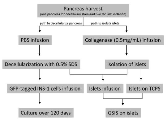
Figure 1: Experimental procedures for pancreas harvesting and decellularization, cells including
islets infusion as well as scaffold and cells/islets characterization. One full pancreas is used for decellularization and two for islet solation.………...37
Figure 2: Pictures of the pancreas during decellularization over 12 hours………...……41 Figure 3: Hematoxylin/eosin (H&E) histological staining of (A) native pancreas and (B)
decellularized pancreas. Alcian blue/nuclear fast red histological staining of (C) native pancreas and (D) decellularized pancreas……….42
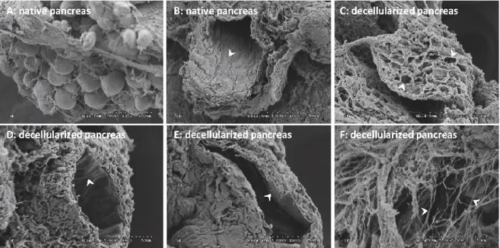
Figure 4: Comparison of native and decellularized pancreata by scanning electron microscopy
(SEM). A) Clusters of pancreatic cells along with an ECM network. B) Intact duct with ductal cells (white arrow). C) Decellularized pancreas showing empty cellular spaces. D) Scalloped structure of inner wall of blood vessel. E) Basal lamina of decellularized ductal port. F) Structures showing collagen fibrils………..……….…..43
Figure 5: (5A) Glucose-stimulated insulin secretion (GSIS) assay on islets maintained in tissue
culture polystyrene (TCPS) plates and cultured in decellularized pancreata for 48 h, insulin concentration given per islet. (5B) Immunohistochemistry staining positive for insulin and DAPI, Green - Insulin and Blue - DAPI. ……….………...……….45
Figure 6: Green fluorescent protein (GFP)-transfected INS-1 cells (A) on a decellularized
pancreas at Day 0. Cells were visible in the duct at (B) Day 3 and (C) Day 10. D) Cells at Day 60 forming pseudo-islets (indicated by white arrows). E) Cells at Day 120. F) Decellularized pancreas releasing pseudo-islets from the matrix (indicated by white arrows)………46
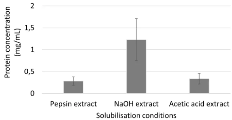
Figure 7: Protein content estimated by a Bradford protein assay of decellularized pancreata
solubilised by pepsin, NaOH and acetic acid………55
Figure 8: Microscopic images of islets after 5 days of culture on a CMD low-fouling layer, TCPS
and a pepsin control surface (i.e., “pepsin control surfaces” refer to activated CMD surfaces incubated in a solution with only pepsin without decellularized pancreata, to check the effect of
vii
immobilized pepsin on cells) and on the biomimetic surfaces bearing decellularized pancreatic extracts obtained from acetic acid, NaOH and pepsin treatments………..57
Figure 9: Stimulation indices (ratios of insulin concentration at high glucose-IBMX concentration
to that at the high-glucose) from glucose-stimulated insulin secretion (GSIS) assays on murine islets cultured on the different surfaces………..………59
Figure 10: Viability of islets cultured on the different surfaces after a GSIS assay, by staining with
fluorescein diacetate (live - green) and propidium iodide (dead - red)………59
Figure 11: (A) Raw insulin secretion data from 58 single islets and 36 pools of 10 islets. Each set
of data points from (A) was used to model the measured cumulative insulin concentration vs time allowing to identify (B) fast, (C) slow, and (D) constant-rate secretors. Each exemplified set of data points and curve is obtained from one glucose concentration. From those curves, time constants as well as as 𝐾𝑝 and 𝐾′𝑝 (fast and slow secretors) and rates of insulin secretion
(constant-rate secretors) were derived………70
Figure 12: (A) Viability of islets measured by staining with fluorescein diacetate (FDA - green)
and propidium iodide (PI - red). Islets viability at time zero: 97.9 % ± 4.3% and at 72 hours: 95.8 % ± 7.8%. Scale bars =100 μm. (B) Pool of ten islets stained with dithizone illustrating the presence of insulin-positive cells. Scale bars =100 μm. (C) Glucose-Stimulated Insulin Secretion (GSIS) from pools of 10 islets………73
Figure 13: Measured cumulative insulin concentration over 72h from single islets maintained in
(A) 2.8mM glucose, (B) 5.6mM glucose, (C) 11.2mM glucose, and (D) 20mM glucose………74
Figure 14: Percentage of islets fitted for insulin secretion kinetics for the four tested glucose
concentrations: A) single islets and B) pools of 10 islets. Overall percentage of islets fitted for insulin secretion kinetics: C) single islets and D) pools of 10 islets. For A and B, 100 percent corresponds to the total number of islets for a given glucose concentration. For C and D, 100 percent corresponds to the total number of islets for either single islets or pools of 10-islet experiments. 58 single islets and 36 pools of 10 islets were used in total………75
Figure 15: Histograms presenting a comparison of the measured cumulative insulin concentration
viii
by islet volume. Three-dimensional histograms were made by combining data points, as those exemplified in Figure 11, from different single islets for a given glucose concentration…………76
Figure 16: Measured cumulative insulin concentration over 72h from pools of 10 islets maintained
ix
LIST OF TABLES
Table 1: Recent studies involving decellularization methods and their outcomes………13 Table 2: Regulatory requirements for 510(k) devices………28 Table 3: X-ray photoelectron spectroscopy (XPS) atomic concentration (%) and atomic ratios of
x
LIST OF ABBREVIATIONS
2D two dimensional
3D three dimensional
ANOVA analysis of variance
BSA bovine serum albumin
CaCl2 calcium chloride
CMD carboxymethyl-dextran
CO2 carbon dioxide
DAB 3,3'-diaminobenzidine
DAPI 4’, 6-diamidino-2-phenylindole, dihydrochloride DMEM Dulbecco's modified Eagle medium
ECM extracellular matrix
ELISA enzyme-linked immunosorbent assay FBS fetal bovine serum
FDA Food and Drug Administration
GAG glycosaminoglycan
GSIS glucose-stimulated insulin secretion HBSS Hank’s balanced salt solution IBMX 3-isobutyl-1-methylxanthine
IEQ islet equivalent
INS-1 rat insulinoma cells
xi
MTT 3-(4,5-dimethylthiazol-2-yl)-2,5-diphenyltetrazolium bromide
P probability
PBS phosphate buffered saline
RPMI Roswell Park Memorial Institute medium
RGD Arg-Gly-Asp
RT room temperature
T1DM type 1 diabetes mellitus T2DM type 2 diabetes mellitus TCPS tissue culture polystyrene
τp time constant
1
Chapter 1
2
In 2015, the International Diabetes Federation estimated that 415 million people worldwide have been diagnosed with diabetes; with an estimated increase to 642 million by 2040. In 2017, the Canadian Diabetes Association reported that there are 11 million Canadians living with diabetes or prediabetes, the treatment of which still remains a huge physiological or medicinal, and socioeconomic problem.
In 1979, the terms type-I (T1DM) and type-II (T2DM) were assigned to classify the diabetic phenotypes; earlier it was called insulin dependent diabetes mellitus or juvenile diabetes, and non-insulin dependent diabetes mellitus or adult-onset diabetes, respectively [1].
T1DM is caused by the autoimmune destruction of insulin-producing β cells of the pancreas [2]. There are several factors including viral, bacterial, environmental, life style, and genetics which have all been associated with the pathophysiology of T1DM [3]. Due to the destruction of these cells, they can no longer produce insulin in response to glucose, thus, creating reliability on an exogenous supply of this hormone indispensable to normalize the blood glucose levels. This process is essential for long-term survival. A possible cure for T1DM came in 1966 when the first transplantation of a whole pancreas was successfully performed. Alike to other major organ transplantations, whole pancreas transplantation continues to have morbid risks including surgical complications, post-op infection, technical failures, donor obesity which all influence the success and long-term survival of such a graft [4]. Furthermore, the recipient also requires lifelong immunosuppressant drugs. Given this, transplanting islets of Langerhans may offer a less invasive, lower risk, alternative cure for T1DM. Recipients of islet transplantation attained insulin independence with normoglyceamia for a stipulated period of time, post-transplantation. This result combined with the less invasive nature of this transplantation reduced the overall post-surgical complications and improved the clinical outcome of the patient using less immunosuppressant drugs [5, 6].
The major limiting factors for islet transplantation include limitation of donors for the source of islets, immune rejection, damages of islets and of the extracellular matrix (ECM) during isolation as well as poor revascularization post transplantation [7].
Islets encapsulation could provide a possible solution to protect isolated islets. Three major advantages of islet encapsulation are: 1. to protect islets from host-immune rejection, 2. to re-establish the lost ECM to avoid anoikis and cell death and 3. to allow diffusion of molecules.
3
This thesis mainly focuses on the improvement of islet culture methods by providing information on islets behaviour in vitro on 2D and 3D ECM and the kinetics of insulin release revealing the full potential of isolated islets in terms of insulin secretion towards glucose stimulation.
Chapter 2 titled ‘Tissue and Organ Decellularization in Regenerative Medicine’ is a review of the scientific literature. Major tissues/organs used for decellularization including the recent techniques and chemicals involved in decellularization and their outcomes are discussed. This review also highlights the inevitability of ECM molecules and the possible immune reactions faced by these ECM scaffolds when transplanted. In addition, applications, status and perspective of decellularized tissues and organs are discussed in detail.
Chapter 3 consists of an experimental paper titled ‘Decellularized Pancreas as a Native Extracellular Matrix Scaffold for Islets’. Mouse pancreata were decellularized and then isolated islets were perfused into the decellularized organ, thus, concluding that these islets were viable and functional after 48h in vitro.
Chapter 4 is an experimental paper titled ‘Solubilisation of Decellularized Pancreata and Immobilization on Low-Fouling Surfaces for Islet Culture’. Briefly, the decellularized pancreata were solubilised using three different methods (pepsin, acetic acid and NaOH treatments). The extracted proteins were immobilized on low-fouling surfaces to culture freshly isolated pancreatic islets for 5 days. The results revealed that the islets were viable and functional after 5 days on the biomimetic surfaces.
Chapter 5 consists of an experimental paper titled ‘Insulin Secretion Kinetics from Single Islets Reveals Distinct Sub-Populations’. 58 islets were freshly isolated from mouse pancreata and cultured on tissue culture polystyrene (TCPS) for 72h at four glucose concentrations. The results revealed that the islets responded to glucose by three different insulin secretion profiles (slow, fast and constant rate secretors), and most of the islets were characterized as slow secretors.
4
Chapter 2
Tissue and Organ Decellularization
in Regenerative Medicine
5
Foreword
Authors and Affiliations:
Rajesh Guruswamy Damodaran: Ph.D. Candidate, Université de Sherbrooke, Département de génie chimique et de génie biotechnologique.
Patrick Vermette: Professor, ingénieur, Université de Sherbrooke, Département de génie chimique et de génie biotechnologique.
Date of Submission: Januray 25th, 2017
State of Acceptance: Under review Journal: Biotechnology Progress Contribution:
This article constitutes the scientific literature review of this thesis. The contents of this work were submitted to Biotechnology Progress. The article was written by Rajesh Guruswamy Damodaran. All work was done under the direction and supervision of Patrick Vermette.
6
Résumé
L’augmentation de la demande en tissus et organes à transplanter a entrainé l’essor de la recherche sur la décellularisation tissulaire. La préparation d’organes et de tissus décellularisés implique l’élimination de leur contenu cellulaire et génomique tout en conservant la structure complexe de la matrice extracellulaire (MEC) pour jouer le rôle de support possédant une fonction physiologique. De nombreux tissus et organes ont été décellularisés avec succès et utilisés en recherche ou comme produits commerciaux. Différentes techniques sont utilisées : traitements mécaniques, chimiques ou biologiques, qui possèdent toutes leurs avantages et inconvénients. Cet article de revue présente les récents développements dans les méthodes de décellularisation, l’importance de la nature des détergents utilisés, ainsi que le rôle de la MEC soit comme support physique, soit comme support et source des signaux pour la survie cellulaire, la différenciation cellulaire et l’homéostasie. Nous présentons également les applications et les statuts actuels des bioproduits commercialisés à partir de tissus et d’organes décellularisés.
Mots clés : décellularisation; recellularisation; ensemencement cellulaire; organes artificiels;
7
Abstract
The advancement and improvement in decellularization methods can be attributed to the increasing demand for tissues and organs for transplantation. Decellularized tissues and organs, which are free of cells and genetic materials while retaining the complex ultrastructure of the extracellular matrix (ECM), can serve as scaffolds to subsequently embed cells for transplantation. They have the potential to mimic the native physiology of the targeted anatomic site. ECM from different tissues and organs harvested from various sources have been applied. Many techniques are currently involved in the decellularization process, which come along with their own advantages and disadvantages. This review focuses on recent developments in decellularization methods, the importance and nature of detergents used for decellularization, as well as on the role of the ECM either as merely a physical support or as a scaffold in retaining and providing cues for cell survival, differentiation and homeostasis. In addition, application, status and perspectives on commercialization of bioproducts derived from decellularized tissues and organs are addressed.
Keywords: Decellularization; Recellularization; Cell seeding; Artificial organs; Detergents;
8
2.1 Introduction
Increased demand for tissues and organs for transplantation has expanded the fields of research on decellularization. Preparation of decellularized tissues and organs involves removal of cells and genetic materials from the tissue or organ and retention of the complex ultrastructure of the extracellular matrix (ECM) to serve as a natural scaffold. The ECM is produced by the native cells and serves as a mechanical support and cues for cell migration, attachment, differentiation and function [8, 9]. Many tissues and organs have been successfully decellularized and used for research and commercial purposes. Different techniques including physical, chemical and biological methods are currently applied in decellularization and these have their own pros and cons.
The two important criteria to be fulfilled during a decellularization procedure are: 1. retention of the native structures and 2. removal of a maximum of cell components. Several tissues and organs such as the small intestine, bladder, placenta and liver, only to name a few, have been decellularized and used as ECM scaffolds [10-13]. Recent advances in regenerative medicine and tissue engineering have paved the path for investigating the use of whole decellularized organs for transplantation. Many in vitro and in vivo studies support that ECM-based scaffolds made of decellularized tissues and organs have beneficial effects on cells, including stem cells, in terms of their attachment, proliferation, viability and functionality. In favor to that, these scaffolds have a low immunogenicity, which facilitates their in vivo applications [13, 14].
Over the past 20 years, decellularized tissues and organs have been commercially available for wound healing. Companies are working to commercialize ECM-based products for organ transplantation, which some are in clinical trials.
The objective of this review is to give an update on the most important tissues and organs that are used for decellularization, the importance of ECM as a complete set of biomolecules, the techniques and chemicals applied in decellularization operations, the rationale behind using ECM-based bioproducts, as well as applications, perspectives and path to commercialization.
9
2.2 Major tissues and organs used for decellularization
2.2.1 Liver
The liver is one of the important organs, which has been decellularized for regenerative medicine. The first successful liver decellularization was done by surface treatment with detergents and the resulting bioproduct was recellularized with rat hepatocytes to assess functionality [15]. Surface treatment refers to decellularizing a tissue or an organ by immersion in decellularizing solutions as opposed to perfusion decellularization, which involves perfusing solutions within the tissue or organ. Studies on liver decellularization report the perfusion of detergents through the network of veins and arteries, then its recellularization and in vivo transplantation [16-18]. For example, seeding rat hepatocytes into decellularized liver and subsequent transplantation in rats showed that less damages occurred to the cells following an 8h transplantation, as revealed by TUNEL staining [16]. In a separate study, when human liver stem cells were seeded in decellularized rat liver, differentiation into functional hepatocytes was observed as well as the presence of epithelial and endothelial-like cells [19]. Research on achieving human-sized whole liver has also been attempted with pigs by decellularizing the organ with SDS (sodium dodecyl sulfate) [20, 21].
2.2.2 Heart
Heart decellularization techniques started initially with heart valves by surface decellularization using detergents and subsequent recellularization with different cell types [22-24]. Introduced in 2008, decellularization of whole hearts from rats was done by perfusion methods and the resulting scaffolds were seeded with cardiac and endothelial cells; contraction in cell patches was observed after 4 days [25]. Recent advances in heart decellularization aim to improve perfusion processes during decellularization and cell seeding with progenitors as well as to achieve decellularization of organs from larger animals [26-30]. Allo-transplanted porcine heart seeded with mesenchymal stem cells (MSCs) showed thrombosis in arteries as well as the presence of inflammatory cells and the observation was similar to decellularized heart transplanted without mesenchymal stem cells [31]. In addition, a 14-day culture of human cardiomyocytes in human decellularized heart showed visible contraction when electrically stimulated [32].
10
2.2.3 Lungs
Decellularized lungs were considered as a source of bioartificial organs for transplantation in end-stage lung diseases. The first successful perfused decellularization was reported in 2010 following methods initially applied to heart and liver [33]. Recent works on decellularizing lung involve the improvement of decellularization process and cell seeding methods, as well as studies on the role of stem cells [33-35]. Attempts to decellularize human lungs are step closer to fill the gap in the demand for organ transplantation [36, 37]. Stabilization of endothelial cells by seeding adipose-derived stem cells into decellularized lungs could be a potential solution to regenerate the vasculature of decellularized lungs [38]. Seeding mesenchymal stromal cells in decellularized lung in suspension bioreactor resulted in differentiation of the cells into collagen-1 alpha-1 producing cells [39]. In another study, human-induced pluripotent stem cells (iPSC)-derived endothelium and ventralized iPSC were seeded in decellularized lungs resulting in retention of epithelial progenitors phenotype by expressing Nkx21 and endothelial phenotype by expressing CD31 [40]. Transplantation of decellularized rat lung seeded with epithelial and endothelial cells maintained the animals alive without ventilation for 6h [33].
2.2.4 Pancreas
The first perfused decellularization of pancreas was carried out in 2009 with a porcine organ [41] and then applied to mouse [42]. The decellularized tissues were used to seed cells and islets and their in vitro functions were studied as well as the biocompatibility of these bioartificial pancreata was investigated in vivo [41-44]. Later, rat pancreata were decellularized and a new method was introduced to infuse islets into the ducts, veins and arteries [45]. Decellularized pancreata were seeded with human stem cells, an AR427 acinal cell line, MIN-6 cells and islets, and cell attachment as well as basic cell functions were studied [42-45]. Recently published work from our group demonstrated that islets infused into the ductal system of decellularized pancreata were functional by releasing insulin in response to glucose stimulation after 48 days in vitro [46].
11
2.2.5 Small intestinal submucosa (SIS)
One of the first usages of SIS was during 1989, as a vascular graft for dogs [47]. SIS biomaterials are widely used in regenerative medicine, for example to reconstruct urethra, cornea, oesophageal, and heart [48-51]. Recently, SIS seeded with urothelial cells showed formation of new vessels, proliferation of smooth muscle cells, epithelization and maintenance of the opening of urethra when compared to unseeded scaffold [52]. Reconstruction of cornea was accomplished successfully in 106 cases by preserving vision at third month of post-surgery using porcine SIS [53]. Seeding autologous oral mucosa epithelial cells along with SIS resulted in muscular regeneration and re-epithelization after 8 weeks of surgery and the results were superior to SIS with no cells [54]. Applying FDA-approved SIS ECM seeded with mesenchymal stem cells resulted in a lower response of the adaptive immune system in a porcine model, when compared to SIS-ECM with no cells [55].
2.2.6 Bladder
Urinary bladder has been one of the oldest organs selected for decellularization. Recent studies on ECM derived from decellularized bladder has indicated a beneficial effect on neuron survival, stem cells, spinal cord and open wounds [56-59]. Urinary bladder-derived ECM has shown greater advantage than cardiac-derived ECM in remodelling and for attracting site-specific cells (cardiomyocytes) after implantation [60]. The implantation of large segments (>24cm2) of urinary
bladder into porcine bladder showed infiltration of smooth muscle cells and promoted angiogenesis after 30 days [61]. Vesicles derived from urinary bladder ECM had a positive effect on neuron neurite growth and on spinal cord injury repair [57, 58]. Urinary bladder-derived ECM also had a better effect on the treatment of open wounds when compared to conventional therapies [56]. Bioproducts made from urinary bladder-derived ECM are available in different formulations such as sheets (Cytal® Burn Matrix, also marketed as MatriStem® Burn Matrix, ACELL (Columbia, USA); the product is applied directly on wounds), powder (MicroMatrix®, ACELL (Columbia, USA)) and hydrogels (not available commercially).
12
2.2.7 Kidneys
The initial method of decellularization of whole kidneys was achieved by perfusion techniques and the resulting products were used to seed and differentiate stem cells [41, 62]. Advances in kidney decellularization include in vitro and in vivo studies on rat and mouse kidneys to investigate the functionality of recellularized kidneys with endothelial, epithelial and renal progenitor cells [63, 64].
2.2.8 Bones and cartilage
The successful removal of cells from bones and cartilages was achieved in 2011[65]. Recent advancements in cartilage and bone repair include utilizing adipose-derived stem cells, multipotent stromal cells seeded on decelluarized cartilage ECM to study the mechanical properties and bone regenerative capacity [66, 67].
2.2.9 Plants
Recent advancements in tissue and organ decellularization have extended their boundaries to apply perfused decellularization protocols on plant leaves and eventually recellularize them with cardiomyocytes resulting in contractile function successfully for 21 days [68].
2.3 Recent decellularization methods
Over the years, several studies have been conducted to understand the utility of decellularized tissues and organs in regenerative medicine. Table 1 below provides a summary of recent studies highlighting the various techniques and their corresponding results on decellularized tissues and organs.
13
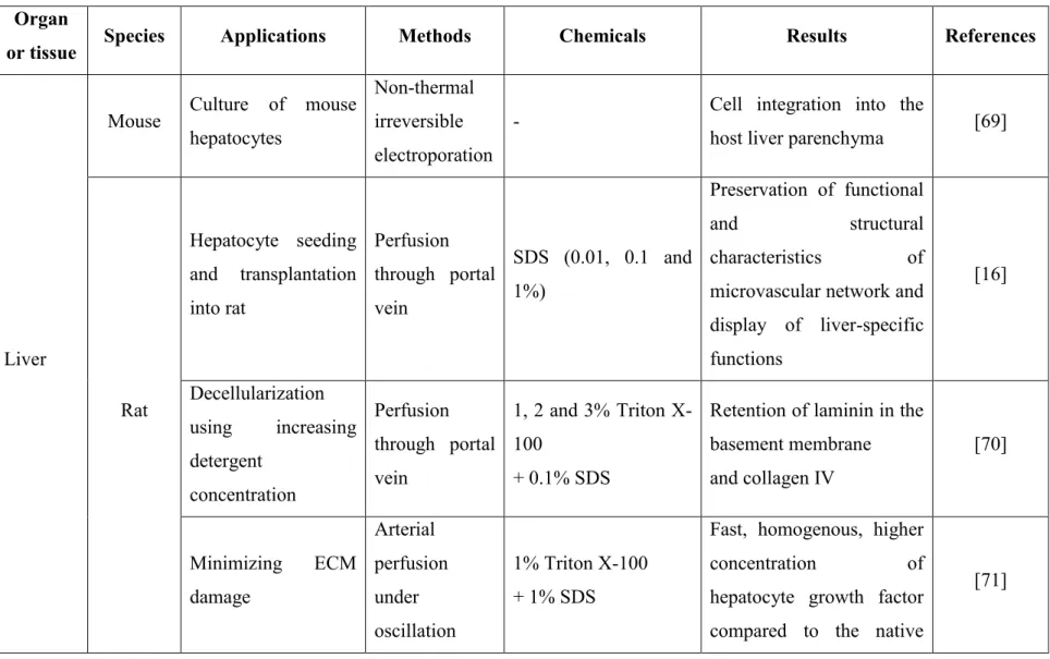
Table 1: Recent studies involving decellularization methods and their outcomes. Organ
or tissue Species Applications Methods Chemicals Results References
Liver
Mouse Culture of mouse hepatocytes
Non-thermal irreversible electroporation
- Cell integration into the
host liver parenchyma [69]
Rat Hepatocyte seeding and transplantation into rat Perfusion through portal vein SDS (0.01, 0.1 and 1%) Preservation of functional and structural characteristics of microvascular network and display of liver-specific functions [16] Decellularization using increasing detergent concentration Perfusion through portal vein 1, 2 and 3% Triton X-100 + 0.1% SDS
Retention of laminin in the basement membrane and collagen IV [70] Minimizing ECM damage Arterial perfusion under oscillation 1% Triton X-100 + 1% SDS
Fast, homogenous, higher concentration of hepatocyte growth factor compared to the native
14
Organ
or tissue Species Applications Methods Chemicals Results References
pressure conditions
tissue, high GAG concentration and gentle method of decellularization Pig Comparison of three detergent mixtures for decellularization and recellularization with rat hepatocytes
Perfusion de-cellularization and freeze-thaw 1% SDS; 1% Triton X-100 + 1% sodium deoxycholate; 1% sodium deoxycholate + 1% SDS
Cell removal, preservation of ECM structure and biocompatibility, achieved by 1% Triton X-100 + 1% sodium deoxycholate [18] Kidneys Pig Study on decellularization methods Perfusion using a high-throughput de-cellularization apparatus water and 0.5% SDS Preservation of important ECM components, intact vasculature tree, in vitro biocompatibility for cells and preservation of gross anatomy after 2 weeks of implantation [72] Heart Rat Recellularization with aortic
endothelial cells and
Coronary
perfusion 1% SDS
Performance of basic
15
Organ
or tissue Species Applications Methods Chemicals Results References
rat neonatal cardiomyocytes Zebra fish + Mouse In vitro proliferation and migration of human cardiac precursor cells Freeze-thaw cycles Exhibition of pro-proliferative and chemotactic effect with human cardiac precursor cells by Zebra fish ECM including structural preservation and cardiac contractile function
[74]
Pig Comparison of two detergents Surface treatment 1% Triton X-100 + nucleases + trypsin and sodium
deoxycholate with and without nucleases
Complete cell removal and preservation of ECM structures by Triton X-100 + nucleases + trypsin [75] Myocard -ium Pig Comparison of decellularizing agents and recellularization with Surface treatment 1% SDS; 1% Triton X-100; 0.5% trypsin Effective decellularization by SDS and retention of ECM microstructures. [76]
16
Organ
or tissue Species Applications Methods Chemicals Results References
rat myocardial fibroblasts
Display of different beating magnitudes of seeded cells: largest beating magnitude in trypsin, moderate in SDS and none in Triton X-100-treated scaffolds Teeth Human Comparison of decellularization methods Surface treatment 10% formaldehyde, PBS + EDTA + 2.5% sodium hypochlorite; PBS + EDTA + 40v (40 volume or 12%) hydrogen peroxide; PBS + 2.5% sodium hypochlorite + Ryozime®; PBS + EDTA + 40 v hydrogen peroxide + Ryozime® Maximal removal of biological particles and less damage to the structures obtained with PBS + EDTA + 40 v hydrogen peroxide + Ryozime®
17
Organ
or tissue Species Applications Methods Chemicals Results References
Bones and cartilages Rat Calvaria regeneration in vivo and recellularization with mesenchymal stem cells Surface treatment 0.5% SDS + 0.1% ammonium hydroxide
Formation of new bone and merging with the host bone after 3 months. Proliferation and osteogenic differentiation of mesenchymal stem cells
[78] Pig Tissue characterization and recellularization with murine fibroblasts and porcine chondrocytes Surface treatment 1% SDS Retention of compatible tension properties of intact tissue but reduction in compression. No cytotoxicity on recellularized cells [79] Bovine Tissue characterization and recellularization with human adipose mesenchymal stem cells Surface treatment 10 mM Ethylenediaminetetra-acetic acid disodium salt dihydrate (Na2EDTA, pH 7.2–
7.4)
Chondrogenic
differentiation of human adipose mesenchymal stem cells [80] Skeletal Muscle Pig Comparison of decellularization Surface treatment 1% SDS; 1% SDS Formation of jelly-like hydrogel by using [81]
18
Organ
or tissue Species Applications Methods Chemicals Results References
methods and formulation of hydrogel scaffolds + 0.2% sodium deoxycholate; 1% SDS + 1% Triton X-100; 1. 0.2% Trypsin/0.1% EDTA, 2. 0.5% Triton X-100, 3. 1% Triton X-100/0.2% sodium deoxycholate 0.2% trypsin/0.1% EDTA, 0.5% Triton X-100, and 1% Triton X-100/0.2% sodium deoxycholate treatment
Nerve Rat Reconstruction of long gap nerve injury
Surface treatment
(Dulbecco’s modified Eagle medium + 10% fetal bovine serum and 4%
penicillin/streptomyci n/amphotericin B) + (DMEM with 10% fetal bovine serum and 2%
penicillin/streptomyci n/
amphotericin) + PBS
Exhibition of functional
nerve regeneration in vivo [82]
Skin Mouse Comparison of detergent-free decellularization Surface treatment (50nM latrunculin B + Dulbecco’s modified Eagle medium) + 0.6 Retention of matrix composition and biomechanical property [83]
19
Organ
or tissue Species Applications Methods Chemicals Results References
with ionic and anionic detergents for decellularization mol/L potassium chloride + 1.0 M potassium iodide with detergent-free decellularization method, but, bio-functionally all methods responded equally
Trachea Rabbit In vivo
transplantation
Freeze-drying, detergent and sonication
1% SDS
Exhibition of necrosis and animal death in 7-24 days and integration of decellularized scaffold into the host structure was observed [84] Spleen Rat Recellularization of bone marrow mesenchymal stem cells (BMSCs) Freeze-thaw cycles and perfusion decellularizatio n 0.1% trypsin, 0.05% EDTA, 3% Triton X-100 Differentiation of BMSCs into functional hepatocyte-like cells [85] Culture of rat hepatocytes Perfusion decellularizat-ion 0.1% SDS
Survival and secretion of urea and albumin for 10 days in culture
20
Organ
or tissue Species Applications Methods Chemicals Results References
Pancreas
Mouse
Recellularization of MIN-6 cells and AR42J Perfusion decellularizat-ion 0.5% SDS Upregulation of insulin gene in vitro, less immune reaction and angiogenesis after 14 days in vivo
[42]
Recellularization of MIN-6 cells and in vivo implantation Freeze-thaw at -800C for one day followed by perfusion decellularizat-ion 1% Triton X-100 + 0.1% ammonium hydroxide Biocompatibility and angiogenesis induction [44] Rat Comparison of three perfusion routes for decellularization and recellularization of islets Perfusion decellularizat-ion 1% Triton X-100 + 0.5% SDS
Reach of islets into parenchymal space of the pancreas after infusion through ductal system
[45]
BMSCs: bone marrow mesenchymal stem cells; ECM: extracellular matrix; GAG: glycosaminoglycans; PBS: phosphate-buffered saline; SDS: sodium dodecyl sulfate
21
2.4 Chemicals used for decellularization
Any decellularizing procedure involves disruption of the ECM. The selection of decellularizing agents causing the least possible damage to the ECM is vital. The choice of decellularizing agents depends on the nature of the tissue or organ. A decellularization operation involves notable changes in the tissue and organ color, starting from its original color to become translucent. The process consists in removing cellular components and nuclear materials. Destruction of the cell membrane, dissolution of cellular components and removal of nucleic acid materials are the key steps.
Alkalis and acids dissolve cellular components and wash nucleic acids [87]. Detergents solubilize cell membranes and remove cellular components and nucleic acids [88].
Detergents are soluble amphiphiles, which can solubilize biological membranes. They have both hydrophilic and hydrophobic groups with a more hydrophilic nature than biological membranes. They can be classified according to their charges: non-ionic, anionic, cationic and zwitterionic detergents [89, 90]. Triton X-100, a non-ionic detergent, is commonly used for cell lysis and permeabilization [91, 92]. The hydrogen bonds in lipid bilayers are disrupted by the polar head group of Triton 100, breaking the cell plasma membrane [89]. In some studies, Triton X-100 has been reported to be the best decellularizing agent compared to other detergents and that for many tissues and organs including ligaments, small intestine, annulus fibrosus, liver and aortic valves [49, 93-96]. On the other hand, some studies demonstrated that Triton X-100 had poor cell removal capacity for many tissues and organs such as veins, cornea, urethra, heart and kidneys [25, 97-100].
Another detergent commonly used in decellularization protocols is sodium dodecyl sulfate (SDS). SDS is an anionic detergent, used mainly in commercial applications such as in cleaning and in the making of cosmetics. SDS is also well known in biochemistry for its use in the SDS-PAGE technique. Its application in decellularization can be counterintuitive when aiming to preserve the tissue or organ ECM structure intact, as SDS is known to denature proteins [101]. SDS solubilizes membrane proteins and thus helps effectively in the decellularization process. SDS incorporates into the outer membrane layer by increasing its curvature, creating a stress which results in membrane disruption [102]. SDS proved to be superior when compared to other
22
detergents in removing cells and genetic materials in many tissues and organs including aorta, veins, heart, kidneys and urethra [25, 97, 99, 100, 103]. Triton X-100 and SDS have also shown negative influence on tissues and organs used for decellularization but, on the other hand, when these two detergents were used in combination, the mixture allowed efficient decellularization and preservation of ECM structures and this has been tested on tendons, kidneys, small intestinal submucosa and liver [18, 50, 104, 105].
Another ionic detergent/bile salt frequently used in decellularization is sodium deoxycholate (SDC). After adhering to the membrane, SDC incorporates its cholate moiety into the lipid membrane, resulting in pore formation and membrane disruption [106]. SDC, alone or in combination with other detergents, has shown to be effective in removing cells while retaining ECM structures in decellularized tissues [23, 107].
CHAPS, 3-[(3-cholamidopropyl)dimethylammonio]-1-propanesulfonate hydrate, is another detergent used in decellularization. CHAPS is a zwitterionic detergent with a hydrophilic side and a hydrophobic back. CHAPS does not result in pore formation; instead, the molecules seem to directly break the lipid membrane [108]. CHAPS has proven to be milder and to have greater ability to retain mechanical strength in lungs [34, 37, 109].
Other chemicals applied in decellularization protocols are acids and bases. Both acids and bases had a negative impact on the ECM mechanical strength [110]. Acetic acid-treated bovine pericardia had reduced tensile strength but, on the other hand, it supported the growth of human mesenchymal stem cells and, hence, proved to be biocompatible [111]. Peracetic acid along with ethanol was used to sterilize decellularized scaffold materials, a well-known and FDA-approved method for medical devices [13]. However, the use of peracetic acid for decellularization resulted in incomplete cell removal from tumor ECM, porcine liver and kidneys [112, 113]. Decellularization of skin using NaOH had a cytotoxic effect on fibroblasts, however, prolonged washing of the scaffold helped in decreasing this cytotoxic effect [114]. Alcohols or acetone solutions can be used in the pre-treatment of tissues for lipid removal. It is easier to lyse and remove cells after eliminating lipid contents [115]. Acetone/alcohol combinations had a negative effect on tissue dehydration and greater influence on modifying mechanical properties and morphology of temporomandibular joint from pig jaws, as compared to SDS-treated tissues [116].
23
Along with chemicals, the use of enzymes in decellularizing techniques can be advantageous. It is not possible to decellularize tissues or organs only with enzymes. However, enzymes are involved in the removal of cell debris. The most commonly used enzymes are trypsin, nucleases, collagenase, thermolysin, α-galactosidase and dispase [110, 117].
2.5 Rationale for using extracellular matrix (ECM)-based products
ECM proteins have shown positive influence on cell growth, function and differentiation and examples of biomimetic surfaces have been reviewed by us [118].
An important class of ECM-derived proteins is that of collagens, as reviewed elsewhere [119]. Collagen is in fact an abundant source of ECM proteins. For example, collagen microspheres can differentiate oligodendrocyte progenitors into oligodendrocytes and collagen-immobilized nanowires had a positive influence on human microvascular endothelial cells [120, 121]. Fibronectin and laminin showed numerous positive effects on cell survival and functions both in vitro and in vivo [122, 123]. In the past years, our group has verified the positive effects of various ECM peptides and proteins. For example, immobilization of fibronectin, fibrinogen and fibrin on polymer surfaces enhanced the proliferation of smooth muscle cells [124]. Also, embedding pancreatic islets and endothelial cells in fibrin showed increased insulin secretion and preservation of islet integrity [125]. Culturing young porcine islets in fibrin exhibited the protective nature of fibrin towards hydrogen peroxide [126].
In addition to whole ECM proteins, functional peptides derived from the ECM can influence cell behaviour [127-130]. Peptide motifs from collagen I (GTPGPQGIAGQRGVV), collagen IV (MNYYSNS), fibronectin (PHSRN) and laminin (YIGSR) can enhance cell attachment [131, 132]. Arginine-glycine-aspartic acid (RGD), a peptide sequence found in fibronectin, vitronectin, laminin, collagen types I and IV, has a high affinity for integrin-mediated cell attachment and spreading, functionality and tissue development [133-137]. The RGD sequence co-immobilized with the SVVYGLR sequence was used to enhance the adhesion of endothelial cells[130]. Three ECM-derived peptides, laminin (IKLLI and PDSGR) and cadherin (HAVDI) supported the adhesion and survival of insulin-containing cultures for 5 days [129]. Rat insulinoma INS-1 cells cultured on immobilized ECM proteins and peptides (RGD &
24
CDPGYIGSR) showed higher glucose-stimulated insulin secretion compared to surfaces bearing no peptides [127].
Hyaluronic acid, a polysaccharide, helped in neuron survival and controlled self-renewal and differentiation of human embryonic stem cells [138, 139]. Many studies demonstrated that proteoglycans from the ECM are involved in molecular events of cell adhesion, proliferation and migration [140].
These are just examples of the effects ECM components can have on cells allowing for appreciating the potential impact of bioproducts made from decellularized tissues and organs. As reviewed by us and others, [118], [141] [142], other ECM proteins have been applied to produce biomimetic materials and surfaces and these include: collagen types I, III and IV, gelatin, fibronectin, vitronectin, polysaccharide nanofibers, proteoglycans, nidogen and laminins, as well as ECM-mimicking peptides including YIGSR,RGD, DGEA, KRSR, IKVAV, P15, and GFOGER, to list a few.
Furthermore, following their implantation, decellularized scaffolds are repopulated by host cells and are even degraded, resulting in a functional tissue containing site-specific cells [143]. Decellularized tissues or organs, with or with no cells, can chemo-attract progenitor cells towards the implantation site [144, 145]. The degrading ECM molecules are also involved in various biological activities and growth factor signaling [146, 147]. Therefore, these matrices made from decellularized tissues and organs act as bioactive scaffolds by modifying the host environment to create a more suitable solution for healing, repair and even regeneration.
ECM scaffolds made from decellularized tissues and organs also provide mechanical support in many conditions. They possess different physical characteristics, which are often difficult to reproduce with synthetic materials [148]. The ECM provides a mechanical support for the recellularizing cells to attach to the matrix. It is very important for scaffolds made from decellularized tissues and organs to have certain mechanical properties matching the site of implantation of the tissue or organ to be substituted. Creating less damage to the tissue during decellularization could yield better mechanical properties and more intact vasculature structure, facilitating the induction of angiogenesis post-implantation [44]. Also, the presence of ECM proteins and functional peptides in 3D scaffolds allows for recreating environments more physiological for cell culture, as opposed to traditional 2D cultures on flat surfaces.
25
2.6 Immunogenicity of ECM-based scaffolds
Most decellularization protocols involve removing cell materials, which could cause major immunogenic reactions: hyperacute rejection, acute immune rejection, chronic immune rejection, and inflammatory reactions. Hyperacute rejection is a severe reaction of vascularized xenografts, triggered a few minutes to a few hours after implantation [149, 150]. This hyperacute rejection is mainly caused by the α-Gal epitope (galactose-α(1,3)-galactose) expressed on many cell surfaces and is less common in human and old-world monkeys. Pigs have been modified genetically to remove the Gal epitope to avoid xenograft rejections, but the application of this method is resulting into an acute immune rejection by non-Gal antibodies circulating at low levels, a few days to a few weeks after implantation [151]. Chronic immune rejection is associated with allograft transplantation by introducing donor-specific antibodies [152]. Also, the presence of DNA in decellularized tissues and organs can potentially cause inflammatory reactions [153].
Collagens with lower immunogenicity is achieved by removing N-and C- terminal telopeptides by pepsin type-I treatment, which are referred to as atelocollagen [154]. Atelocollagens have been clinically applied in wound healing and as bone substitutes [154].
Host response towards bioproducts made from ECM materials are different and are based on the product’s properties, species and methods of preparation; five different commercially available products were tested on rats and resulted in distinct morphological appearance of the implantation site[155]. Similarly, the immune reaction from the decellularized heart tissue (SynerGraft®) had a mixed reaction after implantation. In one study [156], SynerGraft® exhibited much less HLA (human leukocyte antigen) class I and II antibodies, as claimed by the company; however, in another study [117], macrophage infiltration after implantation was observed into the tissue decellularized using the SynerGraft® process [149]. In addition to that, porcine small intestinal submucosa had both positive and negative inflammatory responses when implanted into rodents [157-159].
Another important factor to be studied in detail is the response of host innate and adaptive immune response towards implanted xenograft materials. The polarization of T helper cells 1 (Th1) and macrophages (M1) results in a pro-inflammatory response and T helper cells 2 (Th2)
26
and macrophages (M2) are involved in the anti-inflammatory response, which leads to constructive tissue remodelling[160].
Many studies have different conclusions on host-decellularized tissues/organs interactions and responses. Therefore, a detailed scientific understanding of the outcomes following decellularized product implantation including the responses of host tissues towards the implanted decellularized materials is vital [161].
2.7 Applications, status and perspective of decellularized tissues and
organs
Engineered substitutes could be a possible cure for patients in the last stage of tissue or organ failure. The increase demand for tissues and organs for transplantation and the encountered immune rejection of xenograft transplantations have pushed researchers to find alternative tissue and organ sources.
Decellularized tissues and organs have a great potential to serve as carriers for transplanting autologous tissue made from the host cells or site-specific functional cells. Regardless of the availability of genetically engineered animals to harvest tissues or organs for transplantation, the rejection of the transplanted graft by the host appears inevitable [162]. This situation can potentially be solved with scaffolds and other bioproducts made from decellularized tissues and organs since antigenic elements of cell components are supposed to be completely removed. Recent advances involving recellularization of human-induced pluripotent stem cells in decellularized scaffolds could be a way to avoid the intake of immunosuppressive drugs required after organ transplantation [40, 163].
Achieving a functional tissue or organ is the goal of any decellularized scaffolds carrying either functional cells or stem cells. Failure of re-endothelialization of the organ with functional endothelium is one of the main reasons for organ loss in transplantation and a major hindrance in developing functional decellularized scaffolds. However, many studies have shown promising results on endothelialization and these could be considered as a potential solution to solve re-endothelialization problems [164, 165].
27
Materials and scaffolds made from decellularized tissues and organs have been commercially available for many applications. ECM-derived products from animal sources have been commercially available for more than 20 years and are applied in the treatment of many tissue regeneration processes and statistically, many years ago, over 200,000 patients have been implanted with xenogeneic decellularized scaffolds [166]. Many animal and human tissue-derived decellularized commercial products like Oasis® (porcine small submucosa, Cook Biotech, Inc., Indiana, USA), GraftJacket® (human dermis, Acelity L.P. Inc., Texas, USA), DermACELL® (human dermis, Novadaq Technologies Inc., Mississauga, Canada), Alloderm® (human dermis, Allergan plc (NYSE: AGN, Dublin, Ireland)), NeoFormTM (human dermis, California, USA),
StratticeTM (porcine dermis, Allergan plc (NYSE: AGN, Dublin, Ireland)), RestoreTM (porcine
small intestine (DePuy Orthopedics, Inc., Indiana, USA), PrimaTM Plus (porcine heart valve,
Edwards Life Sciences LLC, California,USA), AlloSkin™ AC (human dermis, AlloSource, Centennial, CO, USA), MatriStem® (mucosa of urinary bladder, ACell, Columbia, USA), Biodesign® (small intestine, COOK MEDICAL INC., Bloomington, IN, USA), Lyoplant® (pericardium, Aesculap, Inc., Center Valley, PA, USA)[167, 168].
Most of the biological scaffolds are marketed as surgical mesh devices [168]. Scaffolds made from decellularized tissues or organs have the potential to be marketed under the category of 510(K) of the FDA (Food and Drug Administration) [169]. Under 510(K) clearance, a company must register to notify (PreMarket Notification or PMN) the FDA 90 days in advance of marketing a medical device. The FDA would assess the notified device to make sure it falls under the already existing three classifications of devices in the market, for which a premarketing approval (PMA) is not required. The claim must be made by the company to support its device is substantially equivalent to the ones already available in the market. 510(K) devices can fall under 3 classes: class I, class II and class III, depending on the assurance of safety and effectiveness.
28
Table 2: Regulatory requirements for 510(k) devices [170]. Regulatory
requirements Class I Class II Class III
General controls
All general controls are complied with and provide reasonable assurance
General controls are insufficient to provide reasonable assurance
General controls are insufficient to provide reasonable assurance
Secondary controls No further secondary controls are required
Information required to perform secondary controls are sufficient and hence secondary
controls are
performed for gaining reliable assurance of the device
Information required to perform secondary
controls are
insufficient and hence secondary controls cannot be performed for gaining reliable assurance of the device
Premarket
notification/approval
Eligible directly for premarket notification
Eligible directly for premarket notification
Requires premarket approval prior to notification
Device/product risk Low Medium High
Note: General controls refer to a comprehensive set of regulatory authorities to be complied for by any medical device to be marketed. Special controls are controls to be complied where general controls are insufficient.
Oasis®, MiroMesh® and PhotoFix® are a few examples of 510(K)-cleared commercially available bioproducts derived from decellularized tissues and organs [167, 171].
Decellularized tissues which are intended to be implanted or transplanted have the potential to be marketed under the category Human Cells, Tissues, and Cellular and Tissue-Based Products
29
(HCT/Ps) of the FDA. HCT/Ps are products regulated by two different divisions, namely, the Center for Devices and Radiological Health (CDRH) & the Center for Biologics Evaluation and Research (CBER), or sometimes as a combination of both [172]. DermACELL® (a decellularized human dermis) and Allopatch HD® (an acellular human dermis) are examples of HCT/Ps products [167, 173]. Miromatrix Medical Inc. (Eden Prairie, Minneapolis), a USA-based biotechnology company founded in 2009, is involved in developing fully functional human organs by perfusion technology, primarily applied to the liver and, upon completion, extended to the kidneys. Miromesh™ and Miroderm™ (derived from porcine liver) are products launched by the company. AxoGen Inc. (Alachua, Florida) from the USA develops a technology to regenerate peripheral nerve injuries. AxoGuard® is derived from pigs and is used as a nerve connector. Another USA-based company, Humacyte, Inc. (Morrisville, North Carolina), develops acellular grafts to repair vascular damages; some of their products are under Phase III clinical trials.
2.8 Conclusions
The selection of a technique or chemicals to decellularize a specific tissue or organ is an important decision to achieve complete decellularization and to retain intact the ECM. Perfusion decellularization has allowed fast and complete decellularization of many whole tissues and organs using either SDS or Triton X-100. Many whole organs have been successfully decellularized and subsequently seeded with different cell types. Recent advances in recellularizing decellularized tissues and organs involve the use of induced pluripotent stem cells to attain the native function of the tissue. Decellularized tissues or organs offer both mechanical support and cues from the ECM for cell growth and function in vivo. Many bioproducts derived from decellularized tissues and organs are commercially available, along with that, regenerative medicine companies are working to market functional whole organs to meet the demand. Extracting purified proteins from ECM or synthesizing new motifs are costly affairs compared to decellularizing a tissue, therefore, decellularized tissues and organs could be a potential source of substitutes for transplantation, provided they are devoid of cellular components, maintain the necessary functionality and are less immunogenic.
30
2.9 Acknowledgements
This research project was supported by the Université de Sherbrooke and NSERC through a Discovery Grant awarded to Patrick Vermette (Grant # 250296-2012).
31
Chapter 3
Decellularized pancreas as a native
extracellular matrix scaffold for pancreatic islet
seeding and culture
32
Foreword
Authors and Affiliations:
Rajesh Guruswamy Damodaran: Ph.D. Candidate, Université de Sherbrooke, Département de génie chimique et de génie biotechnologique.
Patrick Vermette: Professor, ingénieur, Université de Sherbrooke, Département de génie chimique et de génie biotechnologique.
Date of Submission: July 1st, 2017
State of Acceptance: Published
Journal: Journal of Tissue Engineering and Regenerative Medicine Contribution:
This article constitutes an experimental study of this thesis. The content of this work is published in Journal of Tissue Engineering and Regenerative Medicine. All experimental work was performed by Rajesh Guruswamy Damodaran. All data were analysed by Rajesh Guruswamy Damodaran and Patrick Vermette. The article was written by Rajesh Guruswamy Damodaran. All work was performed under the direction and supervision of Patrick Vermette.
33
Titre en français :
Pancréas décellularisé comme support naturel pour les îlots
Résumé
Le diabète se caractérise par une perte de fonction ou du nombre de cellules productrices d’insuline, les cellules-β, dans les îlots pancréatiques. La transplantation d’îlots est une des thérapies étudiées comme traitement du diabète, et des développements dans les méthodes d’ingénierie tissulaire peuvent améliorer la survie et la fonctionnalité des îlots transplantés. La transplantation des îlots les soumet à l’anoïkis, à l’hypoxie et aux attaques du système immunitaire, ce qui les fragilise et peut conduire à la destruction du greffon. Les avancées récentes en ingénierie tissulaire permettent d’utiliser des organes décellularisés comme supports aux cellules greffées. Le pancréas décellularisé pourrait être un bon support pour la transplantation d’îlots dans la mesure où l’on peut conserver correctement sa matrice extracellulaire (MEC) et son système vasculaire. Pour cette étude, le pancréas de souris a été décellularisé par perfusion d’une solution de 0,5% de dodecyl sulfate de sodium (SDS). Avec différentes techniques de caractérisation, nous montrons que le pancréas perd l’intégralité de ses cellules mais conserve une part importante de sa MEC. Des îlots isolés sont injectés dans le pancréas décellularisé via le canal collecteur et la fonctionnalité des îlots est confirmée par réponse à la stimulation au glucose (GSIS) après 48h. La re-cellularisation du pancréas par des cellules INS-1 exprimant la GFP montre que ce support est biocompatible et non-toxique après 120 jours. Ces données suggèrent que le pancréas décellularisé ensemencé par des cellules endocrines pourrait devenir un organe bioartificiel potentiellement utilizable dans le traitement du diabète.
Mots clés : matrice extracellulaire; biomatériaux; pancréas décellularisé; fonction des îlots
34
Abstract
Diabetes Mellitus involves the loss of function and/or absolute numbers of insulin-producing β-cells in pancreatic islets. Islet transplantation is currently being investigated as a potential cure, and advances in tissue engineering methods can be used to improve pancreatic islets survival and functionality. Transplanted islets experience anoikis, hypoxia and inflammation-mediated immune response, leading to early damage and subsequent failure of the graft. Recent development in tissue engineering enables the use of decellularized organs as scaffolds for cell therapies. Decellularized pancreas could be a suitable scaffold as it can retain the native extracellular matrix (ECM) and vasculature. In this study, mouse pancreata were decellularized by perfusion using 0.5% sodium dodecyl sulphate (SDS). Different characterizations revealed that the resulting matrix was free of cells and retained part of the pancreas ECM including the vasculature and its internal elastic basal lamina, the ducts with their basal membrane as well as the glycosaminoglycan and collagen structures. Islets were infused into the ductal system of decellularized pancreata and glucose-stimulated insulin secretion (GSIS) results confirmed their functionality after 48h. Also, re-cellularizing the decellularized pancreas with GFP-tagged INS-1 cells and culturing the system over 120 days confirmed the biocompatibility and non-toxic nature of the scaffold. GFP-tagged INS-1 cells formed pseudo-islets which were, over time, budding out of the decellularized pancreata. Decellularized pancreatic scaffolds seeded with endocrine pancreatic tissue could be a potential bioengineered organ for transplantation.
Keywords: extracellular matrix (ECM) scaffolds; pancreas decellularization; pancreatic islets
![Table 2: Regulatory requirements for 510(k) devices [170].](https://thumb-eu.123doks.com/thumbv2/123doknet/5429431.127155/41.918.106.818.146.806/table-regulatory-requirements-k-devices.webp)