The role of Caulobacter crescentus XerC and XerD
recombinases in site-specific recombination
par
Hua Liu
Département de microbiologie et immunologie
Faculté de Médecine
Mémoire présenté à la Faculté des études supérieures
en vue de l’obtention du grade de Maître ès sciences (M. Sc.)
en microbiologie et immunologie
D
é
cembre, 2010 ©Hua Liu, 2010Ce Mémoire intitulée :
The role of Caulobacter crescentus XerC and XerD
recombinases in site-specific recombination
Présentée par :
Hua Liu
a été évalué par un jury composé des personnes suivantes :
...Dr Patrick Hallenbeck...
président-rapporteur
...Dr George Szatmari...
Directeur de recherche
...Dr Luke Masson...
Membre du jury
XerC et XerD, deux recombinases impliquées dans la recombinaison site spécifique, résolvent les multimères d’ADN en monomères. Cette réaction se produit au niveau du site dif du chromosome, et nécessite le domaine C-terminale de la protéine de division cellulaire FtsK. Caulobacter crescentus est une bactérie aquatique de type Gram-négative qui se retrouve dans plusieurs environnements. Elle présente un cycle cellulaire asymétrique avec deux types de cellules distinctes. Cette propriété peut être utilisée pour synchroniser la croissance d’une population bactérienne pour permettre l’étude de l’expression de gènes à travers le temps et les liens entre le cycle cellulaire et le développement de la bactérie. La liaison à l’ADN et la capacité de former des complexes covalents (phosphotyrosyl) avec le site dif de C. crescentus (ccdif) ont été testé pour les recombinases de C. crescentus (ccXerC et ccXerD). Les deux recombinases ont eu une meilleure liaison au demi-site gauche de ccdif et sont incapable d’effectuer une liaison coopérative, contrairement à ce qui se produit au niveau du site dif de E. coli. La formation de complexes covalents a été testé en utilisant des «substrats suicides avec bris» marqués à la fluorescence ainsi que des protéines de fusion (marquées ou non à la fluorescence). Des complexes ADN-protéines résistants à la chaleur et au SDS ont été observé lors de la réaction de ccXerC et ccXerD de type sauvage avec ccdif, mais pas lors de la réaction de mutants avec le même ADN. Des complexes covalents phosphotyrosine sont formés de façon plus efficace sur les substrats suicides avec un bris au niveau du brin supérieur que ceux ayant un bris au niveau du brin inférieur. Dans les deux cas, c’est ccXerC qui est resté lié de façon covalente à l’ADN de ccdif.
Mots-clés : Recombinasion spécifique de site /tyrosine recombinase/XerC/XerD/dif
Summary
In most bacteria, the chromosomal dimer resolution process is mediated by two tyrosine recombinases, XerC and XerD, which bind cooperatively and perform the recombination reaction at the dif site near the terminus of replication. This reaction also requires the C-terminal domain of the cell division protein FtsK. Caulobacter crescentus is an aquatic Gram-negative bacterium found in various environments. This bacterium has an asymmetric cell cycle which can be used to synchronize cell growth in order to study the temporal expression of a gene and the interconnection between the cell cycle and development. The binding activity and the formation of phosphotyrosyl complex of the C. crescentus recombinases, ccXerC and ccXerD, were tested on the C. crescentus dif (ccdif) site. Both ccXerC and ccXerD bound preferentially to the left half-site of ccdif and showed reduced cooperative binding, unlike what was found with the E. coli dif site. Covalent complex formation activity was tested by using fluorescently labelled linear “nicked suicide substrates” and labelled proteins. Heat and SDS-resistant protein-DNA complexes were formed when both wild-type ccXerC and ccXerD reacted with ccdif but not in the presence of active-site tyrosine mutant proteins. Phosphotyrosine complexes formed on the top-nicked suicide substrate were found to be more efficient than on the bottom-top-nicked suicide substrates and surprisingly ccXerC remained bound to both top and bottom-nicked ccdif suicide substrates.
Keywords: Site-specific recombination/tyrosine recombinase/XerC/XerD/dif/Caulobacter crescentus
TABLE OF CONTENTS
Title page……….…...i Identification of jury……….…….ii Résumé……….……….……iii Summary………..………...iv Table of content……….v List of tables..……….………..…...viii List of figures………...…….ix List of abbreviations………...xiCHAPTER I: INTRODUCTION
………..………1 1. Site-specific recombination ………...1 1.1. Generalities ………..11.2. The resolvase/invertase family ………....5
1.3. Lambda integrase family ………..9
1.3.1. Generalities..………...9
1.3.2. The recombination reaction ……….11
2. Xer site-specific recombination ………12
2.1. Generalities ………....12
2.2. XerC and XerD ………...16
2.2.1. XerC………..21 2.2.1.1. Generalities ………...21 2.2.1.2. Function ………...21 2.2.2. XerD ………...22 2.2.2.1. Generalities ……….22 2.2.2.2. Function ………...23
2.2.3. The catalytic mechanism of XerC and XerD ………...24
2.3. The site of action of the Xer recombinases ………....33
2.3.1.1. Escherichia coli dif site ………...36
2.3.1.1.1. Position and polarity ……….36
2.3.1.1.2. Structure ………....41
2.3.1.1.3. Co-Location Model………....45
2.4. FtsK ……….47
2.5. Regulation of Xer recomnibation ………...56
2.5.1. DAZ and FtsK control ……….56
2.5.2. Homologous recombination control ………...57
3. Caulobacter crescentus ………...59
3.1. C. crescentus--A dimorphic polarized bacterium ………...59
3.2. The C. crescentus cell cycle and regulation ………...63
3.2.1. CtrA ………..65
3.2.2. DnaA ………67
3.2.3. GcrA ………...69
3.3. FtsK in C. crescentus .………...72
3.4. Xer/dif system in C. crescentus ………..73
4. The Master’s project ………...76
CHAPTER II: ARTICLE
………..………...………..77Abstract ………...78
1. Introduction ………79
2. Materials and methods ………..83
2.1 Bacterial strains and plasmids………....83
2.2 Growth condition and DNA manipulations ………..83
2.3 PCR conditions………...84
2.4 Oligonucleotides ………...85
2.5 Purification of Xer-MBP fusion proteins ………..85
2.6 Protein labeling ………...86
2.7. DNA-binding assay ………...86
2.8 In vitro cleavage assay ………...86
3.1 ccXerC and ccXerD bind to the ccdif………88
3.2 In vitro cleavage of ccdif suicide substrate………..………..90
3.3 ccXerC forms phosphotyrosyl complexes on both top and bottom nick strand of ccdif……….92
4. Discussion ………..………...……….…………94
5. Acknowledgements ………...……….………...…...……100
6. References ………...……….……….….…...101
7. Figure legends ………...109
CHAPTER III: DISCUSSION
……….…...1171. Binding activity of ccXerC and ccXerD with Caulobacter dif site ………....117
2. ccXerC and ccXerD bind to both half-sites of ccdif with different affinities...121
3. Reduced cooperativity binding between ccXer proteins with ccdif site ………...124
4. The ability of ccXerC and ccXerD to form phosphotyrosine covalent complexes on ccdif suicide substrate………..128
5. Phosphotyrosine covalent complex with ccdif could not be formed with active site tyrosine mutant proteins………130
6. ccXerC forms phosphotyrosyl complexes on both top and bottom-nicked strand of ccdif ……….133
7. Complementation assay ……….137
8. Perspectives..……….………..…...……..140
REFERENCES
………..…..144LIST OF TABLES
CHAPTER I: INTRODUCTION
TABLE 1: Aligment of dif site from different bacteria and
LIST OF FIGURES
INTRODUCTION
FIGURE 1: Outcomes from site-specific recombination………..………3
FIGURE 2: Recombinational mechanism of the integrases and resolvases……….5
FIGURE 3: Model of the action of the serine recombiases ……….9
FIGURE 4: Sequential strand exchange by the tyrosine recombinases .………12
FIGURE 5: Schematic of a model for the control of catalytic activity within the XerCD–DNA complex ………...18
FIGURE 6: Overall structure of the XerD protein………...26
FIGURE 7: Comparison of the structures of the C-terminal domains of XerD, λ Int and HP1Int ………27
FIGURE 8: Model of XerD bound to DNA ………29
FIGURE 9: A model of the complex between XerC, XerD, and DNA ………...31
FIGURE 10: Control of Catalysis in Xer Recombination ………...32
FIGURE 11: Positions of Ter sites in E. coli……….….………..38
FIGURE 12: Alignment of half-sites from some of the known recombination loci ……...44
FIGURE 13: Hierarchy of specificity determinants in the XerC and XerD binding sites of dif ………..44
FIGURE 14: The co-location model ………46
FIGURE 15: Pseudomonas aeruginosa FtsK domain organization and conservation …....48
FIGURE 16: Crystal Structures of the FtsKγ Domain with and without DNA …………...52
FIGURE 17: FtsK-Dependent and independent Pathways of Xer Recombination at dif …55 FIGURE 18: Sister chromatid exchange (SCE) leads to circular dimer chromosomes …..59
FIGURE 19: Caulobacter cell cycle ………61
FIGURE 20: Model for the temporal control of DNA replication initiation in Caulobacter ……….71
ARTICLE FIGURE 1: Alignment of dif recombination sites and gel retardation analysis of ccXerC and ccXerD binding to ccdif ………111
FIGURE 2: Reduced cooperative binding of ccXerC and ccXerD with ccdif …………..112 FIGURE 3: ccXerC and ccXerD bind preferentially to the left half-site of ccdif …....…..113 FIGURE 4: Phosphotyrosyl complex formation
on ccdif DNA using suicide substrates ………...114 FIGURE 5: Y-F recombinase mutants do not form
phosphotyrosyl complexes with suicide substrates ………115 FIGURE 6: XerC, but not XerD forms phosphotyrosyl complexes
on suicide substrate …………...…….………....116
DISCUSSION
FIGURE 7: Binding activity of ccXerC and ccXerD with ecdif ………...120 FIGURE 8: Binding activity of ccXerC and ccXerD to the
left and right half-site of ccdif ………...122 FIGURE 9: Cooperative binding between ccXerC and ccXerD with ecdif ………..126 FIGURE 10: Comparison of wild-type recombinases and
mutant recombinases binding activity ……….132 FIGURE 11: Alignment of C. crescentus and E. coli
FtsK protein C-terminal domains ………..137 FIGURE 12: Complementation assay ………140
List of Symbols and Abreviations
AMINO ACIDS MEASUREMENT UNITS
A: alanine Å: angstrom unit C: cysteine bp: base pair D: aspartic acid cm: centimetre E: glutamic acid Da: Dalton F: phenylalanine g: gram G: glycine h: hour H: histidine kb: kilobase I: isoleucine kDa: kilodalton K: lysine μg: microgram
L: leucine μl: microlitre
M: methionine μΜ: micromolar
N : asparagine min: minute
P: proline ml : millilitre Q: glutamine mM : millimolar
R: arginine ng: nanogram
S: serine s: second
T: threonine ×g: centrifugation speed V: valine °C: Degree Celsius
W: tryptophan v/cm: volt per centimeter
OTHERS
Ap: ampicillin ATP: adenosine triphosphate
ATPase: adenosine triphosphatase BSA: bovine serum albumin
ccdif: Caulobacter dif site ccXerC: XerC of Caulobacter crescentue ccXerD: XerD of Caulobacter crescentue C-terminal: carboxyl-terminal
DAPI: 4’, 6-diamidino-2-phenylindole ecdif: Escherichia coli dif site
ecXerC: XerC of Escherichia coli ecXerD: XerD of Escherichia coli DIG: digoxygenin DNA: deoxyribonucleic acid
EDTA:ethylenedinitrolotetraacetic acid HJ: Holliday junction
IPTG: isopropyl B-D thiogalactopyranoside LB: Luria-Bertani
MBP: maltose binding protein NaCl: sodium chloride
NEB: New England Biolabs PBS: phosphate bufferred saline
PCR: polymerase chain reaction SDS: sodium dodecyl sulfate
TBE: tris-borate EDTA buffer THA: Todd-Hewitt broth with agar
Ts: thermosensitive α: alpha β: beta λ: lambda
UV: ultraviolet Y: tyrosine λ Int: λ phage integrase 3’OH: three prime hydroxyl 3’PO4: three prime phosphate
ChapterI
INTRODUCTION
1. Site-specific recombination
1.1. Generalities
Genetic recombination is central to DNA metabolism. It promotes sequence diversity and maintains genome integrity in all organisms. Recombination is the breaking and rejoining of DNA in new combinations. This genetic exchange occurs between DNA molecules from the two parents or between two DNA segments within the same molecule. Such recombination may be general, occurring between two DNA substrates with extensive homology, which is called general homologous recombination, or site-specific, occurring between two site-specific, relatively short DNA targets, which is designated site-specific recombination.
The process of site-specific recombination can be divided into a series of conceptually simple steps. Firstly, the recombinase binds to the two recombination sites. The two recombinase-bound sites pair, forming a synaptic complex with crossover sites juxtaposed. The recombinase then catalyzes cleavage, strand exchange, and the rejoining of the DNA within the synaptic complex. Finally, the synaptic complex breaks down, releasing the recombinant products. From this description, it follows that the minimal components of a site-specific recombination system are a recombinase and a pair of recombination sites. In site-specific recombination, DNA strand exchange takes place between segments possessing only a limited degree of sequence homology (Kolb, 2002;
Coates et al., 2005; Landy, 1989). The recombination sites are typically between 30 and 200 nucleotides in length and consist of two motifs with a partial inverted-repeat symmetry, to which the recombinase binds, and which flank a central crossover sequence at which the recombination takes place. The pairs of sites between which the recombination occurs are usually identical, but there are exceptions e.g. attP and attB of λ integrase (Landy, 1989). In the site-specific recombination reaction, recombinases perform rearrangements of DNA segments by recognising and binding to short DNA sequences (sites), at which they: (1) cleave the DNA backbone, (2) exchange the two DNA helices involved and (3) rejoin the DNA strands (Stark et al., 1992). While in some site-specific recombination systems having just a single recombinase enzyme together with the recombination sites is perfectly adequate to be able to perform all these reactions (Bourgeois et al., 2007), in some other systems a number of accessory proteins and accessory sites are also needed.
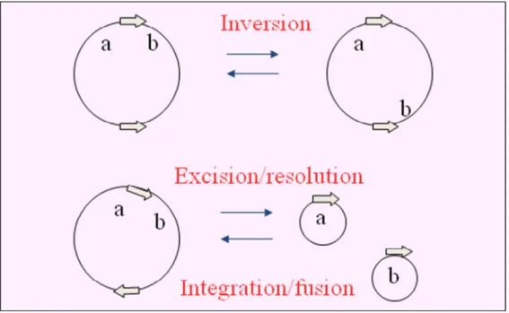
The reaction catalyzed by the recombinase may lead to different outcomes which are dictated mainly by the relative location and the orientation of sites that are to be recombined, but also by the innate specificity of the site-specific system in question. Intramolecular recombination between inverted or directly repeated sites will invert or excise respectively the intervening DNA segment. Recombination between sites on separate DNA molecules will integrate one molecule into the other (Fig. 1).
Figure 1. Outcomes from site-specific recombination. Arrows show the
orientation of the recombination sites. a and b indicate the position of distinct genetic markers and the recombination loci. ‘Excision’ and ‘integration’ refer to recombination events involving genetic entities of different size and /or function (e.g., the bacterial chromosome and a phage genome), whereas ‘resolution’ and ‘fusion’ apply to equivalent DNA molecules, (e.g., two plasmids) (Hallet and Sherratt, 1997; with permission).
Most site-specific recombination systems are highly specialised catalyzing only one of these different types of reactions and have evolved to ignore the sites that are in the ‘wrong’ orientation. The natural site-specific recombination systems are highly specific, fast and efficient, even when faced with complex eukaryotic genomes (Sauer, 1998). As such, site-specific recombination systems are employed in a number of programmed DNA-rearrangement reactions in both prokaryotes and eukaryotes. The different structural consequences of site-specific recombination lead to various biological functions. It includes helping to specify developmental pathways in bacteria and bacteriophages (Sato et al., 1990; Landy, 1993; Carrsco, 1994); determing cell type and virus host range (Zieg and Simon, 1980; Klippel, 1988; Tominaga et al., 1991); processing the products of genetic transposition (Arthur and Sherratt, 1979); and
controlling circular replicon copy number and inheritance (Summers and Sherratt, 1984; Blakely et al., 1991; Stark et al., 1992).
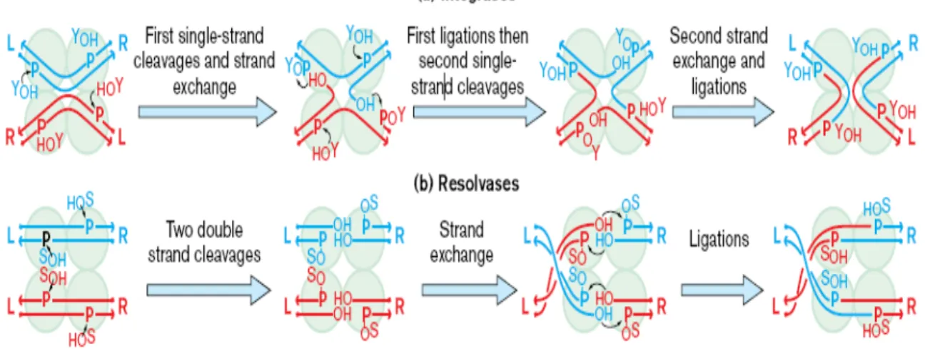
Based on amino acid sequence homology and mechanistic relatedness most site-specific recombinases are grouped into two distinct families: the tyrosine recombinase family or the serine recombinase family. The names stem from the conserved nucleophilic amino acid residue that is used to attack the DNA and which becomes covalently linked to it during strand exchange. The serine recombinase family is also sometimes known as resolvase/invertase family, named after the cointegrate-resolving protein encoded by the transposons γδ and Tn3. Tyrosine recombinases are also known as the integrase family, named after the prototypical phage λ integrase (Argos et al., 1986; Hatfull and Grindley, 1988; Sadowski, 1986; Stark, 1992). The integrase family includes λ and many other phage integrases, phage P1 Cre, the bacterial proteins XerC and XerD, and the FLP protein encoded by the yeast 2 μm plasmid. The resolvase family includes most transposon-encoded resolvases and the DNA-invertases such as Hin and Gin. Enzymes of both families catalyze conservative DNA break-join reactions that proceed by two-step transesterifications in which protein phosphodiesters act as reaction intermediates. These two families are unrelated in protein sequence or structure, and employ different recombinational mechanisms, as illustrated in Fig. 2.
Figure 2. (a) λ integrase and its relatives make ordered and sequential pairs of
single strand exchanges between the two recombinational partners; the first pair of exchanges form a four-way Holiday junction, the second pair resolves the junction to complete the recombination. The nucleophile used for cleavage and formation of the covalent recombinase–DNA intermediate is a conserved tyrosine (YOH). The cleavage
sites on each DNA duplex are separated by 6–8 base pairs with a 5′ stagger, and the tyrosine joins to the 3′ phosphate. (b) γδ resolvase and its relatives make double strand breaks in both recombinational partners, then exchange ends and rejoin them. The resolvase nucleophile is a serine (SOH) and it cleaves the DNA at sites that are separated
by 2 base pairs with a 3′ stagger, attaching to the 5′ phosphate (Grindley, 1997; with permission).
1.2. The resolvase/invertase family
The resolvase/invertase family forms a rather homogenous group of related proteins in which a conserved serine residue plays a key catalytic role (Hatfull and Grindley, 1988; Leschziner et al., 1995). There are currently approximately 40 different members, ranging in size from 180 to nearly 800 amino acid (aa) residues, and with unexpected variations in domain organization (Smith and Thorpe, 2002). The best-characterized recombinases of this family are the invertases Gin from bacteriophage Mu and Hin from Salmonella sp. and the resolvases of Tn3 and γδ transposons (Stark et al., 1992; Van de Putte and Goosen, 1992; Grindley, 1994; Arciszewska and Sherratt, 1995; Johnson, 1991). Some information regarding serine recombinase domain structure and
function given here has come from the prototypical recombinase, γδ resolvase. This 183-residue protein has an N-terminal catalytic domain of 100 183-residues, linked by a long (36aa) α-helix (the E-helix) and an unstructured segment (10aa) to a typical helix-turn-helix DNA-binding domain at the C terminus (Yang and Steitz, 1995). The serine nucleophile is close to the N terminus at position 10. γδ resolvase is a dimer in solution, with the N-terminal portion of the E-helix forming the bulk of the dimer interface. DNA binding (at least to the crossover site) involves not only the H-T-H domain but also the C-terminal portion of the E-helix and the intervening segment. The dimer's H-T-H domains bind symmetrically to the DNA, making sequence-specific major groove contacts ~10bp from the central cleavage point; E-helix residues (particularly the conserved Arg-125) hold the DNA (via phosphate and minor groove contacts) close to the cleavage site and the 3′ end of the DNA after cleavage (Li et al., 2005); and the unstructured segment snakes along the minor groove between the two (Yang and Steitz, 1995). The H-T-H domain appears to play no important roles outside of DNA binding because it could be replaced by a zinc finger DNA recognition domain in Tn3 resolvase without loss of recombination activity (Akopian et al., 2003).
In a recombination catalyzed by serine recombinases, double strand breaks staggered by 2bp occur at the middle of the two paired core sites, giving rise to recessed 5’ ends and 3’-OH overhangs (Fig. 3). One recombinase subunit is linked to each of the 5’ ends through the conserved serine residue of the family (Reed and Moser, 1984; Klippel et al., 1988). This serine presumably provides the primary nucleophile hydroxyl group in the cleavage reaction (Leschziner et al., 1995). The ligation step that follows strand exchange can be viewed as the converse of the cleavage: the protein-DNA
phosphoseryl bond of one strand is attacked by the 3’-OH end of the partner to release the enzyme and reseal the DNA backbone in the recombinant configuration (Fig. 3). Thus, recombination by a resolvase/invertase family occurs by a mechanism in which four DNA strands are broken and rejoined in a concerted manner. This mechanism is quite distinct from that of the tyrosine recombinases that proceed through the formation and resolution of a Holliday junction (HJ) intermediate, during which the DNA strands are transiently attached to recombiase subunits through phospho-tyrosine linkages (Landy, 1989; Stark et al., 1992; Gopaul and Duyne, 1999; Chen et al., 2000). In the serine recombinase reaction, all catalytic processes usually occur within a synaptic complex with two crossover sites and four recombinase subunits (although the Sin recombinase appears to be at least one exception to this, Rowland et al., 2002). It is now clear that, in synaptic complexes formed by the serine recombinases, the crossover sites are located on the outside, separated by the catalytic domains (see review Grindley et al., 2006). The recently solved crystal structure of a minimal synaptic complex formed by γδ resolvase has elegantly confirmed the “DNA-out” configuration of the crossover site synapse and has thrown new light on the processes of synapsis and strand exchange (Li et al., 2005).
Sin is a resolvase of the serine recombinase family that is encoded by various S. aureus multiresistance plasmids (Paulsen et al., 1994; Rowland and Dyke, 1989). Sin is only distantly related to known resolvases and DNA invertases (e.g. Hin, Gin), although sequence alignment implies that it has a structural fold essentially the same as that of γδ resolvase (31% identical to pI9789 Sin) (Yang and Steitz,1995). The Sin recombination system differs from that of Tn3 and γδ resolvase (Rowland et al., 2002; 2005). First is its res site, although complex is only 86bp long and binds just two dimers of Sin, and site II
consists of direct (head-to-tail) repeats of the 12bp binding sequence. Second, recombination requires an architectural, DNA-bending protein such as E. coli HU or Bacillus subtilis Hbsu. Nevertheless, like the transposon-encoded cointegrate resolvases, Sin is specific for an excision reaction (its biological role is likely to be reducing plasmid dimers to monomers to ensure their stability; another possible role, related to the dimer to monomer conversion role, is to generate monomer plasmids after conjugal transfer, where the DNA is transferred by a rolling-circle type mechanism. This could potentially generate multimeric forms which must be converted into monomers.), and the product of recombination in vitro is a pair of singly linked, catenated circles. Furthermore, another difference between Sin and Tn3/γδ resolvase is that Sin is catalytically active in the absence of synapsis (presumably as a dimer) and is able to cleave and rejoin isolated crossover sites (without site II or Hbsu) (Rowland et al., 2002). Thus, for Sin, synapsis, which is essential for the resolution reaction, may simply be a way of bringing together a pair of recombination sites in a controlled (that is excision-specific) manner. By contrast, for Tn3/γδ resolvase, synapsis not only brings the crossover sites together but also activates the recombinase.
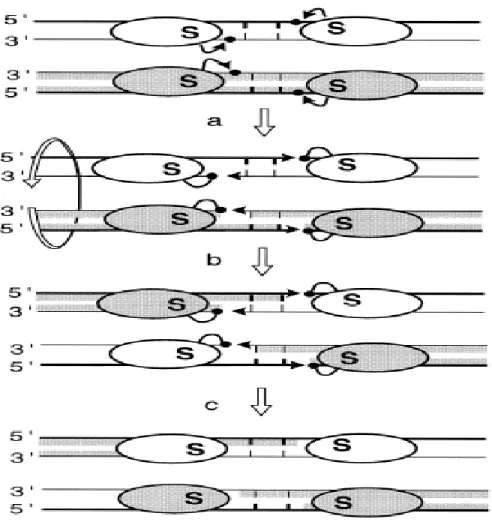
Figure 3. Model of the action of the serine recombianses. The subunit rotation
model is shown. The ovals represent recombinase subunits with the conserved catalytic serine’S’. Thick and thin lines are the top and bottom strands of the recombination sites, respectively. The short vertical bars are the 2bp of the overlap region between the two cleavage points. Black arrows represent the nucleophilic attacks of phosphates (black dots) by hydroxyl groups (arrowheads). The four DNA strands are cleaved (a), exchanged by 180 º rotations of the half-site bound subunits (b) and religated in the recombinant configuration (c) (Hallet and Sherratt, 1997; with permission).
1.3. Lambda Integrase Family
1.3.1. Generalities
The lambda integrase or ‘tyrosine recombinase’ family includes over 100 members identified based on sequence similarity (Nunes-Düby et al., 1998). Tyrosine recombinases are most widespread among prokarytoes but are also found in archaea and
even eukarytoes, where examples have been described in fungi, ciliates, and, most recently certain families of retrotransposons (Nunes-Düby et al., 1998; Poulter and Goodwin, 2005). The most well-studied examples include, in addition to the integrase protein from bacteriophage λ (Int) (Landy, 1989), the bacterial XerC and XerD recombinases (Sherratt et al., 1995), Cre recombinase from bacteriophage P1 (Hoess et al., 1985), and the Flp recombinase from the Saccharomyces cerevisiae 2μ circle (Sadowski, 1995). These recombinases carry out site-specific recombination in a stepwise manner, exchanging one pair of DNA strands to form a HJ intermediate (Craig, 1988) and then resolving the HJ to products by exchange of the second pair of strands. So, unlike the recombinases of the resolvase/invertase family, tyrosine recombinases exchange the two pairs of DNA strands separately and sequentially (Fig. 2).
Tyrosine recombinases share only limited sequence similarity and are much more divergent, with only four completely invariant residues intimately involved in catalysis: the RHRY tetrad (Argos et al., 1986; Abremski et al., 1992; Blakely et al., 1996). Alignments of this integrase family of proteins identified some conserved motifs, which are related to their catalytic function (Nunes-Düby et al., 1998; Esposito et al., 1997). Although some family members, such as FimB and FimE, contain only this domain, in most the catalytic domains are preceded by a variable N-terminal domain that helps bind DNA. All proteins harbor two conserved regions, Box I and Box II, with marked sequence similarity, originally identified from the alignment of only eight recombinases (Argos et al., 1986). Box I includes the fourth conserved residue R, and Box II contains other three conserved residues, the triad H-R-Y, which includes the active site tyrosine (Abremski and Hoess, 1992; Nunes-Düby et al., 1998). The
conservation of Box I is striking in prokaryotic recombinases and it extends with some variation to eukaryotic recombinases. Box II is also relatively strongly conserved among the prokaryotic recombinases, but less so between prokaryotic and eukaryotic proteins. Whereas the active tyrosine is absolutely conserved, the surrounding residues are rather divergent, allowing for quite different secondary structures.
1.3.2. The recombination reaction
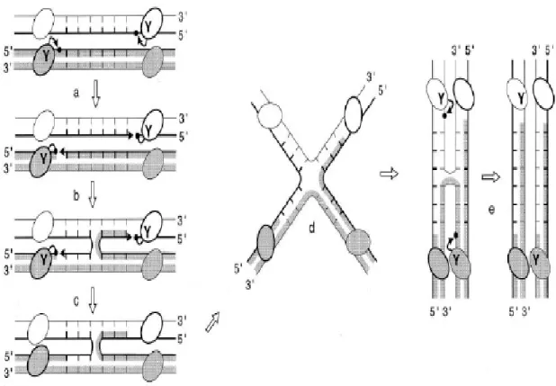
To initiate the first strand exchange, the tyrosine residue of the conserved catalytic motif RHRY attacks the phosphate of the scissile phosphodiester in one strand (defined here after as the top strand) of each recombination core sites, thereby forming a 3’phosphotyrosyl-linked- recombinase-DNA complex and generating a free 5’-OH end (Fig. 4). The polarity of this cleavage reaction is thus reversed when compared to that of the resolvase/invertase-mediated cleavages. In the second step, the recombinase-DNA phosphotyrosyl bond is attacked by the 5’-OH end from the partner duplex to generate a four-way branched structure, or HJ intermediate, in which only two DNA strands have recombined. To resolve this intermediate and complete the recombination reaction, the two other (bottom) strands are exchanged by repeating the cleavage/religation process 6-8bp downstream of the first strand cleavage position (Hallet and Sherratt, 1997).
Figure 4. Sequential strand exchange by the tyrosine recombinases. The DNA
strand swapping /isomerisation model is presented. The letter ‘Y’ refers to the conserved catalytic tyrosine. The ovals represent recombinase subunits. Thick and thin lines are the top and bottom strands of the recombination sites, respectively. Black arrows represent the nucleophilic attacks of phosphates (black dots) by hydroxyl groups (arrowheads). The top strands (thick lines) are cleaved first (a), swapped between the two partners (b), and then religated (c). The branch point of the generated HJ intermediate is positioned at the middle of the (6bp) overlap region and the top strands are crossed. Isomerisation of the HJ to a recombination configuration in which the bottom strands are crossed requires the reorganization of the DNA helices and the four half-sites-bound recombinase subunits within the complex (d). The resulting HJ isoform is resolved by repeating steps a to c in order to exchange the bottom strands (e) (Hallet and Sherratt, 1997; with permission).
2. Xer Site-Specific Recombination
2.1 Generalities
The physical state of circular chromosomes, unlike linear chromosomes, can be altered by homologous recombination. Odd numbers of homologous recombination
events between circular replicons during or after replication, produce dimers that need to be converted to monomers before they can be segregated normally at cell division (Austin et al., 1981; Blakely et al., 1991; Kuempel et al., 1991). Plasmid dimers can also arise as a consequence of rolling circle replication during conjugal transfer and sometimes during vegetative replication (Warren and Clark, 1980; Erickson and Meyer, 1993). The Xer site-specific recombination system was initially discovered in 1984 through its role in converting multimers of ColE1-related multicopy plasmids to monomers and hence ensuring their stable inheritance within E. coli (Summers and Sherratt 1984). A model for the coordination of chromosome dimer resolution and cell division has been elaborated in E. coli based on a substantial accumulation of in vivo and in vitro data. In E. coli, the Xer site-specific recombination system is composed of two paralogous tyrosine recombinases, XerC and XerD, which cooperately catalyze strand exchanges at a 28bp DNA sequence, the dif site (deletion-induced-filamentation), which must be located at the junction of the two replichores to be functional (Pérals et al., 2000; Blakely et al., 1993; Hallet et al., 1999). In addition, it has been demonstrated that FtsK protein is required for chromosome dimer resolution in vivo (Boyle et al., 2000; Steiner et al., 1999) and site-specific recombination at other ectopic dif sites (Sciochetti et al., 2001). Deletion of the E. coli dif site or mutations in xerC or xerD result in the development of a subpopulation of filamentous cells containing abnormally partitioned nucleoids. Homologues of XerCD and FtsK are found in most eubacterial phyla and some archeal lineages (Recchia et al., 1999) as well as the canonical dif site (Hendrickson et al., 2007). Moreover, interactions between the E. coli dif site and the XerCD recombinases of Haemophilus influenza (Neilson et al., 1999), Pseudomonas aeruginosa
(Blakely et al., 2000), Bacillus subtilis (Sciochetti et al., 2001), Proteus mirabilis (Villion and Szatmari, 2003), and Caulobacter crescentus (Jouan and Szatmari, 2003) have been experimentally demonstrated in vitro. These observations lead to the general view that Xer recombination is a function conserved among bacteria harboring circular chromosome(s).
The classical Xer site-specific recombination system is an atypical member of the integrase family because, instead of using a single recombinase, it uses two related recombinases, XerC and XerD, each of which catalyses the exchange of one specific pair of strands (Blakely et al., 1993, 1997; Colloms et al., 1996; reviewed in Sherratt, 1993; Sherratt et al., 1995). The use of two recombinases, XerC and XerD, by the classical Xer site-specific recombination system is unusual but not unique. For example, FimB and FimE of E. coli mediate an inversion gene switch that regulates expression of type I fimbriae (Klemm, 1986), whereas in Staphylococcis aureus, two related tyrosine recombinases of Tn554 mediate promiscuous site-specific recombination (Murphy, 1989). The use of two recombinases that bind to related yet different half-sites had provided a powerful tool for determining the precise role of different molecules as the recombination reaction proceeds. This system has evolved to ensure that a complete recombination reaction is complete only when very special conditions are met (Colloms et al., 1996). Besides the classical dif/Xer system, studies with Streptococci and Lactococci (Le Bougeois et al., 2007) indicate that these bacteria carry alternative Xer recombination machinery; an atypical 31bp dif recombination site associated with a single dedicated tyrosine recombinase (XerS). In these cases, either one Xer protein has been lost or, assuming that xerC and xerD genes arose from a single ancestral gene, these
organisms diverged from other bacterial lineages prior to this duplication (Le Bougeois et al., 2007). Recently, Carnoy and Roten (2009) analyzed 234 chromosomes from 156 proteobacterial species and showed that a subgroup of ε-proteobacteria display a sequence (difH) which is homologous to difSL from Streptococci and Lactococci and harbor a single Xer-like recombinase (XerH) (Carnoy and Roten, 2009). However, no phylogenic association between XerS and XerH could be found, which strongly suggests the existence of two unrelated dif/Xer systems: the classical machinery found in most species and an atypical system present in a sub-group of ε-proteobacteria.
Xer recombination is also distinguished from most other site-specific recombination systems by its different requirements and outcomes, depending on whether it is recombining natural plasmid-borne recombination sites (for example, cer and psi located on plasmids ColE1 and pSC101 respectively) or the chromosomal site dif. Recombination in vivo at plasmid-borne dif sites occurs intermolecularly and intramolecularly, and is not known to require proteins in addition to the two recombinases. In contrast, recombination in vivo at cer and psi is preferentially intramolecular and requires, in addition to the approximately 30bp recombination core site, about 200bp of adjacent accessory sequences, with which accessory proteins interact in order to assemble a synaptic complex that has a precise architecture and entraps three or four negative supercoils (Alén et al., 1997; Colloms et al., 1997). Xer recombination on synaptic complexes of precise topology restricts recombination to intermolcular events between directly repeated recombination sites, and therefore acts to convert dimers to monomers (Colloms et al., 1996, 1997).
2.2 XerC and XerD
The XerC recombinase was initially identified by its role in resolution of ColE1 plasmid multimers (generated by homologous recombination) to monomers. This recombination is necessary for the stable inheritance of this naturally occurring high copy number plasmid and its relatives (Summers and Sherratt, 1984; Colloms et al., 1990). A second recombinase, XerD, was identified by sequence homology to XerC and is encoded in an operon with recJ and dsbC (Blakely et al., 1993; Lovett and Kolodner 1991; Missiakas et al., 1994). Both XerC and XerD are identified as members of the tyrosine recombinase family because of the characteristic four strictly conserved amino acids required for catalysis: the Arg-His-Arg triad and the tyrosine nucleophile (Esposito et al., 1997; Sherratt and Wigley, 1998). All four residues were found to be in the C-terminal halves of the recombinase proteins, which showed more sequence similarity than the N-terminal halves. XerC and XerD are encoded at 4024kb and 3050kb on the E. coli chromosome respectively. Each recombinase is expressed with at least two other proteins that don’t appear to have a role in Xer recombination (Colloms et al., 1990; Blakely et al., 1993). Although E. coli XerC and XerD share only 37% identity, they are the closest relatives to each other among the highly diverged tyrosine recombinase family. Similarly, in other eubacteria, XerC-XerD homologues are readily identified by their homology to each other and to their E. coli recombinases (Hayes et al., 1997; Neilson et al., 1999; Recchia et al., 1999; and Sciochetti et al., 1999). The genes encoding XerC and XerD are invariably located in different regions of the chromosome, where they often lie adjacent to and may be coexpressed with genes involved in recombination, repair, and cellular response to stress (Recchia et al., 1999).
XerC and XerD proteins can be divided into two domains that make a C-shaped clamp into which the DNA is bound (Gopaul et al., 1999). Based on analogy with Cre, α-helices B and D of the N-terminal domain are expected to interact with the major groove formed by the inner palindromic nucleotides of the recombinase binding sites. The N-terminal domain is also expected to contain determinants for protein-protein interaction, whereas the larger, more conserved C-terminal domain contains the catalytic residues and determinants for specific major groove DNA binding and protein-protein interactions. The residues proposed to be involved in specific major groove DNA binding are located within α-helices G and J, with R221 and Q222 being implicated in providing specificity (Subramanya et al., 1997). XerD shows a stronger affinity for its binding site than XerC, each of them binding more tightly to its site in the presence of the partner (Blakely et al., 1993, 1997; Spiers et al., 1999).
In site-specific recombination mediated by tyrosine recombinases, two pairs of DNA strand exchanges are separated in space and time with HJ being a recombination intermediate. In Xer recombination (Fig. 5), one pair of strand exchanges is catalyzed by XerC, while the other pair of strand exchanges is mediated by XerD (Neilson et al., 1999; Colloms et al., 1996; Arciszewska et al., 1995). Recombination between psi sites is initiated by XerC-mediated strand exchange to give an HJ intermediate that is resolved to recombination products by XerD. Strand exchange by XerC initiates recombination at cer, although the resulting HJ intermediate is resolved by Xer-independent celluar processes rather than by XerD (Colloms et al., 1996; McCulloch et al., 1994). In the absence of FtsK, XerC can catalyse the formation of HJ intermediates between dif sites and their conversion back to substrate (Barre et al., 2000). The presence of FtsK and ATP
leads to a remodeling of the nucleoprotein synaptic complex, so that reaction between two duplex dif sites is now initiated by XerD, forming an HJ intermediate that can be acted on by XerC, thereby completing the dimer resolution reaction (Aussel et al., 2002; Recchia et al., 1999). A switch in the preferred order of strand exchange at psi occurs when the core site is inverted with respect to the accessory sequences (Bregu et al., 2002).
Figure 5. Schematic of a model for the control of catalytic activity within the
XerCD–DNA complex. On binding to recombination site DNA, the XerCD–DNA complex preferentially adopts a conformation in which XerC (blue ovals) has its C-terminal end region extended across the wide angle of DNA arms to the receptor region of XerD (purple ovals). Recombinase-mediated synapsis of two such duplexes forms a complex, which is primed for nucleophilic attack (red arrows) by XerC. In this conformation, monomers of XerD, whose C-terminal regions span the acute angle between the DNA arms, have their C-terminal regions compacted and are inactive. A pair of coordinated strand exchanges gives HJ intermediate, which XerC can reconvert to substrate. Alternatively, the HJ complex can undergo a conformational change to form HJ that is primed for catalysis by XerD. A pair of strand exchanges by XerD can then generate complete recombinant products. The conversion of a XerCD–DNA synaptic complex, or a XerCD HJ, to a conformation that allows catalysis by XerD is facilitated by accessory proteins like FtsK and PepA. It is proposed that one role of the accessory
factors is to allow the recombining complex to adopt the energetically less favourable conformation required for catalysis by XerD. The inset shows a dif site with the arrows indicating the cleavage sites for catalysis by XerC (top) and XerD (bottom) (Ferreira et al., 2003; with permission).
Structural, biochemical, and genetic experiments have contributed to our understanding of the ways in which the activities of the four recombinase molecules of a recombining complex are coordinated so that the two pairs of strand exchanges occur at the correct time and in the correct place. Crystal structures of the Cre–loxP synaptic and HJ complexes reveal a tetramer of recombinase molecules assembled on two, antiparallel DNA duplexes in a four-way, almost planar conformation (Guo et al., 1997,1999; Gopaul et al.,1998, 1999). The structures of both the synaptic and HJ complexes are similar and indicate the existence of two conformationally different forms of the Cre–DNA complex, each appropriate for exchange of one specific pair of DNA strands. Only a subtle change in the overall architecture of the complex is required to make a switch between the two conformational states. Genetic and biochemical experiments with XerCD support this general model. Special mutations in either the presumptive donor or acceptor regions of XerCD, and indeed at other specific positions, lead to mutant phenotypes that exhibit either reciprocal stimulation of catalysis by partner and impairment of catalysis by self (the SPIS phenotype), or reciprocal impairment of partner catalysis and stimulation of catalysis by self (the IPSS phenotype) on synthetic HJ substrates (Arciszewska et al., 2000; Hallet et al., 1999; Spiers et al., 1999). In the model of Xer recombination, the C-terminal arms of the XerC monomers are preferentially in an extended conformation that allows them to span the greater angle between adjacent DNA arms in the HJ intermediate, and position the tyrosine nucleophiles for attack at the scissile phosphate groups on the more acute “crossing” strands. The C-terminal regions of XerD are in the
non-extended conformation, but likely make interactions important for duplex synapsis. Small movements in DNA and protein allow the change in conformation that reciprocally activates the second pair of recombinases, XerD, while inactivating the other recombinase pair, XerC.
A range of experimental data have supported the view that the XerCD-DNA complex preferentially assumes that conformation for XerC strand exchange, irrespective of whether the DNA substrate is two synapsed duplexes or a HJ intermediate(Colloms et al., 1996; McCulloch et al., 1994). This appears to be an intrinsic consequence of the XerC and XerD structures and the way they interact with each other and their substrate DNA. The nucleotide sequence of the central region can influence this preference, at least on HJ intermediates. In particular, the purine richness of a strand in the central region predisposes it to be a preferential substrate for the particular recombinase that acts on that strand (subject to the natural bias resulting from the two recombinases and their interactions) (Arciszewska et al., 1997; Azaro et al., 1997).
A major conclusion from these experiments is that the relative concentrations of the two HJ intermediate forms determine the relative levels of catalysis mediated by XerC and XerD. The use of two recombinases in the Xer system not only insures that XerC will initiate catalysis, but provides an editing mechanism; catalysis by XerD is only favored on substrates that can adopt a conformation in which the XerD substrate strands are crossed. Even then, additional factors are required to overcome the thermodynamic and/or kinetic barriers required to form the appropriate substrate for catalysis by XerD.
2.2.1 XerC
2.2.1.1 Generalities
The xerC gene maps close to the E. coli origin of replication, oriC, at 85 min (3700kb). This gene encodes a protein with a calculated molecular mass of 33.8kDa. The translated protein sequence of XerC contains two regions, which are homologous to the two conserved domains of the lambda integrase family of site-specific recombinases (Argos et al., 1986; Colloms et al. 1990). Domain 2 of the XerC sequence has three totally conserved amino acids, histidine (H), arginine (R), and tyrosine (Y), as well as other less conserved amino acids. The XerC sequence has 32% amino acid identity to the E. coli proteins FimB and FimE in an alignment covering about 160 amino acids. These two proteins are involved in inverting a segment of the E. coli chromosome to switch fimbrial antigen (Klemm 1986). Within conserved domain 2, the XerC sequence shows considerable similarity (66% identity) to an integrase-like inferred protein sequence from plasmid R46 (Hall et al., 1987). Given this similarity to the lambda integrase family, XerC presumably catalyzes recombination by a mechanism similar to that of these other recombinases.
2.2.1.2 Function
The stable inheritance of natural muticopy plasmids related to ColE1 requires the function of the Xer site-specific recombination system that convert multimers to monomers, thus increasing the number of segregating units (for example, cer in ColE1; Summers and Sherratt, 1984). Two E. coli chromosomal genes, argR and pepA, which were absolutely required for site-specific recombination at cer were found (Stirling et al.,
1988, 1989). The E. coli xerC gene was the third unlinked chromosomal gene to be identified as necessary for site-specific recombination at ColE1 cer (Colloms et al., 1990). Together, ArgR (originally XerA), PepA (originally XerB), XerC (and XerD) are required to maintain ColE1 and related plasmids in a monomeric state, thus ensuring their stable inheritance (Stirling et al., 1988, 1989; Summers, 1989).
In addition to its role in converting multimers of plasmid ColE1 to monomers, XerC also has a role in the segregation of replicated chromosome at cell division. xerC mutants form filaments with aberrant nucleoids that appear unable to partition properly. A DNA segment (dif) from the replication terminus region of E. coli binds XerC and acts as a substrate for Xer-mediated site-specific recombination when inserted into multicopy plasmids. This dif segment contains a region of 28bp with sequence similarity to the crossover region of ColE1 cer (Blakely et al., 1991). Therefore, XerC not only functions in maintaining ColE1-like plasmids in the monomeric state, but also has a role in normal E. coli chromosomal metabolism, which resolves chromosome dimers to monomers prior to cell division.
2.2.2 XerD
2.2.2.1 Generalities
During the characterization of the RecJ exonuclease of E. coli, an open reading frame was reported and showed sequence similarity to the integrase family of site-specific recombination (Lovett and Kolodner, 1991). This gene, designated xerD (originally designated xprB) (Blakely et al., 1993), appeared to be part of the same transcriptional unit as another open reading frame (designatd xprA) and recJ. The
predicted amino acid sequence of the XerD protein showed a 37% amino acid identity to XerC. Both XerC and XerD are predicted to have 298 amino acids. A high degree of sequence conservation between XerC and XerD is present in domain I and II, regions highly conserved in all integrase family recombinases (Blakely et al., 1993).
2.2.2.2 Function
The fact that XerD contain the same conserved residues as in XerC implies that both proteins are required for catalysis in Xer site-specific recombination. Experiments showed that both xerD and xerC mutants failed to support Xer-mediated site-specific recombination, as judged by their failure to recombine either cer- or dif-contianing reporter plasmids. Furthermore, a plasmid containing a functional xerD gene complemented the defect of xerD mutant strains, but not that of the xerC mutant. Conversely, a plasmid containing a functional xerC gene complemented the xerC mutant but not the xerD mutant. The xerD gene was cotranscribed with two other genes, xprA and recJ. Insertion of Tn10-9 into xprA and recJ did not generate a Xer¯ phenotype. In contrast, an insertion in the xerD gene gave a Xer¯ phenotype suggesting xerD is transcribed from its own promoter. A plasmid containing a deletion that removes most of the xerD gene fails to complement the xerD2 mutation (insertion of a transposon named Tn10-9 in the xerD gene after nucleotide 846), whereas a plasmid deleted for more than half of the xprA gene complemented the xerD2 defect. Therefore, it was concluded that, not only XerC, but also XerD were required for site-specific recombination at cer and dif (Blakely et al., 1993). The two genes that were coexpressed with xerD (xprA and recJ) have no apparent role in Xer site-specific recombination. The putative catalytic sites of both XerC and XerD are required for normal Xer site-specific recombination in vivo.
XerC and XerD bind separately and cooperatively to the dif and cer sites in vitro. The cooperativity could occur as a consequence of specific interactions between XerC and XerD once they have bound to their respective half sites, or by changes in DNA structure that arise as a consequence of binding either recombinase. XerC has a higher affinity for the left-half site of dif than the right half-site. XerD binds preferentially to the dif right-half site (Sherratt et al., 1997). Efficient recombination at the dif site requires the presence of both recombinases, XerC and XerD, though only one is needed to be catalytically active during each pair of strand exchanges, as judged by experiments using mutants defective in the active site tyrosine or the domain II arginine (Arcizewska and Sherratt, 1995; Colloms et al., 1996). Nevertheless, a given recombinase can influence the catalytic activity of its partner. A requirement for both recombinases to be bound at a recombination site to render either XerC or XerD competent for strand cleavage provides a means of controlling recombination by preventing initiation of strand exchange when only a single recombinase molecule is bound to a duplex recombination site.
2.2.3 The Catalytic Mechanism of XerC and XerD/Structure and function
Site-specific recombination mediated by the E. coli Xer system requires two related proteins, XerC and XerD, each of which is responsible for the exchange of one pair of strands in Xer recombination. Both recombinases encode functions necessary for sequence-specific DNA-binding, co-operative XerC/XerD interactions, synapsis and catalysis. In Xer site-specific recombination, DNA strands are cleaved and rejoined through the formation of a transient DNA–protein covalent intermediate involving a conserved tyrosine as the catalytic nucleophile. The same mechanism is used by the related type IB topoisomerases (reviewed in Sherratt and Wigley, 1998). However, type
IB topoisomerases break and reseal the same phosphodiester bond to remove supercoils in DNA, whereas XerC and XerD catalyze two consecutive pairs of strand exchanges, with the formation of a HJ as a recombination intermediate. Each reciprocal strand exchange reaction is a concerted two-step process in which the 3' phosphotyrosyl DNA– protein bonds generated by cleavage of one DNA strand in each recombination site are subsequently attacked by the free 5' OH ends of the partner sites. DNA strands are exchanged by swapping of a few central region nucleotides (Nunes-Düby et al., 1995; reviewed in Guo et al., 1999). This mechanism implies that specific pairs of active sites are sequentially switched on and off in the recombinase tetramer to ensure that appropriate DNA strands will be exchanged at both reaction steps.
How do XerC and XerD interact with their recombination site DNA and then mediate recombination? The convergence of sustained biochemical efforts over many years (Landy, 1993) and the more explosive advances in structural studies (Guo et al., 1997; Hickman et al., 1997; Kwon et al., 1997; Subramanya et al., 1997; Gopaul et al., 1998) now offer a very detailed view of tyrosine recombinases. The structure of XerD has been solved at 2.5Ǻ resolution and reveals that the protein comprises two domains (Subramanya et al., 1997). Domain 1 consists of residues 1-107, while domain 2 comprises residues 108-298. Domain 1 contains four α-helices. Domain 2 is also mainly α-helical, but with a three–stranded antiparalled β-sheet along one edge (Fig. 6). The fold of this domain is similar to that determined for λ and HP1 integrase (Hickman et al., 1997; Kwon et al., 1997). Domain 1 and Domain 2 of XerD correspond to domains of λ Int, HP1 Int and FLP identified by limited proteolysis (Moitoso de Vargas et al., 1988;
Evans et al., 1990; Chen et al., 1991; Pan and Sadowski, 1993; Sadowski, 1995; Hickman et al., 1997; Kwon et al., 1997).
Figure 6. Overall structure of the XerD protein. The numbering refers to the
beginning and end of secondary structural elements. Residues that are not defined are located at the N- and C-termini and in three disordered loops (residues 64–70, 101–110 and 269-270). (Subramanya et al., 1997; with permission).
The region of structural homology within the C-terminal regions of XerD, λ Int and HP1 Int spans ~170 residues (Fig. 7). Two conserved sequence motifs are located in domain 2 of XerD. The locations of motif I and the N-terminal portion of motif II are similar in the structure of XerD (residues 145–159 and 244–281, respectively) and those of λ and HP1 integrases (Hickman et al., 1997; Kwon et al., 1997). However, the extreme C-terminal portions of these proteins, which include the C-terminal portion of motif II, are quite different (Fig. 7). In λ Int, these C-terminal residues (334–356) form a flexible loop that is disordered in one of the two molecules in the asymmetric unit, but is more ordered in the other, where the final 15 residues form two additional β-strands
along one edge of the antiparallel sheet. In HP1 Int, this region (residues 307-337) forms an extended structure which protrudes from the surface of the protein molecule and contains two short helices. This region is involved in crystal contacts which are proposed to be representative of one of the protein dimer interfaces during the recombination reaction. By contrast, in XerD this region (residues 271–298) forms a turn followed by a long alpha-helix, containing the active site tyrosine, which extends almost to the C-terminus (Subramanya et al., 1997).
Figure 7. Comparison of the structures of the C-terminal domains of XerD, λ Int
and HP1 Int. Regions of the C-terminal domains of the proteins that show the greatest structural similarity are shown in grey. The major structural differences (shown in magenta) are located in the polypeptide segments that extend from conserved motif II (Argos et al., 1986) to the C-terminus of the proteins. (Subramanya et al., 1997; with permission).
The structures of the catalytic domains of λ and HP1 integrases suggested how DNA might interact with the C-terminal region of these proteins (Hickman et al., 1997; Kwon et al., 1997). Combined with the observation of electrostatic surface potential of domains of XerD and biochemical footprinting data, it supports the view that both domain 1 and domain 2 contribute to the DNA-binding surface of the protein. Hence, in order for XerD to bind to DNA, there has to be a large conformational change to allow
access of the DNA to the active site region (Subramanya et al., 1997). The proposed base-specific contacts between the XerD recognition helix, αJ and recombination site DNA are very similar to the comparable CAP-DNA contacts (Schultz et al., 1991), with both the CAP and the XerD recognition helices being oriented in the same way. In the model showed in Fig. 8, XerD residues 220R and 221Q could make base-specific contacts at precisely the position that have been identified as being important for XerD binding and XerD-XerC binding specificity. Moreover, examination of known XerD and XerC recombinases shows that all XerD recombinases have the equivalent of position 220R and 221Q, whereas the XerC recombinase has a conserved R in place of Q at the equivalent of position 221 and a non-conserved residue in the preceding position (Subramanya et al., 1997). Other putative Xer recombinase sequences present in the databases have either RQ at the positions corresponding to 220 and 221, respectively, or a conserved R at the position corresponding to 221, preceded by a non-conserved residue. This indicates that these presumptive recombinases can be classified as either XerC or XerD proteins on the basis of the amino acid sequence at positions corresponding to 220 and 221, and that these amino acids may provide much of the discrimination that directs XerC and XerD to their specific DNA-binding sites. The weaker binding of XerC, and the reduced bending it appears to induce, may be a consequence of fewer base-specific contacts. Furthermore, the high conservation of amino acid residues at these two positions in XerD recombinases from different bacteria (and at the one position in different XerC enzymes) suggests a very strong functional selection for the maintenance of specific recombinase-DNA contacts in these enzymes.
Figure 8. Model of XerD bound to DNA. Model of XerD domain 2 bound at its
recognition sequence, derived from the CAP–DNA complex. The protein is shown in green as a ribbon, while the DNA is shown as a space-filling representation. Residues of the DNA within the OP-Cu footprint are shown in cyan, and those outside of the footprint in beige. Specific contacts, as shown by interference binding analysis, are overlaid in orange (phosphates), blue (adenine minor groove contacts) and magenta (thymine, major groove). The scissile phosphate is shown in red. (Subramanya et al., 1997; with permission).
The DNA-binding properties of deletion and pentapeptide insertion mutants also agree well with the model above (Fig. 8) in which helix αJ of domain 2 is the recognition helixes that interact with DNA. A truncated XerD derivative containing residues 1-233 is proficient in DNA binding and retains all but the last residue of helix αJ. In contrast, an even shorter XerD derivative, that is deleted for the six C-terminual residues of helix αJ, is binding-deficient (Spiers and Sherratt, 1997). The 11bp XerD- and XerC-binding sites can be subdivided into two regions; the inner four nucleotides, that are dyad-symmetrical in the XerC- and XerD-binding sites, and the outer seven nucleotides, at least four of which contribute to specific XerD binding (Blakely and Sherratt, 1994; Blake et al., 1997; Sherratt et al., 1997., Hayes and Sherratt, 1997). 1, 10-Phenanthroline-Copper (OP-Cu) intercalates into DNAthrough the minor groove, from where it can cleave the DNA backbone. The OP-Cu “footprint” made by XerD covers the whole of the XerD
DNA-binding site as indicated in Fig. 8. The resistance of the outer part of the site to cleavage by OP-Cu could be the consequence of a widening of the minor groove because of the proposed link in this region, thus preventing intercalation of the footprinting reagent. The resistance of the minor groove, on the inner part of the site and the proximal part of the central region, to OP-Cu cleavage could result from the interactions of residues 236-245 with the minor groove on the “front” face of the DNA viewed in Fig. 8, as well as interactions from “behind” the DNA by the antiparallel strands β2 and β3.
The model for DNA binding to XerD presented in Fig. 8 may also have important implications for the cooperative interactions that occur between XerC and XerD on DNA binding (Blakely et al., 1993; Blakely and Sherratt, 1996b; Spiers and Sherratt, 1997). Fig. 9 shows how XerC and XerD might interact when bound together at a dif site (Subramanya et al., 1997). A truncated XerD protein containing residues 1-268 is able to bind to DNA and to interact cooperatively with XerC, while a protein containing residues 1-262 lacks cooperativity, although it binds DNA normally (Spiers and Sherratt, 1997). Pentapeptide insertions into XerD also define this region as being important for cooperative interaction with XerC (Cao, et al., 1997). A second region likely to be involved in XerC-XerD interactions is defined by a XerD mutant containing a tripeptide substitution at residues 256-258(Hallet et al., 1999). The mutant protein is proficient in XerD cleavage and strand exchange, and can undergo cooperative interactions with XerC. Nevertheless, it is unable to promote efficient catalysis by XerC, thus identifying a region of XerD involved in activation of XerC catalysis. Moreover, XerC appears to induce a smaller bend than XerD (Blakely and Sherratt, 1996b). It is possible that the
XerD-XerC interactions utilize amino acid differences between the recombinases in the interface region.
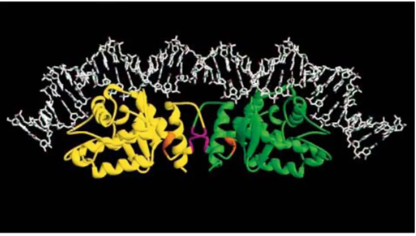
Figure 9. A model of the complex between XerC, XerD, and DNA. Ribbon
representation of the catalytic domains of the two recombinase proteins at a dif site. Regions of the proteins implicated in XerC-XerD interactions are coloured in orange (residues 256-258) and magenta (residues 263-267) (Subramanya et al., 1997; with permission).
It is evident from this model indicated in Fig. 9 that the C-terminal helix, which contains the active-site tyrosine at one end, forms a major part of the interaction of XerD with XerC. The C-terminal domains of XerC and XerD interact with one face of their DNA substrate and carry the determinants for sequence-specific DNA binding, for catalysis, and for recombinase-recombinase interactions. Alteration of the interaction in which XerC donates its C-terminus to XerD impairs the activation of XerC and stimulates XerD catalytic activity, whereas alteration of the reciprocal interaction between XerD C-terminus and XerC acceptor region inhibits XerD and stimulates XerC (Ferreira et al., 2003). Therefore, the catalytic activity of XerC and XerD is controlled by allosteric-type interactions in which donor–acceptor region interactions between adjacent recombinase molecules act as molecular springs in the switch that leads to sequential and synchronized activation/inactivation of pairs of recombinase subunits during
recombination (Fig. 10) (Hallet et al., 1999). These results support the hypothesis that the “normal” state in the heterotetrameric complex, in which XerC is catalytically active and XerD is inactive, depends on the interactions between the C-terminal end of XerC and its receptor region within the C-terminal domain of XerD; Interference with these interactions leads to a switch in the catalytic state, so that XerD is now preferentially active (Ferreira et al., 2003).
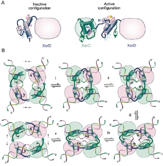
Figure 10. Control of Catalysis in Xer Recombination. (A) Proposed
reconfiguration of XerD C-terminus upon assembly of the recombination complex on DNA. (B) A model for the reciprocal control of catalysis by XerC and XerD Color code is as in (A). The ball-and-socket joint depicts the interaction between the donor and
acceptor regions of adjacent subunits. Step i to step v is the recombination pathway in which XerC strand exchange occurs first. (i) Interactions between XerC and XerD molecules bound on a same duplex, possibly coupled with additional interprotomer interactions across the synapse, force the DNA to bend in a configuration where the top (green) strand of the recombination site central region is exposed toward the outside of the duplex. The torsion energy stored in the bent DNA may act on the XerC–XerD donor–acceptor interaction so as to activate XerC catalysis by repositioning of the tyrosine nucleophile (arrowhead), and possibly other catalytic residues with respect to the DNA target phosphate (circle). DNA torsion strains released upon cleavage may also promote the unwinding and extrusion of the cleaved strands in order to orient the 5′ OH ends for the rejoining step. (ii) Completion of the strand exchange reaction generates a 2-fold symmetric HJ intermediate in which the top strands are crossing. (iii) Coupled protein and DNA conformation changes convert the complex into a configuration in which the bottom strands (purple) are crossing. (iv)This leads to synchronized inactivation of the XerC subunits and concomitant activation of the XerD subunits. (v) The recombinant duplexes are bent in the opposite direction to that of the initial recombination sites. This inversion of the DNA bending strains may promote the restacking of the DNA helices and the dissociation of the resealed molecules from the complex (Hallet et al., 1999; with permission).
2.3 The Site of Action of the Xer Recombinases
Xer recombinase-mediated recombination occurs in two different recombination sites that have different biological functions. One is at chromosome recombination sites called dif, originally found in E. coli. Another is at plasmid sites such as ColE1 cer and pSC101 psi. The Xer site-specific recombination is conserved in most eubacteria (Recchia and Sherratt, 1999). The alignment of 19 naturally occurring plasmids and some eubacterial chromosomes revealed that the wide existence of the homologues of Xer recombination core site (Table 1) (Hayes et al., 1997; Lesterlin et al., 2004). One difference between the sites is that cer is flanked by accessory sequences, which bind additional proteins and enhance recombination between sites in dimers. This provides directionality so that resolution of dimers to monomers is highly favoured (Blakely and Sherratt, 1996). Flanking sequences are not involved in resolution at dif as the phenotype of a 173kb deletion can be suppressed by insertion of a 33bp dif sequence (Tecklenburg
et al., 1995). A comparision between the binding sites shows that XerC binding sites are more variable whereas XerD binding sites are well conserved. Neither half-site can be used to replace the other half-site for recombination in vivo. The central region of the Xer sites, which displays no consensus and separates XerCD binding sites by a 6 (chromosome site) or 8bp (plasmid site) spacer, is a key determinant of the Xer recombination pathway. It determines the requirements for accessory proteins and accessory sequences on the plasmid recombination site (e.g. ColE1 cer site or pSC101 psi site). It also determines the presence of FtsK in chromosome dimer resolution (Barre et al., 2001). Several sets of data, obtained on Xer systems and other tyrosine recombinase systems, indicated that this region is an important determinant of the comformation of the recombinase-core sequence complexes (Azaro and Landy, 1997; Gopaul et al., 1998; Arciszewska et al., 2000; Lee and Sadowski, 2001; Capiaux et al., 2002). Based on the dyad symmetry of the half-sites and by anology with the cleavage positions from other recombinases (Hoess et al., 1986; Bruckner et al., 1986), the boundaries of the central region and recombinase binding sites have been proposed to contain the bases involved in strand nicking and exchange ( Summers, 1989 ).