A 12-Week Exercise Program for Pregnant
Women with Obesity to Improve Physical
Activity Levels: An Open Randomised
Preliminary Study
Michèle Bisson1,2☯, Natalie Alméras2‡, Sébastien S. Dufresne3‡, Julie Robitaille4‡,
Caroline Rhéaume5‡, Emmanuel Bujold6‡, Jérôme Frenette3‡, Angelo Tremblay2‡,
Isabelle Marc1☯*
1 Department of Pediatrics, Centre hospitalier universitaire (CHU) de Québec, Laval University, Québec City, Province of Québec, Canada, 2 Department of Kinesiology, Laval University, Quebec City, Province of Québec, Canada, 3 Department of Rehabilitation, CHU de Québec, Laval University, Québec City, Province of Québec, Canada, 4 Department of Food Science and Nutrition, Laval University, Québec City, Province of Québec, Canada, 5 Department of Family Medicine and Emergency Medicine, Laval University, Québec City, Province of Québec, 6 Department of Obstetrics and Gynecology, CHU de Québec, Laval University, Québec City, Province of Québec, Canada
☯These authors contributed equally to this work. ‡These authors also contributed equally to this work. *isabelle.marc@crchudequebec.ulaval.ca
Abstract
Objective
To evaluate whether a 12-week supervised exercise program promotes an active lifestyle throughout pregnancy in pregnant women with obesity.
Methods
In this preliminary randomised trial, pregnant women (body mass index ! 30 kg/m2) were
allocated to either standard care or supervised training, from 15 to 27 weeks of gestation. Physical activity was measured by accelerometry at 14, 28 and 36 weeks, while fitness (oxygen consumption (VO2) at the anaerobic threshold), nutrition (caloric intake and
macro-nutrients percentage) and anthropometry were assessed at 14 and 28 weeks of gestation. Analyses were performed using repeated measures ANOVA.
Results
A total of fifty (50) women were randomised, 25 in each group. There was no time-group interaction for time spent at moderate and vigorous activity (pinteraction= 0.064), but the
exer-cise group’s levels were higher than controls’ at all times (pgroup effect= 0.014). A significant
time-group interaction was found for daily physical activity (p = 0.023); similar at baseline ((22.0 ± 6.7 vs 21.8 ± 7.3) x 104counts/day) the exercise group had higher levels than the
control group following the intervention ((22.8 ± 8.3 vs 19.2 ± 4.5) x 104counts/day, p = a11111
OPEN ACCESS
Citation: Bisson M, Alméras N, Dufresne SS, Robitaille J, Rhéaume C, Bujold E, et al. (2015) A 12-Week Exercise Program for Pregnant Women with Obesity to Improve Physical Activity Levels: An Open Randomised Preliminary Study. PLoS ONE 10(9): e0137742. doi:10.1371/journal.pone.0137742 Editor: David B. Allison, University of Alabama at Birmingham, UNITED STATES
Received: May 27, 2015 Accepted: August 19, 2015 Published: September 16, 2015
Copyright: © 2015 Bisson et al. This is an open access article distributed under the terms of the Creative Commons Attribution License, which permits unrestricted use, distribution, and reproduction in any medium, provided the original author and source are credited.
Data Availability Statement: Data from the "Inter GO FIT" study are available from the Dryad Digital Repository (URLhttp://dx.doi.org/10.5061/dryad. 87f03).
Funding: This study was funded by an operating grant from the Fondation des Étoiles (grant number F-61359, URL:http://www.fondationdesetoiles.ca/fr). Salaries were supported by the following sources: MB is a doctoral scholarship holder from the Canadian Institutes of Health Research (URL http://www.cihr-irsc.gc.ca/e/193.html). IM and EB are recipients of a Fonds de Recherche du Québec-Santé clinician
0.020) and at 36 weeks of gestation ((19.2 ± 1.5 vs 14.9 ± 1.5) x 104counts/day, p = 0.034).
Exercisers also gained less weight than controls during the intervention period despite simi-lar nutritional intakes (difference in weight change = -0.1 kg/week, 95% CI -0.2; -0.02, p = 0.016) and improved cardiorespiratory fitness (difference in fitness change = 8.1%, 95% CI 0.7; 9.5, p = 0.041).
Conclusions
Compared with standard care, a supervised exercise program allows pregnant women with obesity to maintain fitness, limit weight gain and attenuate the decrease in physical activity levels observed in late pregnancy.
Trial Registration
ClinicalTrials.govNCT01610323
Introduction
Physical activity during pregnancy can increase cardiorespiratory fitness [1], decrease gesta-tional weight gain [2] and lower the risk of preeclampsia [3]. However, such benefits remain uncertain in women with obesity. These women are spontaneously less active than their lean counterparts [4], which may exacerbate their already low fitness levels [5] and risk of excessive gestational weight gain [6]. Consequently, exercise programs targeting this population are needed, as they can potentially decrease the risk of perinatal complications.
Increasing exercise levels in pregnant women with obesity appears challenging, as adherence to exercise programs has been of concern in previous trials [7,8]. Moreover, the efficacy of such interventions to improve physical activity levels throughout pregnancy is usually not objectively measured. Although recommendations have been proposed for pregnant women with obesity [9,10], their impact on maternal fitness have been poorly studied and accordingly, the type, volume and intensity of physical activity required to maintain fitness in this popula-tion is unknown.
As face-to-face, individualized physical activity interventions have the potential to improve adherence to an active lifestyle [11], we sought to investigate its effect in pregnant women with obesity. The primary objective of this study was to evaluate whether an individually supervised, 12-wk moderate-intensity exercise program during the 2ndtrimester of pregnancy results in higher physical activity levels throughout pregnancy in women with obesity.
Materials and Methods
Study design
Recruitment for this randomized controlled parallel-group study with a 1:1 allocation ratio was performed at the Centre Hospitalier Universitaire (CHU) de Québec and the Centre de santé et de services sociaux de la Vieille-Capitale, from October 2011 to November 2013, with follow-ups completed in June 2014. The intervention and fitness tests took place at the Pavillon de pré-vention des maladies cardiaques (PPMC, Institut Universitaire de Cardiologie et Pneumologie de Québec). Research Ethics Board of these institutions approved the study, and all participants provided written informed consent. The protocol of the study was registered in ClinicalTrials. gov, following the enrollment of the first participants (NCT01610323, URLhttps://
scientist award (URLhttp://www.frqs.gouv.qc.ca/en/ accueil). The funders had no role in study design, data collection and analysis, decision to publish, or preparation of the manuscript.
Competing Interests: The authors have declared that no competing interests exist.
clinicaltrials.gov/ct2/show/study/NCT01610323?term=NCT01610323&rank=1). As imple-menting an exercise intervention can be challenging, initial recruitments for this preliminary study aimed at confirming the intervention feasibility. Registration was delayed until funding allowed us to continue the recruitment for this preliminary study. At the time of registration, only 5 participants had completed the primary outcome assessment, which was originally cardiorespiratory fitness following the intervention as mentioned in the original protocol. However, cardiorespiratory testing in pregnant women with obesity raised feasibility issues, especially at 28 weeks, and accordingly the primary outcome was modified for the impact of the intervention on physical activity levels, in accordance with the registered protocol and the sample size requirement for the physical activity outcome (as describe in the “Sample size” sec-tion). There are no ongoing trials for this intervention.
Participants
Participants were recruited before the end of the 14thweek of gestation at family practice, obstetrical and ultrasound clinics and in the community. Women with a pre-pregnancy body mass index (BMI) ! 30.0 kg/m2, regardless of previous physical activity levels, were eligible if
they were 18 years or older, presented a singleton pregnancy and planned to deliver in partici-pating hospitals. Women with diabetes or chronic hypertension before pregnancy were excluded. The absence of physical activity contraindications was verified with the women’s physician and the Physical Activity Readiness Medical Examination for Pregnancy (PARmed-X [12]).
Randomisation
Following baseline assessment, participants were randomly allocated to either the exercise intervention or usual activity. Randomization was stratified according to parity and based on a computer-generated random numbers table. Sealed envelopes were kept in a secure place by a research assistant not involved in the study and provided to a kinesiologist at the time of alloca-tion. Due to the nature of the intervention, kinesiologists in charge of training and participants were not blinded to group assignment. However, all assessors and research assistants in charge of data entry and analyses were blinded to participants’ allocation (defined as “group 1” and “group 2”).
Study protocol
At 14 weeks of gestation (Visit 1), participants were assessed for physical activity, anthropome-try, fitness and fetal growth. Physical activity prior to pregnancy, socio-demographic character-istics and obstetrical history were collected by a trained research assistant. Food intakes were also documented, but no recommendations were made regarding them.
The same measurements were performed at 28 weeks (Visit 2), following the intervention period. Finally, women were met at 36 weeks (Visit 3) to document physical activity during the third trimester. Within 72h following delivery, newborn’s anthropometry was evaluated by a trained research assistant. Medical charts were reviewed to collect perinatal outcomes and birth weight.
Study groups
The exercise group was offered a supervised exercise program starting at the 15thweek of gesta-tion with free membership in a hospital-based condigesta-tioning centre, where kinesiologists were always available for counselling. Participants were individually supervised once a week and
invited to complete two more sessions/wk. Consistent with the American College of Sports Medicine Guidelines [10], exercise prescription consisted of 3 weekly 1h sessions, for a total of 36 prescribed sessions over 12 wks.
Each session included a 5–10 min warm-up on a stationary ergocycle, a 15–30 min treadmill walk, a 20 min muscular work-out and a cool-down period. Duration of the cardiovascular training increased progressively from 15 min during the first week to 30 min by the end of the first month. The muscular work-out included dynamic exercises for both lower and upper limbs using the participant’s own body weight, small weights, exercising balls and strength equipment with selective charges. Participants started with 1 set of 10–15 repetitions per exer-cise and progressed to 2 sets of 15 repetitions, with intensity adjusted to their tolerance level. To enhance motivation, the muscular work-out was modified every 4 weeks (twice during the intervention period). Exercise intensity was self-monitored with heart rate monitors (Polar FT4, Polar Electro, Finland) and the modified Borg Scale [13], with targets at 70% of peak heart rate (measured during the fitness test), and/or at a perceived exertion score of 3-5/10. Participants recorded duration and mean heart rate of each session from their monitors on their exercise log. On non-training days, women were advised to be as active as possible.
The control group was told to continue usual activities without being restrained from doing physical activity. Both groups were given a pamphlet (from Kino-Québec, an agency promot-ing physical activity) about the benefits of physical activity and appropriate exercises for preg-nant women [14].
Outcomes assessment
Physical activity was measured by accelerometry at 14, 28 and 36 weeks of gestation. Women were instructed to wear the accelerometer (GT3X+, ActiGraph, USA) on the hip for 7 consecu-tive days, with permission to remove it before bedtime. The primary outcome was defined as the time spent at moderate and vigorous physical activity (MVPA) at 36 weeks of gestation. As the number of days with wear time varied across subjects, reporting activity data per day (instead of per week) was more appropriate. Accordingly, we also reported the number of accelerometry counts/day (reflecting total activity), daily time spent at MVPA in periods !10 min (minimum duration required to improve fitness [15]) and daily step counts. MVPA was calculated using the Matthews’ cut point [16], previously used in pregnant women with obesity [17]. Accelerometers were operated according to the manufacturer’s specifications, and analy-ses were performed using Actilife software. Non-wear time (60 min or more of consecutive zeros [18]) was assessed from accelerometry data, with spurious data removed [19]. Per proto-col, if accelerometers were worn for less than 8h daily and for less than 5 days, data were excluded [17] from the main analyses. In addition to this analysis, sensitivity analyses without a minimum wear time requirement were conducted, with and without removal of spurious data [19].
Physical activity in the previous month was also measured at each visit using the Pregnancy Physical Activity Questionnaire (PPAQ) [17,20] which specifies the type of physical activity performed, adding to data collected through accelerometry [21]. Time spent at each activity was multiplied by its intensity (in Metabolic Equivalent of Task (MET)) [22] and summed to obtain a weekly energy expenditure in METs"h"wk-1.
Adherence to exercise prescription was calculated as the number of completed sessions dur-ing the intervention, as collected in the participants’ log and verified with heart rate monitors’ recordings.
Maternal weight was measured using an electronic scale (InBody 520, Biospace, USA) at each visit. Height was measured at Visit 1, and skinfolds (Harpenden skinfold calliper, Baty,
UK) were measured at 14 and 28 weeks of gestation by an experienced exercise physiologist, as described elsewhere [5]. Skinfolds were used to estimate fat percentage using the Jackson and Pollock’s equation for women [23]. Weight gain outcomes were weight gain from 14 to 36 weeks of gestation, and weight gain from 14 to 28 weeks (at the end of the intervention period). To account for the different time period between two weight evaluations, the rate of weekly weight gain was reported (weight gain divided by the number of weeks between the two weight evaluations). Total gestational weight gain (difference between the last weight before delivery and pre-pregnancy weight as reported in the medical charts) was also calculated.
Cardiorespiratory fitness, defined as oxygen uptake at the anaerobic threshold (VO2AT),
was assessed at 14 and 28 weeks of gestation by a qualified exercise physiologist during a peak treadmill exercise test with gas exchange analysis (Quark B2, version 8.1a, Cosmed, Italy). A standardized procedure was followed [24], using the modified Bruce ramp protocol [25]. VO2
AT was identified by two independent exercise physiologists using the V-slope method [26]. Muscular testing included handgrip strength (Model 78010, Lafayette Instrument Company, USA) and isokinetic strength and endurance of the quadriceps (Biodex System 4, Biodex Medi-cal Systems Inc., USA) following standardized procedures [5,27–29].
Dietary intakes over the last month were measured at 14 and 28 weeks of gestation using an interviewer-administered food frequency questionnaire [30] with use of food models for esti-mation of portions.
Fetal growth and uterine arteries mean pulsatility index were evaluated by a certified techni-cian during Doppler studies at 14 and 28 weeks of gestation (Voluson E8 Expert system, GE Healthcare Inc., USA). Neonatal anthropometry included length, head circumference (Models 212 and 416, Seca corp, Germany) and skinfolds (Lange skinfold caliper, Beta Technology, USA). Fat mass and percentage were calculated with a validated equation [31], and birth weight Z-scores adjusted for sex and gestational age were based on Canadian references [32].
Sample size
Sample size was calculated a priori based on previously published accelerometry data reporting that in the third trimester, women with obesity spent 16 ± 16 min/d doing MVPA in bouts !10 min [17]. Based on a t-test, a sample size of 21 participants per group allowed detecting an increase of 14 min/d in the intervention group compared to controls, justified by current rec-ommendations (i.e. 30 min/d [33]), with an 80% power and two-sided alpha level at 0.05. With an estimated 15% of losses, 50 participants were recruited.
Statistical analyses
Data are presented as means ± standard deviation and percentage for continuous and cate-gorical variables, respectively. Analyses were performed using SAS statistical package 9.4 on an intention-to-treat basis. In order to perform a comprehensive analysis of all physical activity measures over time (at 14, 28 and 36 weeks of gestation), repeated measures ANOVA using a linear mixed model were conducted to compare the effect of group alloca-tion (exercise vs control), time (baseline, 28 weeks, 36 weeks) and their interacalloca-tion on physi-cal activity levels. If a significant “time-group” interaction was found, comparison over time was made for each group separately, and comparison between groups was made at each indi-vidual time. Otherwise, main effects were presented. The covariance structure for repeated measures ANOVA was chosen separately for each outcome. The structure with the lowest Akaike Information Crieterion corrected for finite sample (AICc) amongst several of the most popular structures was chosen. The normality assumption of the residuals of the ANOVA was verified by checking the distribution of scaled residuals obtained by the linear
mixed model. Skewness and kurtosis coefficients, as well as histograms and Kolmogorov-Smirnov and Shapiro-Wilk tests, were evaluated and confirmed that the assumption was met. Post hoc tests with the Bonferroni correction were performed to take into account mul-tiple comparisons and keep the familywise error rate at 5%. Sensitivity analyses without a minimum wear time requirement for accelerometry were also conducted, with and without removal of spurious data. As pre-pregnancy physical activity levels could influence the ical activity profile during pregnancy, a sensitivity analysis stratified by pre-pregnancy phys-ical activity level was performed for physphys-ical activity outcomes, with women dichotomized as “previously active” or “previously inactive” based on the median value of the pre-preg-nancy self-reported energy expenditure spent at sports and exercise. For other outcomes (exploratory analyses for weight gain and neonatal outcomes), groups were compared by Student t test, Wilcoxon rank sum test, χ2or Fisher’s exact test.
Results
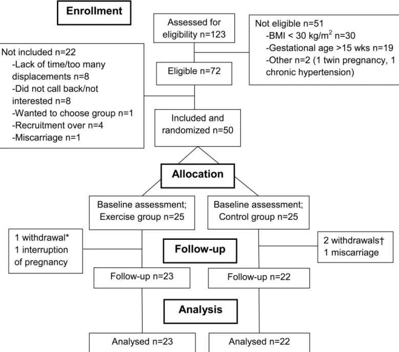
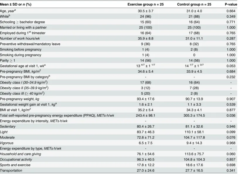
Among 123 interested women, 72 were eligible and 50 were randomized in one of the two study arms (25 per group, seeFig 1). Both groups were similar with respect to baseline socio-demographic characteristics (Table 1). Self-reported physical activity prior to pregnancy was also similar between groups, although the control group reported higher total energy expendi-ture than the exercise group (Table 1).
Physical activity assessments
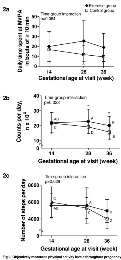
Physical activity data can be found inTable 2. For daily MVPA in bouts !10 min (Fig 2a), there was no significant time-group interaction, although a trend was present. There was a sig-nificant group effect (p = 0.014), meaning that the exercise group spent more time doing MVPA in bouts !10 min than the control group at all times.
For total activity reported by the number of counts/day (Fig 2b), there was a significant time-group interaction. Similar at baseline, the exercise group was significantly more active than the control group at both 28 and 36 weeks of gestation (p = 0.020 and p = 0.034, respec-tively). A significant decline in the number of counts/day between each time point was also found in the control group, whereas the exercise group only decreased their activity levels between 28 and 36 weeks.
A significant time-group interaction was also present for daily step counts (Fig 2c). Although not significantly different between groups at any time, there was a trend towards a higher step counts at 36 weeks in the exercise group compared to controls (p = 0.072). Also, while there was a significant difference in the number of steps per day only between 28 and 36 weeks in the exercise group, the control group showed a step counts at 36 weeks that was signif-icantly lower than those at baseline and at 28 weeks.
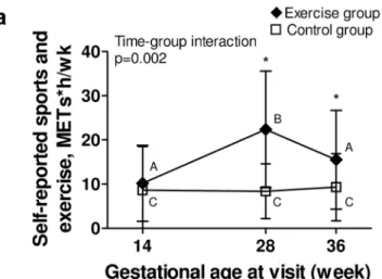
Data from the PPAQ corroborated accelerometry findings (Fig 3a and 3b), as the exercise group reported significantly more time than controls doing sports and exercise activities and vigorous activities at 28 and 36 weeks, respectively. For other domains and intensities of activ-ity from the PPAQ, groups were comparable (Table 2).
Average accelerometer’s daily wear time was 16.1 ± 3.4 h, 15.5 ± 2.8 h and 14.2 ± 1.9 h at 14, 28 and 36 weeks, respectively. Due to drop-outs (n = 5) or insufficient wear time based on our pre-specified requirement (n = 5, 7 and 11 at 14, 28 and 36 weeks, respectively), accelerometry was not available for all participants. However, non-completers’ characteristics were similar in both groups.
Sensitivity analyses without wear time requirement, with and without removal of spurious data, confirmed and even strengthened the results (S1 Table). Moreover, analyses stratified for
pre-pregnancy physical activity levels (“previously active” or “previously inactive”) did not sug-gest significant interactions between pre-pregnancy physical activity levels and physical activity patterns over time in any group (data not shown).
Adherence to the intervention. The exercise group performed a total of 18.5 ± 10.1 ses-sions (1.5 sesses-sions/wk), with 15 (60%) and 5 participants (20%) reaching at least 50% and 75% of the 36 prescribed sessions, respectively. Mean duration was 58.8 ± 4.3 min and exercise heart rate was 121 ± 11 beats"min-1(70.0 ± 5.5% maximal heart rate). There were no adverse events related to the intervention.
Fig 1. Flowchart. *One participant withdrew after randomization (lack of time); †Two participants withdrew after randomization (unsatisfied with group allocation).
Weight gain
Pre-pregnancy weight and weight gain prior to inclusion in the study did not differ between groups (Table 1). However, the rate of weekly weight gain showed a different profile over time between groups (p-value for interaction = 0.024,Fig 3c). Prior to inclusion in the study, the rate of weekly weight gain did not differ between groups (0.11 ± 0.15 kg/wk vs 0.08 ± 0.23 kg/ wk for the exercise and control groups, respectively), while during the program, the exercise group gained less weight per week than the control group (0.35 ± 0.14 kg/wk vs 0.46 ± 0.15 kg/ wk for the exercise and control groups, respectively, p = 0.018), despite similar nutritional intakes (Table 3). The control group experienced an increase in fat percentage during this period, as compared to the exercise group (Table 3). However, the rate of weekly weight gain did not differ between groups following the end of the intervention until Visit 3 (0.47 ± 0.24 Table 1. Participants’ characteristics at 14 weeks (Visit 1).
Mean ± SD orn (%) Exercise group n = 25 Control group n = 25 P-value
Age,yeara 30.5 ± 3.7 31.0 ± 4.0 0.664
Whiteb 24 (96) 21 (88) 0.349
Schooling ! bachelor degree 15 (60) 16 (64) 0.771
Married or living with a partner 25 (100) 25 (100) 1.000
Employed during 1sttrimester 16 (64) 17 (68) 0.765
Number of work hours/wk 35.9 ± 8.8 31.0 ± 11.1 0.287
Preventive withdrawal/mandatory leave 9 (36) 8 (32) 0.765
Smoking before pregnancy 1 (4) 2 (8) 1.000
Smoking during pregnancy 1 (4) 0 1.000
Parity ! 1 14 (56) 14 (56) 1.000
Gestational age at visit 1,wka 134/7±11/7 141/7±10/7 0.053
Pre-pregnancy BMI,kg/m2 34.6 ± 5.4 33.9 ± 4.5 0.684
Pre-pregnancy BMI by categoryb - - 0.232
Obesity class I (30–34.9 kg/m2) 17 (68) 16 (64)
-Obesity class II (35–39.9 kg/m2) 3 (12) 7 (28)
-Obesity class III (! 40 kg/m2) 5 (20) 2 (8)
-Pre-pregnancy weight,kg 93.4 ± 17.6 90.7 ± 13.9 0.907
Gestational weight gain at visit 1,kga 1.6 ± 2.1 1.1 ± 3.3 0.539
BMI at visit 1,kg/m2 35.2 ± 5.4 34.3 ± 4.1 0.877
Total self-reported pre-pregnancy energy expenditure (PPAQ),METs"h/wk 243.4 ± 98.1 305.3 ± 174.5 0.036
Energy expenditure by intensity,METs"h/wk - -
-Sedentary 80.4 ± 26.7 81.1 ± 32.6 0.946
Light 83.7 ± 46.3 110.1 ± 58.1 0.099
Moderate 72.8 ± 71.2 104.7 ± 117.8 0.076
Vigorous 6.5 ± 7.5 9.4 ± 14.3 0.968
Energy expenditure by type,METs"h/wk - -
-Household and care giving 76.1 ± 54.6 113.6 ± 75.7 0.060
Occupational activity 96.3 ± 40.5 104.8 ± 104.3 0.857
Sports and exercise 17.8 ± 12.2 18.6 ± 17.6 0.698
Transportation 27.0 ± 24.6 27.7 ± 16.5 0.341
aStudent t-test (other continuous variables evaluated using Wilcoxon rank sum test) bFisher exact test (other categorical variables evaluated using χ2).
kg/wk vs 0.45 ± 0.29 kg/wk for the exercise and control groups, respectively), nor did total ges-tational weight gain for the entire pregnancy (S2 Table).
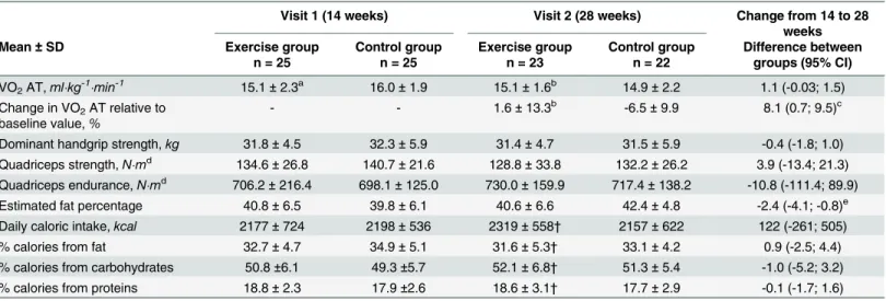
Changes in fitness
At baseline, cardiorespiratory fitness was similar between groups. Following the intervention, VO2AT increased slightly in the exercise group, whereas it decreased in the control group
(Table 3). There was no difference between groups for muscular strength and endurance fol-lowing the intervention (Table 3).
Perinatal and neonatal outcomes. At 28 weeks, no differences were found between groups for either mean uterine arteries pulsatility index (data not shown) or estimated fetal weight (1205 ± 169 vs 1219 ± 230 g for exercise and control groups, respectively). There were no differences between groups for birth weight, gestational age at delivery, rate of hypertensive disorders, gestational diabetes or caesarean delivery (S2 Table).
Discussion
A supervised exercise intervention from 15 to 27 weeks of pregnancy was effective in attenuat-ing the decline in physical activity observed in women with obesity. Indeed, the intervention allowed women to maintain or increase their physical activity levels through the 28thweek of Table 2. Physical activity levels throughout the study.
Baseline at 14 weeks End of program at 28 weeks Follow-up at 36 weeks ANOVA result Mean ± SD orn (%) Exercise
group Controlgroup Exercisegroup Controlgroup Exercisegroup Controlgroup P-value forinteraction
Accelerometry, n 23 22 20 17 18 16
-MVPA in bouts,min/d 19.9 ± 15.0 16.8 ± 17.6 25.4 ± 20.4 11.7 ± 9.5 18.9 ± 14.1 9.5 ± 9.8 0.064a Counts per day (n x 104) 22.0 ± 6.7 21.8 ± 7.3 22.8 ± 8.3 19.2 ± 4.5 20.1 ± 6.2 15.8 ± 5.2 0.023 Steps per day 5587 ± 1472 5984 ± 1806 5598 ± 2094 5298 ± 1252 4947 ± 1349 4006 ± 1157 0.028
Self-reported PA, n 25 25 23 22 23 22
-Total energy expenditure,
METs"h/wk 194.6 ± 71.2 226.0 ± 60.0 218.1 ± 67.8 207.8 ± 72.6 185.0 ± 50.8 186.8 ± 83.6 0.070 b Energy expenditure by intensity,METs"h/wk - - - -Sedentary 74.4 ± 27.3 79.9 ± 27.3 69.1 ± 28.9 66.4 ± 30.9 62.9 ± 25.3 63.5 ± 25.5 0.65c Light 71.8 ± 46.7 86.7 ± 32.5 89.7 ± 41.2 86.3 ± 35.2 73.6 ± 27.7 78.7 ± 43.6 0.23 Moderate 46.3 ± 33.7 56.8 ± 37.0 48.5 ± 30.8 54.4 ± 45.1 41.6 ± 25.3 43.8 ± 37.9 0.64 Vigorous 2.2 ± 3.4 2.6 ± 8.0 10.7 ± 7.1 0.8 ± 2.0 6.9 ± 5.9 0.8 ± 1.9 <0.0001
Energy expenditure by type,
METs"h/wk - - -
-Household and care giving 71.7 ± 61.5 91.7 ± 55.4 82.5 ± 52.8 86.7 ± 60.4 75.3 ± 43.4 86.2 ± 76.8 0.39 Occupational activity 55.6 ± 41.4 62.3 ± 43.2 62.1 ± 47.3 48.8 ± 55.4 39.7 ± 39.9 27.7 ± 39.1 0.21d Sports and exercise 10.2 ± 8.625 8.6 ± 9.9 22.4 ± 13.2 8.4 ± 6.2 15.5 ± 11.2 9.3 ± 7.6 0.002 Transportation 21.6 ± 16.9 22.2 ± 12.9 23.3 ± 15.8 25.1 ± 18.4 20.4 ± 12.6 21.0 ± 16.1 0.97 MVPA = moderate and vigorous physical activity; PA = physical activity
asignificant group effect, p = 0.014; values significantly higher in the exercise vs control group at all time
bsignificant time effect, p = 0.028; values significantly lower at time 3 compared with time 2 in both groups (adjusted p = 0.027) csignificant time effect, p = 0.012; values significantly lower at time 3 compared with time 1 in both groups (adjusted p = 0.012)
dsignificant time effect, p = 0.007; values significantly lower at time 3 vs time 1 and time 2 in both groups (adjusted p = 0.001 and p = 0.010). doi:10.1371/journal.pone.0137742.t002
Fig 2. Objectively measured physical activity levels throughout pregnancy. Black lozenge: exercise group. White square: control group. Fig 2a. Daily time spent at moderate and vigorous physical activity in bouts of at least 10 min; Fig 2b. Total activity per day, expressed as the daily number of accelerometry counts; Fig 2c. Number of steps per day. P-value is for time-group interaction significance;* Indicates a significant difference (p<0.05) between groups at a specific time point; Different capital letters (A, B, C, D, E) within a group indicate significant differences between time points.
Fig 3. Self-reported physical activity and rate of weekly weight gain throughout pregnancy. Black section: exercise group. White section: control group. Fig 3a. Energy expenditure spent at sports and exercise in the previous month, from the PPAQ; Fig 3b. Energy expenditure spent at vigorous intensity activity in the past month, from the PPAQ; Fig 3c. Rate of weekly gestational weight gain, in kg. P-value is for time-group interaction significance;* Indicates a significant difference (p<0.05) between groups at a specific
pregnancy, whereas it decreased in the control group. This improvement was also supported by a maintained cardiorespiratory fitness level and limited weight gain during the intervention period in the exercise group, compared to controls.
The exercise group also remained more active than the control group during the third tri-mester, as demonstrated by higher accelerometry counts and self-reported energy expenditure. Despite these higher levels in the exercise group, both groups decreased their activity levels between 28 and 36 weeks of gestation, with values near baseline levels and significantly lower than baseline levels for the exercise and control groups at 36 weeks, respectively. This probably reflects the end of the intervention and the fact that some activities become less comfortable as pregnancy progresses. Therefore, to maintain higher levels of physical activity throughout pregnancy, a follow-up until delivery appears necessary. The advantage of our 12-wk interven-tion in mid-pregnancy was that it allowed establishing that a supervised exercise program could increase fitness and physical activity levels, with the assessment of a retention effect fol-lowing the end of the intervention. Seizing the opportunity of pregnancy to promote healthy life habits is important, but taking into account the reality of pregnant women with obesity is also crucial. In that sense, creativity and alternatives to individual, center-based intervention might be needed in late pregnancy to sustain the newly acquired physical activity habit (e.g. fol-low-up to reinforce behavior, walking club or group activities, or home-based practice).
Increasing physical activity levels with a goal of reaching physical activity recommendations throughout pregnancy is important, as it might help pregnant women in achieving adequate gestational weight gain [34,35] through an increased energy expenditure, lower their risk of gestational diabetes [35] and fetal macrosomia [36] through a higher muscular glucose uptake time point; Different capital letters (A, B, C, D, E) within a group indicate significant differences between time points.
doi:10.1371/journal.pone.0137742.g003
Table 3. Maternal fitness, anthropometry and nutritional intakes at 14 and 28 weeks of gestation.
Visit 1 (14 weeks) Visit 2 (28 weeks) Change from 14 to 28
weeks
Mean ± SD Exercise group
n = 25 Control group n = 25 Exercise group n = 23 Control group n = 22 Difference between groups (95% CI) VO2AT,ml"kg-1"min-1 15.1 ± 2.3a 16.0 ± 1.9 15.1 ± 1.6b 14.9 ± 2.2 1.1 (-0.03; 1.5) Change in VO2AT relative to
baseline value,% - - 1.6 ± 13.3
b -6.5 ± 9.9 8.1 (0.7; 9.5)c Dominant handgrip strength,kg 31.8 ± 4.5 32.3 ± 5.9 31.4 ± 4.7 31.5 ± 5.9 -0.4 (-1.8; 1.0) Quadriceps strength,N"md 134.6 ± 26.8 140.7 ± 21.6 128.8 ± 33.8 132.2 ± 26.2 3.9 (-13.4; 21.3) Quadriceps endurance,N"md 706.2 ± 216.4 698.1 ± 125.0 730.0 ± 159.9 717.4 ± 138.2 -10.8 (-111.4; 89.9) Estimated fat percentage 40.8 ± 6.5 39.8 ± 6.1 40.6 ± 6.6 42.4 ± 4.8 -2.4 (-4.1; -0.8)e Daily caloric intake,kcal 2177 ± 724 2198 ± 536 2319 ± 558† 2157 ± 622 122 (-261; 505)
% calories from fat 32.7 ± 4.7 34.9 ± 5.1 31.6 ± 5.3† 33.1 ± 4.2 0.9 (-2.5; 4.4)
% calories from carbohydrates 50.8 ±6.1 49.3 ±5.7 52.1 ± 6.8† 51.3 ± 5.4 -1.0 (-5.2; 3.2) % calories from proteins 18.8 ± 2.3 17.9 ±2.6 18.6 ± 3.1† 17.7 ± 2.9 -0.1 (-1.7; 1.6) VO2AT = oxygen consumption at the anaerobic threshold
an = 24 bn = 22
cp<0.05, Wilcoxon rank sum test
dn = 22 and 24 at baseline, and n = 19 and 20 at 28 weeks in exercise and control groups, respectively ep<0.05, Student t test.
[37] and lower their risk of preeclampsia [3] through an anti-inflammatory effect on markers such as C-reactive protein [38] and cytokines [39]. Although the present study was not designed to test these hypotheses, our results remain important as they highlight the feasibility for pregnant women with obesity to as least maintain their physical activity levels during preg-nancy and that such levels, even below current recommendations, can induce benefits on cardiorespiratory fitness and gestational weight gain. Indeed, based on the present findings, a combined 1h cardiovascular and muscular moderate-intensity training performed 3 times every two weeks by pregnant women with obesity appears sufficient to maintain fitness and to have a marginal impact on weekly weight gain. Nevertheless, this does not mean that obese pregnant women should stop following current physical activity guidelines; this should be viewed as a minimal threshold to attain in order to reap some health benefits, while more bene-fits can be expected with higher levels of physical activity [40].
Few studies have focused solely on exercise interventions in pregnant women with obesity, limiting our understanding of the isolated effects of physical activity on maternal and neonatal outcomes. A previous study in pregnant women (BMI ! 25 kg/m2) did not report significant
effects on physical activity levels or weight gain with exercise compared to standard care [7]. However, less than 20% of their participants achieved half the exercise sessions, compared to 60% in the present study. Individual coaching and availability of exercise specialists accus-tomed to the management of patients with specific needs in the present study might explain these differences. Still, with a goal of 3 exercise sessions/wk, we were expecting women to com-plete at least 2 sessions/wk. All women were individually supervised once a week, but they had difficulty completing other sessions on their own, suggesting that having an incentive such as a scheduled session with an exercise specialist might be needed to further increase physical activ-ity levels in these women. Other physical activactiv-ity modalities might facilitate adherence in this population, such as home-based training. Indeed, a recent study showed a 96% adherence to a 6-wk home-based exercise program in diabetic pregnant women [41]. As in the present study, flexible supervision appears as an important component of a successful intervention with preg-nant women, either with obesity or high-risk pregnancy.
Although this study focused on physical activity, the absence of nutritional counselling might have reduced the potential for lowering gestational weight gain [42]. The exercise group remained more active than the control group in the 3rdtrimester, but the effect on weight gain
observed during the intervention did not persist until delivery. It is also important to recognize that even if the intervention had a significant impact on the rate of gestational weight gain dur-ing the traindur-ing period, it was not sufficient to allow women to gain within the Institute of Medicine’s recommended levels for weekly weight gain (0.2–0.3 kg/wk) and for total gesta-tional weight gain (5–9 kg) [43]. However, our single behavior intervention had positive effects on women’s health, without adverse effects on nutritional intakes and no apparent effect on fetal growth. Nevertheless, due to our sample size, conclusions cannot be drawn about the effects of our intervention on weight gain during pregnancy.
Following the 12-wk intervention period, VO2AT decreased by 6.5% in the control group
while it increased by 1.6% in the exercise group. This small change in the exercise group could be due to the lower than expected volume of exercise performed by participants (1.5 vs 3 ses-sions per week), as a dose-response relationship is usually expected between exercise volume and fitness improvement [40]. Indeed, a previous study performed in overweight pregnant women found an 18% increase in VO2AT in their exercise group following a 12-wk
interven-tion [44]. The better adherence found in their study (28 ± 15 sessions over 12 weeks) could partly explain these different findings, as well as the differences in study population character-istics and in the method used to determine the anaerobic threshold. Other potential reasons for the small increase in fitness seen in the present study include the variation in baseline fitness
levels between subjects, as those presenting lower levels were probably less active initially, which gave them a better potential for improvement compared to those with a higher fitness level [45], and the interindividual heterogeneity in responsiveness to training (i.e. genetic pre-dispositions) [46]. Nevertheless, although the change over time in VO2AT was relatively small
in the exercise group, a training effect was still observed, considering the decreased VO2AT in
the control group.
The present study has some limitations. Despite a low drop-out rate (10%), some partici-pants did not adequately complete accelerometry measurements [17], reducing power to show a difference between groups. However, our results remain robust as non-completers were not different between groups and because our results were corroborated by sensitivity analyses and by concordant findings with subjective measures. The social support/interaction with the study staff may have been partially responsible for some observed differences in outcomes between the study arms. However, our trial was pragmatic and objective measurements such as accel-erometry and fitness data are less prone to be affected by the support given to participants or by a desirability bias. Fat percentage estimates were based on widely used equations although not validated during pregnancy, as no consensus exists on which anthropometric method should be used to reliably determine body composition during pregnancy [47]. Because it was not possible to have skinfold measures at 36 weeks made by the same assessor as for the first two visits and to avoid high inter-observer variability [48], this assessment was not performed. Finally, results may not be generalizable to all pregnant women with obesity, as our sample included mostly white women with higher education and living with a partner.
Conclusion
This preliminary study suggests the feasibility of an exercise intervention during pregnancy for women with obesity to enable them to maintain and even increase their physical activity levels, following a supervised exercise program during mid-pregnancy. From a practical perspective, pregnant women with uncomplicated pregnancy should be encouraged to lead an active preg-nancy and referred to competent specialists. A minimum of 3 exercise sessions every two weeks appears necessary to maintain fitness in pregnant women with obesity, but a higher vol-ume of exercise might induce greater benefits on other outcomes such as gestational weight gain. Larger trials are needed to determine short and long term benefits of exercise during preg-nancy on maternal and child health.
Supporting Information
S1 CONSORT Checklist. Consort Checklist. (DOC)
S1 Protocol. Study protocol—French version. (PDF)
S2 Protocol. Study protocol—English version. (PDF)
S1 Table. Comparison of main accelerometry results using various definitions of non-wear time.
(DOCX)
S2 Table. Obstetrical and perinatal outcomes. (DOCX)
Acknowledgments
The authors thank Dr Paul Poirier, PPMC’s medical director, for his kind cooperation, Guy Fournier, research assistant at IUCPQ, who performed the cardiorespiratory testing, and Anne-Sophie Julien, biostatistician at the CHU de Québec, for statistical support.
Author Contributions
Conceived and designed the experiments: MB IM AT NA JF. Performed the experiments: MB IM CR SSD NA JF. Analyzed the data: MB NA SSD JR CR EB JF AT IM. Contributed reagents/ materials/analysis tools: IM NA JF. Wrote the paper: MB NA SSD JR CR EB JF AT IM.
References
1. Kramer MS, McDonald SW. Aerobic exercise for women during pregnancy. Cochrane Database Syst Rev. 2006; 19(3):CD000180.
2. Thangaratinam S, Rogozinska E, Jolly K, Glinkowski S, Duda W, Borowiack E, et al. Interventions to reduce or prevent obesity in pregnant women: a systematic review. Health Technol Assess. 2012; 16(31):iii–iv, 1–191. doi:10.3310/hta16310PMID:22814301
3. Aune D, Saugstad OD, Henriksen T, Tonstad S. Physical activity and the risk of preeclampsia: a sys-tematic review and meta-analysis. Epidemiology. 2014; 25(3):331–43. doi:10.1097/EDE.
0000000000000036PMID:24713878
4. Renault K, Norgaard K, Secher NJ, Andreasen KR, Baldur-Felskov B, Nilas L. Physical activity during pregnancy in normal-weight and obese women: compliance using pedometer assessment. J Obstet Gynaecol. 2012; 32(5):430–3. doi:10.3109/01443615.2012.668580PMID:22663312
5. Bisson M, Almeras N, Plaisance J, Rheaume C, Bujold E, Tremblay A, et al. Maternal fitness at the onset of the second trimester of pregnancy: correlates and relationship with infant birth weight. Pediatr Obes. 2013; 8(6):464–74. doi:10.1111/j.2047-6310.2012.00129.xPMID:23281128
6. Holowko N, Mishra G, Koupil I. Social inequality in excessive gestational weight gain. Int J Obes (Lond). 2014; 38(1):91–6.
7. Oostdam N, van Poppel MNM, Wouters M, Eekhoff EMW, Bekedam DJ, Kuchenbecker WKH, et al. No effect of the FitFor2 exercise programme on blood glucose, insulin sensitivity, and birthweight in preg-nant women who were overweight and at risk for gestational diabetes: results of a randomised con-trolled trial. Bjog-an International Journal of Obstetrics and Gynaecology. 2012; 119(9):1098–107. doi: 10.1111/j.1471-0528.2012.03366.xPMID:22616913
8. Vinter CA, Jensen DM, Ovesen P, Beck-Nielsen H, Jorgensen JS. The LiP (Lifestyle in Pregnancy) Study A randomized controlled trial of lifestyle intervention in 360 obese pregnant women. Diabetes Care. 2011; 34(12):2502–7. doi:10.2337/dc11-1150PMID:21972411
9. Mottola MF. Exercise prescription for overweight and obese women: pregnancy and postpartum. Obstet Gynecol Clin North Am. 2009; 36(2):301–16, viii. doi:10.1016/j.ogc.2009.03.005PMID: 19501315
10. Exercise Prescription for Healthy Populations and Special Considerations: Pregnancy. In: Thompson WR, Gordon NF, Pescatello LS, editors. ACSM's Guidelines for Exercise Testing and Prescription, 8th edition. Philadelphia: Lippincott, Williams & Wilkins; 2010. p. 183–7.
11. Richards J, Hillsdon M, Thorogood M, Foster C. Face-to-face interventions for promoting physical activ-ity. Cochrane Database Syst Rev. 2013; 9:CD010392. doi:10.1002/14651858.CD010392.pub2PMID: 24085592
12. PARmed-X. PARmed-X for pregancy, Physical activity readiness medical examination. 2002; Avail-able:http://www.csep.ca/cmfiles/publications/parq/parmed-xpreg.pdf.
13. Wilson RC, Jones PW. A comparison of the visual analogue scale and modified Borg scale for the mea-surement of dyspnoea during exercise. Clin Sci (Lond). 1989; 76(3):277–82.
14. Kino-Québec. Active pour la vie: L'activité physique pendant et après la grossesse. In: Québec Gd, edi-tor.: Ministère de l'Éducation, du Loisir et du Sport; 2012.
15. US. Physical Activity Guidelines for Americans. Washington, DC: US Department of Health and Human Services; 2008. p. 76.
16. Matthew CE. Calibration of accelerometer output for adults. Med Sci Sports Exerc. 2005; 37(11 Suppl): S512–22. PMID:16294114
17. Chandonnet N, Saey D, Almeras N, Marc I. French Pregnancy Physical Activity Questionnaire com-pared with an accelerometer cut point to classify physical activity among pregnant obese women. PLoS One. 2012; 7(6):e38818. doi:10.1371/journal.pone.0038818PMID:22701717
18. Matthews CE, Chen KY, Freedson PS, Buchowski MS, Beech BM, Pate RR, et al. Amount of time spent in sedentary behaviors in the United States, 2003–2004. Am J Epidemiol. 2008; 167(7):875–81. doi:10.1093/aje/kwm390PMID:18303006
19. Evenson KR, Terry JW Jr. Assessment of differing definitions of accelerometer nonwear time. Res Q Exerc Sport. 2009; 80(2):355–62. PMID:19650401
20. Chasan-Taber L, Schmidt MD, Roberts DE, Hosmer D, Markenson G, Freedson PS. Development and validation of a Pregnancy Physical Activity Questionnaire. Med Sci Sports Exerc. 2004; 36(10):1750–-60. PMID:15595297
21. Bell R, Tennant PW, McParlin C, Pearce MS, Adamson AJ, Rankin J, et al. Measuring physical activity in pregnancy: a comparison of accelerometry and self-completion questionnaires in overweight and obese women. Eur J Obstet Gynecol Reprod Biol. 2013; 170(1):90–5. doi:10.1016/j.ejogrb.2013.05. 018PMID:23849310
22. Ainsworth BE, Haskell WL, Whitt MC, Irwin ML, Swartz AM, Strath SJ, et al. Compendium of physical activities: an update of activity codes and MET intensities. Med Sci Sports Exerc. 2000; 32(9 Suppl): S498–504. PMID:10993420
23. Jackson AS, Pollock ML, Ward A. Generalized equations for predicting body density of women. Med Sci Sports Exerc. 1980; 12(3):175–81. PMID:7402053
24. Bisson M, Rheaume C, Bujold E, Tremblay A, Marc I. Modulation of blood pressure response to exer-cise by physical activity and relationship with resting blood pressure during pregnancy. J Hypertens. 2014; 32(7):1450–7. doi:10.1097/HJH.0000000000000185PMID:24721929
25. Kaminsky LA, Whaley MH. Evaluation of a new standardized ramp protocol: the BSU/Bruce Ramp pro-tocol. J Cardiopulm Rehabil. 1998; 18(6):438–44. PMID:9857276
26. Wasserman K, Stringer WW, Casaburi R, Koike A, Cooper CB. Determination of the anaerobic thresh-old by gas exchange: biochemical considerations, methodology and physiological effects. Zeitschrift fur Kardiologie. 1994; 83 Suppl 3:1–12.
27. Bohannon RW. Dynamometer measurements of hand-grip strength predict multiple outcomes. Percept Mot Skills. 2001; 93(2):323–8. PMID:11769883
28. Harbo T, Brincks J, Andersen H. Maximal isokinetic and isometric muscle strength of major muscle groups related to age, body mass, height, and sex in 178 healthy subjects. Eur J Appl Physiol. 2012; 112(1):267–75. doi:10.1007/s00421-011-1975-3PMID:21537927
29. Danneskiold-Samsoe B, Bartels EM, Bulow PM, Lund H, Stockmarr A, Holm CC, et al. Isokinetic and isometric muscle strength in a healthy population with special reference to age and gender. Acta Phy-siol (Oxf). 2009; 197 Suppl 673:1–68.
30. Goulet J, Nadeau G, Lapointe A, Lamarche B, Lemieux S. Validity and reproducibility of an interviewer-administered food frequency questionnaire for healthy French-Canadian men and women. Nutr J. 2004; 3:13. PMID:15363100
31. Catalano PM, Thomas AJ, Avallone DA, Amini SB. Anthropometric estimation of neonatal body-composition. American Journal of Obstetrics and Gynecology. 1995; 173(4):1176–81. PMID:7485315 32. Kramer MS, Platt RW, Wen SW, Joseph KS, Allen A, Abrahamowicz M, et al. A new and improved
pop-ulation-based Canadian reference for birth weight for gestational age. Pediatrics. 2001; 108(2):E35. PMID:11483845
33. ACOG. ACOG committee opinion. Exercise during pregnancy and the postpartum period. Number 267, January 2002. American College of Obstetricians and Gynecologists. Int J Gynaecol Obstet. 2002; 77-(1):79–81. PMID:12053898
34. Choi J, Fukuoka Y, Lee JH. The effects of physical activity and physical activity plus diet interventions on body weight in overweight or obese women who are pregnant or in postpartum: a systematic review and meta-analysis of randomized controlled trials. Prev Med. 2013; 56(6):351–64. doi:10.1016/j. ypmed.2013.02.021PMID:23480971
35. Sanabria-Martinez G, Garcia-Hermoso A, Poyatos-Leon R, Alvarez-Bueno C, Sanchez-Lopez M, Mar-tinez-Vizcaino V. Effectiveness of physical activity interventions on preventing gestational diabetes mellitus and excessive maternal weight gain: a meta-analysis. BJOG. 2015.
36. Wiebe HW, Boule NG, Chari R, Davenport MH. The effect of supervised prenatal exercise on fetal growth: a meta-analysis. Obstet Gynecol. 2015; 125(5):1185–94. doi:10.1097/AOG.
37. Bessinger RC, McMurray RG, Hackney AC. Substrate utilization and hormonal responses to moderate intensity exercise during pregnancy and after delivery. Am J Obstet Gynecol. 2002; 186(4):757–64. PMID:11967503
38. Hawkins M, Braun B, Marcus BH, Stanek E 3rd, Markenson G, Chasan-Taber L. The impact of an exer-cise intervention on C—reactive protein during pregnancy: a randomized controlled trial. BMC Preg-nancy Childbirth. 2015; 15:139. doi:10.1186/s12884-015-0576-2PMID:26104503
39. van Poppel MN, Peinhaupt M, Eekhoff ME, Heinemann A, Oostdam N, Wouters MG, et al. Physical activity in overweight and obese pregnant women is associated with higher levels of proinflammatory cytokines and with reduced insulin response through interleukin-6. Diabetes Care. 2014; 37(4):1132–9. doi:10.2337/dc13-2140PMID:24296847
40. Garber CE, Blissmer B, Deschenes MR, Franklin BA, Lamonte MJ, Lee IM, et al. American College of Sports Medicine position stand. Quantity and quality of exercise for developing and maintaining cardio-respiratory, musculoskeletal, and neuromotor fitness in apparently healthy adults: guidance for pre-scribing exercise. Med Sci Sports Exerc. 2011; 43(7):1334–59. doi:10.1249/MSS.0b013e318213fefb PMID:21694556
41. Halse RE, Wallman KE, Newnham JP, Guelfi KJ. Home-based exercise training improves capillary glu-cose profile in women with gestational diabetes. Med Sci Sports Exerc. 2014; 46(9):1702–9. doi:10. 1249/MSS.0000000000000302PMID:24518194
42. Thangaratinam S, Rogozinska E, Jolly K, Glinkowski S, Roseboom T, Tomlinson JW, et al. Effects of interventions in pregnancy on maternal weight and obstetric outcomes: meta-analysis of randomised evidence. BMJ. 2012; 344:e2088. doi:10.1136/bmj.e2088PMID:22596383
43. Weight Gain During Pregnancy: Reexamining the Guidelines. Rasmussen KM, Yaktine AL, editors. Washington (DC): Institute of Medicine and National Research Council Committee to Reexamine IOM Pregnancy Weight Guidelines; 2009.
44. Santos IA, Stein R, Fuchs SC, Duncan BB, Ribeiro JP, Kroeff LR, et al. Aerobic exercise and submaxi-mal functional capacity in overweight pregnant women: a randomized trial. Obstet Gynecol. 2005; 106-(2):243–9. PMID:16055571
45. Sisson SB, Katzmarzyk PT, Earnest CP, Bouchard C, Blair SN, Church TS. Volume of exercise and fit-ness nonresponse in sedentary, postmenopausal women. Med Sci Sports Exerc. 2009; 41(3):539–45. doi:10.1249/MSS.0b013e3181896c4ePMID:19204597
46. Bouchard C, Rankinen T. Individual differences in response to regular physical activity. Med Sci Sports Exerc. 2001; 33(6 Suppl):S446–51; discussion S52-3. PMID:11427769
47. Robic T, Benedik E, Fidler Mis N, Bratanic B, Rogelj I, Golja P. Challenges in determining body fat in pregnant women. Ann Nutr Metab. 2013; 63(4):341–9. doi:10.1159/000358339PMID:24603563 48. Fuller NJ, Jebb SA, Goldberg GR, Pullicino E, Adams C, Cole TJ, et al. Inter-observer variability in the